Article

Relationships between Freezing of Gait Severity and Cognitive Deficits in Parkinson’s Disease

1
Division of Basic Biomedical Sciences, Sanford School of Medicine, University of South Dakota, Vermillion, SD 57069, USA
2
Center for Brain and Behavior Research, University of South Dakota, Vermillion, SD 57069, USA
3
Department of Neurology, University of Iowa, Iowa City, IA 52242, USA
4
Department of Neurosciences, Sanford School of Medicine, University of South Dakota, Sioux Falls, SD 57105, USA
5
Avera Therapy, Sioux Falls, SD 57105, USA
6
Department of Physical Therapy, University of South Dakota, Vermillion, SD 57069, USA
*
Author to whom correspondence should be addressed.
Brain Sci. 2021, 11(11), 1496; https://doi.org/10.3390/brainsci11111496
Submission received: 12 October 2021 / Revised: 27 October 2021 / Accepted: 27 October 2021 / Published: 12 November 2021
(This article belongs to the Section Neurorehabilitation)

Abstract

:
Freezing of gait (FOG) is one of the most debilitating motor symptoms experienced by patients with Parkinson’s disease (PD), as it can lead to falls and a reduced quality of life. Evidence supports an association between FOG severity and cognitive functioning; however, results remain debatable. PD patients with (PDFOG+, n = 41) and without FOG (PDFOG–, n = 39) and control healthy subjects (n = 41) participated in this study. The NIH toolbox cognition battery, the Montreal Cognitive Assessment (MoCA), and the interval timing task were used to test cognitive domains. Measurements were compared between groups using multivariable models and adjusting for covariates. Correlation analyses, linear regression, and mediation models were applied to examine relationships among disease duration and severity, FOG severity, and cognitive functioning. Significant differences were observed between controls and PD patients for all cognitive domains. PDFOG+ and PDFOG– exhibited differences in Dimensional Change Card Sort (DCCS) test, interval timing task, and MoCA scores. After adjusting for covariates in two different models, PDFOG+ and PDFOG– differed in both MoCA and DCCS scores. In addition, significant relationships between FOG severity and cognitive function (MoCA, DCCS, and interval timing) were also found. Regression models suggest that FOG severity may be a predictor of cognitive impairment, and mediation models show the effects of cognitive impairment on the relationship between disease severity and FOG severity. Overall, this study provides insight into the relationship between cognitive and FOG severity in patients with PD, which could aid in the development of therapeutic interventions to manage both.

1. Introduction

Freezing of gait (FOG) has attracted attention within clinical and scientific groups, as it is one of the most mismanaged motor symptoms in Parkinson’s disease (PD) [1,2]. Approximately 80% of patients with advanced-stage PD develop FOG, increasing risk of falls, loss of independence, reduced quality of life, and the expression of mood disorders [3]. FOG also occurs in ~26% of patients in early-stage PD [4]; however, not all patients with PD go on to develop FOG symptoms, regardless of disease severity or duration. This variability suggests that different factors, such as age, depression, anxiety, and sleeping problems, or physiological abnormalities, such as increased beta-band oscillations in the cortico-basal ganglia network, may be involved in the development of FOG in patients with PD [5,6].
FOG has also been described in progressive supranuclear palsy (PSP) and multiple system atrophy (MSA), normal pressure hydrocephalus, and other diseases affecting the frontal or basal ganglia regions [7]. The parkinsonism variant of PSP (PSP-P) and PD, especially in their early stages, show overlying clinical manifestations, therefore, differentiation between both groups becomes difficult [8]. However, FOG has been seen as a clinical feature of PSP and MSA and suggested as an early characteristic that might advance the clinical diagnosis of PSP and MSA [9,10]. In contrast, FOG in PD population increases with the disease duration [11].
A recent study suggested the existence of PD patient subgroups exhibiting FOG (PDFOG+) on the basis of three predominant freezing triggers: motor type (occurrence of FOG during turn), cognitive type (occurrence of FOG during dual-task), or limbic type (occurrence of FOG during anxiety) [12]. A relationship between cognitive deficits and FOG in patients with PD has been proposed [13,14,15]; however, it remains undetermined whether cognitive impairment is the main contributor in FOG, or just one of the factors compounding FOG. Gait parameters (specifically, pace, variability, and postural control), rather than cognition, can be employed as a predictor for deterioration in cognitive domains in patients with early-stage PD [14]. Therefore, it is critical to further explore the relationship between FOG and cognitive function in PD.
Cognitive impairment is common in patients with PD as the disease advances. Previous studies have proposed that FOG can result from frontal malfunction or a disconnect between the frontal lobe and the basal ganglia [5,6,16,17]. In addition to PD, the impact of frontal malfunction has been discussed among patients with PSP-P/PSP-Richardson syndrome (PSP-RS), who seem to more commonly experience FOG. Previous studies have also shown that cognitive impairment in the PSP group can be more severe than PD and healthy groups [18,19]. In PSP patients, FOG incidence increases with disease duration but no relationship between executive function and FOG was observed [20].
Recently, reduced power of midfrontal low-frequency oscillation has been shown during leg movements in PDFOG+ compared to PD patients who do not exhibit FOG (PDFOG–). As gait requires active cognitive and motor control systems [13,17,21], frontal theta- and beta-band oscillations in the cortico-basal ganglia network may be critical in initiating and executing gait and may also be involved in freezing among PD patients [6,17]. Aside from physiological factors, studies have shown correlations between FOG and reduction in executive function, attention, memory, and visuospatial function, as well as with hallucinations [22,23]. Though many previous studies did not account for age, disease severity, or disease duration, a recent study showed no significant difference in cognitive function between PDFOG+ and PDFOG– when adjusting for those covariates, particularly disease severity [15]. Aside from the previously discussed exploration into the one-sided relationship between cognitive dysfunction and FOG in patients with PD [16,23], few studies have modeled the relationship between FOG and cognitive deficits [15,24], or determined the directionality of this relationship.
The current study sought to further explore the differences in cognitive measurements between older healthy control participants, PDFOG–, and PDFOG+ via multivariable statistical models, including FOG severity, after adjusting for appropriate covariates. Regression analysis was also performed to determine if FOG severity could be used to predict cognitive impairment in patients with PD.

2. Materials and Methods

2.1. Study Participants and Identification of PDFOG+

A total of 121 participants were recruited into this study: 81 patients with PD (41 PDFOG+ and 39 PDFOG–) and 41 healthy, age-matched controls. All the questionnaires and the methodology for the current study followed the guidelines approved by the local ethics committees of the University of Iowa and the University of South Dakota. Clinical demographics are shown in Table 1.
Several criteria were implemented to identify and assign patients with PD to PDFOG+ or PDFOG– groups. Initially, patients confirmed they had difficulty in starting, stopping, and turning during a movement, required assistance while walking, or were wheelchair bound; and/or their score on the FOG questionnaire number 3 was greater than zero, suggestive of at least one episode of FOG in the past month [25]. Finally, an subjective examination of the patient consisting of an unassisted walk with rapid turning was completed prior to study initiation. Participants were excluded from this study based on the following criteria: (i) participants were unable to give their informed consent; (ii) participants were unable to perform cognitive tasks; and (iii) participants with significant vascular changes, i.e., prior history of stroke.
FOG episodes in PDFOG+ may cause fall risk during the off-medication state. Therefore, to match with real-word gait and cognitive functions, all PD participants were assessed with levodopa medication. Medication “ON” state was confirmed by participants’ motor performance and self-assessments, as well as expert movement disorders assistant. All assessments were performed when patients were in their best “ON” medication state.

2.2. Clinical, Motor, and Cognitive Assessments

Age, gender, and years of education were documented for each participant. Anxiety and depression were measured via the Beck Anxiety Inventory (BAI) and the Geriatric Depression Scale (GDS) for each subject, respectively. Sleep abnormality via the Parkinson’s Disease Sleep Scale (PDSS), disease duration in years, and levodopa equivalent daily dose were determined for patients with PD. To assess PD severity, the motor portion of the Unified Parkinson’s Disease Rating Scale (mUPDRS) [26] and the Hoehn and Yahr scale were determined. All clinical assessments were performed similar to our previous reports [17,27].
Initially, cognitive function was determined using the Montreal Cognitive Assessment (MoCA), which includes items measuring short-term memory; visuospatial abilities; executive functions; attention, concentration, and working memory; language; and orientation to time and place. Afterwards, five of seven instruments comprising the cognition battery of the NIH toolbox for assessment of neurological and behavioral function (NIHTB-CB) were used to compile scores for specific cognitive domains [28]. Episodic memory was measured using the NIHTB-CB Picture Sequence Memory (PSM) test. The NIHTB-CB Dimensional Change Card Sort (DCCS) and the Flanker Inhibitory Control and Attention (FICA) instruments measured executive function and attention. Language domain results were captured using the NIHTB-CB Picture Vocabulary (PV) test, and the NIHTB-CB Pattern Comparison Processing Speed Test (PCPS) measured processing speed. Fully Corrected T-Scores, corrected for age and other demographic characteristics (education, gender, and race/ethnicity), were calculated using the NIH toolbox. A 7 s interval timing task was used to measure temporal performance for the continuous memory functioning index [27,29]. The response time coefficient of variability (RTCV) was used for the analysis, as our previous study showed significant differences in RTCV between controls and PD patients, rather than mean response time [27].

2.3. Statistical Analysis

Demographic and clinical assessments were compared between PD and controls and between PDFOG+ and PDFOG– using independent t-tests (gender with chi-squared test). Motor and cognitive measurements were first compared using independent t-tests, and then with ANCOVAs by applying analysis of variance for linear regression models while controlling for covariates. We used “fitlm” followed by “anova” Matlab (MathWorks, Inc., Natick, MA, USA) functions for ANCOVA models. Covariates of age, gender, and years of education, disease duration, as well as disease severity were selected for modeling. Spearman correlations and Spearman partial correlations were used to determine associations between cognitive measurements and FOG scores in patients with PD after controlling for covariates.
Furthermore, a fit linear regression model using “fitlm” function in Matlab was applied, followed by predictions for each observation using “predict” function to observe the relationship between FOG severity (as measured by FOG scores), cognitive function (as measured by MoCA scores), and disease severity (as measured by mUPDRS scores). Slopes of predicted values may determine the extent of the relationship between FOG severity and development of cognitive deficits. In addition, mediation analyses in “R” were performed to further determine if cognitive deficits mediated the relationship between disease severity and FOG severity. The mediate function in R gives average causal mediation effects (ACME), average direct effects (ADE), and combined indirect and direct effects (total effect). Statistical significance was set at α = 0.05. All values are shown as the mean ± standard deviation.

3. Results

3.1. Differences in Demographic and Clinical Assessments

There were no between-group differences in age, gender, and years of education; however, independent t-tests revealed differences between controls and patients with PD in both anxiety (controls, 1.8 ± 2.1; PD, 14.7 ± 11.0; p < 0.001) and depression (controls, 0.7 ± 1.1; PD, 3.7 ± 3.2; p < 0.001) measures, as well as when comparing PDFOG+ and PDFOG–. The PDFOG+ group exhibited significantly greater anxiety (19.8 ± 11.6) and depression scores (4.6 ± 3.6) compared to PDFOG– (9.5 ± 7.4, p < 0.001 and 2.7 ± 2.4, p = 0.008, respectively). When comparing PDFOG+ and PDFOG–, there were no differences in disease duration; however, differences were found in motor disease severity, medication, and sleep scores (Table 1).

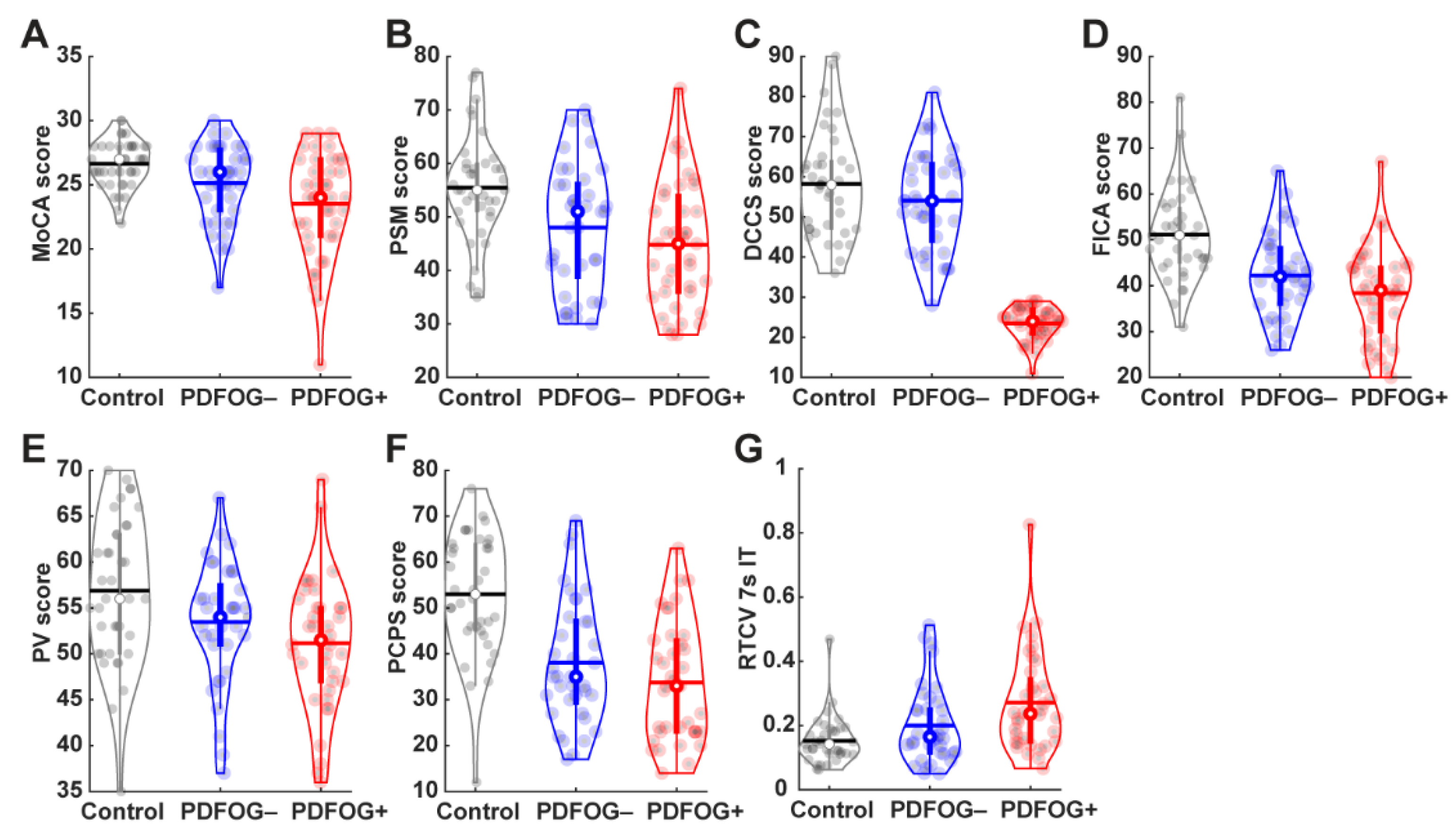
3.2. Differences in Cognitive Measurements

Significant differences between control and patients with PD were found in all cognitive domains (Table 2). Patients with PD showed significantly worse performance in all cognitive tests even after adjusting for covariates of age, gender, and years of education (Table 2).
When comparing PDFOG+ and PDFOG–, uncorrected independent t-tests revealed significant differences in the MoCA (p = 0.044) and DCCS executive function/attention test (p = 0.004) as well as RTCV of 7 s interval timing temporal processing/working memory test (p = 0.03), but no other cognitive measurements (Table 2 and Figure 1). After adjusting for covariates of age, gender, and years of education in the ANCOVA model, significant differences between PDFOG+ and PDFOG– remained for the MoCA (p = 0.03), and DCCS test of executive function and attention (p = 0.007; Table 2). In the next model, when adding disease duration to the covariate adjustments of age, gender, and years of education, significant differences were again observed between PDFOG+ and PDFOG– for the MoCA (p = 0.03), and DCCS measure of executive function and attention (p = 0.008; Table 2). However, similar to a previous study, when adjusting for covariates of age, gender, years of education, and disease severity (mUPDRS) in our complex model, differences between PD groups could not reach statistical significance (Table S1) [15]. Furthermore, we used individual covariates and controlled for them in the analyses in order to show their contribution to cognitive functioning in both PD groups. Overall, our models showed significant differences between PDFOG+ and PDFOG– for the MoCA and DCCS executive function/attention tests (Table S1).

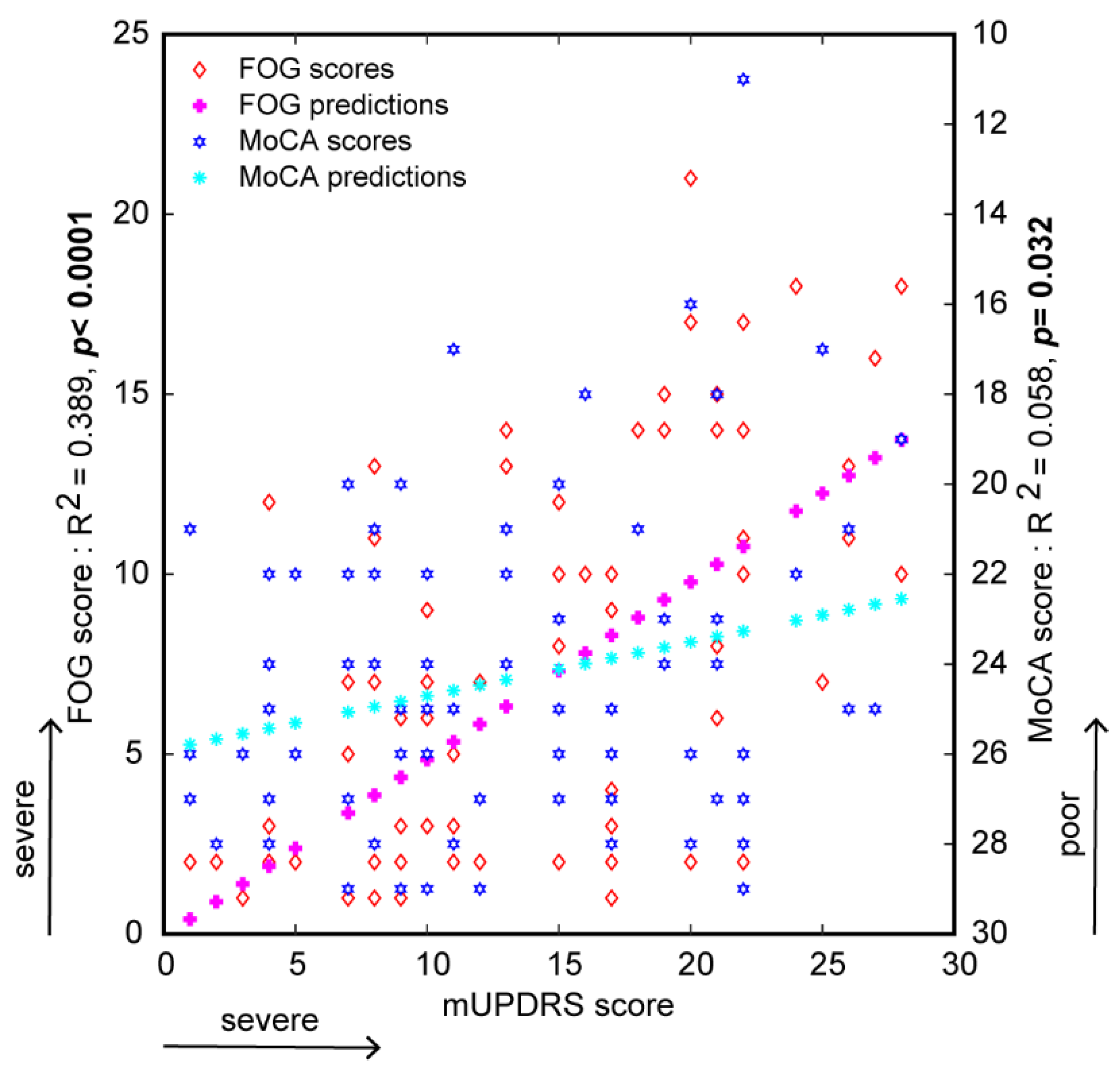
3.3. Relationship between FOG Severity and Cognitive Measurements

Correlation analyses, without controlling for covariates, suggested that FOG severity scores are significantly correlated with poorer cognitive scores for tests of cognitive function (MoCA, p = 0.015), processing speed (PCPS, p = 0.015), executive function/attention (DCCS, p = 0.014), and temporal processing/working memory performance (RTCV of 7 s interval timing, p = 0.002) (Table 3 and Figure S1). Even after controlling for age, gender, years of education, and disease duration, correlation analyses continued to show a significant relationship between FOG severity scores and poorer cognitive tests scores (Table 3).
Predicted responses of regression models showed significant positive relationships between disease severity (as determined by mUPDRS score) and FOG scores (R2 = 0.39, slope = 0.49, p < 0.001; Figure 2) as well as between disease severity and MoCA score (R2 = 0.058, slope = −0.12, p = 0.03; Figure 2). Predicted responses also indicated a significant relationship between disease duration and FOG score (R2 = 0.08, slope = 0.4, p = 0.01; Figure S2); however, predicted responses showed no significant relationship between disease duration and cognitive function, as measured by MoCA score (R2 = 0.0001, slope = −0.003, p = 0.98; Figure S2).
Furthermore, mediation model showed a significant mediation effect of disease severity on the relationship between cognitive function (MoCA score) and FOG severity (FOG score) (ACME estimate = −0.22, p = 0.03) with significant direct effect of cognitive function (ADE estimate = −0.32, p = 0.012) and total effect (p = 0.004). A second mediation model showed a significant mediation effect of cognitive function on the relationship between disease severity and FOG severity (ACME estimate = 0.039, p = 0.046) with significant direct effect of disease severity (ADE estimate = 0.45, p < 0.0001) and total effect (p < 0.0001).

4. Discussion

FOG represents a significant problem for patients with PD. To better understand the relationships between FOG, disease severity and duration, and cognitive function, this study examined patients with PD both with and without FOG compared to controls, and found significant differences between controls and patients with PD for all cognitive domains. PDFOG+ and PDFOG– exhibited decreased performance in several cognitive tests, which persisted after adjusting for covariates that include disease duration, suggesting that FOG severity may be related to cognitive performance difficulties separate from disease duration. A meta-analysis detected a 71% FOG prevalence rate in patients with PD with disease durations ≥ 10 years [30], and patients with >12 years of disease duration developing severe cognitive impairments or PD dementia [31]. The disease duration for PD groups in the current study was not more than 6 years. Similar to our results, a previous study has also shown that PDFOG+ participants exhibit higher LEDD compared to PDFOG– and further suggested that high LEDD, cognitive impairment, and falls and balance problems can be independent determinants of FOG in patients with PD [32].
In general, the advancement of disease severity causes gait abnormalities and cognitive impairment in patients with PD, suggesting the presence of abnormal motor-cognitive networks. Moreover, our and prior studies have reported altered frontal theta and beta oscillations in the cortical-basal ganglia network during the performance of lower-limb motor and cognitive tasks in patients with movement disorders [5,6,17,33,34]. Typically, reduced frontal theta activity correlates with cognitive deficits in patients with PD [35], and similar abnormal activity has been observed at the time of lower-limb movement initiation in PDFOG+ [17], indicating the oscillatory relationship between gait and cognitive networks.
Differences in executive functioning and attention between PDFOG+ and PDFOG- in particular, lend interest to the role of these constructs in FOG. The current study demonstrated significant group-dependent differences in executive functioning that held true when controlling for disease duration, but not with disease severity. It has been suggested by others that release of inhibition associated with executive functioning deficits may be implicated in FOG [23]. Furthermore, our regression models suggest that FOG severity may be a predictor of more global cognitive impairment, and disease severity is correlated with both FOG and cognitive functioning (MoCA) scores. In contrast, disease duration predicted gait severity but not global cognitive impairment. This suggests that the relationships between FOG severity and cognitive impairment are not a direct result of disease duration. Mediation models suggest that cognitive function significantly mediates the relationship between disease severity and FOG severity, further suggesting that the role of cognition as a predictor of both FOG severity and overall PD severity needs to be further explored.
The effects of dopamine in cognitive and lower-limb motor control in patients with PD are not clear; several studies have shown no improvement in cognition and gait functions after levodopa medications [6,17,36]. In the current study, we assessed cognition in patients with PD with ‘on’ dopaminergic medication. Nonetheless, PDFOG+ showed differences in cognitive and FOG assessments compared to PDFOG–, suggesting an overload of the dopaminergic system in PDFOG+ may not improve or may worsen cognition and lower-limb movements, similar to levodopa-induced involuntary movements. Additionally, chronic levodopa treatment may lead to a reduction in synaptic dopamine sensitivity, which may offer an explanation for cognitive and FOG severities in PDFOG+ and why these severities are not adequately improved by levodopa [37]. In our prior report, levodopa intake showed no improvement in frontal theta and beta oscillations in PDFOG+ during lower-limb pedaling motor task with cognitive load [17]; evidence suggests that frontal cognitive and motor functions are not affected by dopaminergic system in the advanced stage of parkinsonism. Therefore, it is critical to explore different pharmacological approaches to improving both cognitive and lower-limb motor functions in patients with advanced PD.
Furthermore, basal ganglia deep brain stimulation (DBS) therapeutic approaches have been implemented to improve cognitive and FOG severity in patients with PD. High-frequency basal ganglia DBS in the advanced PD stage influences only upper-limb motor performances; however, it affects neither lower-limb motor dysfunction (i.e., FOG severity) nor cognitive deficits significantly in the advanced stage of disease [38]. Therefore, another target such as pedunculopontine nucleus (PPN) has been proposed to improve both cognitive and lower-limb motor functions in patients with advanced PD or PDFOG+. Pedunculopontine tegmental nucleus acts as an interface between the basal ganglia-cerebellum network, and PPN-DBS has the potential to influence cognitive and gait motor functions [39]. In addition, low-frequency DBS has been suggested to improve both cognition and FOG in patients with PD. This approach can normalize frontal low-frequency and beta-band oscillations via cortico-basal ganglia networks [38,40]. In our study, DCCS results showed the profound differences between PDFOG+ and other two groups, even after adjusting different variables. These results clearly demonstrate that PDFOG+ participants exhibit deficits in executive function and attention. Our previous reports have also shown the presence of abnormal theta and beta oscillations in the cortical and subcortical regions in PDFOG+ during attentional cognitive and motor tasks [5,6,17,27,35]. Altogether, we suggest that appropriate neuromodulation approaches can be used to improve these oscillations and behavioral performances in PDFOG+.
The current study had some limitations which should be considered: our patients were assessed ‘on’ medication, and, therefore, our results should be interpreted accordingly compared to previous studies; we recruited only patients without DBS, so the effects of DBS on the relationship between cognitive and FOG severity could not be studied; and our patients did not perform gait or dual cognitive-gait task to measure quantitative information of FOG severity with and without cognitive load.
Overall, the current study contributes to the body of evidence observing a link between cognitive function and FOG severity. Understanding the role of cognitive functioning in FOG may aid the development of therapeutic interventions that target cognitive domains such as executive functioning as a means of improving FOG qualities. Such approaches to therapeutic programming have shown initial success and promise toward improving the FOG behaviors in patients with a diagnosis of PD. Therapeutic approaches have successfully targeted explicit learning networks through cuing with the goals of movement recalibration and gait or FOG parameter improvements. Similarly, explicit cues have been used to improve behaviors compromised by executive functioning deficits. Cognitive training has shown initial success in reducing the FOG severity and offers promise that cognitive strategies may be harnessed to improve FOG behaviors in patients with a diagnosis of PD [21].

Supplementary Materials

The following are available online at https://www.mdpi.com/article/10.3390/brainsci11111496/s1, Figure S1: Spearman correlation plots between FOG scores and cognitive measurement scores, Figure S2: Linear regression analysis and predicted responses between disease duration and FOG severity and cognitive assessment (MoCA) scores, Table S1: Cognitive Measurements between PDFOG+ and PDFOG- controlling for variables.

Author Contributions

J.L.S., W.R., M.L., L.A.B., P.B.-P. and A.S. conceptualized this study; J.L.S., A.I.E. and A.S. collected and processed data; J.L.S., M.L., L.A.B., P.B.-P. and A.S. wrote the initial draft; J.L.S., W.R., M.L., L.A.B., P.B.-P. and A.S. supported data interpretation and reviewed the manuscript. All authors have read and agreed to the published version of the manuscript.

Funding

This research received no external funding.

Institutional Review Board Statement

This study was approved by the local Institutional Review Boards (The University of Iowa and the University of South Dakota).

Informed Consent Statement

Informed consent was obtained from all subjects involved in this study.

Data Availability Statement

The data that support the findings of this study are available from the corresponding author, upon reasonable request.

Acknowledgments

A.S. was supported by SD Board of Regents, Research and Commercialization Council.

Conflicts of Interest

The authors declare no conflict of interest.

References

  1. Bloem, B.R.; Hausdorff, J.M.; Visser, J.E.; Giladi, N. Falls and freezing of gait in Parkinson’s disease: A review of two interconnected, episodic phenomena. Mov. Disord. 2004, 19, 871–884. [Google Scholar] [CrossRef]
  2. Lewis, S.J.; Barker, R.A. A pathophysiological model of freezing of gait in Parkinson’s disease. Parkinsonism Relat. Disord. 2009, 15, 333–338. [Google Scholar] [CrossRef]
  3. Huang, Y.F.; Cherng, Y.G.; Hsu, S.P.; Yeh, C.C.; Chou, Y.C.; Wu, C.H.; Chen, T.L.; Liao, C.C. Risk and adverse outcomes of fractures in patients with Parkinson’s disease: Two nationwide studies. Osteoporos. Int. 2015, 26, 1723–1732. [Google Scholar] [CrossRef]
  4. Giladi, N.; McDermott, M.P.; Fahn, S.; Przedborski, S.; Jankovic, J.; Stern, M.; Tanner, C.; Parkinson Study Group. Freezing of gait in PD: Prospective assessment in the DATATOP cohort. Neurology 2001, 56, 1712–1721. [Google Scholar] [CrossRef]
  5. Singh, A. Oscillatory activity in the cortico-basal ganglia-thalamic neural circuits in Parkinson’s disease. Eur. J. Neurosci. 2018, 48, 2869–2878. [Google Scholar] [CrossRef]
  6. Singh, A.; Plate, A.; Kammermeier, S.; Mehrkens, J.H.; Ilmberger, J.; Bötzel, K. Freezing of gait-related oscillatory activity in the human subthalamic nucleus. Basal Ganglia 2013, 3, 25–32. [Google Scholar] [CrossRef]
  7. Elble, R.J. Gait and dementia: Moving beyond the notion of gait apraxia. J. Neural. Transm 2007, 114, 1253–1258. [Google Scholar] [CrossRef]
  8. Alster, P.; Madetko, N.; Koziorowski, D.; Friedman, A. Progressive Supranuclear Palsy-Parkinsonism Predominant (PSP-P)-A Clinical Challenge at the Boundaries of PSP and Parkinson’s Disease (PD). Front. Neurol. 2020, 11, 180. [Google Scholar] [CrossRef]
  9. Osaki, Y.; Morita, Y.; Miyamoto, Y.; Furuta, K.; Furuya, H. Freezing of gait is an early clinical feature of progressive supranuclear palsy. Neurol. Clin. Neurosci. 2017, 5, 86–90. [Google Scholar] [CrossRef] [Green Version]
  10. Gurevich, T.; Giladi, N. Freezing of gait in multiple system atrophy (MSA). Parkinsonism Relat. Disord. 2003, 9, 169–174. [Google Scholar] [CrossRef]
  11. Macht, M.; Kaussner, Y.; Moller, J.C.; Stiasny-Kolster, K.; Eggert, K.M.; Kruger, H.P.; Ellgring, H. Predictors of freezing in Parkinson’s disease: A survey of 6620 patients. Mov. Disord. 2007, 22, 953–956. [Google Scholar] [CrossRef]
  12. Martens, K.A.E.; Shine, J.M.; Walton, C.C.; Georgiades, M.J.; Gilat, M.; Hall, J.M.; Muller, A.J.; Szeto, J.Y.Y.; Lewis, S.J.G. Evidence for subtypes of freezing of gait in Parkinson’s disease. Mov. Disord. 2018, 33, 1174–1178. [Google Scholar] [CrossRef]
  13. Kelly, V.E.; Eusterbrock, A.J.; Shumway-Cook, A. A review of dual-task walking deficits in people with Parkinson’s disease: Motor and cognitive contributions, mechanisms, and clinical implications. Parkinsons Dis. 2012, 2012, 918719. [Google Scholar] [CrossRef] [Green Version]
  14. Morris, R.; Lord, S.; Lawson, R.A.; Coleman, S.; Galna, B.; Duncan, G.W.; Khoo, T.K.; Yarnall, A.J.; Burn, D.J.; Rochester, L. Gait Rather Than Cognition Predicts Decline in Specific Cognitive Domains in Early Parkinson’s Disease. J. Gerontol. A Biol. Sci. Med. Sci. 2017, 72, 1656–1662. [Google Scholar] [CrossRef] [Green Version]
  15. Morris, R.; Smulders, K.; Peterson, D.S.; Mancini, M.; Carlson-Kuhta, P.; Nutt, J.G.; Horak, F.B. Cognitive function in people with and without freezing of gait in Parkinson’s disease. NPJ Parkinsons Dis. 2020, 6, 9. [Google Scholar] [CrossRef]
  16. Amboni, M.; Cozzolino, A.; Longo, K.; Picillo, M.; Barone, P. Freezing of gait and executive functions in patients with Parkinson’s disease. Mov. Disord. 2008, 23, 395–400. [Google Scholar] [CrossRef]
  17. Singh, A.; Cole, R.C.; Espinoza, A.I.; Brown, D.; Cavanagh, J.F.; Narayanan, N.S. Frontal theta and beta oscillations during lower-limb movement in Parkinson’s disease. Clin. Neurophysiol. 2020, 131, 694–702. [Google Scholar] [CrossRef]
  18. Horta-Barba, A.; Pagonabarraga, J.; Martinez-Horta, S.; Busteed, L.; Pascual-Sedano, B.; Illan-Gala, I.; Marin-Lahoz, J.; Aracil-Bolanos, I.; Perez-Perez, J.; Sampedro, F.; et al. Cognitive and behavioral profile of progressive supranuclear palsy and its phenotypes. J. Neurol. 2021, 268, 3400–3408. [Google Scholar] [CrossRef]
  19. Giagkou, N.; Hoglinger, G.U.; Stamelou, M. Progressive supranuclear palsy. Int. Rev. Neurobiol. 2019, 149, 49–86. [Google Scholar] [CrossRef]
  20. Rezvanian, S.; Litvan, I.; Standaert, D.; Jankovic, J.; Reich, S.G.; Hall, D.; Shprecher, D.R.; Bordelon, Y.; Dubinsky, R.; Kluger, B. Understanding the relationship between freezing of gait and other progressive supranuclear palsy features. Parkinsonism Relat. Disord. 2020, 78, 56–60. [Google Scholar] [CrossRef]
  21. Walton, C.C.; Shine, J.M.; Mowszowski, L.; Naismith, S.L.; Lewis, S.J. Freezing of gait in Parkinson’s disease: Current treatments and the potential role for cognitive training. Restor. Neurol. Neurosci. 2014, 32, 411–422. [Google Scholar] [CrossRef]
  22. Snijders, A.H.; Leunissen, I.; Bakker, M.; Overeem, S.; Helmich, R.C.; Bloem, B.R.; Toni, I. Gait-related cerebral alterations in patients with Parkinson’s disease with freezing of gait. Brain 2011, 134, 59–72. [Google Scholar] [CrossRef]
  23. Cohen, R.G.; Klein, K.A.; Nomura, M.; Fleming, M.; Mancini, M.; Giladi, N.; Nutt, J.G.; Horak, F.B. Inhibition, executive function, and freezing of gait. J. Parkinsons Dis. 2014, 4, 111–122. [Google Scholar] [CrossRef] [Green Version]
  24. Yao, Z.; Shao, Y.; Han, X. Freezing of gait is associated with cognitive impairment in patients with Parkinson disease. Neurosci. Lett. 2017, 656, 126–130. [Google Scholar] [CrossRef]
  25. Giladi, N.; Tal, J.; Azulay, T.; Rascol, O.; Brooks, D.J.; Melamed, E.; Oertel, W.; Poewe, W.H.; Stocchi, F.; Tolosa, E. Validation of the freezing of gait questionnaire in patients with Parkinson’s disease. Mov. Disord. 2009, 24, 655–661. [Google Scholar] [CrossRef]
  26. Movement Disorder Society Task Force on Rating Scales for Parkinson’s Disease. The Unified Parkinson’s Disease Rating Scale (UPDRS): Status and recommendations. Mov. Disord. 2003, 18, 738–750. [Google Scholar] [CrossRef]
  27. Singh, A.; Cole, R.; Espinoza, A.I.; Evans, A.; Cao, S.; Cavanagh, J.F.; Narayanan, N.S. Timing variability and midfrontal~ 4 Hz rhythms correlate with cognition in Parkinson’s disease. npj Parkinsons Dis. 2021, 7, 14. [Google Scholar] [CrossRef]
  28. Weintraub, S.; Dikmen, S.S.; Heaton, R.K.; Tulsky, D.S.; Zelazo, P.D.; Slotkin, J.; Carlozzi, N.E.; Bauer, P.J.; Wallner-Allen, K.; Fox, N.; et al. The cognition battery of the NIH toolbox for assessment of neurological and behavioral function: Validation in an adult sample. J. Int. Neuropsychol. Soc. 2014, 20, 567–578. [Google Scholar] [CrossRef] [Green Version]
  29. Singh, A.; Trapp, N.T.; De Corte, B.; Cao, S.; Kingyon, J.; Boes, A.D.; Parker, K.L. Cerebellar Theta Frequency Transcranial Pulsed Stimulation Increases Frontal Theta Oscillations in Patients with Schizophrenia. Cerebellum 2019, 18, 489–499. [Google Scholar] [CrossRef]
  30. Ge, H.L.; Chen, X.Y.; Lin, Y.X.; Ge, T.J.; Yu, L.H.; Lin, Z.Y.; Wu, X.Y.; Kang, D.Z.; Ding, C.Y. The prevalence of freezing of gait in Parkinson’s disease and in patients with different disease durations and severities. Chin. Neurosurg. J. 2020, 6, 17. [Google Scholar] [CrossRef]
  31. Roheger, M.; Kalbe, E.; Liepelt-Scarfone, I. Progression of Cognitive Decline in Parkinson’s Disease. J. Parkinsons Dis. 2018, 8, 183–193. [Google Scholar] [CrossRef] [Green Version]
  32. Vercruysse, S.; Devos, H.; Munks, L.; Spildooren, J.; Vandenbossche, J.; Vandenberghe, W.; Nieuwboer, A.; Heremans, E. Explaining freezing of gait in Parkinson’s disease: Motor and cognitive determinants. Mov. Disord. 2012, 27, 1644–1651. [Google Scholar] [CrossRef]
  33. Singh, A.; Kammermeier, S.; Plate, A.; Mehrkens, J.H.; Ilmberger, J.; Botzel, K. Pattern of local field potential activity in the globus pallidus internum of dystonic patients during walking on a treadmill. Exp. Neurol. 2011, 232, 162–167. [Google Scholar] [CrossRef]
  34. Wang, D.D.; Choi, J.T. Brain Network Oscillations during Gait in Parkinson’s Disease. Front. Hum. Neurosci. 2020, 14, 568703. [Google Scholar] [CrossRef]
  35. Singh, A.; Richardson, S.P.; Narayanan, N.; Cavanagh, J.F. Mid-frontal theta activity is diminished during cognitive control in Parkinson’s disease. Neuropsychologia 2018, 117, 113–122. [Google Scholar] [CrossRef]
  36. Lewis, S.J.; Slabosz, A.; Robbins, T.W.; Barker, R.A.; Owen, A.M. Dopaminergic basis for deficits in working memory but not attentional set-shifting in Parkinson’s disease. Neuropsychologia 2005, 43, 823–832. [Google Scholar] [CrossRef]
  37. Garcia-Ruiz, P.J. Gait disturbances in Parkinson disease. Did freezing of gait exist before levodopa? Historical review. J. Neurol. Sci. 2011, 307, 15–17. [Google Scholar] [CrossRef]
  38. Huang, C.; Chu, H.; Zhang, Y.; Wang, X. Deep Brain Stimulation to Alleviate Freezing of Gait and Cognitive Dysfunction in Parkinson’s Disease: Update on Current Research and Future Perspectives. Front. Neurosci. 2018, 12, 29. [Google Scholar] [CrossRef]
  39. Mori, F.; Okada, K.; Nomura, T.; Kobayashi, Y. The Pedunculopontine Tegmental Nucleus as a Motor and Cognitive Interface between the Cerebellum and Basal Ganglia. Front. Neuroanat. 2016, 10, 109. [Google Scholar] [CrossRef] [Green Version]
  40. Thevathasan, W.; Pogosyan, A.; Hyam, J.A.; Jenkinson, N.; Foltynie, T.; Limousin, P.; Bogdanovic, M.; Zrinzo, L.; Green, A.L.; Aziz, T.Z.; et al. Alpha oscillations in the pedunculopontine nucleus correlate with gait performance in parkinsonism. Brain 2012, 135, 148–160. [Google Scholar] [CrossRef] [Green Version]
Figure 1. Violin plots display the cognitive measurements in healthy controls, PDFOG−, and PDFOG+. (A) Montreal cognitive assessment (MoCA); (B) picture sequence memory (PSM) test; (C) dimensional change card sort (DCCS) test; (D) flanker inhibitory control and attention (FICA) test; (E) picture vocabulary (PV) test; (F) pattern comparison processing speed (PCPS) test; (G) Response Time Coefficient of Variation of 7-s interval timing task (RTCV). The horizontal lines and white circles in the violin plots represent the mean and median values, respectively.
Brainsci 11 01496 g001
Figure 2. Linear regression analysis and predicted responses between disease severity scores and FOG severity and cognitive assessment (MoCA) scores. Bold p-values represent the significance.
Brainsci 11 01496 g002
Table 1. Demographic and clinical assessments in controls vs. patients with PD.
MeasureControl
(n = 41)
PD
(n = 80)
PDFOG−
(n = 39)
PDFOG+
(n = 41)
Control vs. PDPDFOG– vs. PDFOG+
Independent t-TestIndependent t-Test
Gender (M/F) $23/1856/2426/1330/110.120.52
Age (years)71.3 ± 7.668.7 ± 8.068.4 ± 7.768.9 ± 8.31.77 (0.079)−0.26 (0.795)
Education (years)16.6 ± 2.215.8 ± 3.215.6 ± 3.416.0 ± 3.11.35 (0.179)−0.49 (0.623)
BAI1.8 ± 2.114.7 ± 11.09.5 ± 7.419.8 ± 11.6−7.46 (<0.001) ***−4.7 (<0.001) ***
GDS0.7 ± 1.13.7 ± 3.22.7 ± 2.44.6 ± 3.6−5.64 (<0.001) ***−2.73 (0.008) **
Total FOG-6.5 ± 5.71.6 ± 1.311.2 ± 4.1-−13.71 (<0.001) ***
DD (years)-5.15 ± 4.04.3 ± 3.25.9 ± 4.4-−1.83 (0.072)
LEDD (mg)-860.5 ± 450.3710.6 ± 392.91003.0 ± 459.3-−3.05 (0.003) **
mUPDRS-13.4 ± 7.29.4 ± 5.617.2 ± 6.6-−5.71 (<0.001) ***
H and Y-2.07 ± 0.91.5 ± 0.72.5 ± 0.8-−4.89 (<0.001) ***
PDSS-17.5 ± 10.013.1 ± 7.721.7 ± 10.3-−4.26 (<0.001) ***
$ Chi-squared test: presented as p-value. Presented as t-value (p-value). ** p < 0.01; *** p < 0.001. Abbreviations: BAI: Beck Anxiety Inventory; GDS: Geriatric Depression Scale; FOG: Freezing of Gait (maximum 24); DD: Disease duration; LEDD: Levodopa equivalent daily dose; mUPDRS: motor Unified Parkinson’s Disease Rating Scale; H & Y: Hoehn and Yahr; PDSS: Parkinson’s Disease Sleep Scale. “-” symbol represents no values.
Table 2. Cognitive measurements in controls vs. patients with PD.
Control
(n = 41)
PD
(n = 80)
PDFOG−
(n = 39)
PDFOG+
(n = 41)
Control vs. PDPDFOG− vs. PDFOG+
Independent t-Testa ANCOVAIndependent t-Testa ANCOVAb ANCOVA
Cognitive function: MoCA
26.6 ± 1.824.3 ± 3.625.1 ± 323.5 ± 43.9
(<0.001) ***
14.53
(<0.001) ***
2.05 (0.044) *4.55 (0.034) *4.7
(0.034) *
Episodic memory: PSM
55.5 ± 9.946.4 ± 11.748 ± 11.844.8 ± 11.64.11
(<0.001) ***
23.74
(<0.001) ***
1.2
(0.234)
0.049 (0.487)0.43
(0.515)
Executive function/Attention: DCCS
58.2 ± 13.250.1 ± 12.354.1 ± 12.246.1 ± 11.13.26
(0.001) **
12.66
(0.001) ***
2.94 (0.004) **7.78 (0.007) **7.44 (0.008) **
Executive function/Attention: FICA
51.1 ± 9.840.3 ± 9.842.2 ± 9.138.4 ± 10.15.55
(<0.001) ***
30.74
(<0.001) ***
1.74 (0.087)1.64
(0.205)
1.07
(0.304)
Language: PV
56.9 ± 7.952.3 ± 6.953.5 ± 6.551.2 ± 7.23.16
(0.002) **
7.76
(0.006) **
1.45 (0.151)1.21
(0.276)
1.28
(0.262)
Processing speed: PCPS
53 ± 1335.9 ± 13.338.1 ± 13.533.7 ± 12.96.49
(<0.001) ***
44.87
(<0.001) ***
1.44
(0.155)
1.46
(0.232)
1.53
(0.22)
Temporal processing/Working memory: RTCV
0.15 ± 0.070.23 ± 0.140.20 ± 0.120.27 ± 0.16−3.31
(0.001) **
14.34
(<0.001) ***
2.2 (0.031) *2.98 (0.089)2.36
(0.129)
a ANCOVA covariates = Age, Gender, and Education; b ANCOVA covariates = Age, Gender, Education, and Disease Duration; Presented as t-value (p-value) and F-value (p-value).* p < 0.05; ** p < 0.01; *** p < 0.001; Abbreviations: MoCA: Montreal Cognitive Assessment; PSM: Picture Sequence Memory test of the NIHTB-CB; DCCS: Dimensional Change Card Sort test of the NIHTB-CB; FICA: Flanker Inhibitory Control and Attention test of the NIHTB-CB; PV: Picture Vocabulary test of the NIHTB-CB; PCPS: Pattern Comparison Processing Speed test of the NIHTB-CB; RTCV: Response Time Coefficient of Variation of 7-s interval timing task.
Table 3. Correlations between FOG scores and cognitive measures.
VariableSpearman CorrelationSpearman Partial Correlation
a Age, Gender, Educationb Age, Gender, Education, Disease Duration
Cognitive function
MoCA−0.27 (0.015) *−0.27 (0.021) *−0.29 (0.01) **
Episodic memory
PSM−0.11 (0.33)−0.05 (0.69)−0.02 (0.852)
Executive function/Attention
DCCS−0.28 (0.014) *−0.26 (0.032) *−0.25 (0.044) *
FICA−0.20 (0.093)−0.13 (0.313)−0.09 (0.472)
Language
PV−0.14 (0.22)−0.07 (0.567)−0.10 (0.439)
Processing speed
PCPS−0.28 (0.015) *−0.24 (0.05) *−0.25 (0.043) *
Temporal processing/Working memory
RTCV0.36 (0.002) **0.32 (0.009) **0.29 (0.018) *
a controlling for age, gender, years of education; b controlling for age, gender, years of education and disease duration. Presented as correlation coefficients and p-values (in brackets). * p ≤ 0.05; ** p ≤ 0.01.
Publisher’s Note: MDPI stays neutral with regard to jurisdictional claims in published maps and institutional affiliations.

