Gray Matter Morphometry Correlates with Attentional Efficiency in Young-Adult Multiple Sclerosis


Abstract
1. Introduction
2. Materials and Methods
2.1. Study Population
2.2. Attention Network Test-Interactions (ANT-I)
2.3. Symbol Digits Modality Test (SDMT)
2.4. MRI Acquisition
2.5. Automatic Lesion Segmentation and Filling
2.6. Cortical Thickness Estimation
2.7. Subcortical Segmentation
2.8. Statistical Analyses
3. Results
3.1. Demographic and Clinical Characteristics
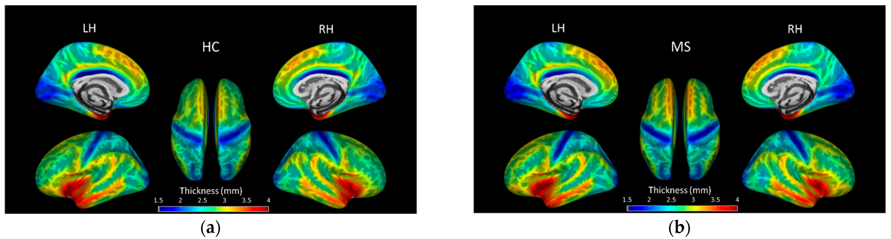
3.2. Cortical Thickness Group Differences
3.3. Subcortical GM Volume Group Differences
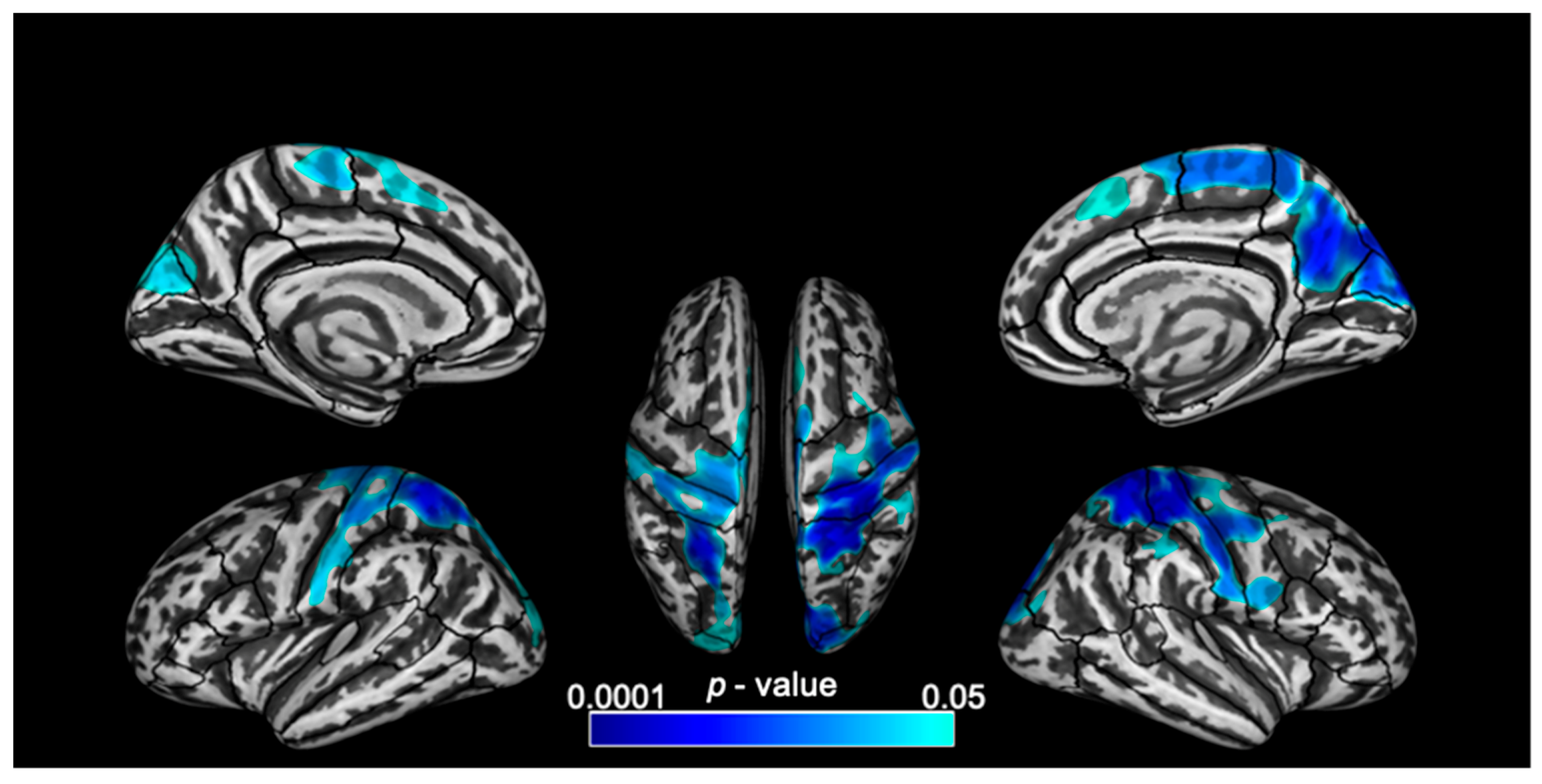
3.4. Vertex-Wise Cortical Thickness Correlations
3.5. Subcortical Volumes Correlations
4. Discussion
4.1. Morphometric Changes
4.2. Attention Network Test-Interactions
4.3. Cortical Thickness Correlation with EXE Network
4.4. Correlation of Subcortical GM Volumes with AE Networks
4.5. Limitation and Future Work
5. Conclusions
Author Contributions
Funding
Institutional Review Board Statement
Informed Consent Statement
Conflicts of Interest
References
- Brochet, B.; Ruet, A. Cognitive Impairment in Multiple Sclerosis With Regards to Disease Duration and Clinical Phenotypes. Front. Neurol. 2019, 10, 261. [Google Scholar] [CrossRef] [PubMed]
- Chiaravalloti, N.D.; DeLuca, J. Cognitive impairment in multiple sclerosis. Lancet Neurol. 2008, 7, 1139–1151. [Google Scholar] [CrossRef]
- Flensner, G.; Landtblom, A.-M.; Söderhamn, O.; Ek, A.-C. Work capacity and health-related quality of life among individuals with multiple sclerosis reduced by fatigue: A cross-sectional study. BMC Public Health 2013, 13, 224. [Google Scholar] [CrossRef] [PubMed]
- Sumowski, J.F.; Benedict, R.; Enzinger, C.; Filippi, M.; Geurts, J.J.; Hamalainen, P.; Hulst, H.; Inglese, M.; Leavitt, V.M.; Rocca, M.A.; et al. Cognition in multiple sclerosis: State of the field and priorities for the future. Neurology 2018, 90, 278–288. [Google Scholar] [CrossRef]
- Drew, M.A.; Starkey, N.J.; Isler, R.B. Examining the Link between Information Processing Speed and Executive Functioning in Multiple Sclerosis. Arch. Clin. Neuropsychol. 2009, 24, 47–58. [Google Scholar] [CrossRef]
- Forn, C.; Belenguer, A.; Ibars, M.A.P.; Ávila, C. Information-processing speed is the primary deficit underlying the poor performance of multiple sclerosis patients in the Paced Auditory Serial Addition Test (PASAT). J. Clin. Exp. Neuropsychol. 2008, 30, 789–796. [Google Scholar] [CrossRef]
- Archibald, C.J.; Fisk, J.D. Information Processing Efficiency in Patients with Multiple Sclerosis. J. Clin. Exp. Neuropsychol. 2000, 22, 686–701. [Google Scholar] [CrossRef]
- Confavreux, C.; Vukusic, S. Age at disability milestones in multiple sclerosis. Brain 2006, 129, 595–605. [Google Scholar] [CrossRef]
- Chitnis, T.; Krupp, L.; Yeh, A.; Rubin, J.; Kuntz, N.; Strober, J.B.; Chabas, D.; Weinstock-Guttmann, B.; Ness, J.; Rodriguez, M.; et al. Pediatric Multiple Sclerosis. Neurol. Clin. 2011, 29, 481–505. [Google Scholar] [CrossRef]
- Belman, A.L.; Krupp, L.B.; Olsen, C.S.; Rose, J.W.; Aaen, G.; Benson, L.; Chitnis, T.; Gorman, M.; Graves, J.; Harris, Y.; et al. Characteristics of Children and Adolescents With Multiple Sclerosis. Pediatrics 2016, 138, e20160120. [Google Scholar] [CrossRef]
- Ozakbas, S.; Kaya, D.; Idiman, E. Early Onset Multiple Sclerosis Has Worse Prognosis Than Adult Onset Multiple Sclerosis Based on Cognition and Magnetic Resonance Imaging. Autoimmune Dis. 2012, 2012, 1–5. [Google Scholar] [CrossRef] [PubMed]
- Julian, L.; Serafin, D.; Charvet, L.; Ackerson, J.; Benedict, R.; Braaten, E.; Brown, T.; O’Donnell, E.; Parrish, J.; Preston, T.; et al. Cognitive impairment occurs in children and adolescents with multiple sclerosis: Results from a United States network. J. Child Neurol. 2013, 28, 102–107. [Google Scholar] [CrossRef] [PubMed]
- Krupp, L.B.; Rintell, D.; Charvet, L.E.; Milazzo, M.; Wassmer, E. Pediatric multiple sclerosis: Perspectives from adolescents and their families. Neurology 2016, 87 (Suppl. 2), S4–S7. [Google Scholar] [CrossRef] [PubMed]
- Ishigami, Y.; Eskes, G.A.; Tyndall, A.V.; Longman, R.S.; Drogos, L.L.; Poulin, M. The Attention Network Test-Interaction (ANT-I): Reliability and validity in healthy older adults. Exp. Brain Res. 2015, 234, 815–827. [Google Scholar] [CrossRef] [PubMed]
- Crivelli, L.; Farez, M.F.; González, C.D.; Fiol, M.; Amengual, A.; Leiguarda, R.; Correale, J. Alerting Network Dysfunction in Early Multiple Sclerosis. J. Int. Neuropsychol. Soc. 2012, 18, 757–763. [Google Scholar] [CrossRef] [PubMed]
- Urbanek, C.; Weinges-Evers, N.; Bellmann-Strobl, J.; Bock, M.; Dörr, J.; Hahn, E.; Neuhaus, A.H.; Opgen-Rhein, C.; Ta, T.M.T.; Herges, K.; et al. Attention Network Test reveals alerting network dysfunction in multiple sclerosis. Mult. Scler. J. 2009, 16, 93–99. [Google Scholar] [CrossRef]
- Wojtowicz, M.; Omisade, A.; Fisk, J.D. Indices of Cognitive Dysfunction in Relapsing-Remitting Multiple Sclerosis: Intra-individual Variability, Processing Speed, and Attention Network Efficiency. J. Int. Neuropsychol. Soc. 2013, 19, 551–558. [Google Scholar] [CrossRef]
- Wojtowicz, M.; Ishigami, Y.; Mazerolle, E.L.; Fisk, J.D. Stability of intraindividual variability as a marker of neurologic dysfunction in relapsing remitting multiple sclerosis. J. Clin. Exp. Neuropsychol. 2014, 36, 455–463. [Google Scholar] [CrossRef]
- Roth, A.K.; Denney, D.R.; Lynch, S.G. Information processing speed and attention in multiple sclerosis: Reconsidering the Attention Network Test (ANT). J. Clin. Exp. Neuropsychol. 2015, 37, 518–529. [Google Scholar] [CrossRef]
- Ayache, S.; Palm, U.; Chalah, M.A.; Nguyen, R.; Farhat, W.H.; Créange, A.; Lefaucheur, J.-P. Orienting network dysfunction in progressive multiple sclerosis. J. Neurol. Sci. 2015, 351, 206–207. [Google Scholar] [CrossRef]
- Omisade, A.; Fisk, J.D.; Klein, R.M.; Schmidt, M.; Darvesh, S.; Bhan, V. Information Processing and Magnetic Resonance Imaging Indices of Brain Pathology in Multiple Sclerosis. Int. J. MS Care 2012, 14, 84–91. [Google Scholar] [CrossRef] [PubMed]
- Vázquez-Marrufo, M.; Galvao-Carmona, A.; Gonzalez-Rosa, J.J.; Hidalgo-Muñoz, A.R.; Borges, M.; Ruiz-Peña, J.L.; Izquierdo, G. Neural Correlates of Alerting and Orienting Impairment in Multiple Sclerosis Patients. PLoS ONE 2014, 9, e97226. [Google Scholar] [CrossRef] [PubMed]
- Audoin, B.; Zaaraoui, W.; Reuter, F.; Rico, A.; Malikova, I.; Confort-Gouny, S.; Cozzone, P.J.; Pelletier, J.; Ranjeva, J.-P. Atrophy mainly affects the limbic system and the deep grey matter at the first stage of multiple sclerosis. J. Neurol. Neurosurg. Psychiatry 2010, 81, 690–695. [Google Scholar] [CrossRef] [PubMed]
- Calabrese, M.; Atzori, M.; Bernardi, V.; Morra, A.; Romualdi, C.; Rinaldi, L.; McAuliffe, M.J.M.; Barachino, L.; Perini, P.; Fischl, B.; et al. Cortical atrophy is relevant in multiple sclerosis at clinical onset. J. Neurol. 2007, 254, 1212–1220. [Google Scholar] [CrossRef]
- Calabrese, M.; Agosta, F.; Rinaldi, F.; Mattisi, I.; Grossi, P.; Favaretto, A.; Atzori, M.; Bernardi, V.; Barachino, L.; Rinaldi, L.; et al. Cortical Lesions and Atrophy Associated With Cognitive Impairment in Relapsing-Remitting Multiple Sclerosis. Arch. Neurol. 2009, 66, 1144–1150. [Google Scholar] [CrossRef]
- Kurtzke, J.F. Rating neurologic impairment in multiple sclerosis: An expanded disability status scale (EDSS). Neurology 1983, 33, 1444. [Google Scholar] [CrossRef]
- Snelbaker, A.J.; Wilkinson, G.S.; Robertson, G.J.; Glutting, J.J. Wide Range Achievement Test 3 (wrat3). In Understanding Psychological Assessment; Dorfman, W.I., Hersen, M., Eds.; Springer: Boston, MA, USA, 2001; pp. 259–274. [Google Scholar]
- Benedict, R.H.B.; Fishman, I.; McClellan, M.M.; Bakshi, R.; Weinstock-Guttman, B. Validity of the Beck Depression Inventory-Fast Screen in multiple sclerosis. Mult. Scler. J. 2003, 9, 393–396. [Google Scholar] [CrossRef]
- Govindarajan, S.T.; Liu, Y.; Corral, M.A.P.; Bangiyev, L.; Krupp, L.; Charvet, L.; Duong, T.Q. White matter correlates of slowed information processing speed in unimpaired multiple sclerosis patients with young age onset. Brain Imaging Behav. 2020. [Google Scholar] [CrossRef]
- Fan, J.; McCandliss, B.D.; Sommer, T.; Raz, A.; Posner, M.I. Testing the Efficiency and Independence of Attentional Networks. J. Cogn. Neurosci. 2002, 14, 340–347. [Google Scholar] [CrossRef]
- Rao, S.M.; Aubin-Faubert, P.S.; Leo, G.J. Information processing speed in patients with multiple sclerosis. J. Clin. Exp. Neuropsychol. 1989, 11, 471–477. [Google Scholar] [CrossRef]
- Smith, A. Symbol Digit Modalities Test; Western Psychological Services: Los Angeles, CA, USA, 1982. [Google Scholar]
- Magon, S.; Gaetano, L.; Chakravarty, M.M.; Lerch, J.P.; Naegelin, Y.; Stippich, C.; Kappos, L.; Radue, E.W.; Sprenger, T. White matter lesion filling improves the accuracy of cortical thickness measurements in multiple sclerosis patients: A longitudinal study. BMC Neurosci. 2014, 15, 1–10. [Google Scholar] [CrossRef] [PubMed]
- Schmidt, P. Bayesian Inference for Structured Additive Regression Models for Large-Scale Problems with Applications to Medical Imaging. Ph.D. Thesis, Ludwig-Maximilians-Universität München, München, Germany, January 2017. [Google Scholar]
- Chard, D.; Jackson, J.S.; Miller, D.H.; Wheeler-Kingshott, C.A. Reducing the impact of white matter lesions on automated measures of brain gray and white matter volumes. J. Magn. Reson. Imaging 2010, 32, 223–228. [Google Scholar] [CrossRef] [PubMed]
- Dahnke, R.; Yotter, R.A.; Gaser, C. Cortical thickness and central surface estimation. NeuroImage 2013, 65, 336–348. [Google Scholar] [CrossRef] [PubMed]
- Wang, H.; Yushkevich, P.A. Multi-atlas segmentation with joint label fusion and corrective learning—an open source implementation. Front. Aging Neurosci. 2013, 7, 27. [Google Scholar] [CrossRef] [PubMed]
- Klein, A.; Tourville, J.A. 101 Labeled Brain Images and a Consistent Human Cortical Labeling Protocol. Front. Behav. Neurosci. 2012, 6, 171. [Google Scholar] [CrossRef] [PubMed]
- Herranz, E.; Giannì, C.; Louapre, C.; Treaba, C.A.; Ms, S.T.G.; Ouellette, R.; Loggia, M.L.; Sloane, J.A.; Madigan, N.; Izquierdo-Garcia, D.; et al. Neuroinflammatory component of gray matter pathology in multiple sclerosis. Ann. Neurol. 2016, 80, 776–790. [Google Scholar] [CrossRef] [PubMed]
- Geisseler, O.; Pflugshaupt, T.; Bezzola, L.; Reuter, K.; Weller, D.; Schuknecht, B.; Linnebank, M. Cortical thinning in the anterior cingulate cortex predicts multiple sclerosis patients’ fluency performance in a lateralised manner. NeuroImage Clin. 2016, 10, 89–95. [Google Scholar] [CrossRef]
- Pravatà, E.; Rocca, M.A.; Valsasina, P.; Riccitelli, G.C.; Gobbi, C.; Comi, G.; Falini, A.; Filippi, M. Gray matter trophism, cognitive impairment, and depression in patients with multiple sclerosis. Mult. Scler. J. 2017, 23, 1864–1874. [Google Scholar] [CrossRef]
- Bakshi, R.; Czarnecki, D.; Shaikh, Z.A.; Priore, R.L.; Janardhan, V.; Kaliszky, Z.; Kinkel, P.R. Brain MRI lesions and atrophy are related to depression in multiple sclerosis. NeuroReport 2000, 11, 1153–1158. [Google Scholar] [CrossRef]
- Bakshi, R.; Benedict, R.H.; Bermel, R.A.; Jacobs, L. Regional brain atrophy is associated with physical disability in multiple sclerosis: Semiquantitative magnetic resonance imaging and relationship to clinical findings. J. Neuroimaging 2001, 11, 129–136. [Google Scholar] [CrossRef]
- Westlye, L.T.; Grydeland, H.; Walhovd, K.B.; Fjell, A.M. Associations between Regional Cortical Thickness and Attentional Networks as Measured by the Attention Network Test. Cereb. Cortex 2011, 21, 345–356. [Google Scholar] [CrossRef] [PubMed]
- Schmidt, E.L.; Burge, W.; Visscher, K.M.; Ross, L.A. Cortical thickness in frontoparietal and cingulo-opercular networks predicts executive function performance in older adults. Neuropsychology 2016, 30, 322–331. [Google Scholar] [CrossRef] [PubMed]
- Johnson, K.A.; Robertson, I.H.; Barry, E.; Mulligan, A.; Dáibhis, A.; Daly, M.; Watchorn, A.; Gill, M.; Bellgrove, M.A. Impaired conflict resolution and alerting in children with ADHD: Evidence from the Attention Network Task (ANT). J. Child Psychol. Psychiatry 2008, 49, 1339–1347. [Google Scholar] [CrossRef] [PubMed]
- Adolfsdottir, S.; Sorensen, L.; Lundervold, A.J. The attention network test: A characteristic pattern of deficits in children with ADHD. Behav. Brain Funct. 2008, 4, 9. [Google Scholar] [CrossRef] [PubMed]
- Kratz, O.; Studer, P.; Malcherek, S.; Erbe, K.; Moll, G.H.; Heinrich, H. Attentional processes in children with ADHD: An event-related potential study using the attention network test. Int. J. Psychophysiol. 2011, 81, 82–90. [Google Scholar] [CrossRef] [PubMed]
- Murphy, C.F.; Alexopoulos, G.S. Attention Network Dysfunction and Treatment Response of Geriatric Depression. J. Clin. Exp. Neuropsychol. 2006, 28, 96–100. [Google Scholar] [CrossRef] [PubMed]
- Urbanek, C.; Neuhaus, A.H.M.; Opgen-Rhein, C.; Strathmann, S.; Wieseke, N.; Schaub, R.; Hahn, E.; Dettling, M. Attention network test (ANT) reveals gender-specific alterations of executive function in schizophrenia. Psychiatry Res. 2009, 168, 102–109. [Google Scholar] [CrossRef]
- Vandenbossche, J.; Deroost, N.; Soetens, E.; Coomans, D.; Spildooren, J.; Vercruysse, S.; Nieuwboer, A.; Kerckhofs, E. Freezing of gait in Parkinson’s disease: Disturbances in automaticity and control. Front. Hum. Neurosci. 2012, 6, 356. [Google Scholar] [CrossRef]
- Fan, J.; McCandliss, B.D.; Fossella, J.; Flombaum, J.; Posner, M. The activation of attentional networks. NeuroImage 2005, 26, 471–479. [Google Scholar] [CrossRef]
- Welton, T.; Constantinescu, C.S.; Auer, D.P.; Dineen, R.A. Graph Theoretic Analysis of Brain Connectomics in Multiple Sclerosis: Reliability and Relationship with Cognition. Brain Connect. 2020, 10, 95–104. [Google Scholar] [CrossRef]
- Yin, X.; Zhao, L.; Xu, J.; Evans, A.C.; Fan, L.; Ge, H.; Tang, Y.; Khundrakpam, B.; Wang, J.; Liu, S. Anatomical Substrates of the Alerting, Orienting and Executive Control Components of Attention: Focus on the Posterior Parietal Lobe. PLoS ONE 2012, 7, e50590. [Google Scholar] [CrossRef] [PubMed]
- Benedict, R.H.; Bakshi, R.; Simon, J.H.; Priore, R.; Miller, C.; Munschauer, F. Frontal cortex atrophy predicts cognitive impairment in multiple sclerosis. J. Neuropsychiatry Clin. Neurosci. 2002, 14, 44–51. [Google Scholar] [CrossRef] [PubMed]
- Calabrese, M.; Rinaldi, F.; Grossi, P.; Mattisi, I.; Bernardi, V.; Favaretto, A.; Perini, P.; Gallo, P. Basal ganglia and frontal/parietal cortical atrophy is associated with fatigue in relapsing—remitting multiple sclerosis. Mult. Scler. J. 2010, 16, 1220–1228. [Google Scholar] [CrossRef] [PubMed]
- Reineberg, A.E.; Andrews-Hanna, J.R.; DePue, B.E.; Friedman, N.P.; Banich, M.T. Resting-state networks predict individual differences in common and specific aspects of executive function. NeuroImage 2015, 104, 69–78. [Google Scholar] [CrossRef] [PubMed]
- Dosenbach, N.U.F.; Fair, D.A.; Miezin, F.M.; Cohen, A.L.; Wenger, K.K.; Dosenbach, R.A.T.; Fox, M.D.; Snyder, A.Z.; Vincent, J.L.; Raichle, M.E.; et al. Distinct brain networks for adaptive and stable task control in humans. Proc. Natl. Acad. Sci. USA 2007, 104, 11073–11078. [Google Scholar] [CrossRef]
- Scolari, M.; Seidl-Rathkopf, K.N.; Kastner, S. Functions of the human frontoparietal attention network: Evidence from neuroimaging. Curr. Opin. Behav. Sci. 2015, 1, 32–39. [Google Scholar] [CrossRef]
- Batista, S.; Zivadinov, R.; Hoogs, M.; Bergsland, N.; Heininen-Brown, M.; Dwyer, M.G.; Weinstock-Guttman, B.; Benedict, R.H.B. Basal ganglia, thalamus and neocortical atrophy predicting slowed cognitive processing in multiple sclerosis. J. Neurol. 2012, 259, 139–146. [Google Scholar] [CrossRef]
- Bisecco, A.; Stamenova, S.; Caiazzo, G.; D’Ambrosio, A.; Sacco, R.; Docimo, R.; Esposito, S.; Cirillo, M.; Esposito, F.; Bonavita, S.; et al. Attention and processing speed performance in multiple sclerosis is mostly related to thalamic volume. Brain Imaging Behav. 2017, 12, 20–28. [Google Scholar] [CrossRef]
- Debernard, L.; Melzer, T.R.; Alla, S.; Eagle, J.; Van Stockum, S.; Graham, C.; Osborne, J.R.; Dalrymple-Alford, J.C.; Miller, D.H.; Mason, D.F. Deep grey matter MRI abnormalities and cognitive function in relapsing-remitting multiple sclerosis. Psychiatry Res. Neuroimaging 2015, 234, 352–361. [Google Scholar] [CrossRef]
- Filippi, M. Linking structural, metabolic and functional changes in multiple sclerosis. Eur. J. Neurol. 2001, 8, 291–297. [Google Scholar] [CrossRef]
- Ge, Y.; Zhang, Z.; Lu, H.; Tang, L.; Jaggi, H.; Herbert, J.; Babb, J.S.; Rusinek, H.; Grossman, R. Characterizing Brain Oxygen Metabolism in Patients with Multiple Sclerosis with T2-Relaxation-Under-Spin-Tagging MRI. Br. J. Pharmacol. 2012, 32, 403–412. [Google Scholar] [CrossRef] [PubMed]
- Harrison, D.; Roy, S.; Oh, J.; Izbudak, I.; Pham, D.; Courtney, S.M.; Caffo, B.; Jones, C.K.; Van Zijl, P.; Calabresi, P.A. Association of Cortical Lesion Burden on 7-T Magnetic Resonance Imaging With Cognition and Disability in Multiple Sclerosis. JAMA Neurol. 2015, 72, 1004–1012. [Google Scholar] [CrossRef] [PubMed]


| Demographics | HC | MS | p Value |
|---|---|---|---|
| Number of subjects | 19 (8M/11F) | 21 (9M/12F) | NA |
| Age ± SD (years) | 22.6 ± 2.3 | 25.7 ± 5.2 * | 0.02 |
| Age range (years) | 19–29 | 18–35 | |
| Age at onset ± SD (years) | - | 19.7 ± 6.55 | NA |
| Age range at onset (years) | - | 10.2–34.0 | NA |
| Disease duration ± SD (years) | - | 5.9 ± 3.5 | NA |
| Disease duration range (years) | - | 1.0–11.8 | NA |
| Expanded Disability Status Scale (EDSS) | - | Median: 2 Range: 0–6 | NA |
| EDSS 0–2 (n, %) | - | 16, 76 | NA |
| EDSS 2.5–3.5 (n, %) | - | 2, 10 | NA |
| EDSS 4–6 (n, %) | - | 3, 14 | NA |
| Symbol Digits Modality Test z-score (SDMTz) | −0.15 ± 0.90 | −0.65 ± 1.31 | 0.17 |
| Attention Network Test-Interaction (ANT-I) scores | |||
| Alerting (AE) | 25.8 ± 25.2 | 35.2 ± 39.6 | 0.4 |
| Orienting (ON) | 49.7 ± 13.5 | 49.3 ± 30.9 | 0.96 |
| Executive control (EXE) | 88.0 ± 19.9 | 98.1 ± 35.5 | 0.3 |
| MRI features | |||
| Average LH cortical thickness (mm) | 2.74 ± 0.1 | 2.73 ± 0.1 | 0.84 |
| Average RH cortical thickness (mm) | 2.73 ± 0.1 | 2.73 ± 0.1 | 0.9 |
| GM Region | Hemisphere | Normalized (Average ± SD) × 10−3 | Uncorrected p Value | |
|---|---|---|---|---|
| HC | MS | |||
| Amygdala | LH | 0.82 ± 0.1 | 0.79 ± 0.09 | 0.36 |
| RH | 0.79 ± 0.1 | 0.80 ± 0.08 | 0.51 | |
| Caudate | LH | 2.36 ± 0.2 | 2.23 ± 0.22 | 0.26 |
| RH | 2.45 ± 0.2 | 2.26 ± 0.21 | 0.04 | |
| Hippocampus | LH | 2.72 ± 0.2 | 2.60 ± 0.20 | 0.07 |
| RH | 2.82 ± 0.2 | 2.77 ± 0.19 | 0.61 | |
| Pallidum | LH | 1.09 ± 0.1 | 1.01 ± 0.09 | 0.01 |
| RH | 1.04 ± 0.1 | 0.99 ± 0.07 | 0.06 | |
| Putamen | LH | 3.31 ± 0.2 | 3.19 ± 0.26 | 0.22 |
| RH | 3.19 ± 0.2 | 3.06 ± 0.23 | 0.16 | |
| Thalamus | LH | 5.90 ± 0.3 | 5.36 ± 0.49 | 0.0019 * |
| RH | 5.75 ± 0.3 | 5.21 ± 0.50 | 0.0015 * | |
| Accumbens | LH | 0.37 ± 0.1 | 0.35 ± 0.04 | 0.18 |
| RH | 0.33 ± 0.0 | 0.32 ± 0.03 | 0.81 | |
| Brain Lobe | Region Name | Number of Significant Vertices | |
|---|---|---|---|
| LH | RH | ||
| Frontal | Superior frontal | 1176 | 1696 |
| Precentral | 2184 | 3959 | |
| Paracentral | 1176 | 2262 | |
| Parietal | Postcentral | 5040 | 5938 |
| Superior parietal | 5040 | 5938 | |
| Precuneus | 336 | 5372 | |
| Supramarginal | 848 | ||
| Occipital | Cuneus | 1008 | 1414 |
| Lateral occipital | 840 | 848 | |
| Total | 16,800 | 28,275 | |
| GM Region | Hemisphere | SDMTz | AE | EXE | |||
|---|---|---|---|---|---|---|---|
| R | p | R | p | R | p | ||
| Pallidum | LH | - | - | −0.57 | 0.01 | - | - |
| RH | - | - | −0.72 | 0.0006 * | −0.51 | 0.025 | |
| Putamen | LH | - | - | - | - | −0.66 | 0.002 |
| RH | - | - | - | - | −0.59 | 0.007 | |
| Thalamus | LH | - | - | −0.59 | 0.007 | −0.46 | 0.048 |
| RH | - | - | −0.51 | 0.027 | - | - | |
| Accumbens | LH | 0.49 | 0.024 | - | - | −0.52 | 0.022 |
| RH | - | - | - | - | - | - | |
Publisher’s Note: MDPI stays neutral with regard to jurisdictional claims in published maps and institutional affiliations. |
© 2021 by the authors. Licensee MDPI, Basel, Switzerland. This article is an open access article distributed under the terms and conditions of the Creative Commons Attribution (CC BY) license (http://creativecommons.org/licenses/by/4.0/).
Share and Cite
Govindarajan, S.T.; Pan, R.; Krupp, L.; Charvet, L.; Duong, T.Q. Gray Matter Morphometry Correlates with Attentional Efficiency in Young-Adult Multiple Sclerosis. Brain Sci. 2021, 11, 80. https://doi.org/10.3390/brainsci11010080
Govindarajan ST, Pan R, Krupp L, Charvet L, Duong TQ. Gray Matter Morphometry Correlates with Attentional Efficiency in Young-Adult Multiple Sclerosis. Brain Sciences. 2021; 11(1):80. https://doi.org/10.3390/brainsci11010080
Chicago/Turabian StyleGovindarajan, Sindhuja T., Ruiqi Pan, Lauren Krupp, Leigh Charvet, and Tim Q. Duong. 2021. "Gray Matter Morphometry Correlates with Attentional Efficiency in Young-Adult Multiple Sclerosis" Brain Sciences 11, no. 1: 80. https://doi.org/10.3390/brainsci11010080
APA StyleGovindarajan, S. T., Pan, R., Krupp, L., Charvet, L., & Duong, T. Q. (2021). Gray Matter Morphometry Correlates with Attentional Efficiency in Young-Adult Multiple Sclerosis. Brain Sciences, 11(1), 80. https://doi.org/10.3390/brainsci11010080