Review

Indian Medicinal Herbs and Formulations for Alzheimer’s Disease, from Traditional Knowledge to Scientific Assessment

1
Department of Neurological Surgery, Washington University School of Medicine, St. Louis, MO 63110, USA
2
Department of Pharmacology, All India Institute of Medical Sciences, New Delhi 110029, India
3
Division of Basic Medical Sciences, Indian Council of Medical Research, Ministry of Health and Family Welfare, Government of India, V. Ramalingaswamy Bhawan, New Delhi 110029, India
*
Authors to whom correspondence should be addressed.
Brain Sci. 2020, 10(12), 964; https://doi.org/10.3390/brainsci10120964
Submission received: 17 October 2020 / Revised: 27 November 2020 / Accepted: 7 December 2020 / Published: 10 December 2020
(This article belongs to the Special Issue Advances in Alzheimer's Disease: Where Do We Stand in 2020?)

Abstract

:
Cognitive impairment, associated with ageing, stress, hypertension and various neurodegenerative disorders including Parkinson’s disease and epilepsy, is a major health issue. The present review focuses on Alzheimer’s disease (AD), since it is the most important cause of cognitive impairment. It is characterized by progressive memory loss, language deficits, depression, agitation, mood disturbances and psychosis. Although the hallmarks of AD are cholinergic dysfunction, β-amyloid plaques and neurofibrillary tangle formation, it is also associated with derangement of other neurotransmitters, elevated levels of advanced glycation end products, oxidative damage, neuroinflammation, genetic and environmental factors. On one hand, this complex etiopathology makes a response to commonly used drugs such as donepezil, rivastigmine, galantamine and memantine less predictable and often unsatisfactory. On the other hand, it supports the use of herbal medicines due to their nonspecific antioxidant and anti-inflammatory activity and specific cholinesterase inhibitory activity. The popularity of herbal medicines is also increasing due to their perceived effectiveness, safety and affordability. In the present article, the experimental and clinical evidence have been reviewed for various Indian herbal medicines such as Centella asiatica, Bacopa monnieri, Curcuma longa, Clitoria ternatea, Withania somnifera, Celastrus paniculatus, Evolvulus alsinoides, Desmodium gangeticum, Eclipta alba, Moringa oleifera and Convolvulus pluricaulis, which have shown potential in cognitive impairment. Some commonly available herbal formulations for memory impairment in India have also been reviewed.

Graphical Abstract

1. Introduction

Ayurveda mentions three aspects of mental abilities, i.e., Dhi (process of acquisition/learning), Dhuti (process of retention) and Smriti (process of recall) [1]. A dysfunction in the process of acquisition/learning, retention or recall is known as dementia. Worldwide, about 40 million elderly are living with dementia [2,3]. In India, an estimated 3.7 million elderly people have dementia, and the prevalence is expected to increase two-fold by 2030 and three-fold by 2050 [4]. Dementia is associated with neurodegenerative disorders such as Alzheimer’s disease (AD), Parkinson’s disease and epilepsy. This review is focused on the potential of herbal medicine in AD since it is responsible for more than two-thirds of all dementia cases [5,6].
Cognitive functions that are mainly affected in AD patients include memory, executive functioning, language, visuospatial functioning and attention. Several hypotheses have been proposed for establishing the cause of AD. Cholinergic hypothesis, which is the oldest theory, describes acetylcholine (ACh) deficiency as the causative factor [7]. Currently available therapies for AD management are based on this hypothesis [8]. The β-amyloid hypothesis, most cogent hypothesis [9,10,11,12] provides the basis for development of new therapeutic strategies for AD treatment [13]. The histopathological hallmarks of AD are neuritic plaque and neurofibrillary tangle (NFT) formation in the brain [14]. Other associated factors that may also contribute to neurodegeneration in AD are elevated levels of advanced glycation end products, oxidative damage and neuroinflammation (Figure 1). The involvement of free radicals and inflammation in pathogenesis of AD hint towards the possible role of antioxidant and anti-inflammatory agents as therapeutic tools [15]. Studies have also reported that antioxidants protect against Aβ induced neuronal toxicity [16,17]. Fuzhisan (FZS), a herbal drug, demonstrated a neuroprotective effect by inhibiting Aβ (25–35)-induced activation of cyclin-dependent kinase 5, calcium influx, calpain activation and tau hyperphosphorylation [18]. Inhibitory effect of an aqueous extract of Ceylon cinnamon (C. zeylanicum) on tau aggregation and filament formation has also been reported [19].

2. Limitations of Currently Approved Cognition Enhancers

Currently approved drugs for AD include cholinesterase inhibitors (donepezil, rivastigmine and galantamine) and NMDA receptor antagonist (memantine). Cholinesterase inhibitors provide only symptomatic relief of behavioral deficits without modifying the complex pathologies in mild to moderate AD patients [20]. However, memantine is mainly recommended for moderate to severe AD cases [20]. Cholinesterase inhibitors significantly improve the cognition in patients with mild to moderate AD but their efficacy for neuropsychiatric symptoms is still questionable. AD patients receiving cholinesterase inhibitors experience adverse effects like nausea, vomiting, diarrhea, dizziness, etc. [21]. The common adverse effects associated with AChE inhibitors are nausea, vomiting, diarrhea, abdominal pain, loss of appetite and weight, though these can be minimized by slow dose escalation and administration with food. Other adverse effects of AChE inhibitors such as extrapyramidal symptoms, sleep disorder and cardiorespiratory adverse effects, are associated with central cholinergic over-activity whereas muscle cramps, weakness and urinary incontinence, are associated with peripheral cholinergic over-activity [22]. These adverse effects are often dose limiting and disabling in nature. Further, AChE inhibitors do not address neuronal degeneration and associated changes in the brain.
Cholinergic dysfunction, amyloid-β neurotoxicity, oxidative damage and inflammation have been targeted for treatment of AD but with limited success [23,24]. Studies indicate that antioxidants (vitamins E and C) and non-steroidal anti-inflammatory drugs slow the progression of AD [25,26,27,28,29,30]. Hormone replacement therapy has also been tried as a therapeutic strategy. Though it performed better than tacrine [31], it is no longer recommended, as it may increase the risk of adverse cardiovascular events and breast cancer [32].
Memantine showed promising anti-Alzheimer effects in preclinical experimental models, however, in clinical studies, it has not shown clear therapeutic efficacy in AD [33]. It is currently being used in the treatment of moderate to severe AD. The rate of decline in behavioral and functional impairment in patients with moderate to severe AD is reduced by memantine [34]. Patients taking memantine experience adverse effects like fatigue, pain, confusion, urinary incontinence, urinary tract infection, peripheral edema, etc. [35]. Thus, the search continues for effective and affordable medicines, which when prescribed for long duration, have acceptable adverse effects or interaction with food and drugs and delay the progression or reverse the disease process.
Herbal medicines, supported by a wealth of traditional knowledge, may serve the purpose as they can target AD pathophysiology at multiple sites, both at cellular and molecular levels. Though the mechanisms of action of herbal medicines are not clear, it has been proposed that they exert their protective effects against cognitive impairment through nonspecific antioxidant and anti-inflammatory activities and through specific action on AChE, β-amyloid fibril formation and tau aggregation (Figure 2) [19,36].
Therefore, the present article reviews selected herbal drugs and formulations commonly studied for the treatment of AD.

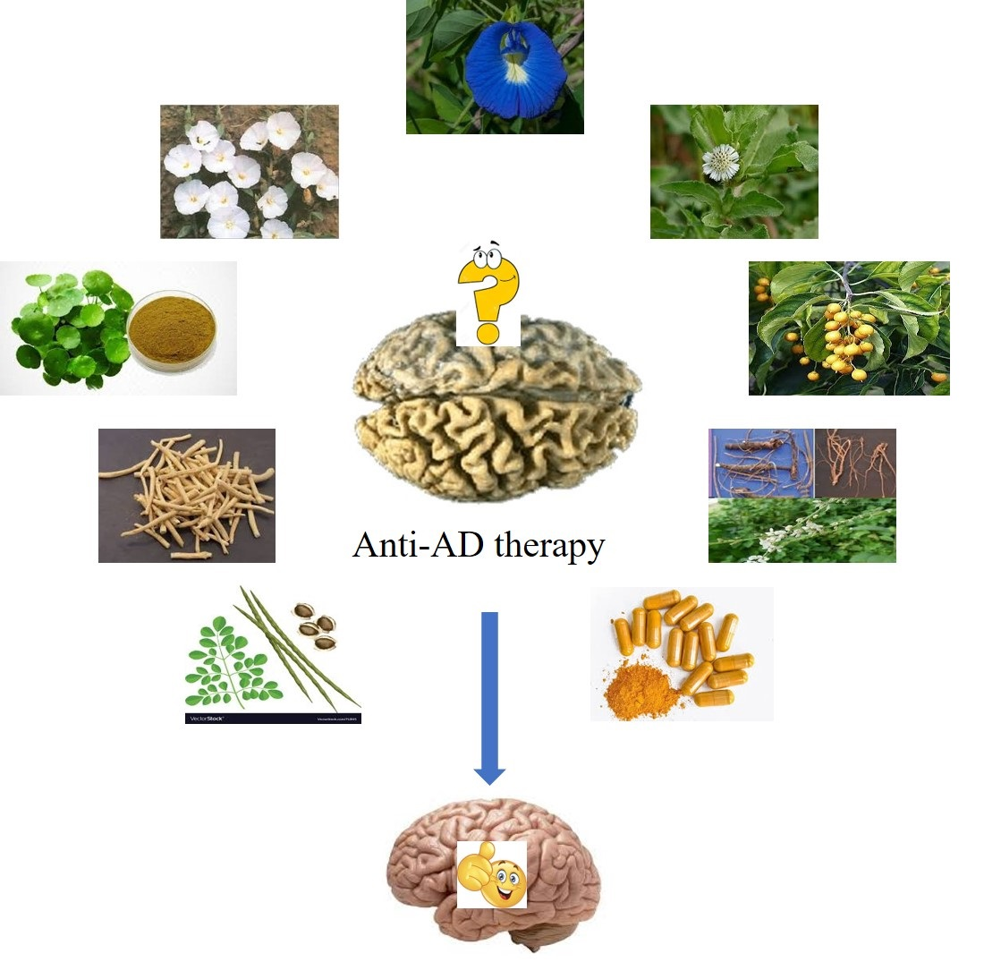
3. Herbal Medicines for Alzheimer’s Disease, Experimental and Clinical Evidence

Herbal drugs and complementary medicines have been used since ancient times for treatment of neurological disorders. Several herbal medicines worldwide have been used for neurodegenerative disorders. For example, Salvia lavandulaefolia (Spanish sage) and Salvia officinalis (common sage) are being used for improving memory in Europe since the 16th century [37]; and are also supported by clinical trials [38,39]. Bacopa monniera (water hyssop) has been used in the Indian Ayurvedic system to improve memory and intellectual functions as an immemorial custom. Centella asiatica (Asiatic pennywort), another Ayurvedic remedy, is given in combination with milk to improve memory [40]. Withania somnifera root, a rejuvenative tonic, is also used in Ayurveda to enhance memory [41,42]. Herbal medicines are becoming popular due to their perceived effectiveness, safety and affordability. Indeed, only recently, scientific studies have started providing evidence and support for the use of herbal medicines in memory related disorders.
Various CNS active Indian herbal medicines like Withania somnifera, Centella asiatica, Celastrus paniculatus and Bacopa monnieri have shown cognitive improvement in experimental models of AD when given as prophylactic treatment [43,44,45,46,47,48]. A randomized, double-blind exploratory trial reported comparable efficacy of a Gingko biloba extract and donepezil in AD patients with associated neuropsychiatric problems. The combination was reported to be superior to donepezil monotherapy in terms of both safety and efficacy [49].

3.1. Centella asiatica

Plant description: Centella asiatica (C. asiatica), a small, annual herb belonging to the family Apiceae is found throughout India and commonly known as mandukparni or jalbrahmi. It has small fan-shaped green leaves with white or light purple-to-pink or white flowers and it bears small oval fruit [50]. The leaves of mandukparni have been used as a memory enhancer in the Ayurvedic system of medicine [51]. Its use has also been described in the African system of medicine, and traditional Chinese medicine. It is used to delay ageing, prevent memory related disorders and is given with milk to enhance memory [40].
Main chemical constituents: The main chemical constituents of C. asiatica are asiaticosides, asiatic acid, madecassoside and madasiatic acid [50]. Other chemical compounds isolated from C. asiatica are brahmoside and brahminoside, isothankuniside, thankuniside and centelloside [50].
Pharmacological activities: C. asiatica is well known for its broad pharmacological activities such anti-inflammatory, antioxidative stress, antiapoptotic effects, neuroprotective effects, wound healing, antipsoriatic, antiulcer, hepatoprotective, antidepressant activity, nootropic activity, anticonvulsant, sedative, immunostimulant, cardioprotective, antidiabetic, cytotoxic and antitumor, antiviral, antibacterial, insecticidal and antifungal [50].
Preclinical studies: Aqueous extract of C. asiatica in 100, 200 and 300 mg/kg doses given orally for 14 days has been reported to dose-dependently improve cognitive functions in normal rats [52]. Pretreatment with the extract for 21 days significantly reversed streptozotocin induced cognitive impairment [51]. The authors attributed the beneficial effect of C. asiatica to antioxidant activity as evidenced by a decrease in malondialdehyde, increase in glutathione, catalase and superoxide dismutase levels. A study by Rao et al. [53] demonstrated that 15 days treatment with C. asiatica at a dose of 200 mg/kg from day 15 to 30 postpartum stimulated learning and memory in rats, which lasted for at least 6 months postpartum. They also observed an increase in dendritic arborization of hippocampal CA3 neurons, which may be one reason for improvement in brain function. Another study showed improved cognitive outcome in elderly subjects following prescribed dose of 500 mg/b.i.d dried C. asiatica for a 6-month period [54]. Dhanasekaran et al. [55] found that an 8 month treatment with 2.5 mg/kg of aqueous extract of C. asiatica significantly decreased amyloid beta 1-40 and 1-42 levels in the hippocampus of PSAPP transgenic mice expressing “Swedish” amyloid precursor protein and M146L presenilin 1 mutations, which result in spontaneous amyloid beta plaque formation. A reduction in Congo red stained fibrillar amyloid plaques was detected on the long-term treatment with 5.0 mg/kg dose.
C. asiatica aqueous leaf extract showed improvement in learning and memory in rats, and modulated dopamine, 5-hydroxytryptamine (5-HT) and noradrenaline systems in the rat brain in-vivo [56]. The leaf extract also had sedative, antidepressant and cholinomimetic activities [57] suggesting its suitability for treatment of AD associated cognitive dysfunction and depression and anxiety. The leaf extract stimulated dendrites of neuronal cells in the rat brain [51] and induced neurite elongation in human SH-SY-5Y cells and accelerated axonal regenerate in rats [58]. Cyclic AMP response element binding property (CREB) and its phosphorylated form are involved in memory formation [59]. Reduced level of phosphorylated CREB has been reported in AD patients and experimental models of AD [60]. The aqueous extract of C. asiatica leaves enhanced phosphorylation of CREB in both neuroblastoma cells, which express inducible Aß and in cortical primary cells, which were chronically exposed to external Aß in-vitro. The extract increased neuronal dendritic arborization and axonal regeneration in rats [51,58,61].
Triterpenoids are the major active component of ethanolic extract of C. asiatica, which consists of many chemical constituents such as asiatic acid, mecadessic acid, asiaticoside, scentellin, asiaticin and centellicin [62,63,64]. Asiatic acid and its derivatives have shown a promising memory improving effect [65] by improving ACh synthesis [66,67]. It has been patented (Hoechst Aktiengesellschaft) for the treatment of dementia and as a cognition enhancer. The exact constituent responsible for cognition enhancing effects of the herb remains to be established. However, studies suggest that perhaps triterpene saponins present in the leaf improve cognitive function by influencing central neurotransmitters.
Clinical evidence: In a randomized, double-blind placebo-controlled, study, C. asiatica extract was administered to healthy volunteers as 250–750 mg once daily dose for 2 months. The high dose enhanced working memory and improved self-rated mood [68].
Thus, clinical and experimental studies support memory enhancing potential of C. asiatica. However, its use for treatment of AD remains to be evaluated.
Toxicity: C. asiatica extract and asiaticoside were found to be well tolerated in experimental studies. Asiaticoside did not cause any toxicity up to 1 g/kg oral dose [69]. In acute toxicity study, C. asiatica extract up to 10 g/kg did not shown any sign of toxicity whereas in the subacute toxicity study, no toxicity was observed when the extract was administered at the doses of 10–1000 mg/kg. In the chronic toxicity study, doses up to 1200 mg/kg/day for six months did not result in significant toxicity in Wistar rats [70]. However, in one study, oral administration of 1000 mg/kg/day dried C. asiatica for 30 days caused hepatotoxicity in albino rats [71].

3.2. Bacopa monnieri

Plant description: Bacopa monnieri (B. monniera), belonging to the Scrophulariaceae family is a small, perennial creeping herb with numerous branches, small oblong leaves and light purple or white flowers. In India, it is commonly called Brahmi and is known for its revitalizing, Medhya rasayana and nootropic activities as it strengthens memory and intellect (Medhya). Bacopa has been used for the treatment of various ailments for thousands of years by the practitioners of the traditional system of medicine of India [72].
Main chemical constituents: The main chemical compounds of B. monniera are triterpenoid saponins known as bacosides. The alkaloids brahmine, nicotine and herpestine have also been reported in this plant. Novel saponins called bacopasides I–XII have also been identified [72].
Pharmacological activities: This medicinal herb possesses various biological activities such as anticonvulsant, antidepressant, anxiolytic, analgesic, anti-inflammatory, antioxidant, antimicrobial, antiulcerogenic, anti-Helicobacter pylori, adaptogenic, antineoplastic, bronchodilatory, hepatoprotective and immunostimulatory [72].
Preclinical studies: The extract of B. monniera has been reported to contain several beneficial bioactive components such as alkaloids, flavonoids, glycoside, triterpenoids saponins and alcohols. The alcoholic extract of B. monniera improved acquisition, consolidation and retention of memory in the foot shock motivated brightness discrimination test, active conditioned avoidance test and Sidman continuous avoidance responses in rats [73,74]. Bacosides A and B (a mixture of 2 saponins) may be responsible for its facilitatory effect on learning and memory. Besides, bacosides has been proven for its antioxidant and anti-inflammatory effects [75] bacosides also attenuated the retrograde amnesia produced by immobilization induced stress, electroconvulsive shock and scopolamine [76]. They enhanced protein kinase activity and increased the protein content in the hippocampus, which may also contribute to their memory enhancing effect [74,77,78]. Administration of bacosides (200 mg/kg) for 3 months in middle-aged and aged rats exerted a protective effect against age associated alterations in the neurotransmission system, behavioral paradigms, hippocampal neuronal loss and oxidative stress markers [79]. The involvement of the microRNA 124-CREB pathway and serotonergic receptor in the memory enhancing mechanism of standardized extract of B. monniera (BESEB CDRI-08) has also been reported [80,81].
The effect of alcoholic extract of Bacopa has been evaluated at the dose of 20, 40 and 80 mg/kg on cognitive functions and neurodegeneration in the animal model of AD induced by bilateral intracerebroventricular administration of AF64A. They found that Bacopa improved the escape latency in the Morris water maze test and prevented the reduction in cholinergic neuron density [47,82]. Besides, oral administration of 40 mg/kg/day of the Bacopa extract for 5 weeks prevented neurotoxicity in rats exposed to aluminum chloride [83]. Cognitive deficit induced by intracerebroventricular (ICV) injection of cholchicine and ibotenic acid into the nucleus basalis magnocellularis was attenuated by standardized Bacopa extract by reversing the depletion of ACh level, reduction in choline acetyl transferase (ChAT) activity and decrease in muscarinic cholinergic receptor binding in frontal cortex and hippocampus [84]. Holcomb et al. [43] reported that administration of ethanolic extract of Bacopa leaves at doses of 40 and 160 mg/kg for 2 and 8 months reduced Aβ 1–40 and 1–42 levels in the cortex of PSAPP mice. Bacopa, at the dose of 50 mg/kg, demonstrated the neuroprotective effect in the colchicine model of dementia through its antioxidant effect and restored the activity of Na+K+ATPase and AChE [85]. The neuronal dendritic growth stimulating property of Bacopa has also been reported which may be responsible for its memory enhancing property [86].
Clinical evidence: In a double-blind, placebo-controlled trial in 38 healthy volunteers (ages 18–60 years), single dose of 300 mg B. monniera extract (containing 55% combined bacosides A and B) did not cause any significant change in cognitive function at 2 h [87]. However, six week Bacopa administration (300 mg for subjects under 90 kg, and 450 mg for subjects over 90 kg, equivalent to 6 g and 9 g dried rhizome, respectively) in a double-blind, randomized, placebo controlled fashion was associated with significant improvement in retention of new information in 40–65 year old healthy adults. Though there was no difference in the rate of acquisition of information [88].
Stough et al. [89] reported significant improvement in verbal learning, memory consolidation and speed of early information processing following Bacopa administration (containing 55% combined bacosides) for 12 weeks at a dose of 300 mg daily in a double-blind placebo-controlled study in healthy volunteers (age 18–60 years, n = 46). Since the effects were not observed until five weeks of treatment, the slow onset of action may be attributed to Bacopa’s antioxidant properties and/or its effect on the cholinergic system. In another randomized, double-blind, placebo-controlled trial in 54 elderly participants without clinical signs of dementia (mean age 73.5 years), similar Bacopa treatment enhanced an auditory verbal learning test, delayed word recall memory scores and a stroop test relative to the placebo [14]. In subjects above 55 years of age with memory impairment, standardized Bacopa extract 125 mg was given twice daily for 12 weeks in a double blind, placebo-controlled manner. There was a significant improvement in mental control, logical memory and paired associated learning [90]. Furthermore, Bacopa extract at the dose of 300 mg/kg, daily for 12 weeks improved memory acquisition and retention in healthy older Australians population [91].
In children (age 6–8 years), Bacopa syrup (350 mg Bacopa powder), when administered three times a day for three months, resulted in significant improvement as compared to the placebo [92]. However, this study was not blinded. Negi et al. [93] carried out a double-blind, randomized, placebo-controlled trial in 36 children diagnosed with attention deficit/hyperactivity disorder (mean age 8.3–9.3 years). Nineteen children received Bacopa extract (standardized to contain 20% bacosides) at a dosage of 50 mg twice daily for 12 weeks. As compared to placebo, a significant improvement in cognitive function was observed in Bacopa-treated children at 12 weeks as evidenced by improvement in sentence repetition, logical memory and paired associate learning tasks, which was maintained at 16 weeks (after four weeks of placebo administration).
Toxicity: The LD50 of orally administered Bacopa extracts in rats was 5 g/kg for aqueous extract and 17 g/kg of the alcoholic extract [77]. The intraperitoneal LD50 was 1000 mg/kg for aqueous extract and 15 g/kg for alcoholic extract [94]. A double-blind, placebo-controlled trial in healthy male volunteers reported safety and tolerability of bacosides in single (20–30 mg) and multiple (100–200 mg) daily doses over a four-week period [77]. A randomized, double-blind, placebo-controlled trial reported that Bacopa treatment (300 mg/kg, daily) for 12 weeks caused increased stool frequency, abdominal cramps and nausea, which may be due to either an upregulation of ACh level or saponin-mediated gastrointestinal tract irritation, or both [91].

3.3. Curcuma longa

Plant description: Curcuma longa (C. longa) Linn is a perennial herb belonging to the family Zingiberaceae. It is grown for commercial use in South and Southeast Asia. Curcumin, also known as turmeric, is obtained from the rhizome of the plant, and is commonly used in India as a food flavoring and coloring agent. Several preparations of the plant have been used for centuries in the Ayurvedic system of medicine [95].
Main chemical constituents: Curcuminoids are main chemical constituents of turmeric, which include mainly curcumin (diferuloyl methane), demethoxycurcumin and bisdemethoxycurcmin. Other chemical compounds reported in this plant are alpha- and beta-tumerone, artumerone, alpha- and gamma-atlantone, curlone, zingiberene and curcumol [96].
Pharmacological activities: Previous studies reported the various pharmacological properties of curcuminoids such as neuroprotective, analgesic, antiproliferative, anti-inflammatory, anticancer, antidiabetic, hypocholesterolemic, antithrombotic, antihepatotoxic, antidiarrheal, carminative, diuretic, antirheumatic, hypotensive, antimicrobial, antiviral, antioxidant, larvicidal, insecticidal, antivenomous and antityrosinase effects [97].
Preclinical studies: It is also one of the most systematically studied plants for various diseases [98]. It has been reported in various experimental studies to possess wide variety of biological and pharmacological activities including antioxidant, anti-inflammatory and cholesterol-lowering properties, all three of which are key processes involved in the pathogenesis of AD.
Water insolubility is a major limitation for curcumin, which has been overcome, to some extent, by synthesis of biodegradable poly (lactic-co-glycolic acid) (PLGA) coated curcumin nanoparticles. These nanoparticles were found to be able to destroy amyloid aggregation and exhibit antioxidative activity without a cytotoxic effect [99,100]. Nanoliposomes of curcumin have high affinity for Aβ1-42 fibrils and were found to inhibit the formation of fibrillar and oligomeric Aβ in-vitro [101,102]. Apolipoprotein E3 mediated poly(butyl) cyanoacrylate nanoparticles containing curcumin (ApoE3-C-PBCA) provided photostability, enhanced the cellular uptake of curcumin and increased its efficacy against Aβ induced cytotoxicity [103]. Curcumin also demonstrated a protective effect against Aβ neurotoxicity by decreasing Aβ production through downregulation of presenilin 1 (PS1) and GSK-3-β expression and accelerating Aβ fibril conversion [104,105].
Curcumin has been shown to reduce both in-vivo and in-vitro Aβ plaque deposition [106,107]. Curcumin treatment for six months significantly decreased the elevated levels of oxidized protein and proinflammatory interleukin-1β in the transgenic APPSw mouse brain (Tg2576) [106]. Plaque formation and the concentration of insoluble and soluble Aβ were also lowered by curcumin in the same study. Pretreatment with curcumin (10, 20 and 50 mg/kg, p.o for 21 days) ameliorated memory impairment in the sporadic AD model in mice [108]. Furthermore, curcumin in diet form improved the spatial memory, oxidative stress and synaptophysin loss via reducing Aβ deposits [109]. Significant cognitive improvement was documented at low (160 ppm) and high (1000 ppm) doses of curcumin after administration for the 6-month period in the double transgenic AD model (APP/PS1) [110]. In-vivo, curcumin may protect cells from beta amyloid attack and subsequent oxidative stress-induced damage [111]. Curcumin can inhibit Aβ aggregation or promote its disaggregation at low concentrations (IC50 = 0.81–1 μM). Monomeric Aβ formed fewer aggregates in the presence of curcumin, whereas increasing doses of curcumin promoted disassembly of preformed Aβ aggregates. Structurally, curcumin is similar to Congo red and can prevent oligomer formation after binding to plaques and recognize secondary structure in fibrillar and oligomeric Aβ. Low dose curcumin significantly lowered the soluble Aβ levels, insoluble amyloid and plaque burden by nearly 40% [106]. Additionally, curcumin treatment for 7 days caused reduction in plaques burden and reversed structural changes in dystrophic dendrites in APPswe/PS1dE9 mouse model of AD [112].
Impaired insulin or insulin-like growth factor-1 (IGF-1) signaling is associated with AD. It leads to hyperphosphorylation of the tau protein, mitochondrial dysfunction, oxidative stress and necrosis, and contributes to cognitive impairment [113,114,115]. Curcumin significantly improved cognitive function by improving the IGF-1 level in the intracerebroventricular (ICV)-streptozotocin (STZ) model of sporadic AD [116]. It also suppressed IL-1 and glial fibrillary acidic protein, reduced oxidative damage and plaque burden and decreased the amount of insoluble amyloid [26]. Another experimental study showed that curcumin treatment restored learning and memory functions in the STZ model of AD by reducing the oxidative stress, enhancing ChAT activity and restoring insulin receptor protein [117,118].
Curcumin suppressed the microgliosis in neuronal layers, but it failed to reduce within plaques microgliosis and even significantly increased microgliosis immediately adjacent to plaques, raising the possibility that it may stimulate microglial phagocytosis of amyloid. Other possible mechanisms for curcumin induced neuroprotective effects include inhibition of IL-1-induced increase in alpha-1-antichymotrypsin (α1ACT) and NFκB-mediated transcription of apolipoprotein E (ApoE). Both α1ACT [119,120] and ApoE [121,122,123,124] have been shown to be proamyloidogenic in APP transgenic mice. Curcumin can also reduce two other proamyloidogenic factors, oxidative damage [125,126] and raised cholesterol levels [127]. The neuroprotective effect of curcuminoid mixture and its individual components on inflammatory and apoptotic gene expression in AD using an Aβ plus ibotenic acid-infused rat model has also been reported [128]. Additionally, Ahmed and colleagues also reported that a curcuminoids mixture (bisdemethoxycurcumin, demethoxycurcumin and curcumin) treatment improved memory function in amyloid fragment induced AD-like conditions in rats [129]. Nonetheless, chronic treatment with curcumin also prevented the colchicine induced cognitive impairment in rats by reducing the oxidative stress [130].
Chronic stress induces impairment of spatial cognition, neuroendocrine and plasticity abnormalities due to an increase in serum corticosterone levels. Curcumin exerts its neuroprotective effect by normalizing the corticosterone response, resulting in downregulating of calcium/calmodulin kinase II and glutamate receptor (NMDA-2B) levels [131]. The protective effect of curcumin on a Aβ1–40 AD model was documented by Wang et al. [132] and Yin et al. [133], where treatment with 300 mg/kg curcumin reversed spatial learning and memory impairment accompanied by hippocampal regeneration. Evidence also suggests that metals are concentrated in the AD brain and curcumin chelates iron and copper (but not zinc) bound to beta amyloid potentially contributing to amyloid reduction [134]. A different approach was followed by McClure et al. [135], where aerosol-mediated treatment of young 5XFAD mice with curcumin averted Aβ buildup and memory deficits in adulthood as compared to the untreated mice.
Thus, this multitarget compound is a promising therapeutic agent for AD and associated cognitive decline. However, despite intensive curcumin related research in various diseases, there is a lack of clinical data on the efficacy of curcumin in AD.
Toxicity: In a phase I trial with 25 healthy subjects, curcumin up to 8000 mg/day for 3 months did not show any toxicity [136]. In an acute toxicity study, ethanolic extract of rhizome of C. longa at the doses of 0.5, 1.0 and 3.0 mg/kg did not cause any sign of toxicity in mice. Moreover, no toxicity was found at 100 mg/kg/day in the 90-day toxicity study in mice [137].

3.4. Clitoria ternatea

Plant description: Clitoria ternatea (C. ternatea) is a perennial tropical climber herb with slender downy stem, found throughout the tropical regions of India, growing wild and in gardens, bearing white or blue flowers. C. ternatea belongs to family Fabaceae commonly called “butterfly”. It is a commonly used Ayurvedic medicine. C. ternatea is called Aparajit (Hindi), Aparajita (Bengali) and Kakkattan in Indian traditional medicine [138]. The extracts of C. ternatea have been used in Ayurveda, as an ingredient in “Medhya rasayana”.
Main chemical constituents: Various phytocomponents such as taraxerol, teraxerone, ternatins, delphinidin-3, delphinidin-3ß-glucoside, malvidin-3ß-glucoside, 3 monoglucoside, 3-rutinoside, 3-neohisperidoside, 3-o-rhamnosyl Glycoside, kaempferol-3-o-rhamnosyl, aparajitin, beta-sitosterol, malvidin-3ß-glucoside, kaemphferol, p-coumaric acid, etc., are isolated from C. ternatea [138].
Pharmacological activities: In previous studies, various biological activities including nootropic, anticonvulsant, antidepressant, antianxiety, antistress, antioxidant, anti-inflammatory, antihyperlipidemic, antidiabetic, antiasthmatic, analgesic, immunomodulatory, cytotoxicity, platelet aggregation inhibitory, antimicrobial, gastroprotective and hepatoprotective of C ternatea have been documented [138].
Preclinical studies: The nootropic activity of methanolic extract of aerial parts of C. ternatea (100 mg/kg, p.o) has been reported by using elevated plus maze and the object recognition test in rats [139]. Taranalli and Cheeramkuzhy evaluated the ethanolic extracts of roots and aerial parts of C. ternatea at the dose of 300 and 500 mg/kg, p.o in amnesia induced by submaximal electroshock [140]. They also estimated the ACh level in the whole brain and different parts of it. The aerial parts extract resulted in improved memory retention and increased brain ACh content, which was more at 300 mg/kg as compared to the 500 mg/kg dose. The root extract exhibited similar but more marked effects, which were almost equal at both doses.
Rai et al. [141] described the learning and memory enhancing effect of the C. ternatea root extract during the growth spurt period in rats. They intubated 7-day old neonatal rats and administered 50 and 100 mg/kg of the aqueous root extract of C. ternatea for 30 days. The extract improved retention in the passive avoidance task and spatial performance in the T-maze test. The behavioral changes were reported to be long lasting as indicated by a 30 days post-treatment evaluation. A previous study also showed that the aqueous root extract (50 and 100 mg/kg, p.o for 30 days) enhanced dendritic arborization of amygdala neurons in rats [142]. This cognition enhancing effect was hypothesized to be due to the presence of growth factors similar to the brain derived neurotrophic factor or nerve growth factor. Increase in hippocampus acetylcholine content [139] may be one of the reasons for nootropic activity of C. ternatea root. In addition, Rai [143] reported that the C. ternatea root extract exhibited the neurogenesis-promoting sequel on the anterior subventricular zone of neural stem cells. More recently, Damodaran et al. [144] documented the neuroprotective effect of the C. ternatea root extract in reversing chronic cerebral hypoperfusion-induced neural damage and memory impairment at doses of 200 and 300 mg/kg. In another study, Mehla and colleagues showed anti-AD effects of C. ternatea in ICV-STZ induced AD-like conditions in rats [145]. These observations suggest that C. ternatea extract exerts its beneficial effect by preventing the progression of cognitive deterioration in AD. However, the potential of C. ternatea extract still needs to be systematically evaluated for human use.
Toxicity: Ethanolic extract of aerials parts and root of C. ternatea have been studied at 200–3000 mg/kg, p.o in mice. A cathartic effect of root extract was observed. Mice treated with a dose above 2000 mg/kg had ptosis and were lethargic. The extract was not lethal orally but resulted in severe CNS depression and death when used intraperitoneally at dose of 2900 mg/kg and above [140]. Taur and Patil [146] reported LD50 of ethanolic extract of C. ternatea root to be more than 1300 mg/kg.

3.5. Withania somnifera

Plant description: Withania somnifera (W. somnifera) is a small woody shrub belonging to the family Solanaceae and is widely grown in India. It is commonly called Indian ginseng or winter cherry or ashwagandha. Its flowers are greenish or yellowish in color and about one centimeter long [147,148]. Ashwagandha is mentioned in ancient Sanskrit writings from India as a “Medhya rasayan”. It is also known as Indian ginseng and is widely used in Ayurveda. It is an ingredient in many formulations prescribed as a general tonic to increase energy, improve overall health and longevity [147,148].
Main chemical constituents: The major phytoconstituents of W. somnifera are isopellertierine, anferine, withanolides, withaferins, sitoindoside VII and VIII and withanoloides. Other chemical compounds are withanine, somniferine, somnine, somniferinine, withananine, pseudo-withanine, tropine, pseudo-tropine, 3-a-gloyloxytropane, choline and cuscohygrine [149,150,151,152].
Pharmacological activities: W. somnifera exhibits a broad range of biological activities like anti-inflammatory, antioxidant, neuroprotective, antischemic, anti-Parkinson’s, antiepileptic, anxiolytic, antidepression, antiarthritic, cardioprotective, antidiabetic, anticancer, antistress, nephroprotective, heptoprotective, antihypoxic, immunomodulatory, hypolipidaemic and antimicrobial [152].
Preclinical studies: Total alkaloid extract (ashwagandholine, AG) of W. somnifera root has been studied for its effects on CNS [153]. W. somnifera attenuated the memory loss induced by STZ through the antioxidant mechanism [154]. The root preparation has been shown to have protective effects in neurodegenerative disorders by reducing stress induced degeneration in the brain hippocampus of rats [155]. The extract containing sitoindosides VII–X and withaferin A (50 mg/kg, p.o for two weeks) reversed ibotenic acid-induced cognitive deficit and reduction in cholinergic markers (e.g., ACh and ChAT) in rats [156]. Sitoindosides VII-X and withaferin differentially (40 mg/kg for 7 days) but favorably altered the AChE activity and enhanced M1- and M2-muscarinic receptor-binding in various brain regions [157]. Withaferin A and Withanolide A suggested to have a potent immunomodulatory effect in BV-2 microglial cells by triggering the Nrf2 pathway, leading to production of the neuroprotective protein, such as heme oxygenase-1 [158].
Withanoside IV, another chemical constituent of Withania, when administered orally at the dose of 10 micromol/kg prevented cognitive impairment in the experimental model of AD [44]. Sominone (1 microM) a metabolite of Withanoside IV, induced axonal and dendritic regeneration and synaptic reconstruction in cultures of rat cortical neurons damaged by the amyloid peptide, Aβ(25–35) [44]. Therefore, withanoside IV may act as a prodrug, with sominone as the active component. The enhancement of spatial memory by sominone may be attributed to neuritic outgrowth, which is mediated by the neurotrophic factor receptor, RET [159]. Methanolic root extract dose dependently enhanced in-vitro dendrite formation in human neuroblastoma cells [159]. A study carried out by Jayaprakasam et al. [160] stated that withanamides (A/C) present in W. somnifera fruits protect pheochromocytoma-(PC-12) from β-amyloid induced toxicity. In the same study, β-amyloid fibril formation was prevented, possibly due to the presence of a serotonin moiety in both withanamide compounds.
Treatment with Withania root extract (1 g/kg, p.o for 30 days) reversed the AD pathology by upregulating the low-density lipoprotein receptor-related protein, which enhanced the Aβ clearance and ameliorated the cognitive deficit in middle-aged and old APP/PS1 mice [161]. Alcoholic extract of the Withania leaf and its component withanone was neuroprotective against scopalmine induced changes in the brain [162]. An in-vitro, inhibitory effect on the fibril formation by Aβ peptide has also been reported [163]. The increase in cortical muscarinic ACh receptor capacity might partly explain the cognition-enhancing and memory-improving effects of Withania. The root extract and their chemical constituents such as glycowithanolides also possess anxiolytic, antidepressant, anti-inflammatory and antioxidant activities, which may be relevant in AD treatment [164,165]. Furthermore, withanone, a chemical constituent from root extract of W. somnifera showed improvement in cognitive functions by inhibiting amyloid processing and reducing the elevated levels of proinflammatory cytokines and oxidative stress markers [166]. W. somnifera (20 mg/mL) treatment mitigated the Aβ toxicity and mediated longevity in the AD model of Drosophila melanogaster [167].
Clinical evidence: A prospective, randomized, double-blind, placebo-controlled study reported that treatment with ashwagandha-root extract (300 mg twice daily for eight weeks) improved immediate and general memory functions and enhanced executive function, attention and information processing speed in adults with a mild cognitive impairment [168]. In a systematic review, Ng and colleagues mentioned that W. somnifera extract ameliorated cognitive impairment and improved executive functions in adults with mild cognitive impairment [169]. There is limited data available on the clinical use of Withania for cognitive impairment.
Toxicity: Different preparations and extracts of W. somnifera root did not cause any toxicity even on chronic treatment [170]. Ashwagandholine 2% suspension in propylene glycol had a LD50 of 465 mg/kg in rats and 432 mg/kg in mice [171]. Whereas intraperitoneal administration of aqueous-methanol root extract caused 50% lethality in mice at a dose of 1076 ± 78 mg/kg [172]. Equimolar combination of sitoindosides VII and VIII and withaferin-A (SG-2) when administered once intraperitoneal, the LD50 was 1564 ± 92 mg/kg [172].

3.6. Celastrus paniculatus

Plant description: Celastrus paniculatus (C. paniculatus) is a large climber of the family Celastraceae. It grows throughout India, on sub-Himalayan slopes and the hilly regions of Punjab and South India. It is commonly known as jyotismati, which comes from the Sanskrit words “jyoti teja” or fire of mind and “mati”—intelligence. Traditionally, the bark and seeds have been used as a brain tonic, to promote intellect and to improve digestion, stimulant and expectorant [173]. In Ayurveda, C. paniculatus has been used to treat many diseases like depression, leprosy, paralysis, fever and arthritis. The seed oil and fruit are commonly used for their tranquilizer, sedative and wound healing properties [174].
Main chemical constituents: C. paniculatus shows the presence of various phytoconstituents such as sesquiterpenoid polyalcohols and esters (malkanguniol, malkangunin, polyalcohol A–D and celapnin); alkaloids (paniculatine and celastrine); phenolic triterpenoids (celastrol and paniculatadiol); fatty acids (oleic, linoleic, linolenic, palmitic, stearic and lignoceric acid) and agarofuran derivatives [175].
Pharmacological activities: Various pharmacological activities such as hypolipidemic, neuroprotective, anti-infertility, antiarthritic, wound healing, anti-inflammatory, antioxidant, analgesic, antimalarial, antibacterial and fungicidal action of C. paniculatus have been reported [176].
Preclinical studies: Celastrus seed extract and oil have been evaluated in different experimental models of cognitive impairment such as scopolamine and sodium nitrite induced amnesia. The aqueous, methanolic, chloroform and petroleum ether extracts of seeds of C. paniculatus were investigated for their effect on cognitive function in rats. The aqueous extract showed significant improvement in cognitive performance at the doses of 200 and 300 mg/kg, p.o for 14 days. In another study, methanolic extract reported to have memory-enhancing activity in rats at doses of 100, 200 and 400 mg/kg [177]. The antioxidant activity of C. paniculatus may be involved in improving the cognitive function [45]. The oil of C. paniculatus seeds when given for 14 days to Wistar rats at a dose of 400 mg/kg resulted in enhanced learning and memory in radial arm maze and decreased the AChE enzyme activity in hypothalamus, frontal cortex and hippocampus [178]. Karanth et al. [179] also demonstrated a similar effect of C. paniculatus at the dose of 400 mg/kg for 3 days. In another study, rats treated with 850 mg/kg of C. paniculatus oil for 15 days had significantly improved retention in two passive avoidance tasks [56]. The seed oil treatment for 14 days at the doses of 50, 200 and 400 mg/kg, p.o reversed scopolamine induced spatial memory impairment in the Morris water maze and increased locomotor activity without affecting AChE activity in rats [180]. The aqueous seed extract improved memory performance in elevated plus maze and in sodium nitrite induced amnesia by reducing the AChE activity [181]. Furthermore, C. paniculatus seed oil treatment showed memory improvement in scopolamine induced amnesia in mice [182]. C. paniculatus has not undergone clinical trials for safety and efficacy. Animal toxicology data is also lacking to date.

3.7. Evolvulus alsinoides

Plant description: Evolvulus alsinoides L. (E. alsionoides, dwarf morning glory), belonging to the family Convolvulaceae, is a perennial herb with small woody and branched rootstock. E. alsionoides is a weed, found mainly in the swampy regions of tropical and subtropical regions of the world. It has numerous branches (greater than 30 cm) with long hairs. The leaves are small, acute, elliptical with small size and blue-colored flowers [183]. It is locally known as Shankhpushpi and is very commonly used in Ayurveda. It is a key ingredient in majority of Medhya Rasayana formulations available in the Indian market. It is traditionally used as a memory enhancer in children and elderly and for neurological disorders like epilepsy [184].
Main chemical constituents: Major chemical constituents are octadecanoic acid, n-hexadecanoic acid, piperine, squalene, ethyl oleate and cholesterol [185].
Pharmacological activities: Studies indicate that Evolvulus alsionoides (E. alsionoides) possesses in-vitro antioxidant [186], immunomodulatory [187], adaptogenic, antiamnesic [188] and antiulcer [189] activities.
Preclinical studies: Nahata et al. [190] reported learning and memory enhancing property of its ethanolic extract and ethyl acetate and aqueous fractions in rats. The ethanolic extract (100 mg/kg, p.o) also protected against scopolamine induced dementia in rats [188]. Three days oral treatment with E. alsionoides (100 mg/kg) was effective in decreasing scopolamine induced deficit in adult male Swiss mice [188]. Pretreatment with hydro-alcoholic extract at the doses of 100, 300 and 500 mg/kg, p.o ameliorated the ICV-STZ induced cognitive impairment by decreasing the oxidative stress and rho kinase (ROCK II) expression in the rat brain [15,145]. In-vitro, aqueous and hydroalcoholic extracts of E. alsinoides showed free radicals scavenging, anti-inflammatory and enzymes (cholinesterase, glycogen synthase kinase-3-β, Rho kinase (ROCKI I), prolyl endopeptidase, catechol-o-methyl transferase and monoglycerol lipase) inhibitory activity, all of which are involved in the pathophysiology of AD [15]. Previous studies also indicated the memory enhancing effect of E. alsionoides in the experimental model of amnesia [191,192]. The methanol and water extract of E. alsinoides documented to exhibit acetylcholinesterase activity, supporting its potential in reverting neuronal dysfunctions and thus in management of AD [193]. E. alsionoides has not been studied systematically for clinical efficacy and toxicological effects.

3.8. Desmodium gangeticum

Plant description: Desmodium gangeticum (D. gangeticum), belonging to the family Fabaceae, commonly known as Salpani in Hindi and is found in abundance throughout India. It is a perennial undershrub, 60–130 cm high with somewhat angular branches. Its leaves are simple, ovateoblong or rounded with purplish or white flowers, 4–7 cm [194]. It has been used in the traditional system of medicine as a bitter tonic, febrifuge, antiemetic, digestive and in various inflammatory conditions due to vata disorder [195]. In Satpuda hills of India, powdered root of D. gangeticum is applied along with honey to treat a mouth ulcer. In Uttat Pradesh state of India, the leaf paste of D. gangeticum and aloe vera are applied to prevent hair fall [196].
Main chemical constituents: D. gangeticum shows the presence of alkaloids (tryptamines and phenylethylamines), pterocarpanoids (gangetin and desmodin), phospholipids, sterols, flavone and glycosides [197].
Pharmacological activities: It shows various pharmacological activities including antileishmanial, immunomodulatory, antioxidant, anti-inflammatory, antinociceptive, cardioprotective, antiulcer, antiamnesic and hepatoprotective [194].
Preclinical studies: Aqueous extract of D. gangeticum when administered orally at the dose of 50, 100 and 200 mg/kg for 7 days improved memory in mice [198,199]. Scopolamine and ageing induced amnesias were also prevented in rats by pretreatment with the aqueous extract of D. gangeticum [198]. Moreover, treatment of mice with the chloroform extract (400 mg/kg) and alkaloidal fraction (50 mg/kg) of D. gangeticum for 6 days alleviated the scopolamine-induced amnesia [200]. Antioxidant, anti-inflammatory and AChE inhibitory activity of D. gangeticum has also been reported [199,201,202]. These pharmacological properties indicate the potential of D. gangeticum in the management of AD related cognitive impairment. Yet, not much clinical evidence is available to this effect. Toxicity studies are also required to establish the safety of this potentially useful herb.

3.9. Eclipta Species

Plant description: Eclipta alba (L.) Hassk (E. alba) is an annual erect or prostrate herb, belonging to the Asteraceae family. There are four major varieties of Eclipta based on the colors of flower like red, yellow, white and blue. The flowers of E. alba are white in color and largely harvested due to its therapeutic activity [203]. Its stem is reddish-purple in color with up-turned hairs and roots are greyish with cylindrical shape [204]. Eclipta alba (E. alba), commonly known as Bringharaj, is well known in the traditional system of medicine for its beneficial effects on learning and memory [205]. Another species of Eclipta, commonly known as false daisy, is E. prostrate. It has also been traditionally used for treatment of memory related disorders, hepatic disorders and atherosclerosis [206].
Main chemical constituents: The major chemical constituents present in E. alba are coumestans, flavonoids, sterols, alkaloids, triterpenoid saponins and volatile oil.
Pharmacological activities: It has good antimicrobial properties like antibacterial, antifungal and antimalarial. It also shows antidiabetic, hepatoprotective, hypolipidemic, anticancer, hair growth promoting and memory enhancement and immunomodulatory properties [207].
Preclinical studies: The ethanolic extract of E. alba resulted in improvement in learning and memory abilities in passive avoidance and the elevated plus maze test in rats after both acute and chronic administration [207]. Saponins, the main chemical constituent of butanol fraction of E. prostrate, prevented ethanol induced memory impairment in rats [208]. Kim et al. [209] also reported that butanol fraction increased ACh content, decreased MAO-B activity and reduced oxidative stress in the rat brain. Lipid lowering and antioxidant activities of Eclipta plants have also been reported [210]. E. alba also possesses antiviral, antinociceptive, anti-inflammatory, bronchodilator, antibacterial, antipyretic, tonic, expectorant and hepatoprotective activity [211,212]. Previous study also reported the improvement in learning and memory functions of rats [213]. Based on the animal data available, the herb needs to be evaluated clinically.
Toxicity: An aqueous extract of E. alba did not cause any toxicity at a dose of 2.0 g/kg orally and 200 mg/kg by intravenous and intraperitoneal routes. The LD50 in mice were 7.841 g/kg, 302.8 and 328.3 mg/kg for oral and intravenous and intraperitoneal routes respectively [137]. The alcoholic extract did not show any toxicity in rats and mice and the minimum lethal dose was found to be greater than 2.0 g/kg when given orally and intraperitonially in mice [214].

3.10. Moringa oleifera

Plant description: Moringa oleifera (M. oleifera) belonging to the family Moringaceae is the commonly distributed species of this family. This plant is native to India and the height of trees can reach up to 10 m. It has fragile branches and bipinnate or tripinnate leaves. It has yellowish white flowers 0.5–1 cm long and around 2 cm broad [215]. It is commonly known as a drumstick. M. oleifera has shown antimicrobial activity and traditionally been used to clarify water due to its coagulant property. Oil of M. oleifera has high stability and contains a large amount oleic acid, hence used as an edible oil, biodiesel and lubrication of machinery [216].
Main chemical constituents: The major chemical constituents in M. oleifera are vitamins (vitamin A and C), polyphenols (flavonoids, chlorogenic acid and phenolic acids), alkaloids, glucosinolates, isothiocyanates, tannins and saponins [217].
Pharmacological activities: Various pharmacological activities like nootropic, anti-inflammatory, hypocholesterolemic, hypotensive and antioxidant effects of its leaves have been reported [218,219,220,221,222]. Additionally, it has also shown hypolipidemic, antiobesity, antidiabetic, anti-inflammatory, immunomodulatory and anticancer effects. M. oleifera is a good source of vitamin, hence prevents night-blindness and delays cataract development [217].
Preclinical studies: Pretreatment with M. oleifera at an oral dose of 250 mg/kg prevented hypoxia induced memory impairment in rats by maintaining the monoamines levels in the brain [223]. The ethanolic leaf extract at a dose of 250 mg/kg, p.o for 14 days provided protection against cognitive impairment induced by ICV–colchicine. It restored colchicine induced changes in the brain norepinephrine, serotonin and dopamine levels [224]. Improvement in learning and memory has been suggested to be due to its antioxidant effect. Other studies also demonstrated the protective effect of M. oleifera against memory impairment in experimental models of dementia [225,226]. Intriguingly, M. oleifera was shown to mitigate hyperphosphorylation and Aβ pathology also in hyperhomocysteinemia-induced AD in rats [227]. The mechanism of action, composition of the herb and difference between different extracts need to be established before it can be taken to clinical trials.
Toxicity: The aqueous leaf extract was found safe in rats after oral administration of 2000 mg/kg [228]. The acute toxicity of aqueous and ethanolic extract of M. oleifera root was evaluated in mice with the LD50 of 15.9 g/kg and 17.8 g/kg, respectively [229].

3.11. Convolvulus pluricaulis

Plant description: Convolvulus pluricaulis (C. pluricaulis) Choisy is a perennial, wild, prostrate herb, which belongs to the Convulvulaceae family and is mainly found in Northern India. It has long branches of about 30 cm and blue flowers. Its leaves are elliptical in shape and located alternately with flowers and branches [230]. It is commonly known as shankhpushpi and is used as a nervine tonic in the Ayurvedic system of medicine, to improve memory and intellect [230]. It is classified as Medhya rasayana (promotes intellectual capacity) and Majjadhatu rasayana (rejuvenates the nervous tissue). The leaves of C. pluricaulis have been used for depression and other mental disturbances [231].
Main chemical constituents: The major chemical components are alkaloids (shankhpushpine and convolamine), volatile oils, favanoid-kampferol, phytosterol, amino acids, fatty acids, scopoletin and beta-sitosterol (Sethiya NK) [232].
Pharmacological activities: Various neuropharmacological actions such nootropic, antistress, antidepressant, anxyiolytic, anticonvulsant and sedative activities of this plant are well reported [233,234,235,236]. Furthermore, it also possesses antiamnesic, antiulcer, anticatatonic, antibacterial, immunomodulatory and cardiovascular activity [232].
Preclinical studies: The ethanolic extract of C. pluricaulis and its ethyl acetate and aqueous fraction at the dose of 100 and 200 mg/kg, p.o showed memory enhancing properties in Cook and Weidley’s Pole Climbing Apparatus, passive avoidance paradigms and active avoidance tests [237]. Convolvine, a chemical constituent of C. pluricaulis potentiated the effect of arecoline (memory enhancer) and improved cognitive dysfunction in AD [238,239]. Sharma et al. [240] also reported that the ethanolic extract at 100 and 200 mg/kg oral dose significantly improved memory in young and aged mice but the retention was better in young mice. C. pluricaulis also possesses antioxidant and hypolipidemic effects, which may be partially responsible for improvement in cognitive function [190,241]. C. pluricaulis administration for 3 months at the dose of 150 mg/kg prevented aluminum chloride induced neurotoxicity by decreasing AChE activity, reducing oxidative stress and preserving the activity of ChAT and Nerve Growth Factor-Tyrosine kinase A receptor (NGF-TrkA) [242]. Alcoholic extract of C. pluricaulis Choisy (leaves) showed Aβ production inhibition in-vitro [243]. Additionally, isolated bioactive coumarins from C. pluricaulis ameliorated scopolamine induced amnesia in mice [244]. Despite detailed experimental studies, the herb has not been evaluated clinically.
Toxicity: C. pluricaulis has not been studied for toxicity. C. microphyllus, another plant of the same family had the LD50 of 1250 mg/kg after oral administration of the whole plant extract [245].

4. Other Plants with Potential Memory Enhancing Activity

Several other plants may improve cognitive functions and be useful in AD. However, very limited, if any, literature is present to review the plants individually. These less explored plants include Acorus calamus (vach), Prunus amygdalus (badam), Orchis mascula (salap), Syzygium aromaticum (lavang), Mukta pishti (pearl), Tinospora cordifolia (guduchi), Picrorrhiza kurroa (kutki), Zingiber officinale (sonth), Boerhaavia diffusa (punarnava), Commiphora wightii (guggal), Piper longum (pippali), Carum copticum (ajwain), Cyperus rotundus (coco-grass), Santalum album (Indian sandalwood), Elettaria cardamomum (cardamom), Foeniculum vulgare (fennel), Rosa damascene (damask rose) and Cinnamomum cassia (cassia).

5. Methodology

Search criteria: Database searches were conducted on PUBMED, and GOOGLE SCHOLAR using keywords: dementia, herbal products/drugs/medicine, Alzheimer’s disease and complementary and alternative medicines. The searches were limited to those plant/plant products, which are mentioned in Indian Ayurvedic literature for their potential use in some form of dementia and literature available online in the English language.
Inclusion criteria: The following studies were included in the present review article: (1) preclinical studies (in-vitro and in-vivo studies); (2) clinical studies and (3) herbal medicine identified with their regional or Hindi name.
Exclusion criteria: The following criteria was used in the present review to exclude the studies: (a) herbal drugs of non-Indian origin; (b) articles available in the language other than English and (c) full text not available.

6. Indian Herbal Formulations Studied in Alzheimer’s Disease

6.1. Mentat

Compound formulations are commonly used in Ayurveda, based on the concept that such combinations provide synergistic therapeutic effect with minimal adverse effects. BR-16A (Mentat) is a polyherbal formulation used as Medhya Rasayana in Ayurveda and is used to improve memory and cognitive deficits associated with chronic illness and aging [156]. The ingredients in BR-16A are Brahmi (Bacopa monnieri), Mandookaparni (Centella asiatica), Ashwagandha (Withania somnifera), Shankapushpi (Evolvulus alsinoides), Jatamansi (Nardostachys jatamansi), Vach (Acorus calamus), Tagar (Valeriana wallachii), Badam (Prunus amygdalus), Salap (Orchis mascula), Lavang (Syzgium aromaticum), Pearl (Mukta pishti), Malkangni (Celastrus paniculatus) and Sonth (Zingiber officinale).
Mentat has been shown to augment acquisition and retention of learning in rats and prevented cognitive deficits induced by variety of insults including prenatal undernutrition, postnatal environmental impoverishment, sodium nitrite hypoxia, aluminum, increasing age and electroconvulsive shock induced antero-grade and retro-grade amnesia [246,247,248]. Administration for 20 days at a dose of 100 mg/kg/day significantly prolonged the shortened step-through latency induced by aluminum administration and also significantly improved retention of learning in aged rats [248]. Ramteke et al. [249] reported that administration of BR-16A facilitated learning and memory in rats on the Hebb Williams complex maze as compared to control. BR-16A also showed dose dependent improvement in learning and memory in scopolamine induced amnesia in rats [250]. Mentat when administered for 2 weeks reversed the cognitive deficit and cholinergic dysfunction induced by colchicine and ibotenic acid model of AD [251].
Clinical evidence: It improved memory quotient of normal subjects in different age groups [252], increased memory span and attenuated fluctuations of attention in normal adults and improved learning ability in children with behavioral problems or minimal brain damage [253].
Toxicity: In an acute toxicity study, Mentat did not show any sign of toxicity up to the dose of 1.5 g/kg. The LD50 was found to be 1.75 g/kg after intraperitoneal injection [254] and 2400 mg/kg after oral administration [250].

6.2. Trasina

Trasina is a polyherbal formulation of some Indian medicinal plants, which are classified as Medhya rasayana in Ayurveda. It consists of Withania somnifera (80 mg), Ocimum sanctum (190 mg), Eclipta alba (10 mg), Tinospora cordifolia (10 mg), Picrorrhiza kurroa (10 mg) and shilajit (20 mg). It has shown significant nootropic effect at a dose of 200 and 500 mg/kg, p.o when administered for 21 days in colchicine and ibotenic acid induced cognitive impairment. Trasina, dose dependently, improved both memory and cholinergic markers like acetylcholine concentration, choline acetyl transferase activity and muscarinic cholinergic receptor binding in the frontal cortex and hippocampus of rat brain after 14 and 21 days of treatment. Thus, its nootropic effect may be attributed to the correction of cholinergic dysfunction [255].

6.3. Memorin

Memorin consists of Mandookparni (60 mg), Shankhpushpi (60 mg), Jatamansi (30 mg), Yashtimadhu (60 mg) and Smruti sagar (60 mg). The effect of memorin was evaluated by Andrade, 1998 in an age related memory disorder and reported beneficial effects in elderly persons who experienced age related memory decline [256]. Additionally, memorin (200 mg/day/kg) was found to attenuate retrograde and anterograde amnesia in rats when tested using passive avoidance learning paradigms in the shuttle box and T-maze test [257].

6.4. Bramhi Ghrita

It is a polyherbal Ayurvedic formulation that contains Bacopa monneri (40% w/w), Evolvulus alsinoids (20% w/w), Acorus calamus (20% w/w), Saussurea lappa (20% w/w) and cow’s ghee (750 mL). Traditionally, it is used as a memory enhancer [258]. Achliya et al. [259] evaluated the learning and memory enhancing effect of this formulation at 30, 50 and 100 mg/kg oral doses. The results of this study showed that Bramhi Ghrita at the doses of 50 and 100 mg/kg, p.o decreased the transfer latency in elevated plus maze and escape latency in Morris water maze test. Additionally, it enhanced the learning and memory of rats indicating the nootropic activity [260].

6.5. Abana

Abana, another polyherbal Ayurvedic formulation, is available in tablet form consisting of Terminalia arjuna (30 mg), Withania somnifera (20 mg), Nepeta hindostana (20), Dashamoola (20 mg), Tinospora cordifolia (10 mg), Phyllanthus emblica (10 mg), Terminalia chebula (10 mg), Eclipta alba (10 mg), Glycyrrhiza glabra (10 mg), Asparagus racemosus (10 mg), Boerhaavia diffusa (10 mg), Shilajeet (20 mg), Centella asiatica (10 mg), Convolvulus pluricaulis (10 mg), Ocimum sanctum (10 mg), Nardostachys jatamansi (10 mg), Piper longum (10 mg), Carum copticum (10 mg), Zingiber officinale (10 mg), Shankh bhasma (10 mg), Makardhwaj (10 mg), Cyperus rotundus (5 mg), Acorus calamus (5 mg), Embelia ribes (5 mg), Syzygium aromaticum (5 mg), Celastrus paniculatus (5 mg), Santalum album (5 mg), Elettaria cardamomum (5 mg), Foeniculum vulgare (5 mg), Rosa damascena (5 mg), Cinnamomum cassia (5 mg), Jaharmohra (10 mg), Abhrak bhasma (5 mg), Akik pishti (5 mg), Yeshab pishti (5 mg), Yakut pishti (5 mg), Praval pishti (5 mg) and Crocus sativus (2 mg).
Abana was administered for 15 days at the doses of 50, 100 and 200 mg/kg orally to young and aged mice and retention memory was tested using elevated plus the maze and passive avoidance test. It was also tested in scopolamine and diazepam induced amnesia at same doses. Abana reduced the brain AChE activity in a dose dependent manner. The results of these studies indicate that Abana improves memory, which may be due to reduction in brain AChE activity. Acute oral administration of Abana in mice did not cause any toxicity up to the dose of 2000 mg/kg [261].

7. Herbal Drugs: Regulatory Status

The regulatory guidelines for herbal medicines differ from country to country. USFDA classifies herbal medicines into dietary supplements and botanical drugs. Safety and efficacy studies are not needed for marketing of dietary supplements, but they should be so labeled. For botanicals, description of the product and documentation of prior human experience of the product is required. The requirements may vary from non-clinical studies to clinical trials, batch effect analysis [262]. In European Union, Committee on Herbal Medicinal Products (HMPC) issues scientific opinions on herbal substances and preparations [263]. The regulatory pathways depend on prior human exposure and range from traditional use registration; well established use marketing authorization and to stand alone or mixed application.
In India, herbal medicines are governed by Drugs and Cosmetics Act 1940, and Rules 1946. The development pathway is similar to other synthetic drugs, if they have to be incorporated into the modern system of medicine as per new drugs and clinical trials rule, 2019 [264]. Licensing, composition, formulation and manufacturing of products, labeling, packing and quality is done as per Schedule T [265]. Safety and efficacy studies are undertaken in accordance with AYUSH GCP guidelines [266].

8. Issues and Challenges with Herbal Drugs

Quality control of herbal drug: Extraction technique and processing step may cause variation in the concentration of active constituents, which necessitates quality control of herbal medicines. The macroscopic and microscopic property of herbal medicines should be examined for quality control. Determination of ash value, heavy metals, pesticide residues and microbial contamination should be carried out.
Herb-drug interaction: Coadministration of herbal drugs with prescribed drugs may result in serious adverse effects. Herbal drugs contain numerous unidentified constituents, which make it difficult to assess the nature of interactions. Additionally, it is generally believed that herbal medicines are safe since they belong to a natural origin but recently many of the herbs were found to exhibit adverse drug reactions [267]. Some reports also showed that adverse events are caused due to the herb–drug interaction [267,268,269]. Heterogeneity in doses and frequency of use also obstruct precise assessment of drug interactions. CYP450 is involved in the metabolism of drugs used for management of AD [270,271]. Hence CYP450 inhibition by herbal drugs should be assessed for predicting potential herb–drug interactions. Ginkgo biloba when given with donepezil cause an increased effect in AD due to additive cholinergic activity. However, when it was given with phenytoin, it causes breakthrough seizures due to the induction of CYP2C19. Curcumin increase the oral bioavailability of celiprolol due to inhibition of intestinal CYP450 enzymes and p-glycoprotein [272]. Moreover, coadministration of curcumin and donepezil (reversible cholinesterase inhibitor) had a synergistic effect on cognition and oxidative stress [273] and good BBB permeability [274]. More experimental and clinical studies need to be performed to evaluate the herb–drug interaction. Such interactions may be prevented with disclosure of concomitant use by the patients and awareness of physicians. In the elderly population, the absorption, metabolism and elimination of drugs are already impaired. Concomitantly use of herbal medicines may worsen the impairment. Therefore, herbal drugs should be used cautiously in elderly patients.
Adulteration: Herbal drugs are many times substituted or adulterated with other inferior products with morphological resemblance of authentic herb. This type of adulteration is more common for herbs with volatile components. The adulterants may not have a therapeutic benefit or may even cause adverse effects. Therefore, quality assurance of herbal medicines should be mandatory.
Labeling of herbal medicines: Proper labeling can reduce the risk of inappropriate use and adverse effects. The label should contain the name and amount of herbal drug and active ingredients, direction for intake, its intended use, storage conditions, shelf life, adverse effects and warnings, if any.
Pharmacovigilance for herbal medicines: Modern times rely more on the systematically studied modern medicines as they must adhere to stringent national and international regulations also. On the other hand, traditional systems of medicine suffer from a lack of or inadequate regulatory guidelines. In addition, herbal medicines are widely perceived to be safe due to their natural origins. However, as reviewed above, several herbal medicines exhibit adverse effects on their own or due to adverse herb–drug interactions with concomitant medicines. Apart from these inherent risks of herbal medicines, several adverse effects associated with them may be due to improper labeling, unknown composition, a lack of standardization, inferior quality, contamination, adulteration, improper use and even quackery.
Patients with Alzheimer’s disease constitute a special subgroup due to their vulnerability and their inability to communicate adverse events, in later stages of the disease. This population needs special attention with respect to the monitoring of adverse effects and drug–drug interactions. Hence, there is a need to integrate pharmacovigilance of herbal medicines with that of the modern medicine, under the national pharmacovigilance programs.

9. Conclusions and Future Prospectus

The alternative systems of medicine have been used since ancient times and different extracts of medicinal plants and herbal formulations have demonstrated potential for use in AD. Medicinal plants provide a fertile ground for new drug discovery because of the presence of various chemical constituents and their ability to act on different biological targets. However, much work remains to be done to translate this potential into actual medicine. Standardization of plant extracts is an urgent need in herbal drug research. Phytoconstituents responsible for pharmacological activities should be isolated, identified and systematically tested. Multicenter clinical trials should be performed to validate the efficacy of these herbal medicines alone or in the form of formulations for the treatment of AD. The present article reviewed the reported efficacy of herbal medicines against AD in experimental and clinical studies.

Funding

This research received no external funding.

Conflicts of Interest

The authors declare no conflict of interest.

References

  1. Dua, J.S.; Prasad, D.N.; Tripathi, A.C.; Gupta, R. Role of traditional medicine in neuropsychopharmacology. Asian J. Pharm. Clin. Res. 2009, 2, 72–76. [Google Scholar]
  2. Liu, P.-P.; Xie, Y.; Meng, X.-Y.; Kang, J.-S. History and progress of hypotheses and clinical trials for Alzheimer’s disease. Signal Transduct. Target. 2019, 4, 29. [Google Scholar] [CrossRef]
  3. 2020 Alzheimer’s disease facts and figures. Alzheimer’s Dement. 2020, 16, 391–460. [CrossRef]
  4. Shaji, K.S.; Jotheeswaran, A.T.; Girish, N.; Bharath, S.; Dias, A.; Pattabiraman, M.; Varghese, M. (Eds.) Alzheimer’s and Related Disorders Society of India; The Dementia India Report 2010, Prevalence, Impact, Costs and Services for Dementia; ARDSI: New Delhi, India, 2010; pp. 10–55. [Google Scholar]
  5. Kumar, A.; Sidhu, J.; Goyal, A.; Goyal, A.; Tsao, J.W. Alzheimer Disease. In StatPearls [Internet]; StatPearls Publishing: Treasure Island, FL, USA, 2020. Available online: https://www.ncbi.nlm.nih.gov/books/NBK499922/ (accessed on 25 November 2020).
  6. Nussbaum, R.L.; Ellis, C.E. Alzheimer’s disease and Parkinson’s disease. N. Engl. J. Med. 2003, 348, 1356–1364. [Google Scholar] [CrossRef] [Green Version]
  7. Davies, P. Selective Loss of Central Cholinergic Neurons in Alzheimer’s Disease. Lancet 1976, 308, 1403. [Google Scholar] [CrossRef]
  8. Anand, P.; Singh, B. A review on cholinesterase inhibitors for Alzheimer’s disease. Arch. Pharmacal Res. 2013, 36, 375–399. [Google Scholar] [CrossRef]
  9. Beyreuther, K.; Masters, C.L. Amyloid precursor protein (APP) and beta A4 amyloid in the etiology of Alzheimer’s disease, precursor-product relationships in the derangement of neuronal function. Brain Pathol. 1991, 1, 241–251. [Google Scholar] [CrossRef]
  10. Hardy, J.; Allsop, D. Amyloid deposition as the central event in the aetiology of Alzheimer’s disease. Trends Pharmacol Sci. 1991, 12, 383–388. [Google Scholar] [CrossRef]
  11. Hardy, J.A.; Higgins, G. Alzheimer’s disease, the amyloid cascade hypothesis. Science 1992, 256, 184–185. [Google Scholar] [CrossRef]
  12. Selkoe, D.J. The molecular pathology of Alzheimer’s disease. Neuron 1991, 6, 487–498. [Google Scholar] [CrossRef]
  13. Selkoe, D.J.; Hardy, J. The amyloid hypothesis of Alzheimer’s disease at 25 years. EMBO Mol. Med. 2016, 8, 595–608. [Google Scholar] [CrossRef]
  14. Calabrese, C.; Gregory, W.L.; Leo, M.; Kraemer, D.; Bone, K.; Oken, B. Effects of a Standardized Bacopa monnieri Extract on Cognitive Performance, Anxiety, and Depression in the Elderly: A Randomized, Double-Blind, Placebo-Controlled Trial. J. Altern. Complement. Med. 2008, 14, 707–713. [Google Scholar] [CrossRef] [Green Version]
  15. Mehla, J.; Pahuja, M.; Dethe, S.M.; Agarwal, A.; Gupta, Y. Amelioration of intracerebroventricular streptozotocin induced cognitive impairment by Evolvulus alsinoides in rats: In vitro and in vivo evidence. Neurochem. Int. 2012, 61, 1052–1064. [Google Scholar] [CrossRef]
  16. Bruce, A.J.; Malfroy, B.; Baudry, M. beta-Amyloid toxicity in organotypic hippocampal cultures: Protection by EUK-8, a synthetic catalytic free radical scavenger. Proc. Natl. Acad. Sci. USA 1996, 93, 2312–2316. [Google Scholar] [CrossRef] [Green Version]
  17. Pappolla, M.A.; Sos, M.; Omar, R.A.; Bick, R.J.; Hickson-Bick, D.L.M.; Reiter, R.J.; Efthimiopoulos, S.; Robakis, N.K. Melatonin Prevents Death of Neuroblastoma Cells Exposed to the Alzheimer Amyloid Peptide. J. Neurosci. 1997, 17, 1683–1690. [Google Scholar] [CrossRef]
  18. Zhang, Z.; Zhao, R.; Tang, Y.; Wen, S.; Wang, D.; Qi, J. Fuzhisan, a Chinese Herbal Medicine, Inhibits Beta-Amyloid-Induced Neurotoxicity and Tau Phosphorylation Through Calpain/Cdk5 Pathway in Cultured Cortical Neurons. Neurochem. Res. 2011, 36, 801–811. [Google Scholar] [CrossRef]
  19. Peterson, D.W.; George, R.C.; Scaramozzino, F.; LaPointe, N.E.; Anderson, R.A.; Donald, J.G.; John, L. Cinnamon extract inhibits tau aggregation associated with Alzheimer’s disease in vitro. J. Alzheimers Dis. 2009, 17, 585–597. [Google Scholar] [CrossRef] [Green Version]
  20. Dou, K.-X.; Tan, M.-S.; Tan, C.-C.; Cao, X.-P.; Hou, X.-H.; Guo, Q.-H.; Tan, L.; Mok, V.; Yu, J.-T. Comparative safety and effectiveness of cholinesterase inhibitors and memantine for Alzheimer’s disease: A network meta-analysis of 41 randomized controlled trials. Alzheimer’s Res. 2018, 10, 126. [Google Scholar] [CrossRef] [Green Version]
  21. Kobayashi, H.; Ohnishi, T.; Nakagawa, R.; Yoshizawa, K. The comparative efficacy and safety of cholinesterase inhibitors in patients with mild-to-moderate Alzheimer’s disease: A Bayesian network meta-analysis. Int. J. Geriatr. Psychiatry 2016, 31, 892–904. [Google Scholar] [CrossRef] [Green Version]
  22. Inglis, F. The tolerability and safety of cholinesterase inhibitors in the treatment of dementia. Int. J. Clin. Pract. Suppl. 2002, 127, 45–63. [Google Scholar]
  23. Quinn, J.; Kaye, J.; Montine, T.; Stackman, R. Phytochemicals in Alzheimer disease, the development of clinical trials. Pharm. Biol. 2004, 42, 64–73. [Google Scholar] [CrossRef] [Green Version]
  24. Zhou, X.; Li, Y.; Shi, X.; Ma, C. An overview on therapeutics attenuating amyloid β level in Alzheimer’s disease: Targeting neurotransmission, inflammation, oxidative stress and enhanced cholesterol levels. Am. J. Transl. Res. 2016, 8, 246–269. [Google Scholar]
  25. Bagi, Z.; Csekő, C.; Toth, E.; Koller, A. Oxidative stress-induced dysregulation of arteriolar wall shear stress and blood pressure in hyperhomocysteinemia is prevented by chronic vitamin C treatment. Am. J. Physiol. Circ. Physiol. 2003, 285, H2277–H2283. [Google Scholar] [CrossRef] [Green Version]
  26. Cole, G.M.; Morihara, T.; Lim, G.P.; Yang, F.; Begum, A.; Frautschy, S.A. NSAID and antioxidant prevention of Alzheimer’s disease, lessons from in vitro and animal models. Ann. N. Y. Acad. Sci. 2004, 1035, 68–84. [Google Scholar] [CrossRef]
  27. Engelhart, M.J.; Geerlings, M.I.; Ruitenberg, A.; van Swieten, J.C.; Hofman, A.; Witteman, J.C.M.; Breteler, M.M.B. Dietary intake of antioxidants and risk of Alzheimer disease. JAMA 2002, 287, 3223–3229. [Google Scholar] [CrossRef] [Green Version]
  28. Esposito, E.; Rotilio, D.; Di Matteo, V.; Di Giulio, C.; Cacchio, M.; Algeri, S. A review of specific dietary antioxidants and the effects on biochemical mechanisms related to neurodegenerative processes. Neurobiol. Aging 2002, 23, 719–735. [Google Scholar] [CrossRef]
  29. Moore, A.H.; O’Banion, M.K. Neuroinflammation and anti-inflammatory therapy for Alzheimer’s disease. Adv. Drug Deliv. Rev. 2002, 54, 1627–1656. [Google Scholar] [CrossRef]
  30. Pavlik, V.N.; Doody, R.S.; Rountree, S.D.; Darby, E.J. Vitamin E use is associated with improved survival in an Alzheimer’s disease cohort. Dement. Geriatr. Cogn. Disord. 2009, 28, 536–540. [Google Scholar] [CrossRef] [Green Version]
  31. Yoon, B.-K.; Kim, D.K.; Kang, Y.; Kim, J.-W.; Shin, M.-H.; Na, D.L. Hormone replacement therapy in postmenopausal women with Alzheimer’s disease: A randomized, prospective study. Fertil. Steril. 2003, 79, 274–280. [Google Scholar] [CrossRef]
  32. Cummings, J.L. Alzheimer’s disease. N. Engl. J. Med. 2004, 351, 56–67. [Google Scholar] [CrossRef]
  33. Folch, J.; Busquets, O.; Ettcheto, M.; Sánchez-López, E.; Castro-Torres, R.D.; Verdaguer, E.; Garcia, M.L.; Olloquequi, J.; Casadesús, G.; Beas-Zarate, C.; et al. Memantine for the Treatment of Dementia: A Review on its Current and Future Applications. J. Alzheimers Dis. 2018, 62, 1223–1240. [Google Scholar] [CrossRef] [Green Version]
  34. Bullock, R. Efficacy and Safety of Memantine in Moderate-to-Severe Alzheimer Disease: The Evidence to Date. Alzheimer Dis. Assoc. Disord. 2006, 20, 23–29. [Google Scholar] [CrossRef]
  35. Grossberg, G.T.; Thomas, S.J. Memantine: A review of studies into its safety and efficacy in treating Alzheimer’s disease and other dementias. Clin. Interv. Aging 2009, 4, 367–377. [Google Scholar] [CrossRef] [Green Version]
  36. Youdim, K.A.; Josepha, J. A possible emerging role of phytochemicals in improving age-related neurological dysfunctions: A multiplicity of effects. Free Radic. Biol. Med. 2001, 30, 583–594. [Google Scholar] [CrossRef]
  37. Perry, N.; Court, G.; Bidet, N.; Court, J.; Perry, E. European Herbs with Cholinergic Activities: Potential in Dementia Therapy. Int. J. Geriatr. Psychiatry 1996, 11, 1063–1069. [Google Scholar] [CrossRef]
  38. Perry, N.S.; Bollen, C.; Perry, E.K.; Ballard, C. Salvia for dementia therapy: Review of pharmacological activity and pilot tolerability clinical trial. Pharm. Biochem. Behav. 2003, 75, 651–659. [Google Scholar] [CrossRef]
  39. Akhondzadeh, S.; Noroozian, M.; Mohammadi, M.; Ohadinia, S.; Jamshidi, A.H.; Khani, M. Salvia officinalis extract in the treatment of patients with mild to moderate Alzheimer’s disease, a double blind, randomized and placebo-controlled trial. J. Clin. Pharm. Ther. 2003, 28, 53–59. [Google Scholar] [CrossRef]
  40. Manyam, B.V. Dementia in Ayurveda. J. Altern. Complement. Med. 1999, 5, 81–88. [Google Scholar] [CrossRef]
  41. Kuboyama, T.; Tohda, C.; Komatsu, K. Neuritic regeneration and synaptic reconstruction induced by withanolide A. Br. J. Pharm. 2005, 144, 961–971. [Google Scholar] [CrossRef] [Green Version]
  42. Abourjaily, P. American Herbal Pharmacopoeia and Therapeutic Compendium (A botanical supplement monograph series). Nutr. Clin. Care 2001, 4, 221–222. [Google Scholar] [CrossRef]
  43. Holcomb, L.A.; Dhanasekaran, M.; Hitt, A.R.; Young, K.A.; Riggs, M.; Manyam, B.V. Bacopa monniera extract reduces amyloid levels in PSAPP mice. J. Alzheimer’s Dis. 2006, 9, 243–251. [Google Scholar] [CrossRef]
  44. Kuboyama, T.; Tohda, C.; Komatsu, K. Withanoside IV and its active metabolite, sominone, attenuate Ab (25–35)-induced neurodegeneration. Eur. J. Neurosci. 2006, 23, 1417–1426. [Google Scholar] [CrossRef]
  45. Kumar, M.; Gupta, Y. Antioxidant property of Celastrus paniculatus Willd: A possible mechanism in enhancing cognition. Phytomedicine 2002, 9, 302–311. [Google Scholar] [CrossRef] [Green Version]
  46. Shinomol, G.K.; Bharath, M.M. Exploring the Role of “Brahmi” (Bacopa monnieri and Centella asiatica) in Brain Function and Therapy. Recent Pat. Endocr. Metab. Immune Drug Discov. 2011, 5, 33–49. [Google Scholar] [CrossRef] [Green Version]
  47. Uabundit, N.; Wattanathorn, J.; Mucimapura, S.; Ingkaninan, K. Cognitive enhancement and neuroprotective effects of Bacopa monnieri in Alzheimer’s disease model. J. Ethnopharmacol. 2010, 127, 26–31. [Google Scholar] [CrossRef]
  48. Kumar, M.H.V.; Gupta, Y. Effect of Centella asiatica on cognition and oxidative stress in an intracerebroventricular streptozotocin model of Alzheimer’s disease in rats. Clin. Exp. Pharm. Physiol. 2003, 30, 336–342. [Google Scholar] [CrossRef]
  49. Yancheva, S.; Ihl, R.; Nikolova, G.; Panayotov, P.; Schlaefke, S.; Hoerr, R. GINDON Study Group. Ginkgo biloba extract EGb 761®, donepezil or both combined in the treatment of Alzheimer’s disease with neuropsychiatric features: A randomised, double-blind, exploratory trial. Aging Ment. Health 2009, 13, 183–190. [Google Scholar] [CrossRef]
  50. Gohil, K.J.; Patel, J.A.; Gajjar, A.K. Pharmacological review on Centella asiatica: A potential herbal cure-all. Indian J. Pharm. Sci. 2010, 72, 546–556. [Google Scholar] [CrossRef] [Green Version]
  51. Rao, K.G.M.; Rao, S.M.; Rao, S.G. Centella asiatica (L.) Leaf Extract Treatment during the Growth Spurt Period Enhances Hippocampal CA3 Neuronal Dendritic Arborization in Rats. Evid. Based Complement. Altern. Med. 2006, 3, 349–357. [Google Scholar] [CrossRef] [Green Version]
  52. Kumar, M.V.; Gupta, Y. Effect of different extracts of Centella asiatica on cognition and markers of oxidative stress in rats. J. Ethnopharmacol. 2002, 79, 253–260. [Google Scholar] [CrossRef]
  53. Rao, S.B.; Chetana, M.; Devi, P.U. Centella asiatica treatment during postnatal period enhances learning and memory in mice. Physiol. Behav. 2005, 86, 449–457. [Google Scholar] [CrossRef] [PubMed]
  54. Tiwari, S.; Singh, S.; Patwardhan, K.; Gehlot, S.; Gambhir, I.S. Effect of Centella asiatica on mild cognitive impairment (MCI) and other common age-related clinical problems. Dig. J. Nanomat. Biostruct. 2008, 3, 215–220. [Google Scholar]
  55. Dhanasekaran, M.; Holcomb, L.A.; Hitt, A.R.; Tharakan, B.; Porter, J.W.; Young, K.A.; Manyam, B.V. Centella asiatica extract selectively decreases amyloid beta levels in hippocampus of Alzheimer’s disease animal model. Phytother. Res. 2009, 23, 14–19. [Google Scholar] [CrossRef] [PubMed]
  56. Nalini, K.; Karanth, K.S.; Rao, A.; Aroor, A.R. Effects of Celastrus paniculatus on passive avoidance performance and biogenic amine turnover in albino rats. J. Ethnopharmacol. 1995, 47, 101–108. [Google Scholar] [CrossRef]
  57. Sakina, M.R.; Dandiya, P.C. A psycho-neuropharmacological profile of Centella asiatica extract. Fitoterapia 1990, 61, 291–296. [Google Scholar]
  58. Soumyanath, A.; Zhong, Y.P.; Gold, S.A.; Yu, X.; Koop, D.R.; Bourdette, D.; Gold, B.G. Centella asiatica accelerates nerve regeneration upon oral administration and contains multiple active fractions increasing neurite elongation in vitro. J. Pharm. Pharmacol. 2005, 57, 1221–1229. [Google Scholar] [CrossRef]
  59. Kandel, E.R. The Molecular Biology of Memory Storage: A Dialogue between Genes and Synapses. Science 2001, 294, 1030–1038. [Google Scholar] [CrossRef] [Green Version]
  60. Yamamoto-Sasaki, M.; Ozawa, H.; Saito, T.; Rösler, M.; Riederer, P. Impaired phosphorylation of cyclic AMP response element binding protein in the hippocampus of dementia of the Alzheimer type. Brain Res. 1999, 824, 300–303. [Google Scholar] [CrossRef]
  61. Xu, Y.; Cao, Z.; Khan, I.; Luo, Y. Gotu Kola (Centella Asiatica) Extract Enhances Phosphorylation of Cyclic AMP Response Element Binding Protein in Neuroblastoma Cells Expressing Amyloid Beta Peptide. J. Alzheimer’s Dis. 2008, 13, 341–349. [Google Scholar] [CrossRef]
  62. Hausen, B.M. Centella asiatica (Indian pennywort), an effective therapeutic but a weak sensitizer. Contact Dermat. 1993, 29, 175–179. [Google Scholar] [CrossRef]
  63. James, J.T.; Dubery, I.A. Pentacyclic Triterpenoids from the Medicinal Herb, Centella asiatica (L.) Urban. Molecules 2009, 14, 3922–3941. [Google Scholar] [CrossRef] [PubMed] [Green Version]
  64. Siddiqui, B.S.; Aslam, H.; Ali, S.T.; Khan, S.; Begum, S. Chemical constituents of Centella asiatica. J. Asian Nat. Prod. Res. 2007, 9, 407–414. [Google Scholar] [CrossRef] [PubMed]
  65. Lee, M.K.; Kim, S.R.; Sung, S.H.; Lim, D.; Kim, H.; Choi, H.; Park, H.K.; Je, S.; Ki, Y.C. Asiatic acid derivatives protect cultured cortical neurons from glutamate-induced excitotoxicity. Res. Commun. Mol. Pathol. Pharmacol. 2000, 108, 75–86. [Google Scholar] [PubMed]
  66. Kim, S.R.; Koo, K.A.; Lee, M.K.; Park, H.-G.; Jew, S.-S.; Cha, K.-H.; Kim, Y.C. Asiatic acid derivatives enhance cognitive performance partly by improving acetylcholine synthesis. J. Pharm. Pharm. 2004, 56, 1275–1282. [Google Scholar] [CrossRef]
  67. Orhan, I.E. Centella asiatica (L.) Urban: From Traditional Medicine to Modern Medicine with Neuroprotective Potential. Evid. Based Complement. Altern. Med. 2012, 2012, 946259. [Google Scholar] [CrossRef] [Green Version]
  68. Wattanathorn, J.; Mator, L.; Muchimapura, S.; Tongun, T.; Pasuriwong, O.; Piyawatkul, N.; Yimtae, K.; Sripanidkulchai, B.; Singkhoraard, J. Positive modulation of cognition and mood in the healthy elderly volunteer following the administration of Centella asiatica. J. Ethnopharmacol. 2008, 116, 325–332. [Google Scholar] [CrossRef] [PubMed]
  69. Karting, T. Herbs, Spices and Medicinal Plants; Cracker, L.E., Simon, J.E., Eds.; Oryx Press: Phoenix, AZ, USA, 1998; Volume 3, pp. 145–173. [Google Scholar]
  70. Chivapat, S.; Chavalittumrong, P.; Attawish, A.; Boonruad, T.; Bansiddhi, J.; Phadungpat, S.; Punyamong, S.; Mingmuang, J. Toxicity study of Centella asiatica (L) urban. J. Thai Trad Alt Med. 2004, 2, 3–17. [Google Scholar]
  71. Oruganti, M.; Kumar Roy, B.; Kumar Singh, K.; Prasad, R.; Kumar, S. Safety assessment of Centella asiatica in albino rats. Phcog. J. 2010, 2, 5–11. [Google Scholar] [CrossRef]
  72. Aguiar, S.; Borowski, T. Neuropharmacological Review of the Nootropic Herb Bacopa monnieri. Rejuvenation Res. 2013, 16, 313–326. [Google Scholar] [CrossRef] [Green Version]
  73. Chaudhari, K.S.; Tiwari, N.R.; Tiwari, R.R.; Sharma, R.S. Neurocognitive effect of nootropic drug Brahmi (Bacopa monnieri) in Alzheimer’s disease. Ann. Neurosci. 2017, 24, 111–122. [Google Scholar] [CrossRef] [Green Version]
  74. Maheshwari, K.K.; Singh, M. Effect of bacosides, alcoholic extract of Bacopa monniera Linn. (brahmi), on experimental amnesia in mice. Indian J. Exp. Boil. 2005, 43, 640–645. [Google Scholar]
  75. Singh, M.; Murthy, V.; Ramassamy, C. Modulation of Hydrogen Peroxide and Acrolein-Induced Oxidative Stress, Mitochondrial Dysfunctions and Redox Regulated Pathways by the Bacopa Monniera Extract: Potential Implication in Alzheimer’s Disease. J. Alzheimer’s Dis. 2010, 21, 229–247. [Google Scholar] [CrossRef] [PubMed]
  76. Joshi, A.; Parle, M. Brahmi rasayana Improves Learning and Memory in Mice. Evid. Based Complement. Altern. Med. 2006, 3, 79–85. [Google Scholar] [CrossRef] [PubMed] [Green Version]
  77. Singh, H.K.; Dhawan, B.N. Neuropsychopharmacological effects of the Ayurvedic nootropic Bacopa monniera Linn. (Brahmi). Indian J. Pharmacol. 1997, 29, S359–S365. [Google Scholar]
  78. Srinath, S. Memory enhancing medicinal herbs. J. Pharm. Sci. Res. 2014, 6, 331. [Google Scholar]
  79. Rastogi, M.; Ojha, R.P.; Prabu, P.C.; Devi, B.P.; Agrawal, A.; Dubey, G.P. Prevention of age-associated neurodegeneration and promotion of healthy brain ageing in female Wistar rats by long term use of bacosides. Biogerontology 2012, 13, 183–195. [Google Scholar] [CrossRef] [PubMed]
  80. Preethi, J.; Singh, H.K.; Charles, P.D.; Rajan, K.E. Participation of microRNA 124-CREB pathway: A parallel memory enhancing mechanism of standardised extract of Bacopa monniera (BESEB CDRI-08). Neurochem. Res. 2012, 37, 2167–2177. [Google Scholar] [CrossRef]
  81. Rajan, K.E.; Singh, H.K.; Parkavi, A.; Charles, P.D. Attenuation of 1-(m-Chlorophenyl)-Biguanide Induced Hippocampus-Dependent Memory Impairment by a Standardised Extract of Bacopa monniera (BESEB CDRI-08). Neurochem. Res. 2011, 36, 2136–2144. [Google Scholar] [CrossRef]
  82. Pandey, S.P.; Singh, H.K.; Prasad, S.B. Alterations in Hippocampal Oxidative Stress, Expression of AMPA Receptor GluR2 Subunit and Associated Spatial Memory Loss by Bacopa monnieri Extract (CDRI-08) in Streptozotocin-Induced Diabetes Mellitus Type 2 Mice. PLoS ONE 2015, 10, e0131862. [Google Scholar] [CrossRef]
  83. Jyoti, A.; Sethi, P.; Sharma, D. Bacopa monniera prevents from aluminium neuro- toxicity in the cerebral cortex of rat brain. J. Ethnopharmacol. 2007, 111, 56–62. [Google Scholar] [CrossRef]
  84. Bhattacharya, S.K.; Kumar, A.; Ghosal, S. Effect of Bacopa monniera on animal models of Alzheimer’s disease and perturbed central cholinergic markers of cognition in rats. In Molecular Aspects of Asian Medicines; Siva Sankar, D.V., Ed.; PJD Publications: New York, NY, USA, 2000. [Google Scholar]
  85. Saini, N.; Singh, D.; Sandhir, R. Neuroprotective Effects of Bacopa monnieri in Experimental Model of Dementia. Neurochem. Res. 2012, 37, 1928–1937. [Google Scholar] [CrossRef] [PubMed]
  86. Vollala, V.R.; Upadhya, S.; Nayak, S. Enhanced dendritic arborization of hippocampal CA3 neurons by Bacopa monniera extract treatment in adult rats. Romanian J. Morphol. Embryol. 2011, 52, 879–886. [Google Scholar]
  87. Nathan, P.J.; Clarke, J.; Lloyd, J.; Hutchison, C.W.; Downey, L.; Stough, C. The acute effects of an extract of Bacopa monniera (Brahmi) on cognitive function in healthy normal subjects. Hum. Psychopharmacol. Clin. Exp. 2001, 16, 345–351. [Google Scholar] [CrossRef] [PubMed]
  88. Roodenrys, S.; Booth, D.; Bulzomi, S.; Phipps, A.; Micallef, C.; Smoker, J. Chronic Effects of Brahmi (Bacopa monnieri) on Human Memory. Neuropsychopharmacol. 2002, 27, 279–281. [Google Scholar] [CrossRef] [Green Version]
  89. Stough, C.; Lloyd, J.; Clarke, J.; Downey, L.A.; Hutchison, C.W.; Rodgers, T.; Nathan, P.J. The chronic effects of an extract of Bacopa monniera (Brahmi) on cognitive function in healthy human subjects. Psychopharmacol 2001, 156, 481–484. [Google Scholar] [CrossRef] [Green Version]
  90. Raghav, S.; Singh, H.; Dalal, P.K.; Srivastawa, J.S.; Asthana, O.P. Randomized controlled trial of standardized Bacopa monniera extract in age associated memory impairment. Indian J. Psychiatry. 2006, 48, 238–242. [Google Scholar]
  91. Morgan, A.; Stevens, J. Does Bacopa monnieri Improve Memory Performance in Older Persons? Results of a Randomized, Placebo-Controlled, Double-Blind Trial. J. Altern. Complement. Med. 2010, 16, 753–759. [Google Scholar] [CrossRef] [Green Version]
  92. Sharma, R.; Chaturvedi, C.; Tewari, P.V. Efficacy of Bacopa monnieri in revitalizing intellectual functions in children. J. Res. Educ. Indian Med. 1987, 1, 12. [Google Scholar]
  93. Negi, K.S.; Singh, Y.D.; Kushwaha, K.P.; Rastogi, C.K.; Rathi, A.K.; Srivastava, J.S. Clinical evaluation of memory enhancing properties of Memory Plus in children with attention deficit hyperactivity disorder. Indian J. Psychiatry. 2000, 42, 42–50. [Google Scholar]
  94. Martis, G.; Rao, A.; Karanth, K.S. Neuropharmacological activity of Herpestis monniera. Fitoterapia 1992, 63, 399–404. [Google Scholar]
  95. Majeed, M.; Badmaev, V.; Murrray, F. Turmeric and the Healing Curcuminoids; Keats Publishing, Inc.: New Canaan, CT, USA, 1996. [Google Scholar]
  96. Perrone, D.; Ardito, F.; Giannatempo, G.; Dioguardi, M.; Troiano, G.; Russo, L.L.; De Lillo, A.; Laino, L.; Muzio, L.L. Biological and therapeutic activities, and anticancer properties of curcumin. Exp. Med. 2015, 10, 1615–1623. [Google Scholar] [CrossRef] [PubMed] [Green Version]
  97. Sharifi-Rad, J.; El Rayess, Y.; Rizk, A.A.; Sadaka, C.; Zgheib, R.; Zam, W.; Sestito, S.; Rapposelli, S.; Neffe-Skocińska, K.; Zielińska, D.; et al. Turmeric and Its Major Compound Curcumin on Health: Bioactive Effects and Safety Profiles for Food, Pharmaceutical, Biotechnological and Medicinal Applications. Front. Pharm. 2020, 11. [Google Scholar] [CrossRef]
  98. Ghosh, S.; Banerjee, S.; Sil, P.C. The beneficial role of curcumin on inflammation, diabetes and neurodegenerative disease: A recent update. Food Chem. Toxicol. 2015, 83, 111–124. [Google Scholar] [CrossRef] [PubMed]
  99. Mathew, A.; Fukuda, T.; Nagaoka, Y.; Hasumura, T.; Morimoto, H.; Yoshida, Y.; Maekawa, T.; Venugopal, K.; Kumar, D.S. Curcumin loaded-PLGA nanoparticles conjugated with Tet-1 peptide for potential use in Alzheimer’s disease. PLoS ONE 2012, 7, e32616. [Google Scholar] [CrossRef] [PubMed] [Green Version]
  100. Tiwari, S.K.; Agarwal, S.; Seth, B.; Yadav, A.; Nair, S.; Bhatnagar, P.; Karmakar, M.; Kumari, M.; Chauhan, L.K.S.; Patel, D.K.; et al. Correction to Curcumin-Loaded Nanoparticles Potently Induce Adult Neurogenesis and Reverse Cognitive Deficits in Alzheimer’s Disease Model via Canonical Wnt/β-Catenin Pathway. ACS Nano 2013, 8, 76–103. [Google Scholar] [CrossRef]
  101. Mourtas, S.; Canovi, M.; Zona, C.; Aurilia, D.; Niarakis, A.; La Ferla, B.; Salmona, M.; Nicotra, F.; Gobbi, M.; Antimisiaris, S.G. Curcumin-decorated nanoliposomes with very high affinity for amyloid-β1-42 peptide. Biomaterials 2011, 32, 1635–1645. [Google Scholar] [CrossRef]
  102. Taylor, M.; Moore, S.; Mourtas, S.; Niarakis, A.; Re, F.; Zona, C.; La Ferla, B.; Nicotra, F.; Masserini, M.; Antimisiaris, S.G.; et al. Effect of curcumin-associated and lipid ligand-functionalized nanoliposomes on aggregation of the Alzheimer’s Aβ peptide. Nanomedicine 2011, 7, 541–550. [Google Scholar] [CrossRef]
  103. Mulik, R.S.; Mönkkönen, J.; Juvonen, R.O.; Mahadik, K.R.; Paradkar, A. ApoE3 Mediated Poly(butyl) Cyanoacrylate Nanoparticles Containing Curcumin: Study of Enhanced Activity of Curcumin against Beta Amyloid Induced Cytotoxicity Using In Vitro Cell Culture Model. Mol. Pharm. 2010, 7, 815–825. [Google Scholar] [CrossRef]
  104. Caesar, I.; Jonson, M.; Nilsson, K.P.R.; Thor, S.; Hammarström, P. Curcumin Promotes A-beta Fibrillation and Reduces Neurotoxicity in Transgenic Drosophila. PLoS ONE 2012, 7, e31424. [Google Scholar] [CrossRef] [Green Version]
  105. Xiong, Z.; Hongmei, Z.; Lu, S.; Yu, L. Curcumin mediates presenilin-1 activity to reduce β-amyloid production in a model of Alzheimer’s disease. Pharm. Rep. 2011, 63, 1101–1108. [Google Scholar] [CrossRef]
  106. Lim, G.P.; Chu, T.; Yang, F.; Beech, W.; Frautschy, S.A.; Cole, G.M. The Curry Spice Curcumin Reduces Oxidative Damage and Amyloid Pathology in an Alzheimer Transgenic Mouse. J. Neurosci. 2001, 21, 8370–8377. [Google Scholar] [CrossRef] [PubMed]
  107. Yang, F.; Lim, G.P.; Begum, A.N.; Ubeda, O.J.; Simmons, M.R.; Ambegaokar, S.S.; Chen, P.P.; Kayed, R.; Glabe, C.G.; Frautschy, S.A.; et al. Curcumin inhibits formation of amyloid beta oligomers and fibrils, binds plaques, and reduces amyloid in vivo. J. Biol. Chem. 2005, 280, 5892–5901. [Google Scholar] [CrossRef] [PubMed] [Green Version]
  108. Awasthi, H.; Tota, S.; Hanif, K.; Nath, C.; Shukla, R. Protective effect of curcumin against intracerebral streptozotocin induced impairment in memory and cerebral blood flow. Life Sci. 2010, 86, 87–94. [Google Scholar] [CrossRef] [PubMed]
  109. Frautschy, S.A.; Hu, W.; Kim, P.; Miller, S.A.; Chu, T.; Harris-White, M.E.; Cole, G.M. Phenolic anti-inflammatory antioxidant reversal of Abeta-induced cognitive deficits and neuropathology. Neurobiol. Aging 2002, 22, 993–1005. [Google Scholar] [CrossRef]
  110. Wang, C.; Zhang, X.; Teng, Z.; Zhang, T.; Li, Y. Downregulation of PI3K/Akt/mTOR signaling pathway in curcumin-induced autophagy in APP/PS1 double transgenic mice. Eur. J. Pharm. 2014, 740, 312–320. [Google Scholar] [CrossRef]
  111. Kim, D.S.; Park, S.Y.; Kim, J.Y. Curcuminoids from Curcuma longa L. (Zingiberaceae) that protect PC12 rat pheochromocytoma and normal human umbilical vein endothelial cells from betaA(1-42) insult. Neurosci. Lett. 2001, 303, 57–61. [Google Scholar] [CrossRef]
  112. Ramassamy, C. Faculty Opinions recommendation of Curcumin labels amyloid pathology in vivo, disrupts existing plaques, and partially restores distorted neurites in an Alzheimer mouse model. Fac. Opin. Post-Publ. Peer Rev. Biomed. Lit. 2008, 102, 1095–1104. [Google Scholar] [CrossRef]
  113. De la Monte, S.M.; Wands, J.R. Review of insulin and insulin-like growth factor expression, signaling, and malfunction in the central nervous system, relevance to Alzheimer’s disease. J. Alzheimers Dis. 2005, 7, 45–61. [Google Scholar] [CrossRef] [Green Version]
  114. Schubert, M.; Brazil, D.P.; Burks, D.J.; Kushner, J.A.; Ye, J.; Flint, C.L.; Farhang-Fallah, J.; Dikkes, P.; Warot, X.M.; Rio, C.; et al. Insulin Receptor Substrate-2 Deficiency Impairs Brain Growth and Promotes Tau Phosphorylation. J. Neurosci. 2003, 23, 7084–7092. [Google Scholar] [CrossRef] [Green Version]
  115. Schubert, M.; Gautam, D.; Surjo, D.; Ueki, K.; Baudler, S.; Schubert, D.; Kondo, T.; Alber, J.; Galldiks, N.; Küstermann, E.; et al. Role for neuronal insulin resistance in neurodegenerative diseases. Proc. Natl. Acad. Sci. USA 2004, 101, 3100–3105. [Google Scholar] [CrossRef] [Green Version]
  116. Isik, A.T.; Celik, T.; Ulusoy, G.K.; Ongoru, O.; Elibol, B.; Doruk, H.; Bozoglu, E.; Kayir, H.; Mas, M.R.; Akman, S. Curcumin ameliorates impaired insulin/IGF signalling and memory deficit in a streptozotocin-treated rat model. AGE 2008, 31, 39–49. [Google Scholar] [CrossRef] [PubMed] [Green Version]
  117. Agrawal, R.; Mishra, B.; Tyagi, E.; Nath, C.; Shukla, R. Effect of curcumin on brain insulin receptors and memory functions in STZ (ICV) induced dementia model of rat. Pharm. Res. 2010, 61, 247–252. [Google Scholar] [CrossRef] [PubMed]
  118. Ishrat, T.; Hoda, M.N.; Khan, M.B.; Yousuf, S.; Ahmad, M.; Khan, M.M.; Ahmad, A.; Islam, F. Amelioration of cognitive deficits and neurodegeneration by curcumin in rat model of sporadic dementia of Alzheimer’s type (SDAT). Eur. Neuropsychopharmacol. 2009, 19, 636–647. [Google Scholar] [CrossRef] [PubMed]
  119. Aksenova, M.V.; Aksenov, M.Y.; Butterfield, D.A.; Carney, J.M. alpha-1-antichymotrypsin interaction with a beta (1-40) inhibits fibril formation but does not affect the peptide toxicity. Neurosci. Lett. 1996, 211, 45–48. [Google Scholar] [CrossRef]
  120. Shoji, M.; Hirai, S.; Yamaguchi, H.; Harigaya, Y.; Ishiguro, K.; Matsubara, E. Alpha 1-antichymotrypsin is present in diffuse senile plaques. A comparative study of beta-protein and alpha 1-antichymotrypsin immunostaining in the Alzheimer brain. Am. J. Pathol. 1991, 138, 247–257. [Google Scholar]
  121. Beffert, U.; Cohn, J.S.; Petit-Turcotte, C.; Tremblay, M.; Aumont, N.; Ramassamy, C.; Davignon, J.; Poirier, J. Apolipoprotein E and beta-amyloid levels in the hippocampus and frontal cortex of Alzheimer’s disease subjects are disease-related and apolipoprotein E genotype dependent. Brain Res. 1999, 843, 87–94. [Google Scholar] [CrossRef]
  122. Weisgraber, K.H.; Mahley, R.W. Human apolipoprotein E, the Alzheimer’s disease connection. FASEB J. 1996, 10, 1485–1494. [Google Scholar] [CrossRef]
  123. Wisniewski, T.; Castano, E.M.; Golabek, A.; Vogel, T.; Frangione, B. Acceleration of Alzheimer’s fibril formation by apolipoprotein E in vitro. Am. J. Pathol. 1994, 145, 1030–1035. [Google Scholar]
  124. Wisniewski, T.; Frangione, B. Apolipoprotein E: A pathological chaperone protein in patients with cerebral and systemic amyloid. Neurosci. Lett. 1992, 135, 235–238. [Google Scholar] [CrossRef]
  125. Friedlich, A.L.; Butcher, L.L. Involvement of free oxygen radicals in beta-amyloidosis, an hypothesis. Neurobiol. Aging 1994, 15, 443–455. [Google Scholar] [CrossRef]
  126. Hensley, K.; Carney, J.M.; Mattson, M.P.; Aksenova, M.; Harris, M.; Wu, J.F.; Floyd, R.A.; Butterfield, D.A. A model for beta-amyloid aggregation and neurotoxicity based on free radical generation by the peptide: Relevance to Alzheimer disease. Proc. Natl. Acad. Sci. USA 1994, 91, 3270–3274. [Google Scholar] [CrossRef] [PubMed] [Green Version]
  127. Soudamini, K.K.; Unnikrishnan, M.C.; Soni, K.B.; Kuttan, R. Inhibition of lipid peroxidation and cholesterol levels in mice by curcumin. Indian J. Physiol. Pharmacol. 1992, 36, 239–243. [Google Scholar] [PubMed]
  128. Ahmed, T.; Gilani, A.H. A comparative study of curcuminoids to measure their effect on inflammatory and apoptotic gene expression in an Aβ plus ibotenic acid-infused rat model of Alzheimer’s disease. Brain Res. 2011, 1400, 1–18. [Google Scholar] [CrossRef] [PubMed]
  129. Ahmed, T.; Enam, S.A.; Gilani, A.H. Curcuminoids enhance memory in an amyloid-infused rat model of Alzheimer’s disease. Neuroscience 2010, 169, 1296–1306. [Google Scholar] [CrossRef] [PubMed]
  130. Kumar, A.; Naidu, P.; Seghal, N.; Padi, S. Effect of Curcumin on Intracerebroventricular Colchicine-Induced Cognitive Impairment and Oxidative Stress in Rats. J. Med. Food 2007, 10, 486–494. [Google Scholar] [CrossRef] [PubMed]
  131. Xu, Y.; Lin, D.; Li, S.; Li, G.; Shyamala, S.G.; Barish, P.A.; Vernon, M.M.; Pan, J.; Ogle, W.O. Curcumin reverses impaired cognition and neuronal plasticity induced by chronic stress. Neuropharmacol 2009, 57, 463–471. [Google Scholar] [CrossRef] [PubMed]
  132. Wang, Y.; Yin, H.; Li, J.; Zhang, Y.; Han, B.; Zeng, Z.; Qiao, N.; Cui, X.; Lou, J.; Li, J. Amelioration of β-amyloid-induced cognitive dysfunction and hippocampal axon degeneration by curcumin is associated with suppression of CRMP-2 hyperphosphorylation. Neurosci. Lett. 2013, 557, 112–117. [Google Scholar] [CrossRef]
  133. Yin, H.L.; Wang, Y.L.; Lin, J.F.; Han, B.; Zhang, X.X.; Wang, Y.T.; Geng, S. Effects of curcumin on hippocampal expression of NgR and axonal regeneration in Abeta-induced cognitive disorder rats. Genet. Mol. Res. 2014, 13, 2039–2047. [Google Scholar] [CrossRef]
  134. Baum, L.; Alex, N.G. Curcumin interaction with copper and iron suggests one possible mechanism of action in Alzheimer’s disease animal models. J. Alzheimers Dis. 2004, 6, 367–377. [Google Scholar] [CrossRef]
  135. McClure, R.; Ong, H.; Janve, V.; Barton, S.; Zhu, M.; Li, B.; Dawes, M.; Jerome, W.G.; Anderson, A.; Massion, P.; et al. Aerosol Delivery of Curcumin Reduced Amyloid-β Deposition and Improved Cognitive Performance in a Transgenic Model of Alzheimer’s Disease. J. Alzheimer’s Dis. 2016, 55, 797–811. [Google Scholar] [CrossRef] [Green Version]
  136. Chainani-Wu, N. Safety and Anti-Inflammatory Activity of Curcumin: A Component of Tumeric (Curcuma longa). J. Altern. Complement. Med. 2003, 9, 161–168. [Google Scholar] [CrossRef] [Green Version]
  137. Qadri, N.M.; Ahmad, S.; Qureshi, S.; Badar, Y. Acute toxicological evaluation of the aqueous extract of Eclipta alba Hassk. Pak. J. Sci. Ind. Res. 2001, 44, 38–41. [Google Scholar]
  138. Mukherjee, P.K.; Kumar, V.; Kumar, N.S.; Heinrich, M. The Ayurvedic medicine Clitoria ternatea—From traditional use to scientific assessment. J. Ethnopharmacol. 2008, 120, 291–301. [Google Scholar] [CrossRef] [PubMed]
  139. Rai, K.; Murthy, K.; Karanth, K.; Nalini, K.; Rao, M.; Srinivasan, K. Clitoria ternatea root extract enhances acetylcholine content in rat hippocampus. Fitoterapia 2002, 73, 685–689. [Google Scholar] [CrossRef]
  140. Taranalli, A.D.; Cheeramkhuzhy, T.C. Influence of Clitoria ternatea extracts on memory and central cholinergic activity in rats. Pharm Biol. 2000, 38, 51–56. [Google Scholar] [CrossRef]
  141. Rai, K.S.; Murthy, K.D.; Karanth, K.S.; Rao, M.S. Clitoria ternatea (Linn) root extract treatment during growth spurt period enhances learning and memory in rats. Indian J. Physiol. Pharmacol. 2001, 45, 305–313. [Google Scholar] [PubMed]
  142. Rai, K.S.; Murthy, K.D.; Rao, M.S.; Karanth, K.S. Altered dendritic arborization of amygdale neurons in young adult rats orally intubated with Clitoria ternatea aqueous root extract. Phytother. Res. 2005, 19, 592–598. [Google Scholar] [CrossRef]
  143. Rai, K.S. Neurogenic potential of Clitoria ternatea aqueous root extract-a basis for enhancing learning and memory. World Acad. Sci. Eng. Technol. 2010, 46, 237–242. [Google Scholar]
  144. Damodaran, T.; Cheah, P.S.; Murugaiyah, V.; Hassan, Z. The nootropic and anticholinesterase activities of Clitoria ternatea Linn. root extract: Potential treatment for cognitive decline. Neurochem. Int. 2020, 139, 104785. [Google Scholar] [CrossRef]
  145. Mehla, J.; Pahuja, M.; Gupta, Y.K. Streptozotocin-induced sporadic Alzheimer’s disease: Selection of appropriate dose. J Alzheimers Dis. 2012, 33, 17–21. [Google Scholar] [CrossRef]
  146. Taur, D.J.; Patil, R.Y. Evaluation of antiasthmatic activity of Clitoria ternatea L. roots. J. Ethnopharmacol. 2011, 136, 374–376. [Google Scholar] [CrossRef] [PubMed]
  147. Bone, K. Clinical Applications of Ayurvedic and Chinese Herbs. Monographs for the Western Herbal Practitioner; Phytotherapy Press: Queensland, Australia, 1996; pp. 137–141. [Google Scholar]
  148. Chatterjee, A.; Pakrashi, S.C. The Treatise on Indian Medicinal Plants. Council for Scientific and Industrial Research; Publications & Information Directorate: New Delhi, India, 1995; Volume 4, pp. 208–212. [Google Scholar]
  149. Dar, N.J.; Hamid, A.; Ahmad, M. Pharmacologic overview of Withania somnifera, the Indian Ginseng. Cell. Mol. Life Sci. 2015, 72, 4445–4460. [Google Scholar] [CrossRef] [PubMed]
  150. Mirjalili, M.H.; Moyano, E.; Bonfill, M.; Cusido, R.M.; Palazón, J. Steroidal Lactones from Withania somnifera, an Ancient Plant for Novel Medicine. Molecules 2009, 14, 2373–2393. [Google Scholar] [CrossRef] [PubMed] [Green Version]
  151. Mishra, L.C.; Singh, B.B.; Dagenais, S. Scientific basis for the therapeutic use of Withania somnifera (ashwagandha): A review. Altern. Med. Rev. 2000, 5, 334–346. [Google Scholar]
  152. Kumar, V.; Dey, A.; Hadimani, M.B.; Marcović, T.; Emerald, M. Chemistry and pharmacology of Withania somnifera: An update. Tang (Humanit. Med.) 2015, 5, e1. [Google Scholar] [CrossRef] [Green Version]
  153. Malhotra, C.L.; Mehta, V.L.; Das, P.K.; Dhalla, N.S. Studies on Withania-ashwagandha, Kaul. V. The effect of total alkaloids (ashwagandholine) on the central nervous system. Indian J. Physiol. Pharmacol. 1965, 9, 127–136. [Google Scholar]
  154. Parihar, M.; Chaudhary, M.; Shetty, R.; Hemnani, T. Susceptibility of hippocampus and cerebral cortex to oxidative damage in streptozotocin treated mice: Prevention by extracts of Withania somnifera and Aloe vera. J. Clin. Neurosci. 2004, 11, 397–402. [Google Scholar] [CrossRef]
  155. Jain, S.; Shukla, S.D.; Sharma, K.; Bhatnagar, M. Neuroprotective effects of Withania somnifera Dunn. In hippocampal sub-regions of female albino rat. Phytother. Res. 2001, 15, 544–548. [Google Scholar] [CrossRef]
  156. Bhattacharya, S.K.; Kumar, A.; Ghosal, S. Effects of glycowithanolides from Withania somnifera on an animal model of Alzheimer’s disease and perturbed central cholinergic markers of cognition in rats. Phytother. Res. 1995, 9, 110–113. [Google Scholar] [CrossRef]
  157. Schliebs, R.; Liebmann, A.; Bhattacharya, S.K.; Kumar, A.; Ghosal, S.; Bigl, V. Systemic administration of defined extracts from Withania somnifera (Indian ginseng) and Shilajit differentially affects cholinergic but not glutamatergic and GABAergic markers in rat brain. Neurochem. Int. 1997, 30, 181–190. [Google Scholar] [CrossRef]
  158. Sun, G.Y.; Li, R.; Cui, J.; Hannink, M.; Gu, Z.; Fritsche, K.L.; Lubahn, D.B.; Simonyi, A. Withania somnifera and Its Withanolides Attenuate Oxidative and Inflammatory Responses and Up-Regulate Antioxidant Responses in BV-2 Microglial Cells. Neuromolecular. Med. 2016, 18, 241–252. [Google Scholar] [CrossRef] [PubMed]
  159. Tohda, C.; Joyashiki, E. Sominone enhances neurite outgrowth and spatial memory mediated by the neurotrophic factor receptor, RET. Br. J. Pharm. 2009, 157, 1427–1440. [Google Scholar] [CrossRef] [Green Version]
  160. Jayaprakasam, B.; Padmanabhan, K.; Nair, M.G. Withanamides in Withania somnifera fruit protect PC-12 cells from beta-amyloid responsible for Alzheimer’s disease. Phytother Res. 2010, 24, 859–863. [Google Scholar] [CrossRef] [PubMed]
  161. Sehgal, N.; Gupta, A.; Valli, R.K.; Joshi, S.D.; Mills, J.T.; Hamel, E.; Khanna, P.; Jain, S.C.; Thakur, S.S.; Ravindranath, V. Withania somnifera reverses Alzheimer’s disease pathology by enhancing low-density lipoprotein receptor-related protein in liver. Proc. Natl. Acad. Sci. USA 2012, 109, 3510–3515. [Google Scholar] [CrossRef] [PubMed] [Green Version]
  162. Konar, A.; Shah, N.; Singh, R.; Saxena, N.; Kaul, S.C.; Wadhwa, R.; Thakur, M.K. Protective Role of Ashwagandha Leaf Extract and Its Component Withanone on Scopolamine-Induced Changes in the Brain and Brain-Derived Cells. PLoS ONE 2011, 6, e27265. [Google Scholar] [CrossRef] [PubMed]
  163. Kumar, S.; Harris, R.J.; Seal, C.J.; Okello, E.J. An Aqueous Extract of Withania somnifera Root Inhibits Amyloid β Fibril Formation In Vitro. Phytother. Res. 2011, 26, 113–117. [Google Scholar] [CrossRef] [PubMed]
  164. Dhuley, J.N. Effect of ashwagandha on lipid peroxidation in stress-induced animals. J. Ethnopharmacol. 1998, 60, 173–178. [Google Scholar] [CrossRef]
  165. Panda, S.; Kar, A. Evidence for free radical scavenging activity of Ashwagandha root powder in mice. Indian J. Physiol. Pharmacol. 1997, 41, 424–426. [Google Scholar]
  166. Pandey, A.; Bani, S.; Dutt, P.; Satti, N.K.; Suri, K.A.; Qazi, G.N. Multifunctional neuroprotective effect of Withanone, a compound from Withania somnifera roots in alleviating cognitive dysfunction. Cytokine 2018, 102, 211–221. [Google Scholar] [CrossRef]
  167. Halim, M.A.; Rosli, I.M.; Jaafar, S.S.M.; Ooi, H.; Leong, P.; Shamsuddin, S.; Najimudin, N.; Azzam, G. Withania somnifera showed neuroprotective effect and increase longevity in Drosophila Alzheimer’s disease model. bioRxiv 2020. [Google Scholar] [CrossRef]
  168. Choudhary, D.; Bhattacharyya, S.; Bose, S. Efficacy and Safety of Ashwagandha (Withania somnifera (L.) Dunal) Root Extract in Improving Memory and Cognitive Functions. J. Diet. Suppl. 2017, 14, 599–612. [Google Scholar] [CrossRef] [PubMed]
  169. Ng, Q.X.; Loke, W.; Foo, N.X.; Tan, W.J.; Chan, H.W.; Lim, D.Y.; Yeo, W.S. A systematic review of the clinical use of Withania somnifera (Ashwagandha) to ameliorate cognitive dysfunction. Phytother. Res. 2019, 34, 583–590. [Google Scholar] [CrossRef] [PubMed]
  170. Arseculeratne, S.N.; Gunatilaka, A.; Panabokke, R.G. Studies on medicinal plants of sri lanka. part 14: Toxicity of some traditional medicinal herbs. J. Ethnopharmacol. 1985, 13, 323–335. [Google Scholar] [CrossRef]
  171. Malhotra, C.L.; Mehta, V.L.; Prasad, K.; Das, P.K. Studies on Withania ashwagandha, Kaul. IV. The effect of total alkaloids on the smooth muscles. Indian J. Physiol. Pharmacol. 1965, 9, 9–15. [Google Scholar] [PubMed]
  172. Grandhi, A.; Mujumdar, A.; Patwardhan, B. A comparative pharmacological investigation of Ashwagandha and Ginseng. J. Ethnopharmacol. 1994, 44, 131–135. [Google Scholar] [CrossRef]
  173. Warrier, P.K.; Ramankutty, C.; Nambiar, V.P.K. Indian Medicinal Plants, A Compendium of 500 Species; Orient Longman Ltd.: Madras, India, 1997; Volume 2, p. 47. [Google Scholar]
  174. Debnath, M.; Biswas, M.; Shukla, V.K.; Nishteswar, K. Phytochemical and analytical evaluation of Jyotishmati (Celastrus paniculatus Willd.) leaf extracts. Ayu 2014, 35, 54–57. [Google Scholar] [CrossRef] [Green Version]
  175. Malik, J.; Karan, M.; Dogra, R. Ameliorating effect of Celastrus paniculatus standardized extract and its fractions on 3-nitropropionic acid induced neuronal damage in rats: Possible antioxidant mechanism. Pharm. Biol. 2017, 55, 980–990. [Google Scholar] [CrossRef] [Green Version]
  176. Ramaiah, C.V.; Kumar, G.S.; Rajendra, W. Traditional, Ethnomedical, and Pharmacological uses of Celastrus paniculatus. Asian J. Pharm. 2018, 12, S1119–S1126. [Google Scholar]
  177. Jakka, A.L. A study on nootropic activity of Celastrus paniculata willd whole plant methanolic extract in rats. Asian J. Pharmaceut. Clin. Res. 2016, 9, 336–341. [Google Scholar]
  178. Lekha, G.; Bhagya, P.; Kumar, S.; Rao, N.; Irudaya, A.; Karthik, M. Cognitive enhancement and Neuroprotective effect of Celastrus paniculatus Willd. seed oil (Jyothismati oil) on male Wistar rats. J. Pharma. Sci. Tech. 2010, 2, 130–138. [Google Scholar]
  179. Karanth, K.S.; Haridas, K.K.; Gunasundari, S.; Guruswami, M.N. Effect of Celastrus paniculatus on learning process. Arogya 1980, 6, 137–139. [Google Scholar]
  180. Gattu, M.; Boss, K.L.; Terry, A.V.; Buccafusco, J.J. Reversal of Scopolamine-Induced Deficits in Navigational Memory Performance by the Seed Oil of Celastrus paniculatus. Pharm. Biochem. Behav. 1997, 57, 793–799. [Google Scholar] [CrossRef]
  181. Bhanumathy, M.; Harish, M.; Shivaprasad, H.; Sushma, G. Nootropic activity of Celastrus paniculatus seed. Pharm. Biol. 2010, 48, 324–327. [Google Scholar] [CrossRef] [PubMed]
  182. Jadhav, K.S.; Marathe, P.A.; Rege, N.N.; Raut, S.B.; Parekar, R.R. Effect of Jyotiṣmatī seed oil on spatial and fear memory using scopolamine induced amnesia in mice. Anc. Sci. Life 2015, 34, 130–133. [Google Scholar] [CrossRef]
  183. Cervenka, F.; Koleckar, V.; Rehakova, Z.; Jahodar, L.; Kunes, J.; Opletal, L.; Hyspler, R.; Jun, D.; Kuca, K. Evaluation of natural substances from Evolvulus alsinoides L. with the purpose of determining their antioxidant potency. J. Enzym. Inhib. Med. Chem. 2008, 23, 574–578. [Google Scholar] [CrossRef]
  184. Chatterjee, A. Treatise of Indian Medicinal Plants; Council for Scientific and Industrial Research; Publications & Information Directorate: New Delhi, India, 1990; p. 327. [Google Scholar]
  185. Gomathi, D.; Kalaiselvi, M.; Ravikumar, G.; Devaki, K.; Uma, C. GC-MS analysis of bioactive compounds from the whole plant ethanolic extract of Evolvulus alsinoides (L.) L. J. Food Sci. Technol. 2015, 52, 1212–1217. [Google Scholar] [CrossRef] [Green Version]
  186. Auddy, B.; Ferreira, M.; Blasina, F.; Lafon, L.; Arredondo, F.; Dajas, F.; Tripathi, P.; Seal, T.; Mukherjee, B. Screening of antioxidant activity of three Indian medicinal plants, traditionally used for the management of neurodegenerative diseases. J. Ethnopharmacol. 2003, 84, 131–138. [Google Scholar] [CrossRef]
  187. Ganju, L.; Karan, D.; Chanda, S.; Srivastava, K.; Sawhney, R.; Selvamurthy, W. Immunomodulatory effects of agents of plant origin. Biomed. Pharm. 2003, 57, 296–300. [Google Scholar] [CrossRef]
  188. Siripurapu, K.B.; Gupta, P.; Bhatia, G.; Maurya, R.; Nath, C.; Palit, G. Adaptogenic and anti-amnesic properties of Evolvulus alsinoides in rodents. Pharmacol. Biochem. Behav. 2005, 81, 424–432. [Google Scholar] [CrossRef]
  189. Asolkar, L.V.; Kakkar, K.K.; Chakre, O.J. Second Supplement to Glossary of India Medicinal Plants with Active Constituents; Council for Scientific and Industrial Research; Publications & Information Directorate: New Delhi, India, 1992; p. 1965. [Google Scholar]
  190. Nahata, A.; Patil, U.K.; Dixit, V.K. Anxiolytic activity of Evolvulus alsinoides and Convulvulus pluricaulis in rodents. Pharm Biol. 2009, 5, 444–451. [Google Scholar] [CrossRef]
  191. Sethiya, N.K.; Nahata, A.; Singh, P.K.; Mishra, S. Neuropharmacological evaluation on four traditional herbs used as nervine tonic and commonly available as Shankhpushpi in India. J. Ayurveda Integr. Med. 2019, 10, 25–31. [Google Scholar] [CrossRef] [PubMed]
  192. Yadav, M.K.; Singh, S.K.; Singh, M.; Mishra, S.S.; Singh, A.K.; Tripathi, J.S.; Tripathi, Y.B. Neuroprotective Activity of Evolvulus alsinoides & Centella asiatica Ethanolic Extracts in Scopolamine-Induced Amnesia in Swiss Albino Mice. Open Access Maced. J. Med Sci. 2019, 7, 1059–1066. [Google Scholar] [CrossRef] [PubMed] [Green Version]
  193. Patel, S.S.; Raghuwanshi, R.; Masood, M.; Acharya, A.; Jain, S.K. Medicinal plants with acetylcholinesterase inhibitory activity. Rev. Neurosci. 2018, 29, 491–529. [Google Scholar] [CrossRef] [PubMed]
  194. Rastogi, S.; Pandey, M.M.; Rawat, A.K.S. An ethnomedicinal, phytochemical and pharmacological profile of Desmodium gangeticum (L.) DC. and Desmodium adscendens (Sw.) DC. J. Ethnopharmacol. 2011, 136, 283–296. [Google Scholar] [CrossRef]
  195. Purushothaman, K.K.; Chandrasekharan, S.; Balakrishna, K.; Connolly, J.D. Gangetinin and desmodin, two minor pterocarpanoids of Desmodium gangeticum. Phytochemistry. 1975, 14, 1129–1130. [Google Scholar] [CrossRef]
  196. Singh, A.; Singh, P. An ethnobotanical study of medicinal plants in Chandauli District of Uttar Pradesh, India. J. Ethnopharmacol. 2009, 121, 324–329. [Google Scholar] [CrossRef]
  197. Mishra, P.K.; Singh, N.; Ahmad, G.; Dube, A.; Maurya, R. Glycolipids and other constituents from Desmodium gangeticum with antileishmanial and immunomodulatory activities. Bioorganic Med. Chem. Lett. 2005, 15, 4543–4546. [Google Scholar] [CrossRef]
  198. Joshi, H.; Parle, M. Anti-amnesic effect of Desmodium gangeticum in mice. Yakugaku Zasshi 2006, 126, 795–804. [Google Scholar] [CrossRef] [Green Version]
  199. Joshi, H.; Parle, M. Pharmacological evidences for the antiamnesic effects of Desmodium gangeticum in mice. Iran. J. Pharm. Res. 2007, 6, 199–207. [Google Scholar]
  200. Mahajan, K.; Kumar, D.; Kumar, S. Antiamnesic Activity of Extracts and Fraction of Desmodium Gangeticum. J. Pharm. Technol. Res. Manag. 2015, 3, 67–77. [Google Scholar] [CrossRef] [Green Version]
  201. Govindarajan, R.; Rastogi, S.; Vijayakumar, M.; Shirwaikar, A.; Rawat, A.K.S.; Mehrotra, S.; Pushpangadan, P. Studies on the antioxidant activities of Desmodium gangeticum. Biol. Pharm. Bull. 2003, 26, 1424–1427. [Google Scholar] [CrossRef] [PubMed] [Green Version]
  202. Rathi, A.; Rao, C.; Ravishankar, B.; De, S.; Mehrotra, S. Anti-inflammatory and anti-nociceptive activity of the water decoction Desmodium gangeticum. J. Ethnopharmacol. 2004, 95, 259–263. [Google Scholar] [CrossRef] [PubMed]
  203. Puri, H.S. Rasayana: Ayurvedic Herbs for Longevity and Rejuvenation: Volume 2 of Traditional Herbal Medicines for Modern Times. J. Altern. Complement. Med. 2003, 9, 331–332. [Google Scholar] [CrossRef]
  204. Kapoor, L.D. Handbook of Ayurvedic Medicinal Plants: Herbal Reference Library; CRC Press: New Delhi, India, 2000; p. 169. [Google Scholar]
  205. Ashok, D.B. The status and scope of Indian Medicinal Plants acting on Central nervous system. Indian J. Pharmacol. 1997, 29, 340–343. [Google Scholar]
  206. Thakur, V.; Mengi, S. Neuropharmacological profile of Eclipta alba (Linn.) Hassk. J. Ethnopharmacol. 2005, 102, 23–31. [Google Scholar] [CrossRef]
  207. Rajani, G.P. Prasad KVSRG. Effect of Eclipta alba Linn on learning and memory in rats. Indian J. Pharm. Educ. Res. 2007, 41, 369–372. [Google Scholar]
  208. Choi, Y.H.; Kim, Y.S.; Yeo, S.J.; Roh, S.H.; Jeong, Y.C.; Kang, J.S.; Ryu, S.Y. Ameliorating effect of balloon flower saponin on the ethanol-induced memory impairment in ice. Phytother. Res. 2008, 22, 973–976. [Google Scholar] [CrossRef]
  209. Kim, D.-I.; Lee, S.-H.; Hong, J.-H.; Lillehoj, H.S.; Park, H.-J.; Rhie, S.-G.; Lee, G.-S. The butanol fraction of Eclipta prostrata (Linn) increases the formation of brain acetylcholine and decreases oxidative stress in the brain and serum of cesarean-derived rats. Nutr. Res. 2010, 30, 579–584. [Google Scholar] [CrossRef]
  210. Kim, D.-I.; Lee, S.-H.; Choi, J.-H.; Lillehoj, H.S.; Yu, M.-H.; Lee, G.-S. The butanol fraction of Eclipta prostrata (Linn) effectively reduces serum lipid levels and improves antioxidant activities in CD rats. Nutr. Res. 2008, 28, 550–554. [Google Scholar] [CrossRef]
  211. Kirtikar, K.R.; Basu, B.D. Indian Medicinal Plants, 2nd ed.; Jayyed Press: Delhi, India, 1975. [Google Scholar]
  212. Rajagopal, V. Standardization of Botanicals; Eastern Publishers: New Delhi, India, 2002; Volume 1. [Google Scholar]
  213. Banji, O.; Banji, D.; Annamalai, A.R.; Manavalan, R. Investigation on the effect of Eclipta alba on animal models of learning and memory. Indian J. Physiol. Pharmacol. 2007, 51, 274–278. [Google Scholar]
  214. Singh, B.; Saxena, A.K.; Chandan, B.K.; Agarwal, S.G.; Bhatia, M.S.; Anand, K.K. Hepatoprotective effect of ethanolic extract ofEclipta alba on experimental liver damage in rats and mice. Phytother. Res. 1993, 7, 154–158. [Google Scholar] [CrossRef]
  215. Dhongade, H.K.J.; Paikra, B.K.; Gidwani, B. Phytochemistry and Pharmacology of Moringa oleifera Lam. J. Pharm. 2017, 20, 194–200. [Google Scholar] [CrossRef] [PubMed]
  216. Rani, N.Z.A.; Husain, K.; Kumolosasi, E. Moringa Genus: A Review of Phytochemistry and Pharmacology. Front. Pharm. 2018, 9, 108. [Google Scholar] [CrossRef] [PubMed] [Green Version]
  217. Vergara-Jimenez, M.; AlMatrafi, M.M.; Fernandez, M.L. Bioactive Components in Moringa Oleifera Leaves Protect against Chronic Disease. Antioxidants 2017, 6, 91. [Google Scholar] [CrossRef] [Green Version]
  218. Caceres, A.; Saravia, A.; Rizzo, S.; Zabala, L.; De Leon, E.; Nave, F. Pharmacologic properties of Moringa oleifera. 2: Screening for antispasmodic, antiinflammatory and diuretic activity. J. Ethnopharmacol. 1992, 36, 233–237. [Google Scholar] [CrossRef]
  219. Faizi, S.; Siddiqui, B.S.; Saleem, R.; Siddiqui, S.; Aftab, K.; Gilani, A.-U.-H. Fully acetylated carbamate and hypotensive thiocarbamate glycosides from Moringa oleifera. Phytochemistry 1995, 38, 957–963. [Google Scholar] [CrossRef]
  220. Ghasi, S.; Nwobodo, E.; Ofili, J. Hypocholesterolemic effects of crude extract of leaf of Moringa oleifera Lam in high-fat diet fed wistar rats. J. Ethnopharmacol. 2000, 69, 21–25. [Google Scholar] [CrossRef]
  221. Mohan, M.; Kaul, N.; Punekar, A.; Girnar, R.; Junnare, P.; Patil, L. Nootropic activity of Moringa oleifera leaves. J. Nat. Remed. 2005, 5, 59–62. [Google Scholar]
  222. Verma, A.R.; Vijayakumar, M.; Mathela, C.S.; Rao, C.V. In vitro and in vivo antioxidant properties of different fractions of Moringa oleifera leaves. Food Chem. Toxicol. 2009, 47, 2196–2201. [Google Scholar] [CrossRef]
  223. Ganguly, R.; Guha, D. Protective role of an Indian herb, Moringa oleifera in memory impairment by high altitude hypoxic exposure, Possible role of monoamines. Biog. Amines. 2006, 20, 121–133. [Google Scholar]
  224. Ganguly, R.; Guha, D. Alteration of brain monoamines & EEG wave pattern in rat model of Alzheimer’s disease & protection by Moringa oleifera. Indian J. Med Res. 2008, 128, 744–751. [Google Scholar] [PubMed]
  225. Sutalangka, C.; Wattanathorn, J.; Muchimapura, S.; Thukham-Mee, W. Moringa oleiferaMitigates Memory Impairment and Neurodegeneration in Animal Model of Age-Related Dementia. Oxidative Med. Cell. Longev. 2013, 2013, 695936. [Google Scholar] [CrossRef] [PubMed] [Green Version]
  226. Zhou, J.; Yang, W.-S.; Suo, D.-Q.; Li, Y.; Peng, L.; Xu, L.-X.; Zeng, K.-Y.; Ren, T.; Wang, Y.; Zhou, Y.; et al. Moringa oleifera Seed Extract Alleviates Scopolamine-Induced Learning and Memory Impairment in Mice. Front. Pharm. 2018, 9, 389. [Google Scholar] [CrossRef] [PubMed] [Green Version]
  227. Mahaman, Y.A.R.; Huang, F.; Wu, M.; Wang, Y.; Wei, Z.; Bao, J.; Salissou, M.T.M.; Ke, D.; Wang, Q.; Liu, R.; et al. Moringa Oleifera Alleviates Homocysteine-Induced Alzheimer’s Disease-Like Pathology and Cognitive Impairments. J. Alzheimer’s Dis. 2018, 63, 1141–1159. [Google Scholar] [CrossRef] [Green Version]
  228. Adedapo, A.A.; Mogbojuri, O.M.; Emikpe, B.O. Safety evaluations of the aqueous extract of the leaves of Moringa oleifera in rats. J. Med. Plants Res. 2009, 3, 586–591. [Google Scholar]
  229. Kasolo, J.N.; Bimenya, G.S.; Ojok, L.; Ogwal-okeng, J.W. Phytochemicals and acute toxicity of Moringa oleifera roots in mice. J. Pharmacog. Phytother. 2011, 3, 38–42. [Google Scholar]
  230. Adams, M.; Gmünder, F.; Hamburger, M. Plants traditionally used in age related brain disorders—A survey of ethnobotanical literature. J. Ethnopharmacol. 2007, 113, 363–381. [Google Scholar] [CrossRef]
  231. Singh, V.K.; Ali, Z.A.; Zaidi, S.T.H.; Siddiqui, M.K. Ethnomedicinal uses of plants of Gonda district forests of Uttar Pradesh, India. Fitoterapia. 1996, 2, 129–139. [Google Scholar]
  232. Sethiya, N.K. An update on Shankhpushpi, a cognition-boosting Ayurvedic medicine. J. Chin. Integr. Med. 2009, 7, 1001–1022. [Google Scholar] [CrossRef]
  233. Ahmad, S.; Zafar, R.-U.; Shahid, M. Anticonvulsant potential of callus cultures of Convolvulus microphyllus Sieb. Orient. Pharm. Exp. Med. 2007, 7, 46–50. [Google Scholar] [CrossRef]
  234. Dhingra, D.; Valecha, R. Evaluation of the antidepressant-like activity of Convolvulus pluricaulis choisy in the mouse forced swim and tail suspension tests. Med. Sci. Monit. 2007, 13, BR155–BR161. [Google Scholar] [PubMed]
  235. Dubey, G.P.; Pathak, S.R.; Gupta, B.S. Combined effect of Brahmi (Bacopa monniera) and Shankhpushpi (Convolvulus pluricaulis) on cognitive functions. Pharmacopsychoecol 1994, 3, 249–251. [Google Scholar]
  236. Sharma, K.; Arora, V.; Rana, A.C.; Bhatnagar, M. Anxiolytic effect of Convolvulus pluricaulis petals on elevated plus maze model of anxiety in mice. J. Herb. Med. Toxicol. 2009, 1, 41–46. [Google Scholar]
  237. Nahata, A.; Patil, U.K.; Dixit, V.K. Effect of Convulvulus pluricaulis Choisy on learning behavior and memory enhancement activity in rodents. Nat. Prod. Res. 2008, 22, 1472–1482. [Google Scholar] [CrossRef] [PubMed]
  238. Asthana, S.; Greig, N.H.; Holloway, H.W.; Raffaele, K.C.; Berardi, A.; Schapiro, M.B.; Rapoport, S.I.; Soncrant, T.T. Clinical pharmacokinetics of arecoline in subjects with Alzheimer’s disease. Clin. Pharm. 1996, 60, 276–282. [Google Scholar] [CrossRef]
  239. Mirzaev, Y.R.; Aripova, S.F. Neuro- and psychopharmacological investigation of the alkaloids convolvine and atropine. Chem. Nat. Compd. 1998, 34, 56–58. [Google Scholar] [CrossRef]
  240. Sharma, K.; Bhatnagar, M.; Kulkarni, S.K. Effect of Convolvulus pluricaulis Choisy and Asparagus racemosus Willd on learning and memory in young and old mice: A comparative evaluation. Indian J. Exp. Boil. 2010, 48, 479–485. [Google Scholar]
  241. Chaturvedi, M.; Mali, P.C.; Dixit, V.P. Hypolipidaemic effect of Convolvulus microphyllus on cholesterol fed gerbils. J. Phytol. Res. 1997, 2, 153–155. [Google Scholar]
  242. Bihaqi, S.W.; Sharma, M.; Singh, A.P.; Tiwari, M. Neuroprotective role of Convolvulus pluricaulis on aluminium induced neurotoxicity in rat brain. J. Ethnopharmacol. 2009, 124, 409–415. [Google Scholar] [CrossRef]
  243. Liu, L.-F.; Durairajan, S.S.K.; Lu, J.-H.; Koo, I.; Li, M. In vitro screening on amyloid precursor protein modulation of plants used in Ayurvedic and Traditional Chinese medicine for memory improvement. J. Ethnopharmacol. 2012, 141, 754–760. [Google Scholar] [CrossRef]
  244. Malik, J.; Karan, M.; Vasisht, K. Attenuating effect of bioactive coumarins from Convolvulus pluricaulis on scopolamine-induced amnesia in mice. Nat. Prod. Res. 2016, 30, 578–582. [Google Scholar] [CrossRef] [PubMed]
  245. Pawar, S.A.; Dhuley, J.; Naik, S. Neuropharmacology of an Extract derived from Convolvulus microphyllus. Pharm. Biol. 2001, 39, 253–258. [Google Scholar] [CrossRef] [Green Version]
  246. Bhattacharya, S.K. Nootropic effect of BR-16A (Mentat), a psychotropic herbal formulation, on cognitive deficits induced by prenatal undernutrition, postnatal environmental impoverishment and hypoxia in rats. Indian J. Exp. Boil. 1994, 32, 31–36. [Google Scholar]
  247. Faruqi, S.; Andrade, C.; Ramteke, S.; Joseph, J.; Venkataraman, B.V.; Rani, M.A.N. Herbal pharmacotherapy for the attenuation of electroconvulsive shock-induced anterograde and retrograde amnestic deficits. Convuls. Ther. 1995, 11, 241–247. [Google Scholar] [PubMed]
  248. Handa, S.S.; Bhargava, V.K. Effect of BR-16A (MentatR) on cognitive deficits in aluminium-treated and aged rats. Indian J. Pharmacol. 1997, 29, 258–261. [Google Scholar]
  249. Ramteke, S.; Andrade, C.; Faruqi, S.; Joseph, J.; Venkataraman, B.V.; Naga Rani, M.A. BR-16A attenuates anterograde amnesia induced by electro-convulsive shocks in slow-learning rats. Indian J. Pharmacol. 1995, 27, 186–188. [Google Scholar]
  250. Verma, A.; Kulkarni, S.K. Effect of a herbal psychotropic preparation, BR-16A (Mentat), on performance of mice on elevated plus-maze. Indian J. Exp. Boil. 1991, 29, 1120–1123. [Google Scholar]
  251. Bhattacharya, S.K.; Kumar, A.; Jaiswal, A.K. Effect of Mentat, a Herbal Formulation, on Experimental Models of Alzheimer’s Disease and Central Cholinergic Markers in Rats. Fitoterapia 1995, 3, 216. [Google Scholar]
  252. Agarwal, A.; Dubey, M.; Dubey, G.P. Effect of Mentat on memory, anxiety scores and neuroticism index in normal subjectsin three age groups. Probe 1991, 3, 257–261. [Google Scholar]
  253. Koti, S.T. Effect of Mentat on school students performance. Probe 1991, 3, 250–252. [Google Scholar]
  254. Jagetia, G.C.; Baliga, M.S. Treatment of mice with a herbal preparation (Mentat) protects against radiation-induced mortality. Phytother. Res. 2003, 17, 876–881. [Google Scholar] [CrossRef] [PubMed]
  255. Bhattacharya, S.; Kumar, A. Effect of Trasina®, an Ayurvedic Herbal Formulation, on Experimental Models of Alzheimer’s Disease and Central Cholinergic Markers in Rats. J. Altern. Complement. Med. 1997, 3, 327–336. [Google Scholar] [CrossRef] [PubMed]
  256. Andrade, C.; Gowda, S.; Chaturvedi, S. Treatment of Age-Related Cognitive Decline with a Herbal Formulation: A Double-Blind Study. Indian J. Psychiatry 1998, 40, 240–246. [Google Scholar] [PubMed]
  257. Vinekar, A.S.; Andrade, C.; Sriprada, V.T.; George, J.; Joseph, T.; Chandra, J.S. Attenuation of ECS-Induced Retrograde Amnesia by Using an Herbal Formulation. J. ECT 1998, 14, 83–88. [Google Scholar] [CrossRef]
  258. Tripathi, B. Caraka Samhita, 3rd ed.; Chaukhamba Surbharati Prakashan: Vanarasi, India, 1994; Volume 2. [Google Scholar]
  259. Achliya, G.; Barabde, U.; Wadodkar, S.; Dorle, A. Effect of Bramhi Ghrita, an polyherbal formulation on learning and memory paradigms in experimental animals. Indian J. Pharmacol. 2004, 36, 159–162. [Google Scholar]
  260. Reddy, K.R.C.; Kumar, V.; Yadav, K.D. Beneficial effect of Brahmi Ghrita on learning and memory in normal rat. Ayu (Int. Q. J. Res. Ayurveda) 2014, 35, 325–329. [Google Scholar] [CrossRef] [Green Version]
  261. Parle, M.; Vasudevan, M. Memory Enhancing Activity of Abana®: An Indian Ayurvedic Poly-Herbal Formulation. J. Health Sci. 2007, 53, 43–52. [Google Scholar] [CrossRef] [Green Version]
  262. USFDA. Botanical Drug Development: Guidance for Industry. Available online: https://www.fda.gov/regulatory-information/search-fda-guidance-documents/botanical-drug-development-guidance-industry (accessed on 19 October 2020).
  263. EMA. Human Regulatory-Herbal Medicinal Products. Available online: https://www.ema.europa.eu/en/human-regulatory/herbal-medicinal-products (accessed on 19 October 2020).
  264. New Drugs and Clinical Trials Rules. Available online: https://cdsco.gov.in/opencms/export/sites/CDSCO_WEB/Pdf-documents/NewDrugs_CTRules_2019.pdf (accessed on 29 October 2020).
  265. ASU Drug Industry. Good Manufacturing Practices for Ayurvedic, Siddha and Unani Medicines; Department of AYUSH, Ministry of Health & Family Welfare, Government of India: New Delhi, India, 2014. [Google Scholar]
  266. Department of AYUSH. Good Clinical Trial Practices for Clinical Trials in Ayurveda, Siddha and Unani Medicine (GCP-ASU); Department of AYUSH, Ministry of Health & Family Welfare, Government of India: New Delhi, India, 2013. [Google Scholar]
  267. Zhou, S.-F.; Zhou, Z.-W.; Li, C.G.; Chen, X.; Yu, X.; Xue, C.C.; Herington, A.C. Identification of drugs that interact with herbs in drug development. Drug Discov. Today 2007, 12, 664–673. [Google Scholar] [CrossRef] [Green Version]
  268. Kennedy, D.A.; Seely, D.M.R. Clinically based evidence of drug-herb interactions: A systematic review. Expert Opin. Drug Saf. 2009, 9. [Google Scholar] [CrossRef]
  269. Izzo, A.A.; Ernst, E. Interactions between herbal medicines and prescribed drugs: An updated systematic review. Drugs 2009, 69, 1777–1798. [Google Scholar] [CrossRef]
  270. Farlow, M.R. Clinical Pharmacokinetics of Galantamine. Clin. Pharm. 2003, 42, 1383–1392. [Google Scholar] [CrossRef] [PubMed]
  271. Shintani, E.Y.; Uchida, K.M. Donepezil: An anticholinesterase inhibitor for Alzheimer’s disease. Am. J. Health Pharm. 1997, 54, 2805–2810. [Google Scholar] [CrossRef] [PubMed]
  272. Wilson, V. Herb-Drug Interactions in Neurological Disorders: A Critical Appraisal. Curr. Drug Metab. 2018, 19, 443–453. [Google Scholar] [CrossRef] [PubMed]
  273. Akinyemi, A.J.; Oboh, G.; Oyeleye, S.I.; Ogunsuyi, O. Anti-amnestic Effect of Curcumin in Combination with Donepezil, an Anticholinesterase Drug: Involvement of Cholinergic System. Neurotox. Res. 2017, 31, 560–569. [Google Scholar] [CrossRef]
  274. Yan, J.; Hu, J.; Liu, A.; He, L.; Li, X.; Wei, H. Design, synthesis, and evaluation of multitarget-directed ligands against Alzheimer’s disease based on the fusion of donepezil and curcumin. Bioorganic Med. Chem. 2017, 25, 2946–2955. [Google Scholar] [CrossRef]
Figure 1. General pathogenesis of Alzheimer’s disease (APP, amyloid precursor protein).
Brainsci 10 00964 g001
Figure 2. Multipronged approach of herbal medicines in Alzheimer’s disease.
Brainsci 10 00964 g002
Publisher’s Note: MDPI stays neutral with regard to jurisdictional claims in published maps and institutional affiliations.

