Article

Comparison of the Pullout Strength of Pedicle Screws According to the Thread Design for Various Degrees of Bone Quality

1
Spine Center and Department of Orthopedic Surgery, Seoul National University College of Medicine and Seoul National University Bundang Hospital, 166 Gumiro, Bundang-gu, Sungnam 463-707, Korea
2
Department of Mechanical Engineering, Yonsei University, 134 Shinchon-dong, Seodaemun-gu, Seoul 100-011, Korea
*
Author to whom correspondence should be addressed.
Appl. Sci. 2019, 9(8), 1525; https://doi.org/10.3390/app9081525
Submission received: 4 March 2019 / Revised: 31 March 2019 / Accepted: 6 April 2019 / Published: 12 April 2019
(This article belongs to the Special Issue Biomaterials and Biofabrication)

Abstract

:
Although dual-threaded pedicle screws have been developed, the advantages over single-threaded screws remain controversial. We aimed to investigate the biomechanical performance of two types of dual-threaded pedicle screw by comparing their pullout strength with that of a single-threaded screw in relation to bone quality. Four types of pedicle screw with different thread patterns were designed. Type I: single-threaded screw; Type II: double-threaded screw; Type III: dual-threaded screw; Type IV: a newly designed double dual-threaded screw. Five types of polyurethane foams simulating various degrees of bone quality were used. These were: Type A: cancellous bone; Type B: cancellous bone with cortical bone in the upper margin; Type C: osteoporotic cancellous bone; Type D: osteoporotic cancellous bone with cortical bone in the upper margin; and Type E: osteoporotic bone with cortical bone in the upper and lower margins. A comparison of the pullout strength of Type I, II, and III screws in Type A, B, C and D bone specimens was performed. Type C and E bone specimens were used for comparisons among Type I, II, and IV screws. Compared to the single-threaded screw, the dual-threaded pedicle screws exhibited higher pullout strength in normal-quality bone and significantly lower pullout strength in compromised osteoporotic bone. However, the double dual-threaded screw exhibited better pullout biomechanics in osteoporotic bone with bi-cortical bone.

1. Introduction

It is well known that a pedicle screw can provide robust initial stability to a spinal segment, supplying critical support during fusion surgeries for various spinal diseases [1,2,3,4]. Accordingly, the pedicle screw has been widely used in fusion surgery for degenerative spinal diseases [4]. However, the versatile use of the pedicle screw is limited by fixation failures resulting from screw loosening, the incidence of which was reported to be 0.6–11% and might be even higher in patients with osteoporosis [5,6].
As the general population continues to grow older, spine surgeons frequently encounter challenging cases in which patients not only present with an osteoporotic spine but also require pedicle screw fixation for successful surgical treatment. The materials of the pedicle screw, such as stainless steel, titanium alloy, and nickel–titanium (NiTi) alloy, may potentially cause corrosion and release some harmful metallic ions into the human body [7,8,9]. In order to improve corrosion resistance and fixation capability, a growing number of coating techniques are being used for biomedical applications [10,11,12]. In addition, because the compromised bone quality caused by osteoporosis increases the risk of an early fixation failure, such as screw loosening, several ways for enhancing device fixation have been explored. These include alterations of screw thread design, optimization of pilot hole size for non-self-tapping screws, modification of the implant’s trajectory, and bone cement augmentation [13].
A pedicle screw with dual thread has been developed for the mechanical improvement of pedicle screws by two different threads: a proximal fine pitch for the proximal shaft within the pedicle, and a distal standard single coarse pitch for cancellous bone. The potential advantage of pedicle screws with two different threads is supported by previous studies [14,15]. However, some researchers suggest that the dual-threaded design did not yield any additional advantage over a single thread, as the cylindrical single-lead thread screws presented better biomechanical anchorage than the dual-lead thread screws in axial loading conditions [14]. Therefore, the selection of optimal thread design for the improvement of device fixation in clinical practice remains controversial. We designed a new type of dual-threaded screw and hypothesized that the screws with the dual-threaded design would exhibit better biomechanical performance compared to a single-threaded screw in terms of pullout strength, because of an increased resistance force to pullout in the cortical bone region.
The purpose of the present study was to investigate the biomechanical performance of dual-threaded pedicle screws along with that of a newly designed dual-threaded screw in laminated polyurethane foam blocks simulating various levels of bone quality and compare it with that of a conventional single-threaded screw.

2. Material and Method

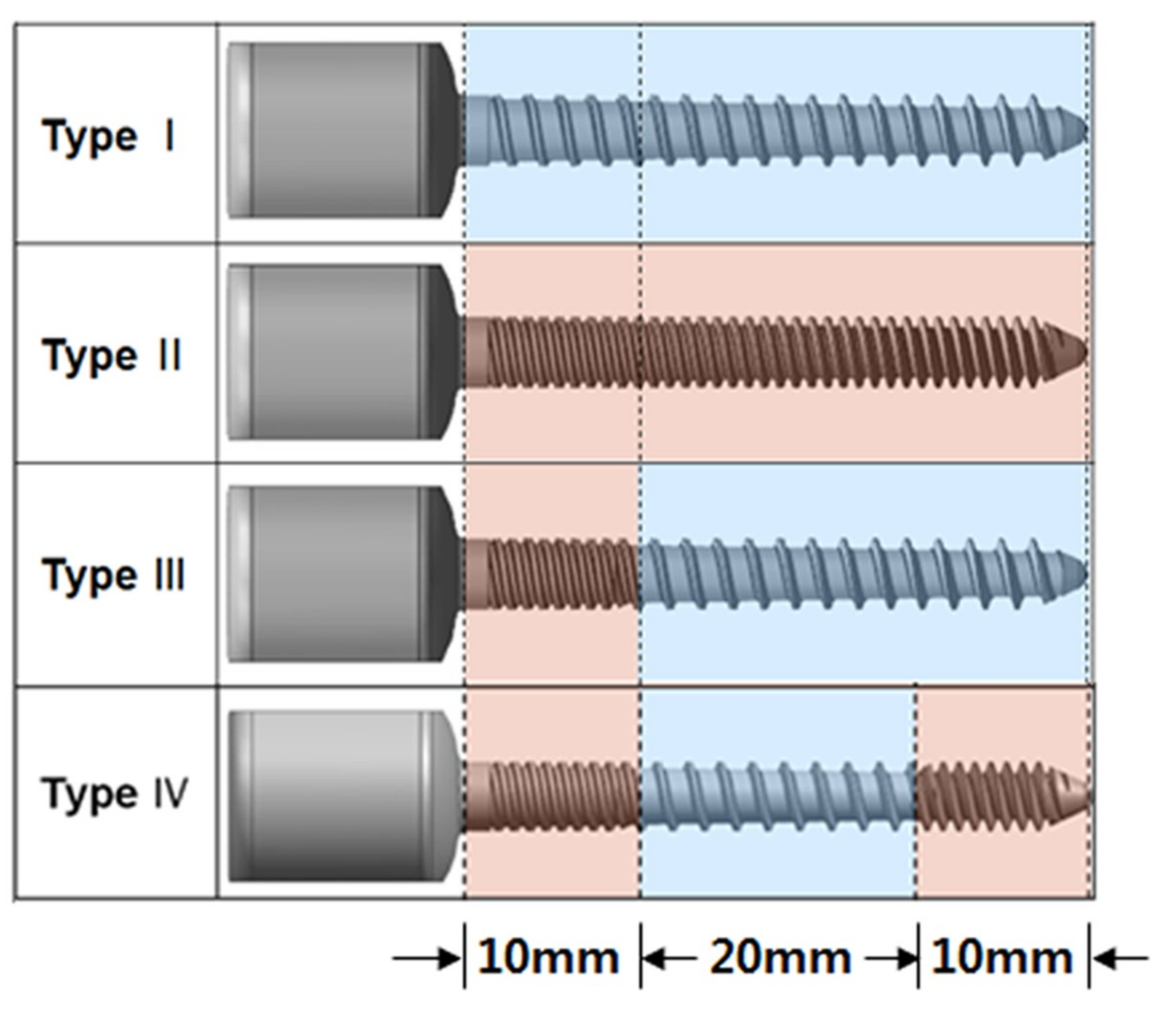
2.1. Types of Pedicle Screw

Four types of screw with different thread patterns were designed to test the axial pullout strength in this study (Figure 1). The screws were of uniform dimensions in length, outer diameter, and proximal and distal core diameter, with values of 40 mm, 4.5 mm, 4.0 mm and 2.7 mm, respectively (Table 1). The differences in pitch length along the screw thread contributed to establishing the different structures of the four screw types (Table 1). All pedicle screws were made of stainless steel.

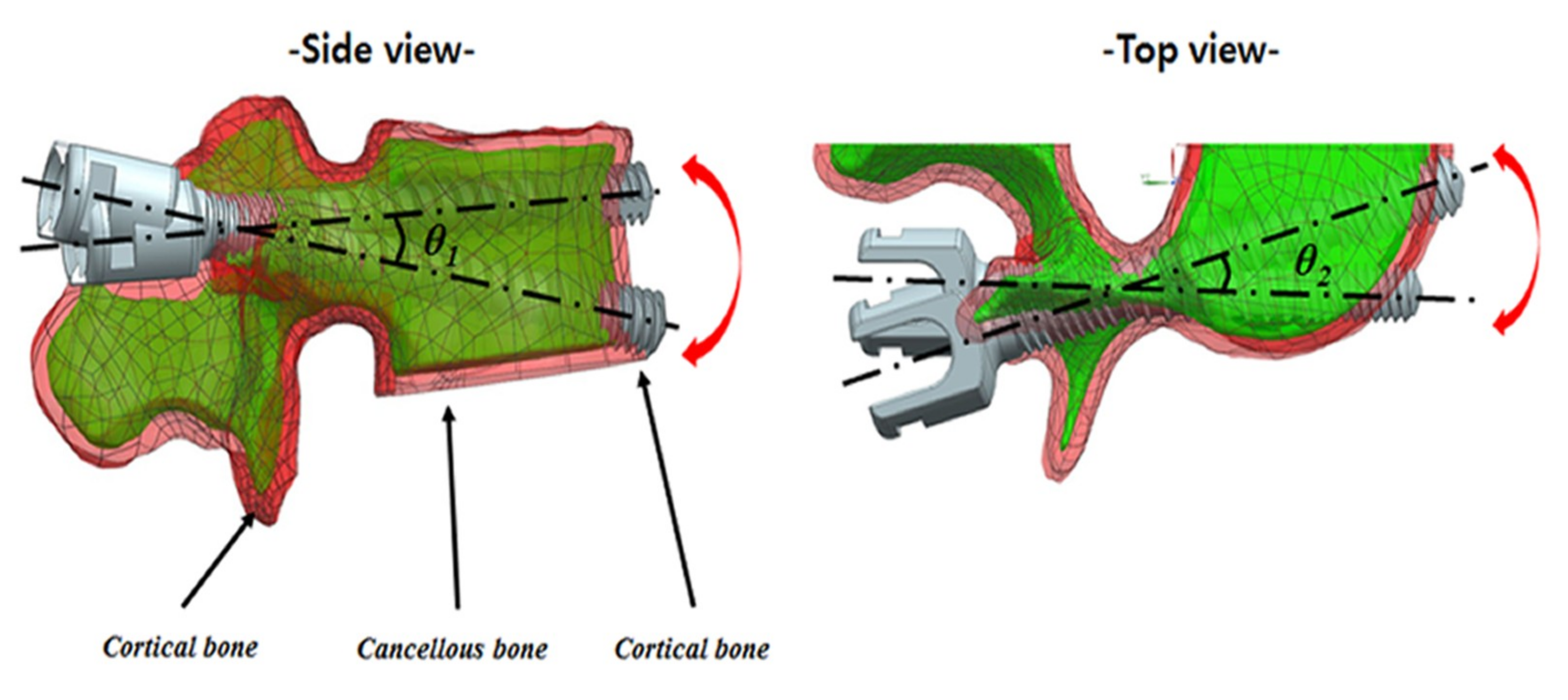
2.2. Preparation of Specimens
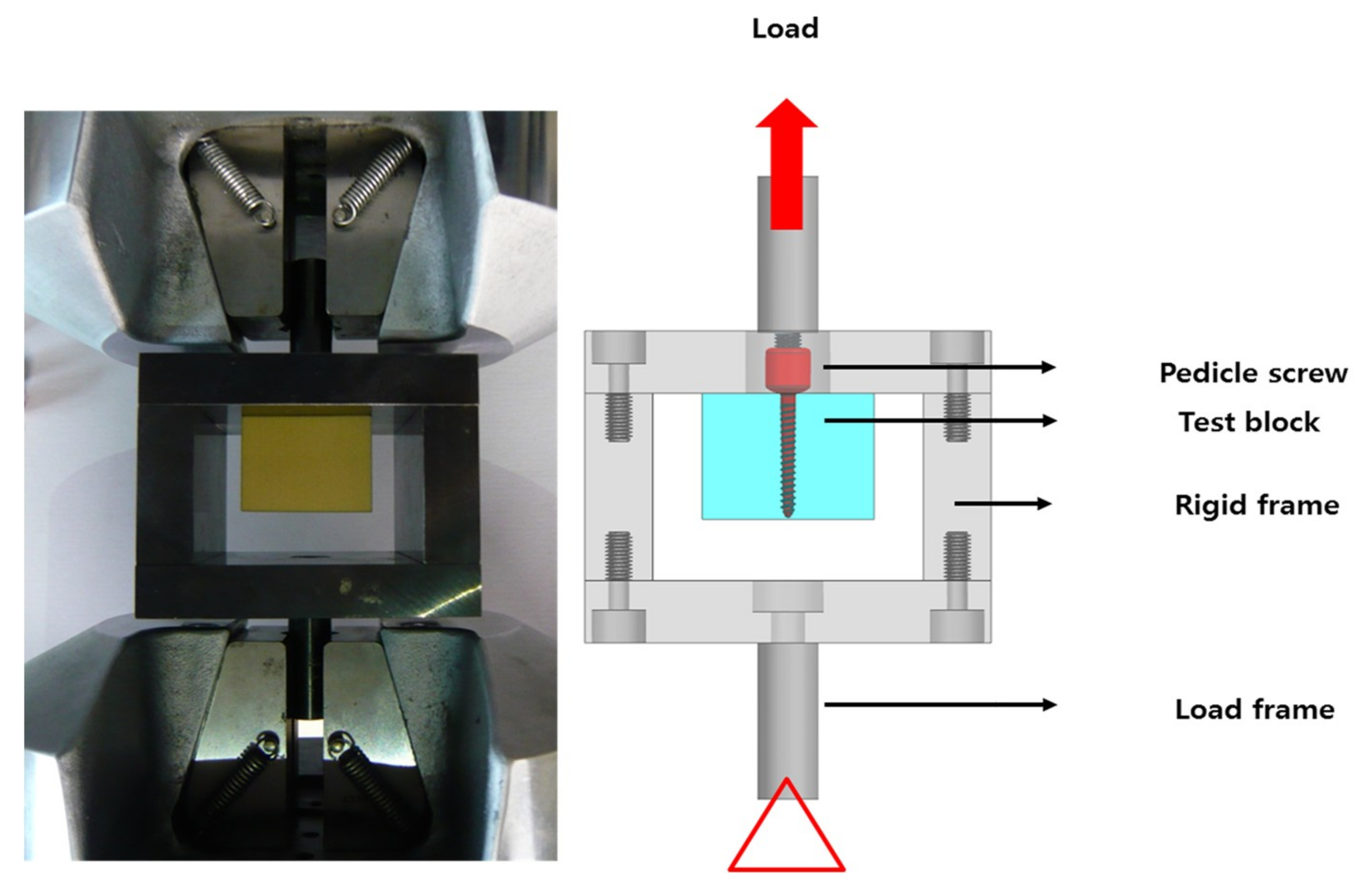
Polyurethane foams have been commonly used as an alternative test medium analogous to human bone [16]. They have shown several advantages compared to human vertebrae, including the reduction of the inter-specimen variability of cadaveric bones and the prevention of deformation or breakage during the mechanical loading test [17]. Cellular polyurethane foams (Sawbones, Pacific Research Corporation, Vashon, WA, USA), with three different densities were used as biomechanical testing material in this study. Foams with a low density of 160 kg/m3, middle density of 320 kg/m3, and high density of 800 kg/m3 represented human osteoporotic cancellous bone, normal cancellous bone, and cortical bone, respectively, in accordance with the American Society for Testing and Materials (ASTM F1839-01) protocol [16]. To simulate various degrees of bone quality, five types of bone specimens were designed using polyurethane foam blocks with various densities (Figure 2).
Type A: normal cancellous bone;
Type B: normal cancellous bone with cortical bone in the upper margin;
Type C: osteoporotic cancellous bone;
Type D: osteoporotic cancellous bone with cortical bone in the upper margin;
Type E: osteoporotic cancellous bone with cortical bone in the upper and lower margins.
To simulate screw insertion in normal-quality and osteoporotic bone using a conventional technique, Type A, B, C, and D bone specimens, measuring 50.8 mm × 50.8 mm × 40 mm, were used to compare the pullout strength among Type I, II, and III screws (Figure 2a). To simulate the screw insertion in osteoporotic bone with and without bi-cortical bone using an anterior cortex purchase technique (Figure 3), bone specimens of Type C and E, measuring 50.8 mm × 50.8 mm × 33 mm, were used for comparisons among Type I, II, and IV screws (Figure 2b).

2.3. Test Apparatus

The experimental configuration used for testing the axial pullout strength of pedicle screws conformed to the requirements of a suitable axial loading test fixture guided by ASTM F543-13 Standard Specification and Test Methods for Metallic Medical Bone Screws [18] and is shown in Figure 4. The pullout test machine (MTS MiniBionix 858, MTS Systems Corp, Eden Prairie, MN, USA) consisted of a rigid frame measuring 130 mm × 100 mm × 100 mm in size. It was fixed to the base of a load frame, with an opening in the upper surface where the screw could pass through. The test block with laminated polyurethane foam and the screw inside was connected to the load frame. The load frame was transferred vertically through the head of the screw and was aligned with the longitudinal axis of the screw. The setup configuration was designed rigid enough so that deflection under the loading conditions was negligible. The pullout test was conducted according to the ASTM F543-13 standard testing protocol [18].

2.4. Biomechanical Testing Procedures

A pilot hole with a 2.7 mm diameter was drilled manually in the center of a polyurethane block. Then, the pedicle screw was inserted into the polyurethane block sample to full depth, until the tip of the screw was leveled or passed through the bottom of the block, depending on the size of the block. The foam block with the pedicle screw inserted in it was completely seated within the rigid fixture frame, which was fixed to the base of the load frame. This design ensured that the direction of the applied load was aligned with the longitudinal axis of the screw. An extraction load was gradually applied to the screw head at a loading rate of 5 mm/min, until the screw was pulled out from the test block. The peak load was defined as the screw pullout strength, and the load-displacement curve was recorded using a data acquisition system (instruNet, GW Instruments, Somerville, MA, USA). The same biomechanical testing procedure was conducted five times per case, and the pullout strength of all types of screw in different bone specimens was recorded for analysis.

2.5. Statistical Analysis

Independent t-tests were performed to assess the significance of the differences of pullout strength between dual-threaded screws and conventional single-threaded screws in the same type of bone specimen. Statistical analyses were performed using the Statistical Package for the Social Sciences (SPSS, version 20.0; SPSS, Inc., Chicago, IL, USA). A p value of <0.05 was considered statistically significant.

3. Results

All of the screws were loaded to failure. Their structures were completely preserved after pullout. Defined as the experimentally measured peak load, the pullout strength of all types of screws is shown in Table 2.

3.1. Comparison among Type I, Type II, and Type III Screws in Type A, B, C, and D Bone Specimens (in Normal and Osteoporotic Bone)

The typical patterns of the load–displacement curves that illustrate the holding characteristics of different bone specimens combined with different screw types are presented in Figure 5a. All load–displacement curves exhibited similar trends, whereby the load increased sharply as the screws were extracted and then decreased rapidly once the screws were pulled out from the specimens. The displacement at the point of peak load was always less than 1.0 mm. In the Type A bone specimen, which represented normal cancellous bone, the pullout strength required for the Type I screw (conventional pedicle screw) was 2.23 ± 0.07 kilo Newtons, while that for the Type III screw (dual-threaded screw), it was 2.28 ± 0.13 kilo Newtons; the difference was not significant (p = 0.471). In the Type B bone specimen, which represented normal cancellous bone with cortical bone, the pullout strengths were 2.65 ± 0.16 kilo Newtons and 2.80 ± 0.08 kilo Newtons for the Type I and Type III screws, respectively, and there was no significant difference (p = 0.106). However, in the Type C bone specimen, which represented osteoporotic cancellous bone, the pullout strength required for the Type III screw was 1.32 ± 0.13 kilo Newtons, which was significantly lower than that for the Type I screw (1.56 ± 0.23 kilo Newtons) (p = 0.046). Furthermore, in the Type D bone specimen, which represented osteoporotic cancellous bone with cortical bone, the Type III screw exhibited a significantly lower pullout strength (1.82 ± 0.09 kilo Newtons) compared to the Type I screw (1.99 ± 0.11 kilo Newtons) (p = 0.031) (Figure 6a).

3.2. Comparison among Type I, Type II, and Type IV screws in Type C and E Bone Specimen (in Osteoporotic Bone with and without Bi-cortical Bone)

Typical load–displacement curves are shown in Figure 5b. The patterns of the load–displacement curves for all screws exhibited similar trends, as described in the above section. In the Type C bone specimen, which represented osteoporotic cancellous bone without bi-cortical bone, the pullout strength for the Type IV screw (double dual-threaded screw) (0.98 ± 0.15 kilo Newtons) was significantly lower than that for the Type I screw (1.56 ± 0.23 kilo Newtons) (p = 0.006). In the Type E bone specimen, which represented osteoporotic cancellous bone with bi-cortical bone, the Type IV screw exhibited a higher pullout strength (2.67 ± 0.07 kilo Newtons) compared to the Type I screw (2.54 ± 0.15 kilo Newtons), with the trend being statistically significant (p = 0.105) (Figure 6b).

4. Discussion

In this experimental testing study, we found that, compared to a conventional pedicle screw, dual-threaded pedicle screws exhibited higher pullout strength in normal-quality bone and significantly lower pullout strength in osteoporotic bone, after controlling for several factors such as screw length, diameter, and insertion technique. However, using an anterior cortex purchase insertion technique, the newly designed double dual-threaded screw exhibited better pullout biomechanics in osteoporotic bone with bi-cortical bone.
The pullout strength of pedicle screws, as one important mechanism responsible for implant failure, has been widely used to assess the strength of the bone–screw interface in different conditions [19,20] and is a popular biomechanical testing parameter. Several factors, including the size and design of the screw, pedicle structures, bone quality, size of pilot holes, and insertion technique, may affect the pullout strength of a pedicle screw [13]. Therefore, these variable factors must be controlled for under similar situations during experimental testing to ensure a reliable comparison of differently designed screws. Polyurethane foam blocks with consistent properties are used to simulate a particular human bone density, and this commonly accepted testing material can limit the bias caused by the variation of human bone quality, pedicle structures, and screw–cortical interface [5]. Furthermore, the foam blocks, which are simple and easy to handle, could prevent deformation and breakage during biomechanical tests [18]. Therefore, to investigate the biomechanical performance of differently designed pedicle screws, we used laminated polyurethane foam blocks to compare the pullout strengths in the current experimental study.
On the basis of the results of our tests, we found that, when compared to the conventional single-threaded screw (Type I), the dual-threaded screw (Type III) exhibited better pullout strength in normal cancellous bones, either with or without cortical (Type B bone specimen just contains cortical bone, which is different from Type E bone specimen consist of bi-cortical bone), although there were no significant differences. The pullout strength of the screw depends on several variables, most of which are related to the biomechanical properties of the bone–screw interface along the length of the screw. With uniformed dimensions in length, diameter, and thread profile of all screws in this study, the quality of bone and the thread pattern were the main factors affecting the pullout strength of the screws [21,22]. As the normal cancellous bone contains bone of high quality, the thread pattern determining the properties of the bone–screw interface was critical for the holding strength of the pedicle screw. Because the double-threaded part of the screw affects the properties of the bone–screw interface more extensively, the screws with larger double-threaded regions may display a higher purchase, resisting the pedicle screw pullout in normal cancellous bone. This possibility is supported by the result of our test showing that the full double-threaded screw (Type II) displayed the highest pullout strength in normal cancellous bone with and without cortical bone (Type B and A bone specimens) (Table 2).
However, the biomechanical testing in the osteoporotic bone model yielded different results. Compared to the conventional single-threaded screw (Type I), the dual-threaded screw (Type III) showed significantly lower pullout strength in osteoporotic bone, either with or without cortical bone (Type D and C bone specimens). This finding is consistent with those of a previous study [23], reasoning that osteoporotic cancellous bone with fragile trabecular would be destroyed easily by the screw thread during insertion. Therefore, the screws with a larger region of double threads would damage more extensively the bone–screw interface, leading to a decreased resistance to the pullout force. This proposed mechanism may explain why the dual-threaded screw exhibited significantly lower pullout strength relative to the conventional single-threaded screw in osteoporotic cancellous bone with and without cortical bone. A previous study is in concordance with the present results and reported that dual-threaded screws increase the insertion torque in poor-quality bone compared to single-threaded screws, without increasing the axial pullout strength [15].
Notably, cortical bone is less affected by osteoporosis relative to cancellous bone [24], and 60% of pullout strength was provided by cortical bone in the pedicle region [15]. Therefore, we designed the double dual-threaded screw with double threads in both the proximal and distal shafts to augment the holding strength in the pedicle and anterior cortex of the vertebral body region. To simulate the screw insertion in a real vertebral body using an anterior cortex purchase technique (Figure 3), we used the foam block with a smaller size (Type C and E bone specimens), in which the tip of the screw could pass through the bottom. Although cancellous bone may experience much larger destruction during insertion of a screw with double threads, the interaction of cortical bone with the double threads in proximal and distal areas could provide enhanced holding power and thus resist the pullout. That may explain why the double dual-threaded screw (Type IV) exhibited higher pullout strength in osteoporotic bone with bi-cortical bone (Type E bone specimen), compared to the conventional single-threaded screw (Type I). Although there was no significant difference because of the small sample size, the trend of statistical significance indicates that the biomechanical performance of the double dual-threaded screw would potentially be superior to that of the conventional pedicle screw.
Although a growing number of spinal surgery interventions are using dual-threaded screws as fixation devices to improve the fixation capability for osteoporotic patients, the prioritization of these devices over conventional pedicle screws remains controversial. According to the results of our study, the dual-threaded screw exhibited significantly lower pullout strength in osteoporotic bone, which indicates that it would not be the optimal fixation device for osteoporotic patients. However, the increased biomechanical performance of the double dual-threaded screw in osteoporotic bone with bi-cortical bone margin indicates that this device could be an alternative option for patients with osteoporosis, using an anterior cortex purchase technique. Therefore, the present experimental study may provide spine surgeons with selection strategies for optimal fixation devices according to patients’ bone quality and screw insertion technique. Nevertheless, it should be kept in mind that these recommendations are based on results obtained under axial loading conditions, and no conclusion could be made for the other loads applied to screws intraoperatively or postoperatively.
Recently, many surgeons have focalized their attention on the selection of pedicle screw material, because corrosion of the external implant would be one of the causes of fixation failure leading to severe side effects in the human body [7,9]. As a solution for implant corrosion, several materials and coating techniques, including PVD/CVD, micro-arc-oxidation (MAO), and sol–gel coating, were explored to improve corrosion resistance and subsequently reduce harmful ion release [10,12,25]. Furthermore, recent studies have reported that coating techniques, such as hydroxyapatite coating and melted polymer sleeve, can be applied to pedicle screws for improving fixation and anchorage [11,26]. However, the effect of screw materials and the application of coating techniques were not a concern of this study. Further studies are needed to investigate the influence of different biomaterials and coating techniques on screw pullout biomechanics.
There are several limitations to this study. Firstly, this study used a relatively small sample of specimens, which might establish a bias on the statistical outcomes. Second, we focused on the pullout strength of the screws under axial loading only, which is not representative of the entire biomechanical performance of these screws. On the basis of the present results, we plan future studies with cadaveric specimens and finite element model (FEM) studies under different loading conditions. Finally, this was a biomechanical testing study, and the results based on laminated polyurethane foam may differ from those obtained in real surgical conditions.

5. Conclusions

The present study demonstrated that dual-threaded pedicle screws yielded higher pullout strength in normal-quality bone and significantly lower pullout strength in compromised osteoporotic bone, compared to a conventional pedicle screw. Considering the trend of statistical significance, the newly designed double dual-threaded pedicle screw exhibited better pullout biomechanics in osteoporotic bone with bi-cortical bone margin. The findings could help spine surgeons to select optimal thread designs according to patients’ bone quality and screw insertion technique, considering axial loading condition.

Author Contributions

F.S. and H.-J.K. designed the study and wrote the manuscript. K.-T.K. performed the experimental tests. Y.J.S. provided critical revision of the manuscript.

Funding

This research was supported by research grants from Je-il Pharm and the Basic Science Research Program through the National Research Foundation of Korea (NRF) funded by the Ministry of Education, Science and Technology (2016R1A2B3012850).

Conflicts of Interest

The authors declare no conflicts of interest.

References

  1. Gertzbein, S.D.; Robbins, S.E. Accuracy of pedicular screw placement in vivo. Spine 1990, 15, 11–14. [Google Scholar] [CrossRef]
  2. Weinstein, J.N.; Rydevik, B.L.; Rauschning, W. Anatomic and technical considerations of pedicle screw fixation. Clin. Orthop. Relat. Res. 1992, 34–46. [Google Scholar] [CrossRef]
  3. Enker, P.; Steffee, A.D. Interbody fusion and instrumentation. Clin. Orthop. Relat. Res. 1994, 300, 90–101. [Google Scholar] [CrossRef]
  4. Baliga, S.; Treon, K.; Craig, N.J.A. Low Back Pain: Current Surgical Approaches. Asian Spine J. 2015, 9, 645–657. [Google Scholar] [CrossRef] [PubMed]
  5. Hsu, C.C.; Chao, C.K.; Wang, J.L.; Hou, S.M.; Tsai, Y.T.; Lin, J. Increase of pullout strength of spinal pedicle screws with conical core: Biomechanical tests and finite element analyses. J. Orthop. Res. 2005, 23, 788–794. [Google Scholar] [CrossRef]
  6. Krishnan, V.; Varghese, V.; Kumar, G.S. Comparative Analysis of Effect of Density, Insertion Angle and Reinsertion on Pull-Out Strength of Single and Two Pedicle Screw Constructs Using Synthetic Bone Model. Asian Spine J. 2016, 10, 414–421. [Google Scholar] [CrossRef]
  7. Serhan, H.; Slivka, M.; Albert, T.; Kwak, S.D.; Ibrahim, H.; Jahadakbar, A.; Dehghan, A.; Moghaddam, N.S.; Amerinatanzi, A.; Elahinia, M. Is galvanic corrosion between titanium alloy and stainless steel spinal implants a clinical concern? Spine J. 2004, 4, 379–387. [Google Scholar] [CrossRef] [PubMed]
  8. Ibrahim, H.; Jahadakbar, A.; Dehghan, A.; Moghaddam, N.S.; Amerinatanzi, A.; Elahinia, M. In vitro corrosion assessment of additively manufactured porous NiTi structures for bone fixation applications. Metals 2018, 8, 164. [Google Scholar] [CrossRef]
  9. Ševčíková, J.; Bártková, D.; Goldbergová, M.; Kuběnová, M.; Čermák, J.; Frenzel, J.; Weiser, A.; Dlouhý, A. On the Ni-ion release rate from surfaces of binary NiTi shape memory alloys. Appl. Surf. Sci. 2018, 427, 434–443. [Google Scholar] [CrossRef]
  10. Tillmann, W.; Stangier, D.; Denkena, B.; Grove, T.; Lucas, H. Influence of PVD-coating technology and pretreatments on residual stresses for sheet-bulk metal forming tools. Prod. Eng. Res. Dev. 2016, 10, 17–24. [Google Scholar] [CrossRef]
  11. Ohe, M.; Moridaira, H.; Inami, S.; Takeuchi, D.; Nohara, Y.; Taneichi, H. Pedicle screws with a thin hydroxyapatite coating for improving fixation at the bone-implant interface in the osteoporotic spine: Experimental study in a porcine model. J. Neurosurg. Spine 2018, 28, 679–687. [Google Scholar] [CrossRef] [PubMed]
  12. Dehghanghadikolaei, A.; Ibrahim, H.; Amerinatanzi, A.; Hashemi, M.; Moghaddam, N.S.; Elahinia, M. Improving corrosion resistance of additively manufactured nickel–titanium biomedical devices by micro-arc oxidation process. J. Mater. Sci. 2019, 54, 7333–7355. [Google Scholar] [CrossRef]
  13. Shea, T.M.; Laun, J.; Gonzalez-Blohm, S.A.; Doulgeris, J.J.; Lee, W.E.; Aghayev, K.; Vrionis, F.D. Designs and techniques that improve the pullout strength of pedicle screws in osteoporotic vertebrae: Current status. Biomed Res Int. 2014, 2014, 748393. [Google Scholar] [CrossRef]
  14. Bianco, R.J.; Arnoux, P.J.; Wagnac, E.; Mac-Thiong, J.M.; Aubin, C.É. Minimizing pedicle screw pullout risks: A detailed biomechanical analysis of screw design and placement. Clin Spine Surg. 2017, 30, E226–E232. [Google Scholar] [CrossRef] [PubMed]
  15. Brasiliense, L.B.; Lazaro, B.C.; Reyes, P.M.; Newcomb, A.G.; Turner, J.L.; Crandall, D.G.; Crawford, N.R. Characteristics of immediate and fatigue strength of a dual-threaded pedicle screw in cadaveric spines. Spine J. 2013, 13, 947–956. [Google Scholar] [CrossRef]
  16. ASTM International. Standard Specification for Rigid Polyurethane Foam for Use as a Standard Material for Testing Orthopedic Devices and Instrument; ASTM F1839-01; ASTM International: West Conshohocken, PA, USA, 2001. [Google Scholar]
  17. Kim, Y.-Y.; Choi, W.-S.; Rhyu, K.-W. Assessment of pedicle screw pullout strength based on various screw designs and bone densities-an ex vivo biomechanical study. Spine J. 2012, 12, 164–168. [Google Scholar] [CrossRef]
  18. ASTM International. Standard Specification and Test Methods for Metallic Medical Bone Screws; ASTM F543-13; ASTM International: West Conshohocken, PA, USA, 2013. [Google Scholar]
  19. Abshire, B.B.; McLain, R.F.; Valdevit, A.; Kambic, H.E. Characteristics of pullout failure in conical and cylindrical pedicle screws after full insertion and back-out. Spine J. 2001, 1, 408–414. [Google Scholar] [CrossRef]
  20. Ono, A.; Brown, M.D.; Latta, L.L.; Milne, E.L.; Holmes, D.C. Triangulated pedicle screw construct technique and pull-out strength of conical and cylindrical screws. J. Spinal. Disord. 2001, 14, 323–329. [Google Scholar] [CrossRef] [PubMed]
  21. Chapman, J.R.; Harrington, R.M.; Lee, K.M.; Anderson, P.A.; Tencer, A.F.; Kowalski, D. Factors affecting the pullout strength of cancellous bone screws. J. Biomech. Eng. 1996, 118, 391–398. [Google Scholar] [CrossRef]
  22. Inceoglu, S.; Ferrara, L.; McLain, R.F. Pedicle screw fixation strength: Pullout versus insertional torque. Spine J. 2004, 4, 513–518. [Google Scholar] [CrossRef]
  23. Krenn, M.H.; Piotrowski, W.P.; Penzkofer, R.; Augat, P. Influence of thread design on pedicle screw fixation. Laboratory investigation. J. Neurosurg. Spine 2008, 9, 90–95. [Google Scholar] [CrossRef]
  24. Santoni, B.G.; Hynes, R.A.; McGilvray, K.C.; Rodriguez-Canessa, G.; Lyons, A.S.; Henson, M.A.; et al. Cortical bone trajectory for lumbar pedicle screws. Spine J. 2009, 9, 366–373. [Google Scholar] [CrossRef]
  25. Dehghanghadikolaei, A.; Ansary, J.; Ghoreishi, R. Sol-gel process applications: A mini-review. Proc. Natl. Res. Soc. 2018, 2, 02008. [Google Scholar] [CrossRef]
  26. Spicher, A.; Lindtner, R.A.; Zimmermann, S.; Stofferin, H.; Schmoelz, W. Ultrasound melted polymer sleeve for improved primary pedicle screw anchorage: A novel augmentation technique. Clin. Biomech. 2019, 63, 16–20. [Google Scholar] [CrossRef]
Figure 1. Illustration of four types of screws with different thread patterns.
Applsci 09 01525 g001
Figure 2. Illustration of bone specimens simulating various degrees of bone quality using polyurethane foam blocks with various densities. (a) Type A, B, C and D bone specimens and (b) Type C and E bone specimens.
Applsci 09 01525 g002
Figure 3. Simulation of screw insertion using an anterior cortex purchase technique.
Applsci 09 01525 g003
Figure 4. Experimental configuration used for testing the axial pullout strength of pedicle screws.
Applsci 09 01525 g004
Figure 5. Load–displacement curves from the axial pullout test. (a) Type I, II, and III screws in Type A, B, C and D bone specimens, and (b) Type I, II, and IV screws in Type C and E bone specimens.
Applsci 09 01525 g005
Figure 6. Comparison of the pullout test results; the error bar shows standard deviation, and the blocks indicate mean values. (a) Comparison between Type I and III screws in Type A, B, C and D bone specimens (p = 0.471, p = 0.106, p = 0.046 and p = 0.031, respectively), and (b) comparison between Type I and Type IV screws in Type C and E bone specimens (p = 0.006 and p = 0.105, respectively). * Significant difference.
Applsci 09 01525 g006
Table 1. Dimensions of the examined screws.
Screw TypeLength (mm)Outer Diameter (mm)Proximal Core Diameter (mm)Distal Core Diameter (mm)Proximal Thread Width (mm)Distal Thread Width (mm)Pitch (mm)
Proximal 1/4MiddleDistal 1/4
Type I404.54.02.70.40222
Type II404.54.02.70.40111
Type III404.54.02.70.40122
Type IV404.54.02.70.40121
Type I: conventional single-threaded screw; Type II: double-threaded screw (control type); Type III: dual-threaded screw; Type IV: newly designed double dual-threaded screw.
Table 2. Pullout strength of all types of screws in various bone specimens.
Bone SpecimensScrew Types
Type I (KN)Type II (KN)Type III (KN)Type IV (KN)
Type A2.23 ± 0.072.38 ± 0.182.28 ± 0.13
Type B2.65 ± 0.162.87 ± 0.132.80 ± 0.08
Type C1.56 ± 0.231.26 ± 0.061.32 ± 0.130.98 ± 0.15
Type D1.99 ± 0.111.80 ± 0.141.82 ± 0.09
Type E2.54 ± 0.152.68 ± 0.14 2.67 ± 0.07
The four types of screw were: Type I: single-threaded screw; Type II: double-threaded screw; Type III: dual-threaded screw; Type IV: double dual-threaded screw. The five types of bone specimens were: Type A: cancellous bone; Type B: cancellous bone with cortical bone in the upper margin; Type C: osteoporotic cancellous bone; Type D: osteoporotic cancellous bone with cortical bone in the upper margin; Type E: osteoporotic cancellous bone with cortical bone in the upper and lower margins. KN: kilo Newtons; values are mean ± standard deviation. Disclosure: the screws were supplied by TJC life biotechnology company (Korea).

