Wireless Magnetoelasticity-Based Sensor for Monitoring the Degradation Behavior of Polylactic Acid Artificial Bone In Vitro


Abstract
1. Introduction
2. Materials and Methods
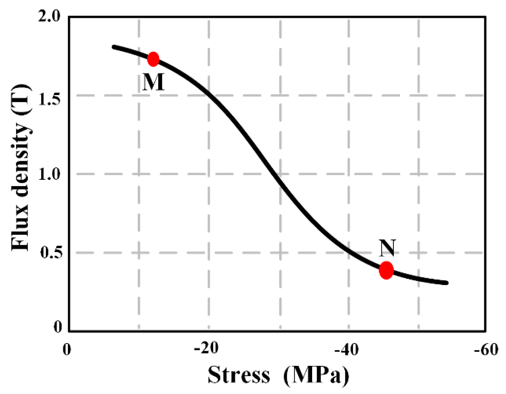
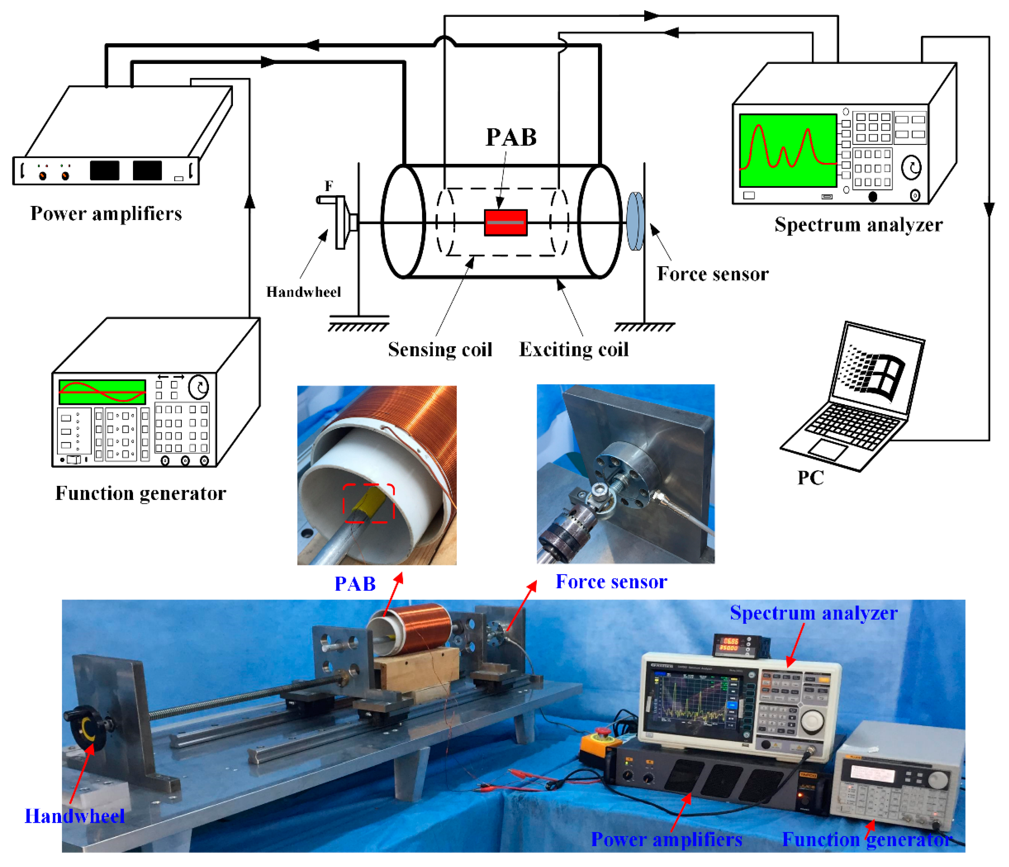
2.1. Theoretical Analysis of the Magnetoelasticity-Based (MB) Sensor
2.2. Fabrication of the PLA artificial bone (PAB)-Coated MB Sensor
2.3. Preparation of PAB Degradation
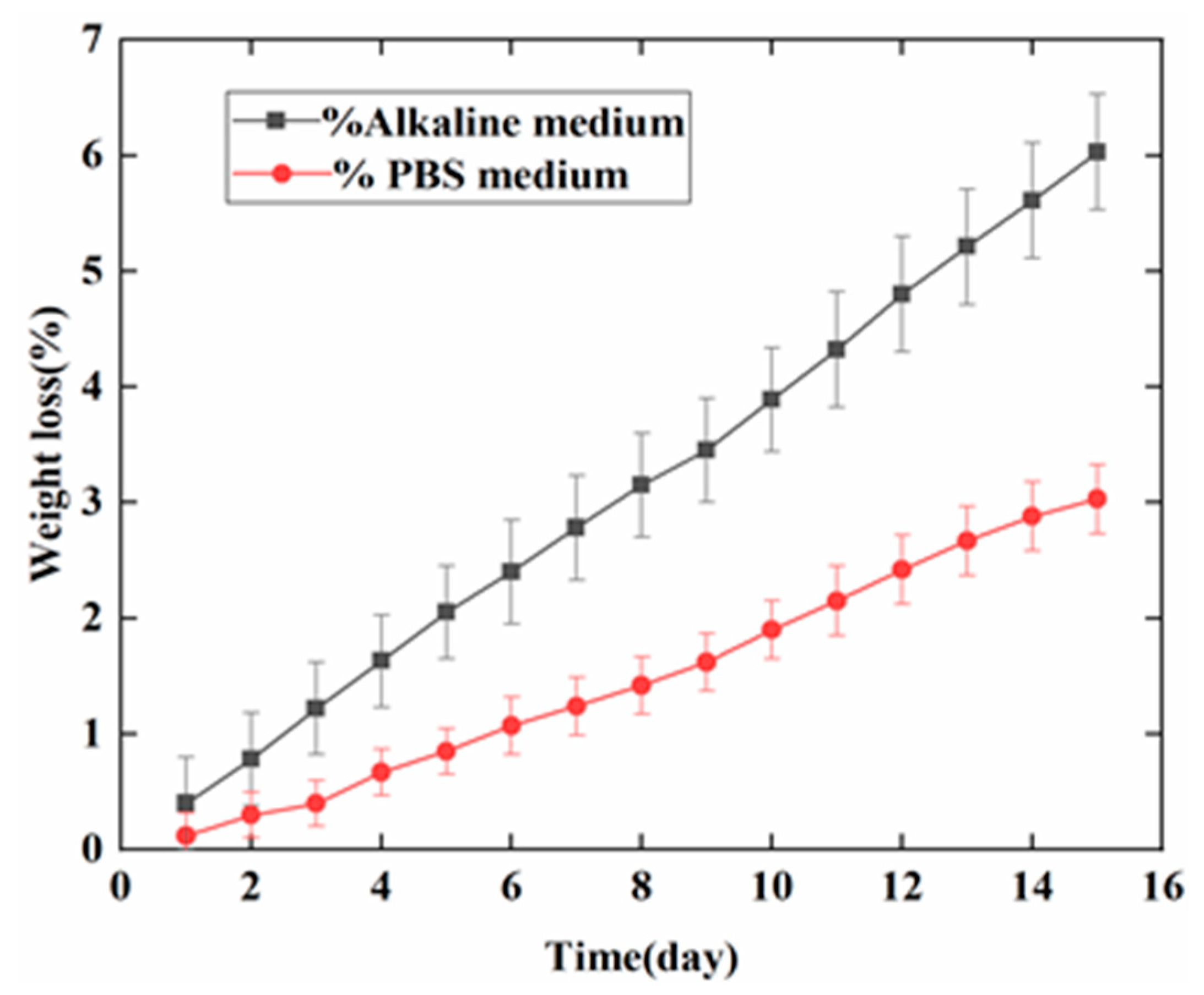
2.4. Weight Loss of PAB
2.5. Surface Characterization
2.6. Monitoring the Degradation Behavior of PAB with the MB Sensor in Vitro
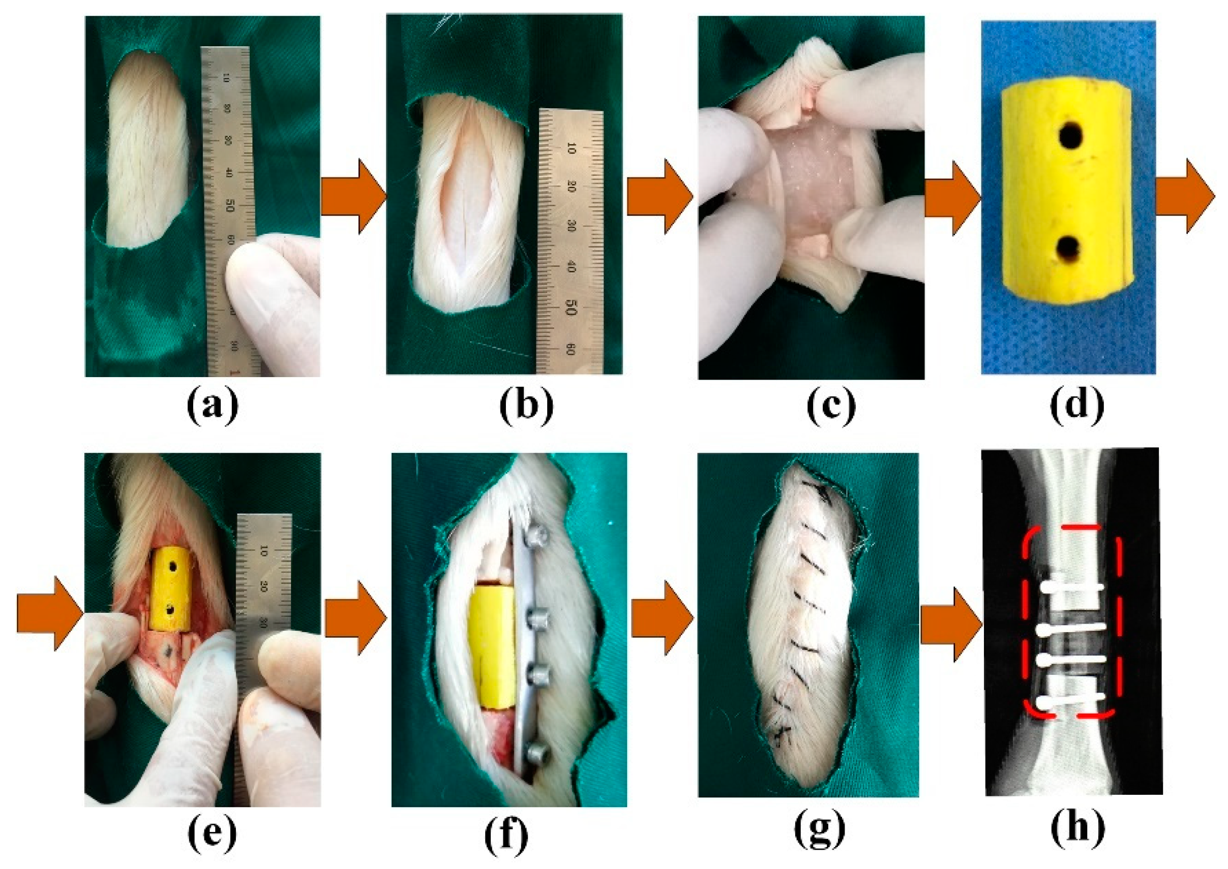
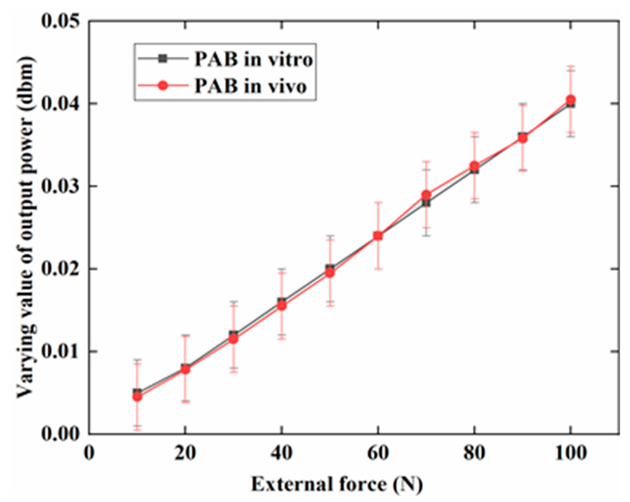
2.7. Simulated In Vivo Testing
3. Results
3.1. Weight Loss of PAB
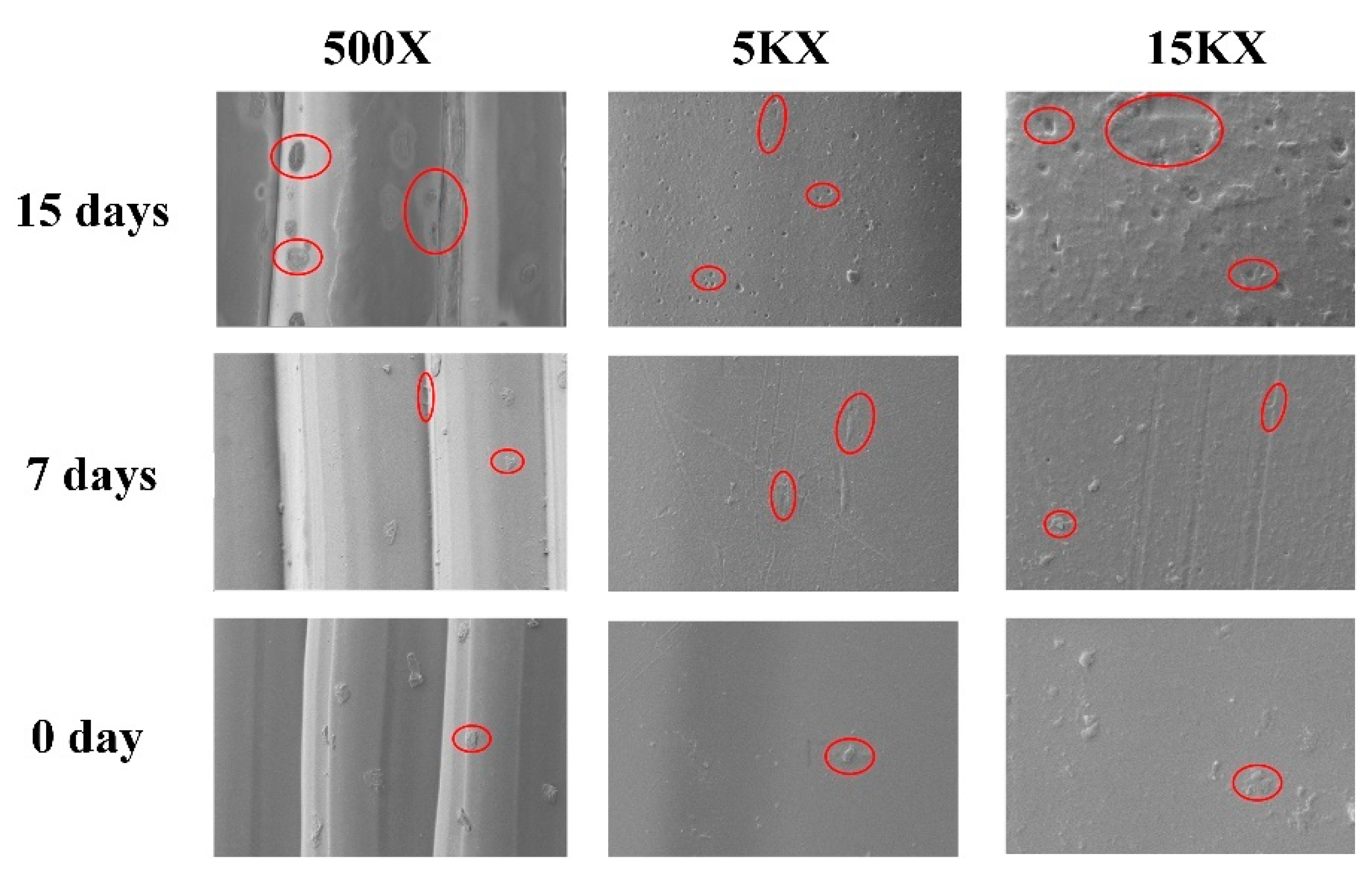
3.2. Morphology of PAB
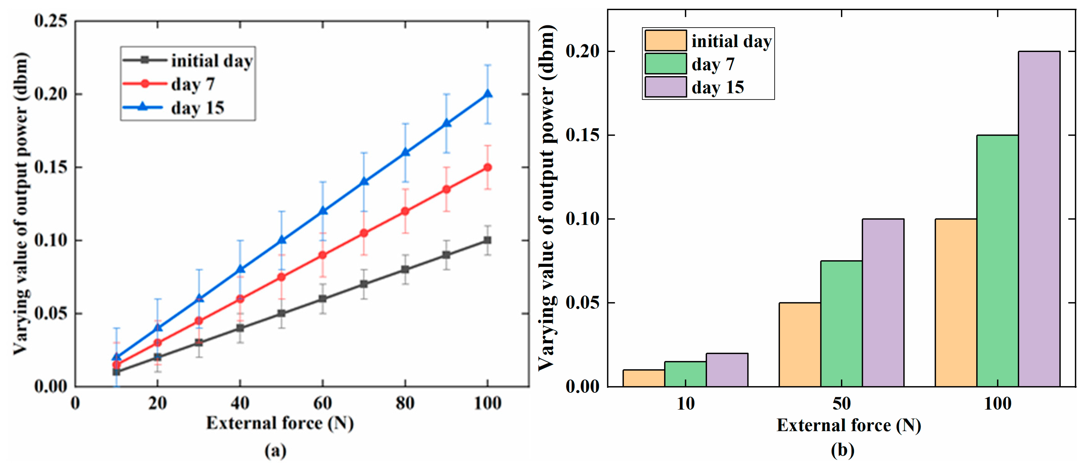
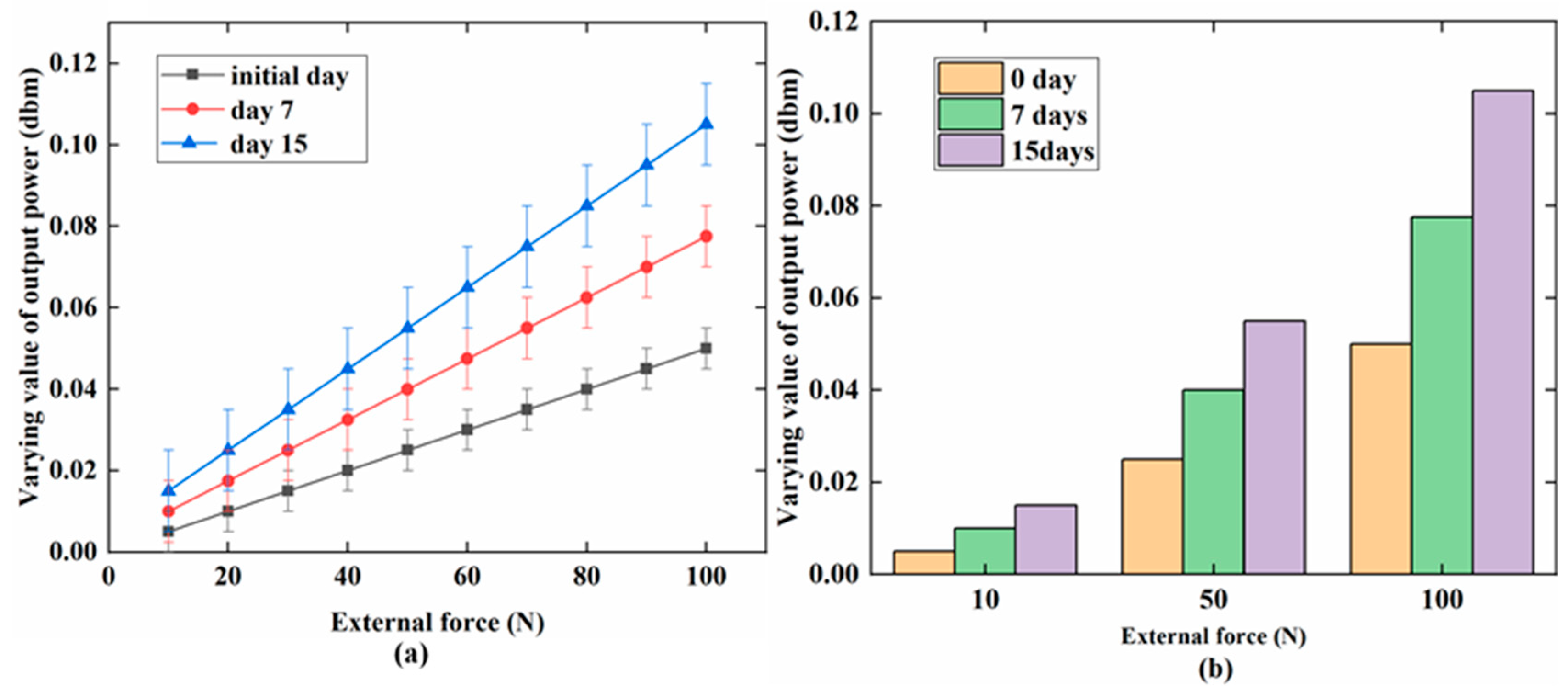
3.3. MB Sensor Monitoring Results
4. Discussion
5. Conclusions
Author Contributions
Funding
Conflicts of Interest
References
- Pihlajamaki, H.; Hirvensalo, E. Poly-L-Lactic of Fractures Pins of Acid Self-Reinforced for Fixation. Bone Joint J. 1992, 74–B, 853–857. [Google Scholar]
- Matsusue, Y.; Nakamura, T.; Iida, H.; Shimizu, K. A long-term clinical study on drawn poly-L-lactide implants in orthopaedic surgery. J. Long. Term. Eff. Med. Implants 1997, 7, 119–137. [Google Scholar] [CrossRef]
- Ho, Y.-C.; Huang, F.-M.; Chang, Y.-C. Cytotoxicity of formaldehyde on human osteoblastic cells is related to intracellular glutathione levels. J. Biomed. Mater. Res. B Appl. Biomater. 2007, 83, 340–344. [Google Scholar] [CrossRef] [PubMed]
- Lu, H.H.; El-Amin, S.F.; Scott, K.D.; Laurencin, C.T. Three-dimensional, bioactive, biodegradable, polymer-bioactive glass composite scaffolds with improved mechanical properties support collagen synthesis and mineralization of human osteoblast-like cells in vitro. J. Biomed. Mater. Res. Part A 2003, 64, 465–474. [Google Scholar] [CrossRef] [PubMed]
- Rokkanen, P.U.; Böstman, O.; Hirvensalo, E.; Mäkelä, E.A.; Partio, E.K.; Pätiälä, H.; Vainionpää, S.; Vihtonen, K.; Törmälä, P. Bioabsorbable fixation in orthopaedic surgery and traumatology. Biomaterials 2000, 21, 2607–2613. [Google Scholar] [CrossRef]
- Middleton, J.C.; Tipton, A.J. Synthetic biodegradable polymers as orthopedic devices. Biomaterials 2000, 21, 2335–2346. [Google Scholar] [CrossRef]
- Göpferich, A. Mechanisms of polymer degradation and erosion. Biomaterials 1996, 17, 103–114. [Google Scholar] [CrossRef]
- Mukherjee, D.P.; Pietrzak, W.S. Bioabsorbable fixation: Scientific, technical, and clinical concepts. J. Craniofac. Surg. 2011, 22, 679–689. [Google Scholar] [CrossRef]
- Cooper, J.A.; Lu, H.H.; Ko, F.K.; Freeman, J.W.; Laurencin, C.T. Fiber-based tissue-engineered scaffold for ligament replacement: Design considerations and in vitro evaluation. Biomaterials 2005, 26, 1523–1532. [Google Scholar] [CrossRef]
- Ouyang, H.W.; Goh, J.C.H.; Mo, X.M.; Teoh, S.H.; Lee, E.H. The efficacy of bone marrow stromal cell-seeded knitted PLGA fiber scaffold for Achilles tendon repair. Ann. N. Y. Acad. Sci. 2002, 961, 126–129. [Google Scholar] [CrossRef]
- Tsuji, H.; Ikada, Y. Properties and morphology of poly(L-lactide) 4. Effects of structural parameters on long-term hydrolysis of poly(L-lactide) in phosphate-buffered solution. Polym. Degrad. Stab. 2000, 67, 179–189. [Google Scholar] [CrossRef]
- Yuan, X.; Mak, A.F.T.; Yao, K. Surface degradation of poly (L-lactic acid) fibres in a concentrated alkaline solution. Polym. Degrad. Stab. 2003, 79, 45–52. [Google Scholar] [CrossRef]
- Shih, C. Chain-end scission in acid catalyzed hydrolysis of poly (d,l-lactide) in solution. J. Control. Release 1995, 34, 9–15. [Google Scholar] [CrossRef]
- Iwata, T.; Doi, Y. Morphology and Enzymatic Degradation of Poly (L-lactic acid) Single Crystals. Macromolecules 1998, 9297, 2461–2467. [Google Scholar] [CrossRef]
- Soller, B.R.; Zhang, S. Optical measurement of tissue pH for surgical and critical care monitoring. In Proceedings of the BiOS ’98 International Biomedical Optics Symposium, San Jose, CA, USA, 23–24 January 1998; Volume 3259, pp. 122–129. [Google Scholar]
- Ohman, H.; Vahlquist, A. In vivo studies concerning a pH gradient in human stratum corneum and upper epidermis. Acta Derm. Venereol. 1994, 74, 375–379. [Google Scholar] [PubMed]
- Prince, S.; Malarvizhi, S. Analysis of spectroscopic diffuse reflectance plots for different skin conditions. Spectroscopy 2010, 24, 467–481. [Google Scholar] [CrossRef]
- Tannock, I.F.; Rotin, D. Acid pH in Tumors and Its Potential for Therapeutic Exploitation Perspectivesin CancerResearch Acid pH in Tumors and Its Potential for Therapeutic Exploitation1. Cancer Res. 1989, 49, 4373–4384. [Google Scholar]
- Sims, C.; Seigne, P.; Menconi, M.; Monarca, J.; Barlow, C.; Pettit, J.; Puyana, J.C. Skeletal muscle acidosis correlates with the severity of blood volume loss during shock and resuscitation. J. Trauma 2001, 51, 1137–1145, discussion 1145–1146. [Google Scholar] [CrossRef] [PubMed]
- Lin, M.H.; Anderson, J.; Pinnaratip, R.; Meng, H.; Konst, S.; DeRouin, A.J.; Rajachar, R.; Ong, K.G.; Lee, B.P. Monitoring the Long-Term Degradation Behavior of Biomimetic Bioadhesive Using Wireless Magnetoelastic Sensor. IEEE Trans. Biomed. Eng. 2015, 62, 1838–1842. [Google Scholar] [CrossRef] [PubMed]
- Tan, Y. A Passive and Wireless Sensor for Bone Plate Strain Monitoring. Sensors 2017, 17, 2635. [Google Scholar] [CrossRef]
- De Rouin, A.; Pacella, N.; Zhao, C.; An, K.N.; Ong, K.G. A wireless sensor for real-time monitoring of tensile force on sutured wound sites. IEEE Trans. Biomed. Eng. 2016, 63, 1665–1671. [Google Scholar] [CrossRef] [PubMed]
- Ueno, T.; Qiu, J.Q.J.; Tani, J. Magnetic force control based on the inverse magnetostrictive effect. IEEE Trans. Magn. 2004, 40, 1601–1605. [Google Scholar] [CrossRef]
- Polanschiitz, W. Inverse magnetostrictive effect and electromagnetic destructive testing methods. NDT Int. 1986, 19, 249–258. [Google Scholar] [CrossRef]
- Modzelewski, C.; Savage, H.T.; Kabacoff, L.T.; Clark, A.E. Magnetomechanical coupling and permeability in transversely annealed metglas 2605 alloys. IEEE Trans. Magn. 1981, 17, 2837–2839. [Google Scholar] [CrossRef]
- O’Handley, R.C. Modern Magnetic Materials: Principles and Applications; Wiley-Interscience: New York, NY, USA, 2000; ISBN 0471155667. [Google Scholar]
- Wun-Fogle, M.; Savage, H.T.; Clark, A.E. Sensitive, wide frequency range magnetostrictive strain gage. Sens. Actuators 1987, 12, 323–331. [Google Scholar] [CrossRef]
- Ren, L.; Yu, K.; Tan, Y. Wireless and Passive Magnetoelastic-Based Sensor for Force Monitoring of Artificial Bone. IEEE Sens. J. 2019, 19, 2096–2104. [Google Scholar] [CrossRef]
- Barandiaran, J.M.; Gutierrez, J.; Gómez-Polo, C. New sensors based on the magnetoelastic resonance of metallic glasses. Sens. Actuators A Phys. 2000, 81, 154–157. [Google Scholar] [CrossRef]
- Narita, F.; Fox, M. A Review on Piezoelectric, Magnetostrictive, and Magnetoelectric Materials and Device Technologies for Energy Harvesting Applications. Adv. Eng. Mater. 2018, 20, 1–22. [Google Scholar] [CrossRef]
- Hafezi, H.; Robertson, T.L.; Moon, G.D.; Au-Yeung, K.Y.; Zdeblick, M.J.; Savage, G.M. An ingestible sensor for measuring medication adherence. IEEE Trans. Biomed. Eng. 2015, 62, 99–109. [Google Scholar] [CrossRef]
- Navarro, M.; Ginebra, M.P.; Planell, J.A.; Barrias, C.C.; Barbosa, M.A. In vitro degradation behavior of a novel bioresorbable composite material based on PLA and a soluble CaP glass. Acta Biomater. 2005, 1, 411–419. [Google Scholar] [CrossRef]
- Araque-Monrós, M.C.; Vidaurre, A.; Gil-Santos, L.; Gironés Bernabé, S.; Monleón-Pradas, M.; Más-Estellés, J. Study of the degradation of a new PLA braided biomaterial in buffer phosphate saline, basic and acid media, intended for the regeneration of tendons and ligaments. Polym. Degrad. Stab. 2013, 98, 1563–1570. [Google Scholar] [CrossRef]
- Puckett, L.G.; Barrett, G.; Kouzoudis, D.; Grimes, C.; Bachas, L.G. Monitoring blood coagulation with magnetoelastic sensors. Biosens. Bioelectron. 2003, 18, 675–681. [Google Scholar] [CrossRef]
- Vlaisavljevich, E.; Holmes, H.R.; Tan, E.L.; Qian, Z.; Trierweiler, S.; Ong, K.G.; Rajachar, R.M. Magnetoelastic vibrational biomaterials for real-time monitoring and modulation of the host response. J. Mater. Sci. Mater. Med. 2013, 24, 1093–1104. [Google Scholar] [CrossRef]
- Klosterhoff, B.S.; Ghee Ong, K.; Krishnan, L.; Hetzendorfer, K.M.; Chang, Y.-H.; Allen, M.G.; Guldberg, R.E.; Willett, N.J. Wireless Implantable Sensor for Noninvasive, Longitudinal Quantification of Axial Strain Across Rodent Long Bone Defects. J. Biomech. Eng. 2017, 139, 111004. [Google Scholar] [CrossRef] [PubMed]
- Ee, L.T.; Pereles, B.D.; Horton, B.; Shao, R.; Zourob, M.; Keat, G.O. Implantable biosensors for real-time strain and pressure monitoring. Sensors 2008, 8, 6396–6406. [Google Scholar] [CrossRef]


| Magnetic Properties | Physical Properties | ||
|---|---|---|---|
| Saturation induction (T) | 0.88 | Density (g/cm3) | 7.90 |
| Maximum D.C. permeability (µ): | Vicker’s hardness (50 g load) | 740 | |
| Annealed | 800,000 | Elastic modulus (GPa) | 100–110 |
| As-cast | >50,000 | Tensile strength (GPa) | 1–2 |
| Saturation magnetostriction (ppm) | 11.7 | Lamination factor (%) | >75 |
| Electrical resistivity (µΩ·cm) | 138 | Continuous service temperature. (°C) | 125 |
| Curie temperature (°C) | 353 | Thermal expansion (ppm/°C) | 11.7 |
| - | - | Crystallization temperature (°C) | 410 |
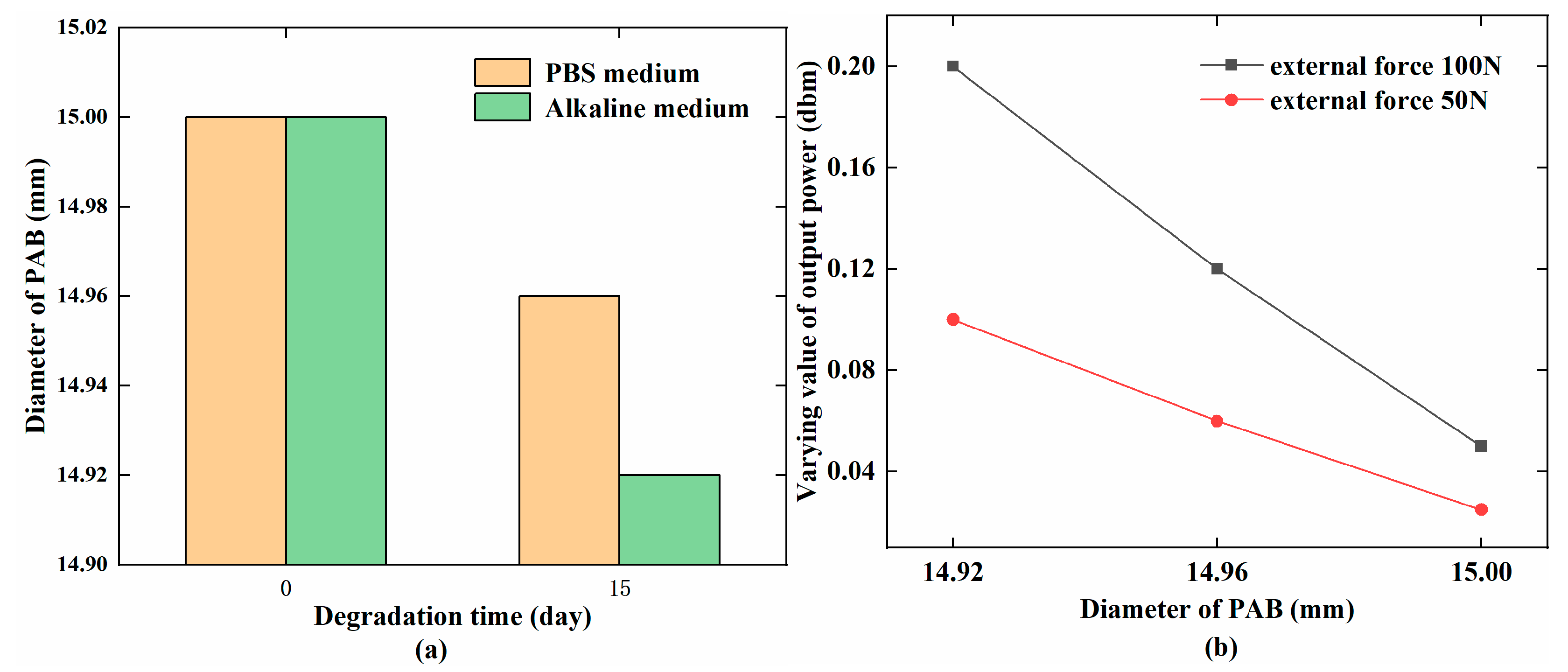
| Time (days) (PAB Condition) | 0 (Without Treatment) | 15 (In Alkaline Media) | 15 (In PBS Media) |
|---|---|---|---|
| Diameter of PAB (mm) | 15 ± 0.05 | 14.92 ± 0.02 | 14.96 ± 0.02 |
| Weight loss (g) | 0 | 0.3 ± 0.03 | 0.15 ± 0.03 |
| Varying value of output power (dbm) in 50 N | 0.02 ± 0.004 | 0.09 ± 0.04 | 0.06 ± 0.02 |
| Varying value of output power (dbm) in 100 N | 0.04 ± 0.004 | 0.2 ± 0.004 | 0.11 ± 0.02 |
| Stress (N/mm2) | 0.566 | 0.579 | 0.569 |
© 2019 by the authors. Licensee MDPI, Basel, Switzerland. This article is an open access article distributed under the terms and conditions of the Creative Commons Attribution (CC BY) license (http://creativecommons.org/licenses/by/4.0/).
Share and Cite
Yu, K.; Ren, L.; Tan, Y.; Wang, J. Wireless Magnetoelasticity-Based Sensor for Monitoring the Degradation Behavior of Polylactic Acid Artificial Bone In Vitro. Appl. Sci. 2019, 9, 739. https://doi.org/10.3390/app9040739
Yu K, Ren L, Tan Y, Wang J. Wireless Magnetoelasticity-Based Sensor for Monitoring the Degradation Behavior of Polylactic Acid Artificial Bone In Vitro. Applied Sciences. 2019; 9(4):739. https://doi.org/10.3390/app9040739
Chicago/Turabian StyleYu, Kun, Limin Ren, Yisong Tan, and Junyao Wang. 2019. "Wireless Magnetoelasticity-Based Sensor for Monitoring the Degradation Behavior of Polylactic Acid Artificial Bone In Vitro" Applied Sciences 9, no. 4: 739. https://doi.org/10.3390/app9040739
APA StyleYu, K., Ren, L., Tan, Y., & Wang, J. (2019). Wireless Magnetoelasticity-Based Sensor for Monitoring the Degradation Behavior of Polylactic Acid Artificial Bone In Vitro. Applied Sciences, 9(4), 739. https://doi.org/10.3390/app9040739