Article

A New “Turn-On” Fluorescence Probe for Al3+ Detection and Application Exploring in Living Cell and Real Samples

1
School of Materials Science and Engineering, Qilu University of Technology, Shandong Academy of Sciences, Jinan 250353, China
2
School of Chemistry and Pharmaceutical Engineering, Qilu University of Technology, Shandong Academy of Sciences, Jinan 250353, China
*
Author to whom correspondence should be addressed.
Appl. Sci. 2019, 9(3), 577; https://doi.org/10.3390/app9030577
Submission received: 10 January 2019 / Revised: 29 January 2019 / Accepted: 4 February 2019 / Published: 10 February 2019
(This article belongs to the Section Applied Biosciences and Bioengineering)

Abstract

:
An excess of Al3+ will lead to biological disorders and even many diseases. Therefore, detecting the levels of Al3+ in the human body has drawn great attention for health monitoring. The fluorescence method has been broadly applied because of high sensitivity, real-time detection, and intracellular imaging. In this work, a new probe with “turn-on” fluorescence based on Schiff base derivative, 3,6-imine-triphenylamine-(9-ethyl) carbazole (ITEC), has been successfully synthesized and studied. The high selectivity and sensitivity of ITEC to Al3+ were verified by fluorescence spectra and the detection limit was 2.19 nmol/L. A 1:2 stoichiometry of ITEC-Al3+ was obtained by the 1H NMR spectra and Job’s plot. Furthermore, ITEC was successfully applied to the detection of Al3+ with different concentrations in living HeLa cells. The analog experiments about nature contamination of Al3+ in cells and real samples were finished.

Graphical Abstract

1. Introduction

Recently, the development and applications of multifarious fluorescence probes for metal ions detection in living organisms and the environment have drawn considerable attentions [1,2,3,4,5]. For all we know, aluminum is the richest metallic element on earth [6]. During cell growth and living body survival, aluminum is not a necessary element. However, it is used broadly in our daily life, such as food ingredients [7], medicines [8], water treatment [9], and the huge manufacturing industry [10], which lead to many aluminum ions discharged in our soil, water, and air [11,12]. Thus, the excess deposition of Al3+ in the human body from the food cycle may disturb the immune system and nervous system, and lead to numerous irreversible diseases including cerebral disease [13], Alzheimer’s disease [14], and Parkinson’s disease [15]. The World Health Organization (WHO) suggested that the daily intake of Al3+ should be approximate 3–10 mg per person [16,17]. In China, the National Food Safety Standard sets the aluminum content not to exceed 100 mg/kg in food and food additives (China GB 29924-2013). In view of the above, it is necessary to detect Al3+ in organisms and the environment [18]. Ion selective electrodes [19], voltammetry [20], and atomic absorption spectroscopy [21] are generally employed to detect metal ions. Nevertheless, these methods commonly have some disadvantages, such as being time-consuming, expensive, needing skilled operations, and having tedious synthetic processes [22]. However, the fluorescence method has attracted much interest in environmental chemistry, biology, and clinical science owing to its high sensitivity and easy operational procedure [16,23].
Currently, Schiff bases compounds are vastly recognized as fluorescence probes for metal ions with the high sensitivity, good selectivity, and high excitation coefficients, but a lot of them displayed fluorescence “on-off” behavior [24,25,26], which is a disadvantage for the visualization study. In recent years, a few “off-on” fluorescence probes have been developed, but the probes applied to detect Al3+ in living cells and real samples were scarce [27,28,29,30]. Hence, it is very meaningful to develop “off-on” probes for Al3+ monitoring in real application [31].
In this manuscript, we reported a feasible method to synthesize a novel Schiff base, 3,6-imine-triphenylamine-(9-ethyl) carbazole (ITEC), as a “turn-on” fluorescence probe for selective detection of Al3+. The ITEC was further applied to detect Al3+ in living cells. In addition, the experiments of Al3+ detection in the real sample have been carried out.

2. Experimental

2.1. Materials and Instrumentation

Carbazole, triphenylamine, and Palladium/C catalysts were purchased from Aladdin. All other experimental materials were derived from the local commercial suppliers. All materials were not further purified. 1H NMR spectra were measured on a Bruker Avance 400 spectrometer. The electrospray ionization (ESI) mass spectra were recorded using an Agilent-6510-Q-TOF spectrometer. A UV-3600 spectrophotometer and a Thermo Scientific Nicolet IS10 Spectrometer were used to measure the UV-visible absorption spectra and Infrared spectra. Fluorescence spectra were obtained using a Hitachi F-7000 Fluorescence Spectrophotometer. The fluorescence images in living cells were obtained by an Olympus FV300 confocal microscope.

2.2. Synthesis of ITEC

Intermediates M1-M4 were synthesized according to the literature reports [32,33] and the detailed procedures were described in supporting information. The synthesis of 3,6-imine-triphenylamine-(9-ethyl) carbazole (ITEC) is shown in Scheme 1.

2.3. The Synthesis Procedure of ITEC

3-(Diphenyl amino) benzaldehyde (M1) (1.99 g, 11.5 mmol) and 3,6-diamino-9-ethyl-carbazole (1.1 g, 5 mmol) (M4) were dissolved in methanol (15 mL), and a drop of acetic acid was added to the above mixture. The mixture was reacted at 70 °C for 48 hours, then filtered and washed three times with ethanol. The brown solid was dried under vacuum. Yield: 65.6%. 1H NMR (400 MHz, DMSO) δ ppm: 8.68 (s, 2 H), 8.16 (s, 2 H), 7.8(d, J = 12 Hz, 4 H), 7.6 (d, J = 8 Hz 2 H), 7.41 (m, 10 H), 7.13 (m, 12 H), 7(d, J = 8 Hz, 4 H), 4.45 (d, J = 8 Hz, 2 H), 1.32 (t, J = 6 Hz, 3 H). IR: (KBr, cm−1): 2970 (N-H), 1687 (-CH=N), 1585 (C=C). Elemental analyses of C52H41N5 (%): calcd for C, 84.90; H, 5.58; N, 9.52, found C, 84.53; H, 5.46; N, 9.39. EI-MS (M−1): calcd for 736.75, found 735.92.

2.4. Absorption and Fluorescence Studies

The stock solutions (2 × 10⁻² mol/L) of the nitrate salts of Cu2+, Co2+, Pb2+, Cd2+, Ag+, Zn2+ Al3+, Hg2+, Ca2+, Na+, Fe3+, and Ni2+ were prepared in ultrapure water. The stock solution of ITEC (1 × 10⁻² mol/L) was prepared in ethanol. Test sample solutions will be diluted to 4 × 10⁻⁵ mol/L for nitrate salts and 2 × 10⁻⁵ mol/L for ITEC before using. The fluorescence spectra of ITEC were excited at 365 nm.

2.5. General Processes for Cell Culture and Fluorescence Imaging

HeLa cells were cultured in H-DMEM at 37 °C in humidified air and 5% CO2. The cells were fully incubated with ITEC or ITEC-Al3+ solution for 20 min at 37 °C, and then washed three times with phosphate buffered saline (PBS) to remove extracellular ITEC and Al3+. Continuously cultured cells with 7.4 umol/L Al3+ daily for 3 days and followed by normal culture for 5 days to mimic bioaccumulation. Finally, confocal imaging was carried out (λex = 405 nm, λem = 400–550 nm).

3. Results and Discussion

3.1. The Colorimetric Detection of ITEC to Al3+ by Naked Eyes

The observing method by the naked eyes is the convenient and inexpensive way of detecting metal ions. ITEC could successfully detect Al3+ by the naked eyes. In Figure 1a, the obviously blue fluorescence was found in the presence of Al3+ at the excitation wavelength of 365 nm when ITEC (20 μmol/L) solutions in ethanol were added various metal ions (40 μmol/L) including Co2+, Cu2+, Pb2+, Cd2+, Ag+, Zn2+ Al3+, Hg2+, Ca2+, Na+, Fe3+, and Ni2+. Moreover, the fluorescence intensities of ITEC solutions were gradually strengthened with the increasing concentrations of Al3+ in the range from 1 × 10⁻⁶ mol/L to 4 × 10⁻⁵ mol/L (Figure 1b). The limiting concentration (7.41 umol/L) of Al3+ is just in the range according to WHO regulations on drinking water, which indicates that it is possible to determine the level of Al3+ by naked eye observation [34].

3.2. Selectivity of ITEC for Metal Ions

In view of the above easy way that ITEC response to Al3+, a further optical investigation was carried out to evaluate potential application of ITEC for Al3+ detection. The absorption and fluorescence spectra of ITEC was investigated after adding different metal ions (Al3+, Ag+, Ca2+, Cd2+, Co2+, Cu2+, Fe3+, Hg2+, Na+, Ni2+, Pb2+, Zn2+). In Figure 2a, a new absorption band at 475 nm was found after adding Cu2+, Fe3+, Hg2+, and Al3+. In Figure 2b, compared with other metal ions, the fluorescence of ITEC exhibited remarkable enhancements in the presence of Al3+. Under the excitation wavelength of 365 nm, though the fluorescence of ITEC was enhanced in the presence of Fe3+, the fluorescent intensity was far lower than that in the presence of Al3+. Moreover, the two spectral shapes were absolutely difference. The possible reason for the enhanced fluorescence was the blocking of the photo-induced electron transfer process in the presence of Al3+ [35,36].
The selective bars of ITEC to various metal ions were shown in Figure 2c. Here, F and F0 are the fluorescent intensity of ITEC in the presence and absence of metal ions. Compared with other metal ions, the fluorescence intensity of ITEC was obviously improved after adding Al3+ (at least 6.6 times). The fluorescence quantum yields of ITEC and ITEC-Al3+ (Mole ratio = 1:2) were calculated to be 0.0023 and 0.016, respectively, by using the solution containing coumarin307 (Φ = 0.56, in methanol) standard as the reference [37]. These results clearly showed that ITEC exhibited excellent selectivity for Al3+.

3.3. Sensitivity of ITEC to Al3+

The fluorescence titration experiments were carried out by adding the various concentrations of Al3+ (Figure 3a). With the increased concentrations of Al3+ in the range from 2 μmol/L to 10 μmol/L, the linear fitting curve with fluorescence intensity of ITEC was plotted in Figure 3b. The concentration of Al3+ (7.41 umol/L) is within this linear range according to WHO regulations on drinking water [34]. According to the basis of 3 σ/k, the detection limit of ITEC was calculated to be 2.19 × 10⁻⁹ mol/L (k = 8.861 × 10⁸, σ = 0.647) [38,39], which was lower than those of many reported fluorescence probes for Al3+ (Table 1).

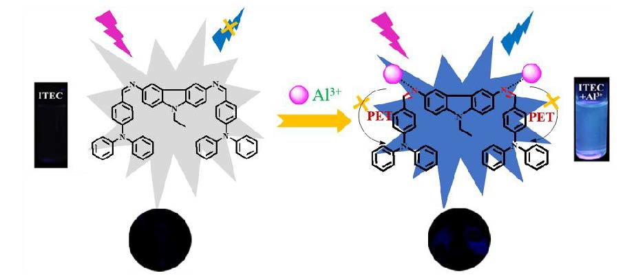
3.4. The Interaction Mechanism of ITEC-Al3+

A Job’s plot was drawn according to our previous method to obtain the complex ratio of ITEC-Al3+ [47]. From Figure 4a, the fluorescent spectrum vertex reached the mole fraction of 0.7, which indicated a 1:2 stoichiometry between ITEC and Al3+ [48,49]. To further investigate interaction mechanism, the 1H NMR spectra of ITEC and ITEC-Al3+ were measured and showed in Figure 4b. The integral intensities of peaks from a to c at 7.71 ppm improve continually with the increase of Al3+ concentrations, and the same phenomena is also found at 6.87, which indicates that the proton signal of HC=N will change with the addition of Al3+. The coordination of Al3+ with the N on amide bond can availably reduce their electron-donating ability, so the photoinduced electron transfer (PET) process from the N to the triphenylamine is suppressed, which leads to a large increase of fluorescence intensity. Based on the above studying, a possible sensing mechanism of ITEC toward Al3+ has been proposed as shown in Figure 4c. At the same time, there was obvious blue fluorescence after adding Al3+. In addition, a 2:1 stoichiometry between ITEC and Fe3+ was found. The binding mode of ITEC and Fe3+ was explored and detailed in Figure S6–S8 are supporting information.

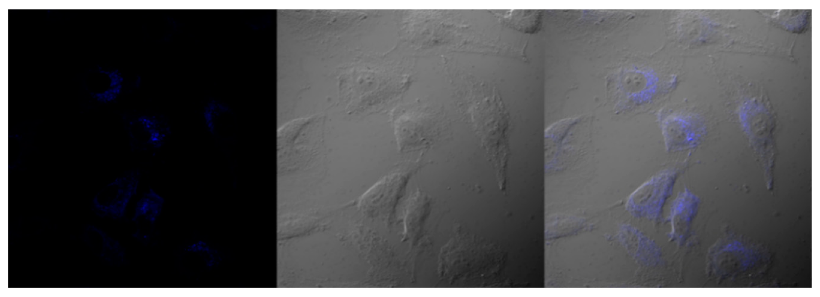
3.5. Fluorescence Imaging in Cells

The application of ITEC for the detection of Al3+ in HeLa cells was studied. The cells were incubated with 5 μmol/L of ITEC and 0 μmol/L, 2 μmol/L, and 5 μmol/L Al3+ solution, respectively, in PBS (2% DMSO, pH = 7.4) for 20 min at 37 °C, then washed three times with PBS to remove the excess ITEC and Al3+. As depicted in Figure 5, no obvious fluorescence was observed in the cells incubated only with ITEC. However, the fluorescence was founded in Figure 5 (2a–3a) after adding Al3+. Moreover, it displayed enhanced fluorescence with the increased concentrations of Al3+ (2 μmol/L, 5 μmol/L) in cells. Therefore, the results in cells indicated that ITEC could be used as an effective Al3+ imaging agent.
As we all know, Al3+ is a non-essential element of the human body. It will accumulate in the body from water or food containing Al3+. Considering the complicated process, we mimic the phenomenon of bioaccumulation and performed cell imaging (Figure 6). The cells were cultured with 7.4 μmol/L Al3+ daily for 3 days and followed by normal culture for 5 days to achieve natural concentration, then incubated with ITEC solution (5 μmol/L) for 20 min. The cell has been successfully imaged, which demonstrates the applicability of the technique.

3.6. The Test of the ITEC in Real Sample

The lake water and tap water samples were gained from the Changqing Lake (Jinan, China) and the Qilu University of Technology, respectively. The bottled water samples were purchased from the local supermarket. The water samples were centrifuged and filtrated, and then added to Al3+ solutions with the concentrations of 3 μmol/L, 5 μmol/L, and 10 μmol/L, respectively. The proposed method was used to detect Al3+ in real samples. The results in Table 2 showed the recoveries were in the range from 97.6% to 109.3% with lower relative standard deviation (RSD < 1%), which clearly indicates the reliability and accuracy of the proposed method.

4. Conclusions

In a word, we developed a new Schiff base derivative, which has good selectivity and sensitivity towards Al3+. The detection limit was 2.19 nmol/L. The fluorescence titration tests and the Job’s plot elucidated that a stoichiometric ratio of 1:2 for ITEC-Al3+ was calculated. Furthermore, ITEC can be successfully regarded as a fluorescence “turn-on” probe for the detection of Al3+ in cells imaging, which supplied the potential application of ITEC to study the effect of Al3+ in biological systems. In addition, ITEC was highly efficient for the monitoring of Al3+ in real samples.

Supplementary Materials

The following are available online at https://www.mdpi.com/2076-3417/9/3/577/s1, Figure S1: The 1H NMR spectrum of intermediate M1 in DMSO; Figure S2: The 1H NMR spectrum of intermediate M4 in DMSO; Figure S3: The 1H NMR spectrum of ITEC in DMSO; Figure S4: Mass spectrum of ITEC; Figure S5: The FTIR spectrum of ITEC; Figure S6: The Job’s plot for determining the stoichiometry between ITEC and Fe3+; Figure S7: 1H NMR (400 MHz) spectra of ITEC and ITEC with Fe3+ (1.0 equiv.); Figure S8: The proposed binding mechanism of ITEC with Fe3+.

Author Contributions

Conceptualization, X.Z. and J.-S.Y.; Data Curation, C.-D.Z. and S.X.; Formal Analysis, X.Z. and J.-S.Y.; Writing—Original Draft Preparation, Z.-Y.G.; Visualization, W.-L.L.; Funding Acquisition, X.Z.

Funding

This research was funded by the National Natural Science Foundation of China (NSFC 51403111, 11774188).

Conflicts of Interest

The authors declare no conflict of interest.

References

  1. Kim, H.; Rao, B.A.; Jeong, J.W.; Mallick, S.; Kang, S.M.; Choi, J.S.; Lee, C.S.; Son, Y.A. A highly selective dual-channel Cu2+ and Al3+ chemodosimeter in aqueous systems: Sensing in living cells and microfluidic flows. Sens. Actuators B Chem. 2015, 210, 173–182. [Google Scholar] [CrossRef]
  2. Anand, T.; Sivaraman, G.; Mahesh, A.; Chellappa, D. Aminoquinoline based highly sensitive fluorescent sensor for lead(II) and aluminum(III) and its application in live cell imaging. Anal. Chim. Acta 2015, 853, 596–601. [Google Scholar] [CrossRef] [PubMed]
  3. Manjunath, R.; Hrishikesan, E.; Kannan, P. A selective colorimetric and fluorescent sensor for Al3+ ion and its application to cellular imaging. Spectrochim. Acta A Mol. Biomol. Spectrosc. 2015, 140, 509–515. [Google Scholar] [PubMed]
  4. Dey, S.; Halder, S.; Mukherjee, A.; Ghosh, K.; Roy, P. Development of highly selective chemosensor for Al3+: Effect of substituent and biological application. Sens. Actuators B Chem. 2015, 215, 196–205. [Google Scholar] [CrossRef]
  5. Xie, J.Y.; Li, C.Y.; Li, Y.F.; Fu, Y.J.; Nie, S.X.; Tan, H.Y. A near-infrared chemosensor for determination of trivalent aluminum ions in living cells and tissues. Dyes Pigments 2017, 136, 817–824. [Google Scholar] [CrossRef]
  6. Asimov, I. The elementary composition of the earth. J. Chem. Educ. 1954, 33, 67. [Google Scholar] [CrossRef]
  7. Soni, M.G.; White, S.M.; Flamm, W.G.; Burdock, G.A. Safety Evaluation of Dietary Aluminum. Regul. Toxicol. Pharmacol. 2001, 33, 66–79. [Google Scholar] [CrossRef]
  8. Kontoghiorghes, G.J. Comparative efficacy and toxicity of desferrioxamine, deferiprone and other iron and aluminium chelating drugs. Toxicol. Lett. 1995, 80, 1–18. [Google Scholar] [CrossRef]
  9. Wang, W.; Li, H.; Ding, Z.; Wang, X. Effects of advanced oxidation pretreatment on residual aluminum control in high humic acid water purification. J. Environ. Sci. 2011, 23, 1079–1085. [Google Scholar] [CrossRef]
  10. Liu, G.; Bangs, C.E.; Müller, D.B. Unearthing potentials for decarbonizing the U.S. aluminum cycle. Environ. Sci. Technol. 2011, 45, 9515. [Google Scholar] [CrossRef]
  11. Delhaize, E.; Ryan, P.R. Aluminum Toxicity and Tolerance in Plants. Plant Physiol. 1995, 107, 315–321. [Google Scholar] [CrossRef] [PubMed] [Green Version]
  12. Tavakoli, O.; Yoshida, H. Effective recovery of harmful metal ions from squid wastes using subcritical and supercritical water treatments. Environ. Sci. Technol. 2005, 39, 2357–2363. [Google Scholar] [CrossRef] [PubMed]
  13. Verstraeten, S.V.; Aimo, L.; Oteiza, P.I. Aluminium and lead: Molecular mechanisms of brain toxicity. Arch. Toxicol. 2008, 82, 789–802. [Google Scholar] [CrossRef] [PubMed]
  14. Good, P.F.; Olanow, C.W.; Perl, D.P. Neuromelanin-containing neurons of the substantia nigra accumulate iron and aluminum in Parkinson’s disease: A LAMMA study. Brain Res. 1992, 593, 343–346. [Google Scholar] [CrossRef]
  15. Flaten, T.P. Aluminium as a risk factor in Alzheimer’s disease, with emphasis on drinking water. Brain Res. Bull. 2001, 55, 187–196. [Google Scholar] [CrossRef]
  16. Shangguan, J.; Huang, J.; He, D.; He, X.; Wang, K.; Ye, R.; Yang, X.; Qing, T.; Tang, J. Highly Fe3+-selective fluorescent nanoprobe based on ultrabright N/P codoped carbon dots and its application in biological samples. Anal. Chem. 2017, 89, 7477–7484. [Google Scholar] [CrossRef] [PubMed]
  17. Shen, K.; Mao, S.; Shi, X.; Wang, F.; Xu, Y.; Aderinto, S.O.; Wu, H. Characterization of a highly Al3+ selective fluorescence probe based on naphthalimide-Schiff base and its application to practical water samples. Luminescence 2018, 33, 54–63. [Google Scholar] [CrossRef]
  18. Fu, Y.; Jiang, X.J.; Zhu, Y.Y.; Zhou, B.J.; Zang, S.Q.; Tang, M.S.; Zhang, H.Y.; Mak, T.C. A new fluorescent probe for Al(3+) based on rhodamine 6G and its application to bioimaging. Dalton Trans. 2014, 43, 12624–12632. [Google Scholar] [CrossRef]
  19. Gupta, V.K.; Jain, A.K.; Maheshwari, G. Aluminum(III) selective potentiometric sensor based on morin in poly(vinyl chloride) matrix. Talanta 2007, 72, 1469–1473. [Google Scholar] [CrossRef]
  20. Mergu, N.; Kumar Singh, A.; Kumar Gupta, V. Highly sensitive and selective colorimetric and off on fluorescent reversible chemosensors for Al3+ based on the rhodamine fluorophore. Sensors 2015, 15, 9097–9111. [Google Scholar] [CrossRef]
  21. Cassella, R.J.; Magalhaes, O.I.; Couto, M.T.; Lima, E.L.S.; Neves, M.A.F.; Coutinho, F.M.B. Synthesis and application of a functionalized resin for flow injection/F AAS copper determination in waters. Talanta 2005, 67, 121–128. [Google Scholar] [CrossRef] [PubMed]
  22. Dong, M.; Dong, Y.M.; Ma, T.H.; Wang, Y.W.; Peng, Y. A highly selective fluorescence-enhanced chemosensor for Al3+ in aqueous solution based on a hybrid ligand from BINOL scaffold and β-amino alcohol. Inorg. Chim. Acta 2012, 381, 137–142. [Google Scholar] [CrossRef]
  23. Gupta, V.K.; Singh, A.K.; Kumawat, L.K. Thiazole Schiff base turn-on fluorescent chemosensor for Al3+ ion. Sens. Actuators B Chem. 2014, 195, 98–108. [Google Scholar] [CrossRef]
  24. Tian, J.; Yan, X.; Yang, H.; Tian, F. A novel turn-on Schiff-base fluorescent sensor for Aluminum (III) ions in living cells. RSC Adv. 2015, 5, 107012–107019. [Google Scholar] [CrossRef]
  25. Yong, S.K.; Lee, J.J.; Ye, W.C.; You, G.R.; Nguyen, L.T.; Noh, I.; Kim, C. Simultaneous bioimaging recognition of cation Al3+ and anion F- by a fluorogenic method. Dyes Pigments 2016, 129, 43–53. [Google Scholar]
  26. Azadbakht, R.; Almasi, T.; Keypour, H.; Rezaeivala, M. A new asymmetric Schiff base system as fluorescent chemosensor for Al3+ ion. Inorg. Chem. Commun. 2013, 33, 63–67. [Google Scholar] [CrossRef]
  27. Maity, S.B.; Bharadwaj, P.K. A Chemosensor Built with Rhodamine Derivatives Appended to an Aromatic Platform via 1,2,3-Triazoles: Dual Detection of Aluminum(III) and Fluoride/Acetate Ions. Inorg. Chem. 2013, 52, 1161–1163. [Google Scholar] [CrossRef]
  28. Boonkitpatarakul, K.; Wang, J.; Niamnont, N.; Liu, B.; Mcdonald, L.; Pang, Y.; Sukwattanasinitt, M. Novel Turn-On Fluorescent Sensors with Mega Stokes Shifts for Dual Detection of Al3+ and Zn2+. ACS Sens. 2015. [Google Scholar] [CrossRef]
  29. Samanta, S.; Nath, B.; Baruah, J.B. Hydrolytically stable Schiff base as highly sensitive aluminium sensor. Inorg. Chem. Commun. 2012, 22, 98–100. [Google Scholar] [CrossRef]
  30. Oliveira, E.; Santos, H.M.; Capelo, J.L.; Lodeiro, C. New emissive dopamine derivatives as fluorescent chemosensors for metal ions: A CHEF effect for Al(III) interaction. Inorg. Chim. Acta 2012, 381, 203–211. [Google Scholar] [CrossRef]
  31. Sen, B.; Pal, S.; Banerjee, S.; Lohar, S.; Chattopadhyay, P. Al3+-Ion-Triggered Conformational Isomerization of a Rhodamine B Derivative Evidenced by a Fluorescence Signal—A Crystallographic Proof. Eur. J. Inorg. Chem. 2015, 2015, 1383–1389. [Google Scholar] [CrossRef]
  32. Kaya, S.; Koyuncu, S. Synthesis and characterization of imine-coupled polyphenols containing carbazole units. J. Appl. Poly. Sci. 2009, 113, 1975–1985. [Google Scholar] [CrossRef]
  33. Monçalves, M.; Rampon, D.D.S.; Schneider, P.H.; Rodembusch, F.S.; Silveira, C.D.C. Divinyl sulfides/sulfones-based D-π-A-π-D dyes as efficient non-aromatic bridges for π-conjugated compounds. Dyes Pigments 2014, 102, 71–78. [Google Scholar] [CrossRef]
  34. Han, T.; Feng, X.; Tong, B.; Shi, J.; Chen, L.; Zhi, J.; Dong, Y. A novel “turn-on” fluorescent chemosensor for the selective detection of Al3+ based on aggregation-induced emission. Chem. Commun. 2012, 48, 416–418. [Google Scholar] [CrossRef] [PubMed]
  35. Guo, A.; Zhu, R.; Ren, Y.; Dong, J.; Feng, L. A “turn-on” fluorescent chemosensor for aluminum ion and cell imaging application. Spectrochim. Acta A Mol. Biomol. Spectrosc. 2016, 153, 530–534. [Google Scholar] [CrossRef] [PubMed]
  36. Zhou, D.; Sun, C.; Chen, C.; Cui, X.; Li, W. Research of a highly selective fluorescent chemosensor for aluminum(III) ions based on photoinduced electron transfer. J. Mol. Struct. 2015, 1079, 315–320. [Google Scholar] [CrossRef]
  37. Orlovskii, Y.V.; Popov, A.V.; Orlovskaya, E.O.; Vanetsev, A.S.; Vagapova, E.A.; Rähn, M.; Sammelselg, V.; Sildos, I.; Baranchikov, A.E.; Grachev, P.V. Comparison of concentration dependence of relative fluorescence quantum yield and brightness in first biological window of wavelengths for aqueous colloidal solutions of Nd3+: LaF3 and Nd3+: Ky3 F10 nanocrystals synthesized by microwave-hydrothermal. J. Alloys Compd. 2018. [Google Scholar] [CrossRef]
  38. Li, C.R.; Qin, J.C.; Wang, B.D.; Fan, L.; Yan, J.; Yang, Z.Y. A Chromone-Derived Schiff-Base Ligand as Al3+ “Turn on” Fluorescent Sensor: Synthesis and Spectroscopic Properties. J. Fluoresc. 2016, 26, 345–353. [Google Scholar] [CrossRef]
  39. Peng, H.; Shen, K.; Mao, S.; Shi, X.; Xu, Y.; Aderinto, S.O.; Wu, H. A Highly Selective and Sensitive Fluorescent Turn-on Probe for Al(3+) Based on Naphthalimide Schiff Base. J. Fluoresc. 2017, 27, 1191–1200. [Google Scholar] [CrossRef]
  40. Qin, J.C.; Yang, Z.Y. Bis-schiff base as a donor-acceptor fluorescent probe: Recognition of Al3+ ions in near 100% aqueous solution. J. Photoch. Photobiol. A 2015, 303–304, 99–104. [Google Scholar] [CrossRef]
  41. Shoora, S.K.; Jain, A.K.; Gupta, V.K. A simple schiff base based novel optical probe for aluminium (iii) ions. Sens. Actuators B Chem. 2015, 216, 86–104. [Google Scholar] [CrossRef]
  42. Wang, G.; Qin, J.; Li, C.; Yang, Z. A highly selective fluorescent probe for Al3+ based on quinoline derivative. Spectrochim. Acta A Mol. Biomol. Spectrosc. 2015, 150, 21–25. [Google Scholar] [CrossRef] [PubMed]
  43. Kang, L.; Liu, Y.T.; Li, N.N.; Dang, Q.X.; Xing, Z.Y.; Li, J.L.; Zhang, Y. A schiff-base receptor based naphthalimide derivative: Highly selective and colorimetric fluorescent turn-on sensor for Al3+. J. Luminesc. 2017, 186, 48–52. [Google Scholar] [CrossRef]
  44. Mu, X.; Li, Q.; Qiao, J.; Ma, H. One-pot synthesis of tyrosine-stabilized fluorescent gold nanoclusters and their application as turn-on sensors for Al3+ ions and turn-off sensors for Fe3+ ions. Anal. Methods 2014, 6, 6445–6451. [Google Scholar] [CrossRef]
  45. Zhu, S.Y.; Yan, B. A novel covalent post-synthetically modified mof hybrid as a sensitive and selective fluorescent probe for Al3+ detection in aqueous media. Dalton Trans. 2017. [Google Scholar] [CrossRef] [PubMed]
  46. Fang, B.Y.; Li, C.; Song, Y.Y.; Tan, F.; Cao, Y.C.; Zhao, Y.D. Nitrogen-doped graphene quantum dot for direct fluorescence detection of Al3+ in aqueous media and living cells. Biosens. Bioelectron. 2017, 100, S0956566317305894. [Google Scholar] [CrossRef] [PubMed]
  47. Meng, X.-L.; Zhang, X.; Yao, J.-S.; Zhang, J.-J.; Ding, B.-Y. Fluorescence and fluorescence imaging of two Schiff derivatives sensitive to Fe3+ induced by single- and two-photon excitation. Sens. Actuators B Chem. 2013, 176, 488–496. [Google Scholar] [CrossRef]
  48. Li, C.R.; Qin, J.C.; Wang, G.Q.; Wang, B.D.; Fu, A.K.; Yang, Z.Y. Development of a simple pyrazine-derived “turn on” Al3+ fluorescent sensor with high selectivity and sensitivity. Inorg. Chim. Acta 2015, 430, 91–95. [Google Scholar] [CrossRef]
  49. Lashgari, N.; Badiei, A.; Ziarani, G.M. A Fluorescent Sensor for Al(III) and Colorimetric Sensor for Fe(III) and Fe(II) Based on a Novel 8-Hydroxyquinoline Derivative. J. Fluoresc. 2016, 26, 1885–1894. [Google Scholar] [CrossRef]
Scheme 1. The synthesis route of 3,6-imine-triphenylamine-(9-ethyl) carbazole (ITEC).
Applsci 09 00577 sch001
Figure 1. (a) Color changes of 3,6-imine-triphenylamine-(9-ethyl) carbazole (ITEC) with various cations under 365 nm UV-light in ethyl alcohol. [ITEC] = 20 μM, [cations] = 40 μM. (b) Color changes of ITEC with different Al3+ concentrations (0 μM, 5 μM, 10 μM, 15 μM, 20 μM, 25 μM, 40 μM) under 365 nm UV-light, [ITEC] = 20 μM.
Applsci 09 00577 g001
Figure 2. (a) Ultraviolet spectra of ITEC (10 μM) in different metal ions (20 μM). (b) Fluorescence responses of ITEC (10 μM) to various metal ions (20 μM) by excitation at 365 nm. (c) The bar graph of fluorescence intensity of ITEC at 431 nm.
Applsci 09 00577 g002
Figure 3. (a) Change in fluorescence spectra of ITEC (10 μM) after adding Al3+ (0 μM, 0.6 μM, 0.8 μM, 2.0 μM, 2.4 μM, 3.0 μM, 4.4 μM, 5.0 μM, 6.4 μM, 7.0 μM, 9.0 μM, 10.0 μM, and 14.0 μM, respectively) with an excitation at 365 nm. (b) The curve was plotted with the fluorescence intensity of ITEC in 460 nm versus Al3+ concentrations (2.4 μM–10 μM).
Applsci 09 00577 g003
Figure 4. (a) The Job’s plot for ITEC-Al3+complex, [ITEC] + [Al3+] = 10 μM; (b) 1H NMR (400 MHz) spectra of ITEC, ITEC with Al3+ (1.0 equiv.), and ITEC with Al3+ (2.0 equiv.) in DMSO; (c) The proposed binding mechanism of ITEC-Al3+ and the color change of ITEC solution (20 μM) after adding Al3+ (40 μM).
Applsci 09 00577 g004
Figure 5. Fluorescence microscope images of ITEC (5 μM) in living HeLa cells with different concentrations of Al3+ (0 μM, 2 μM, and 5 μM, respectively).
Applsci 09 00577 g005
Figure 6. The cell cultured with 7.4 μM Al3+ daily for 3 days followed by normal culture for 5 days, then incubated with 5 μM ITEC for 20 min.
Applsci 09 00577 g006
Table 1. Comparison of probes structure for Al3+ detection by fluorescence method.
Structure of ProbesDetection Limit (mol/L)Modes (probe:Al3+)ApplicationRef.
Applsci 09 00577 i0011.0 × 10−71:1-[40]
Applsci 09 00577 i0024.79 × 10−81:1-[41]
Applsci 09 00577 i0031.0 × 10−71:1-[42]
Applsci 09 00577 i0047.4 × 10−91:1Real samples[43]
Tyrosine-stabilized fluorescent gold nanoclusters3.0 × 10−7-Real samples[44]
Salicylaldehyde modified MOF (UiO-66-NH2-SA)6.98 × 10⁻⁶--[45]
Nitrogen-doped graphene quantum dot1.3 × 10−61:1Cell imaging[46]
Applsci 09 00577 i0057.4 × 10−91:2Cell imagingThis work
Table 2. Determinations of Al3+ in real samples (n = 3).
SampleAdded (μmol/L)Measured (μmol/L)Recovery (%)RSD (%)
River water 132.9999.70.40
River water 255.27105.40.48
River water 31010.3103.30.60
Tap water 133.17105.50.50
Tap water 255.25104.90.12
Tap water 3109.7697.60.36
Bottle water 132.9999.80.88
Bottle water 255.46109.30.23
Bottle water 3109.8898.80.53
RSD: relative standard deviation.

