Low-Background Shielding Box for Autoradiography of Environmental Samples and the α-, β-, and γ-ray Sensitivities of the Imaging Plates


Abstract
:Featured Application
Abstract
1. Introduction
2. Materials and Methods
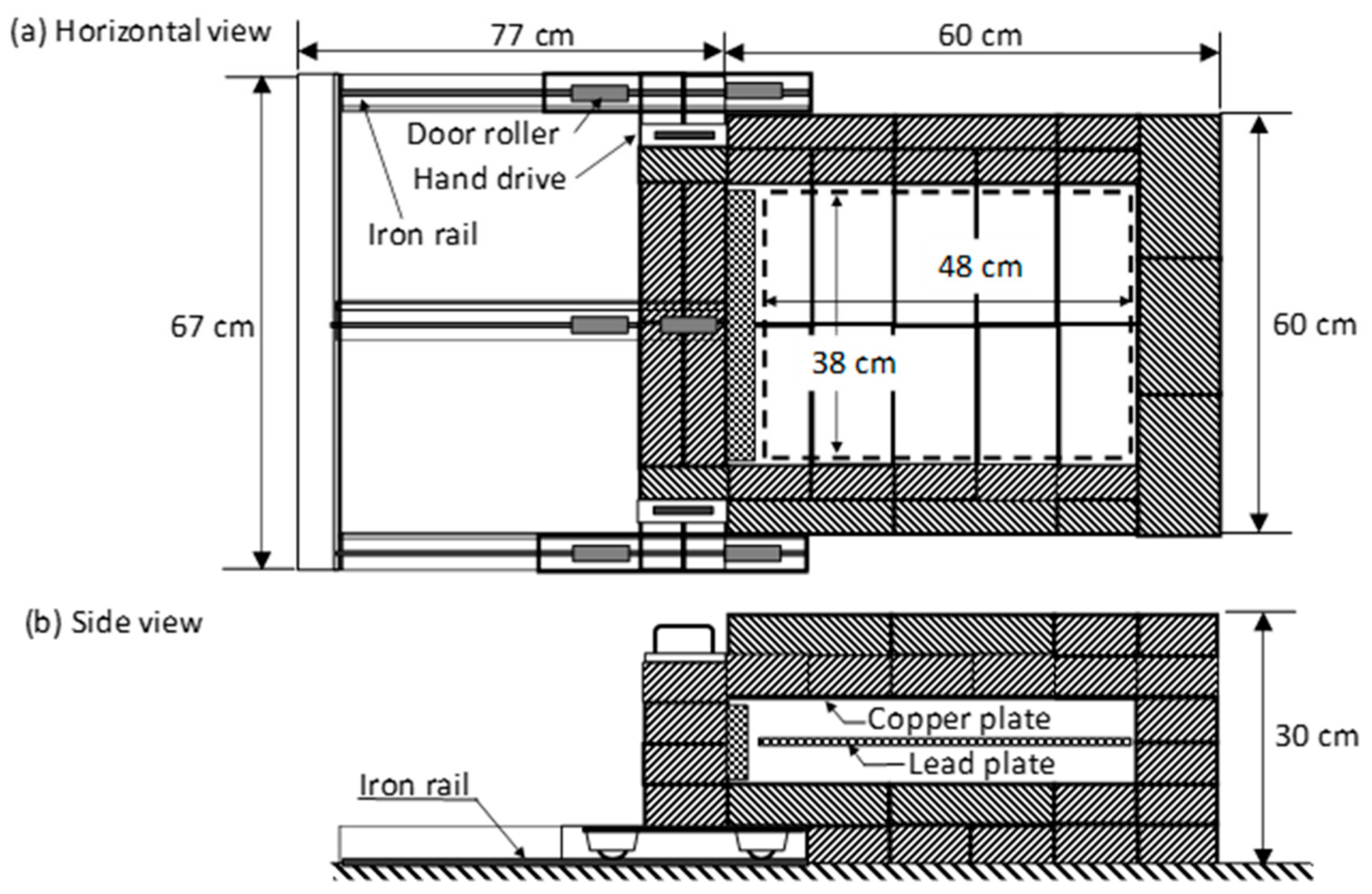
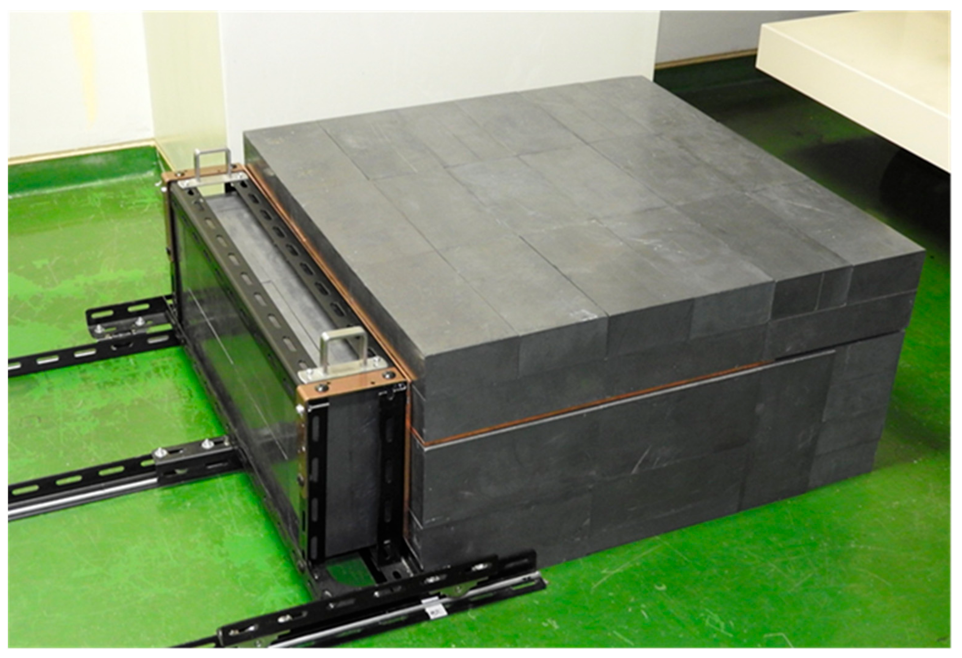
2.1. Design of Low-Background Shielding Box for IPs
2.2. Background Reduction of Radon Progeny Nuclides
2.3. α-ray Sensitivity of IPs
2.4. β- and γ-ray Sensitivity Measurement of IP
3. Results and Discussion
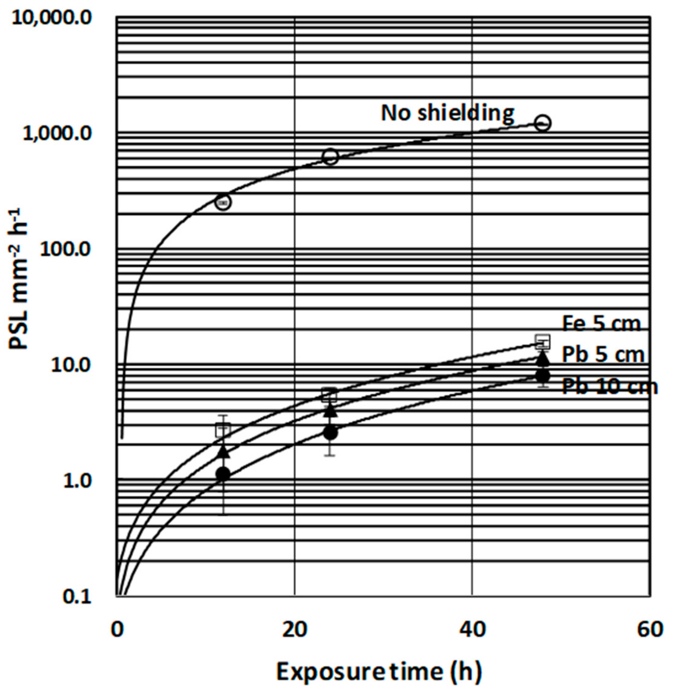
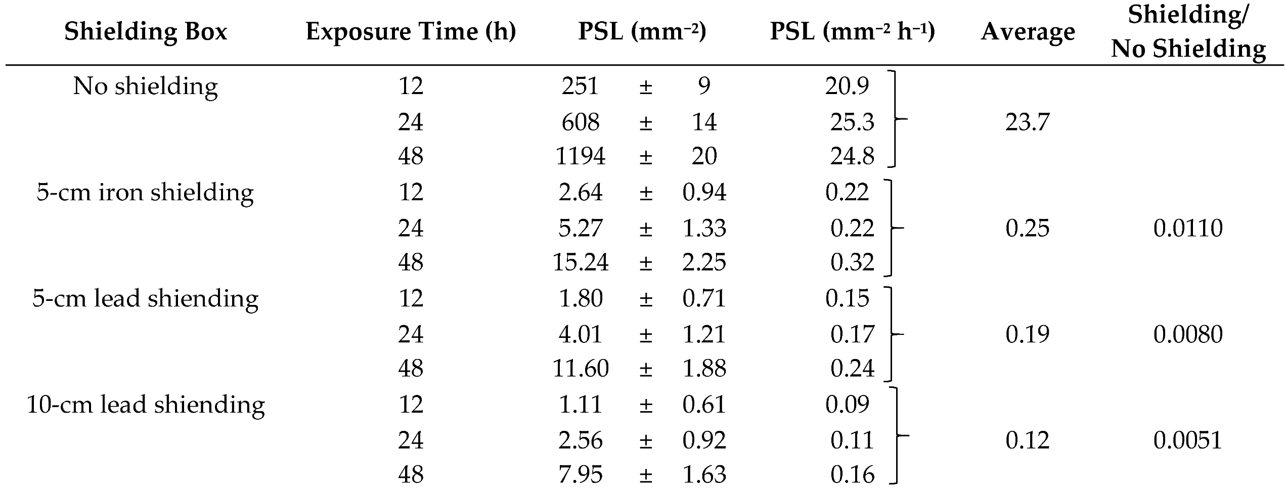
3.1. Background Characteristics of the Shielding Box
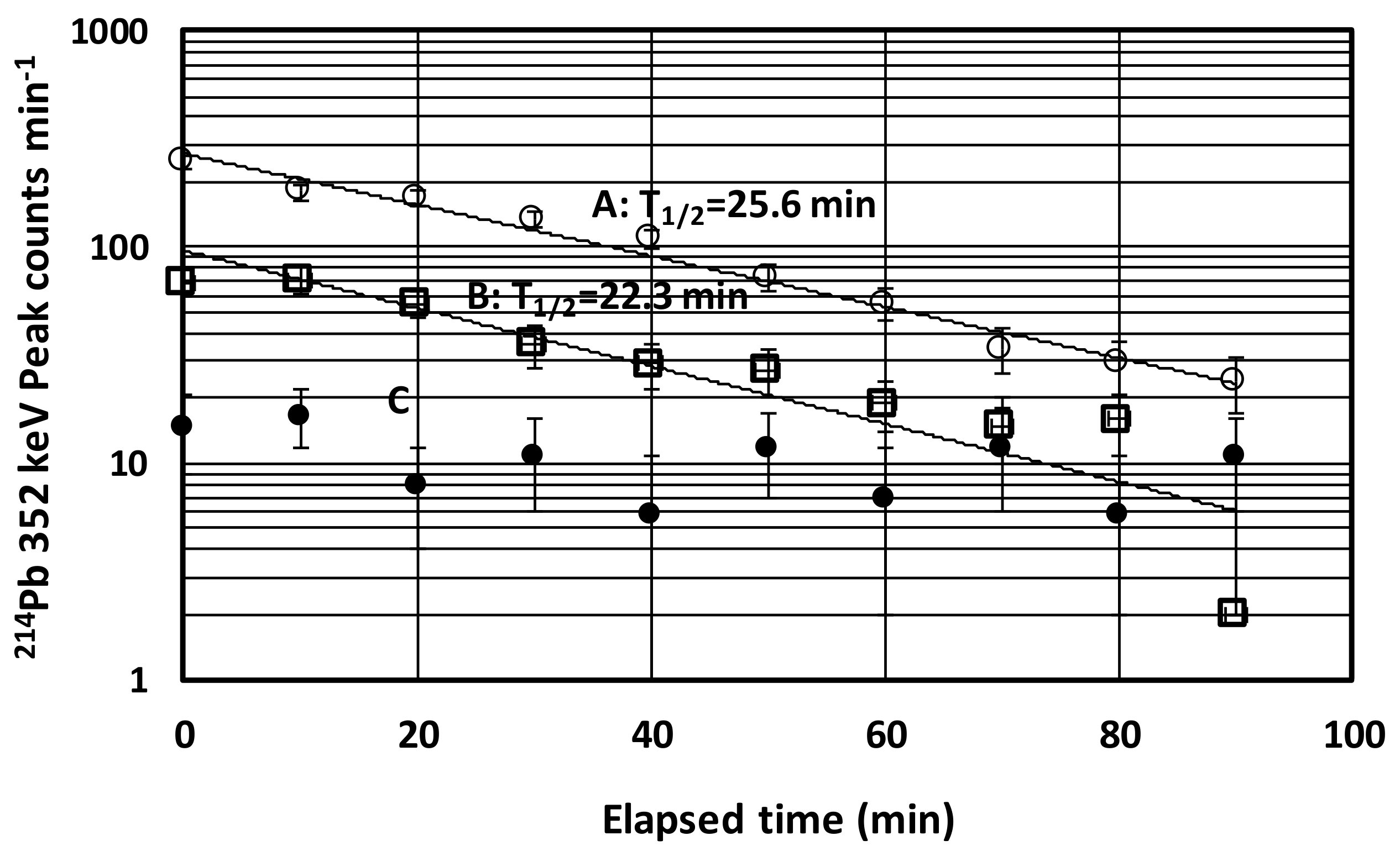
3.2. Background Reduction from Radon Progeny Nuclei
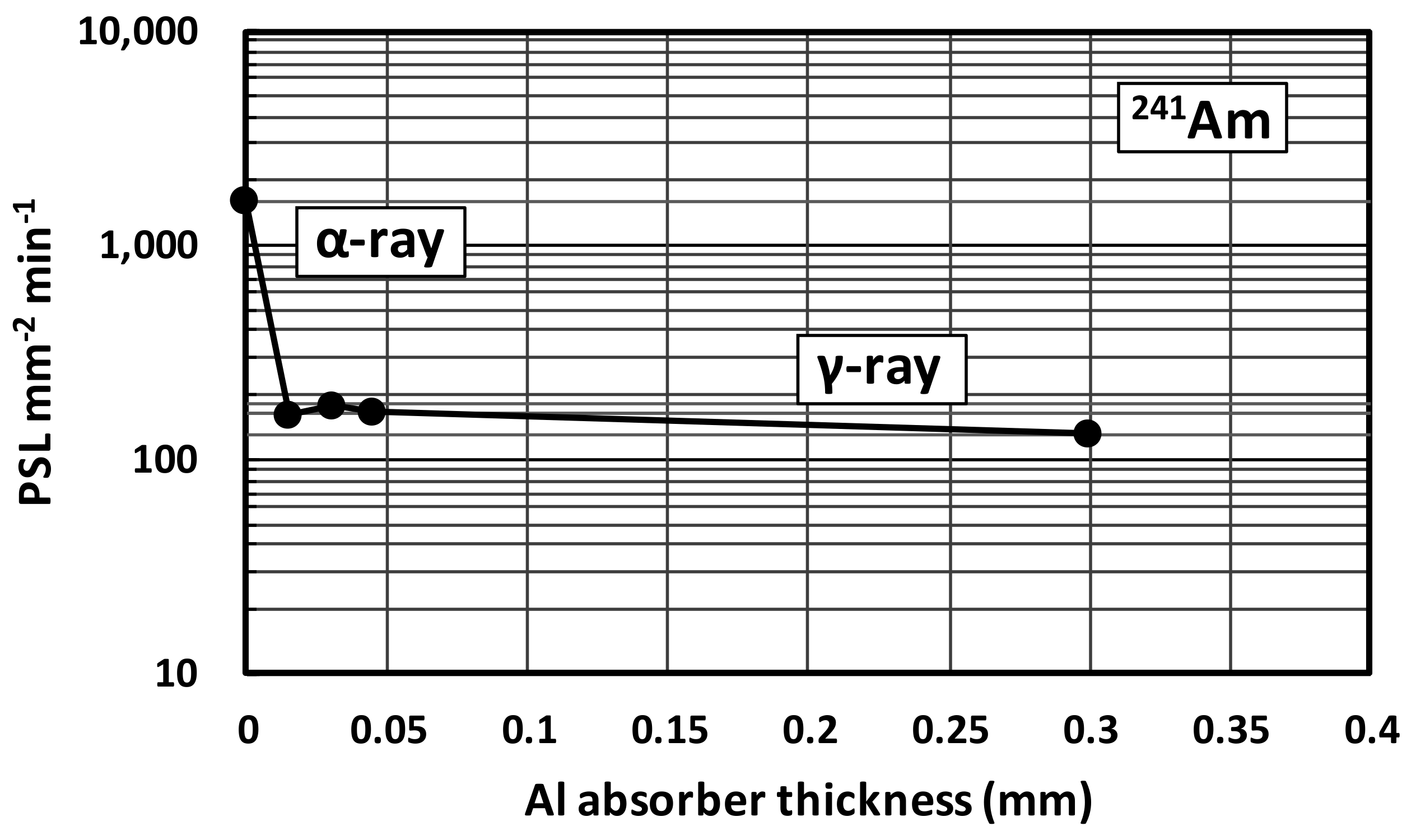
3.3. α-ray Sensitivity of IPs
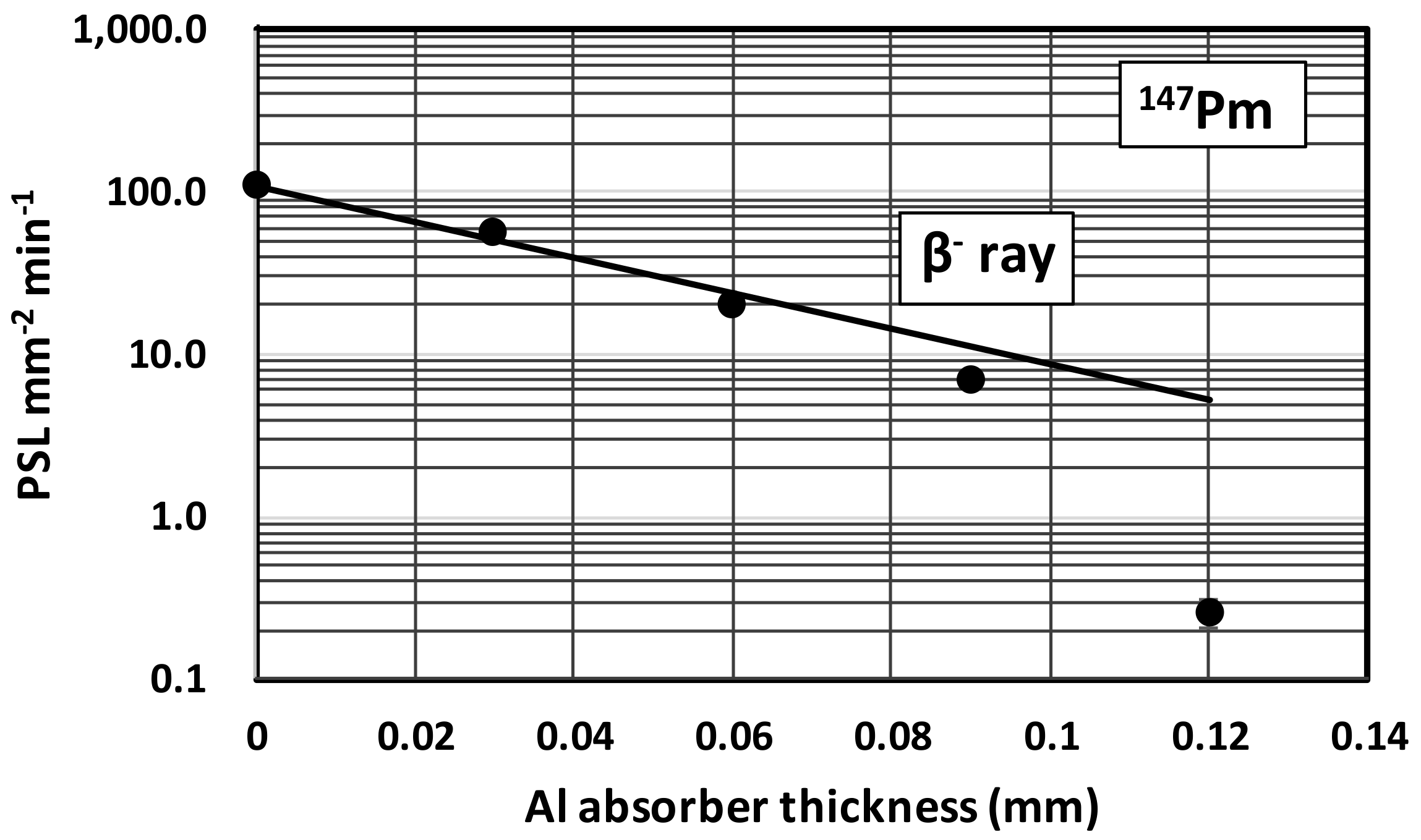
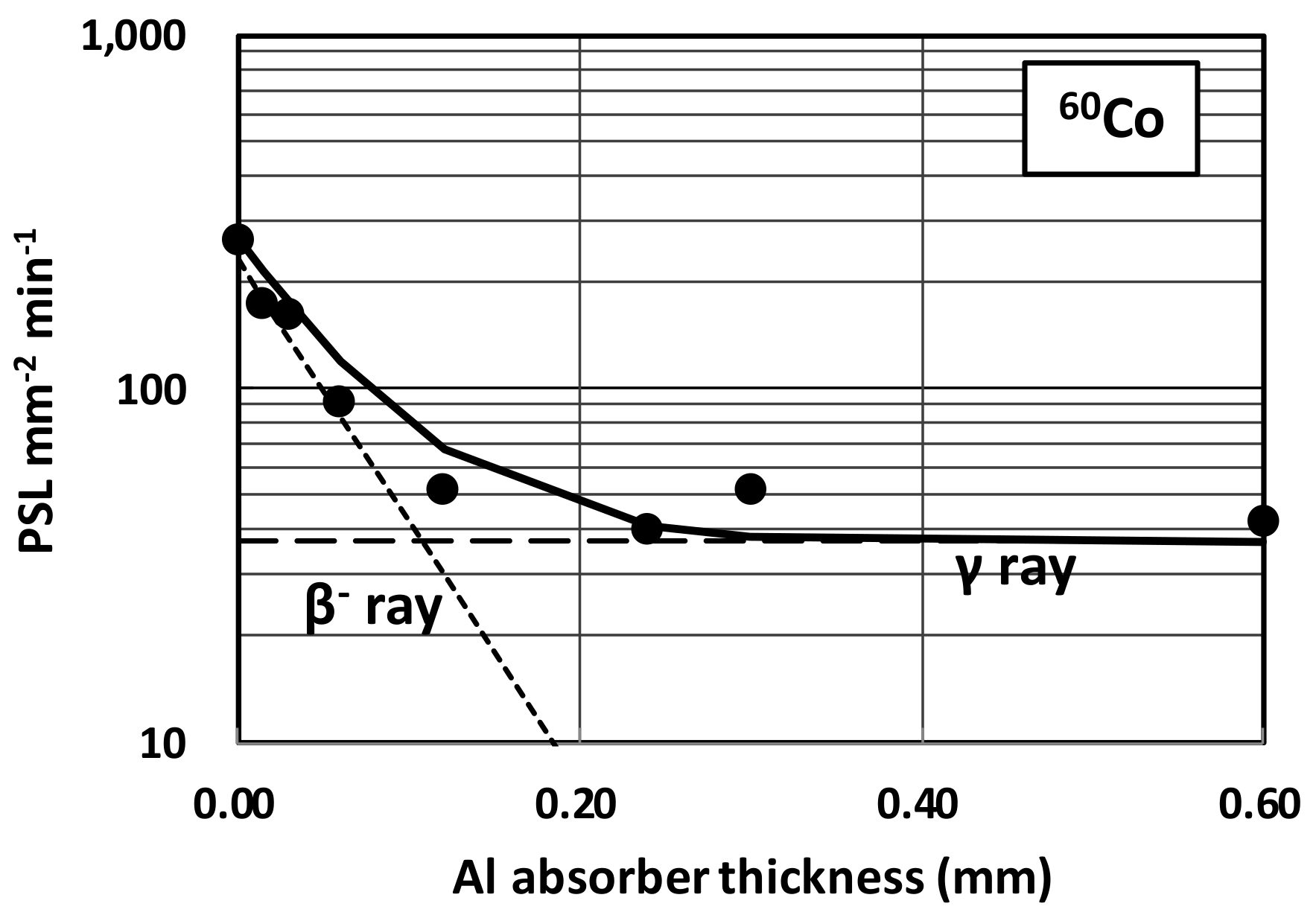
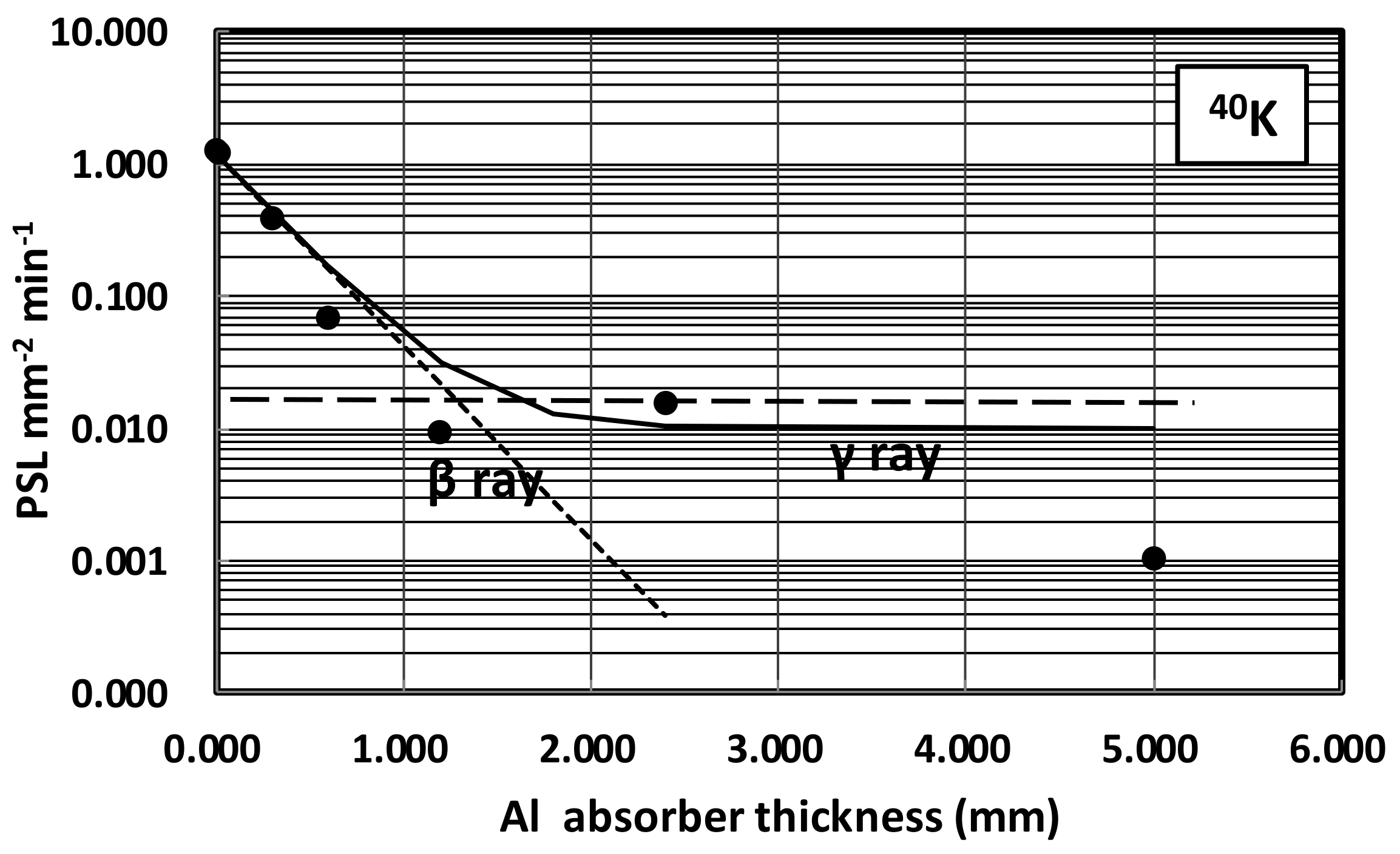
3.4. β- and γ-ray Sensitivity of IPs
4. Applications to Autoradiography of Environmental Samples
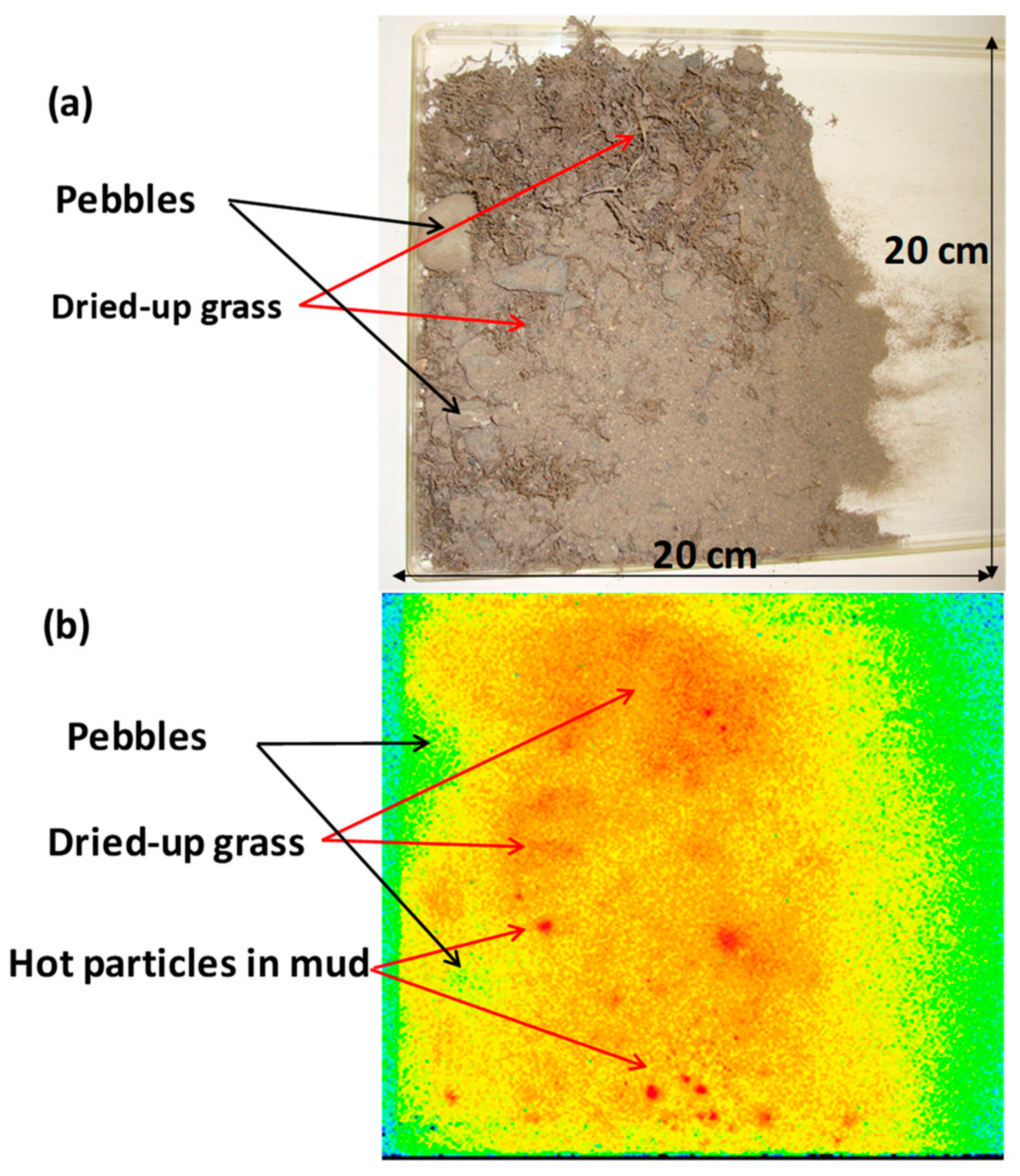
4.1. Sediments
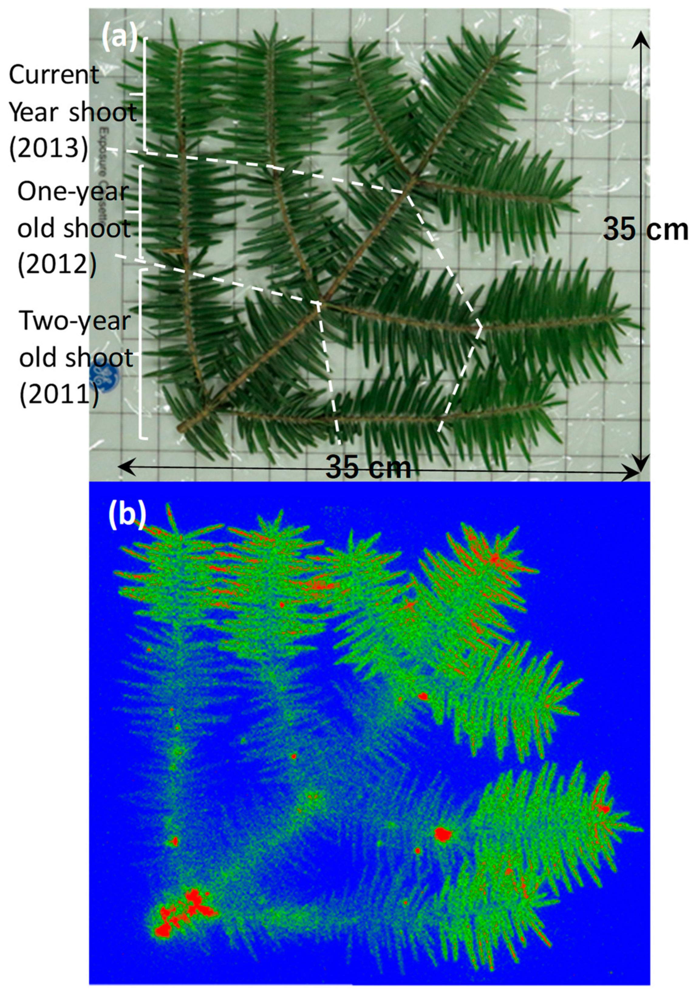
4.2. Momi Fir
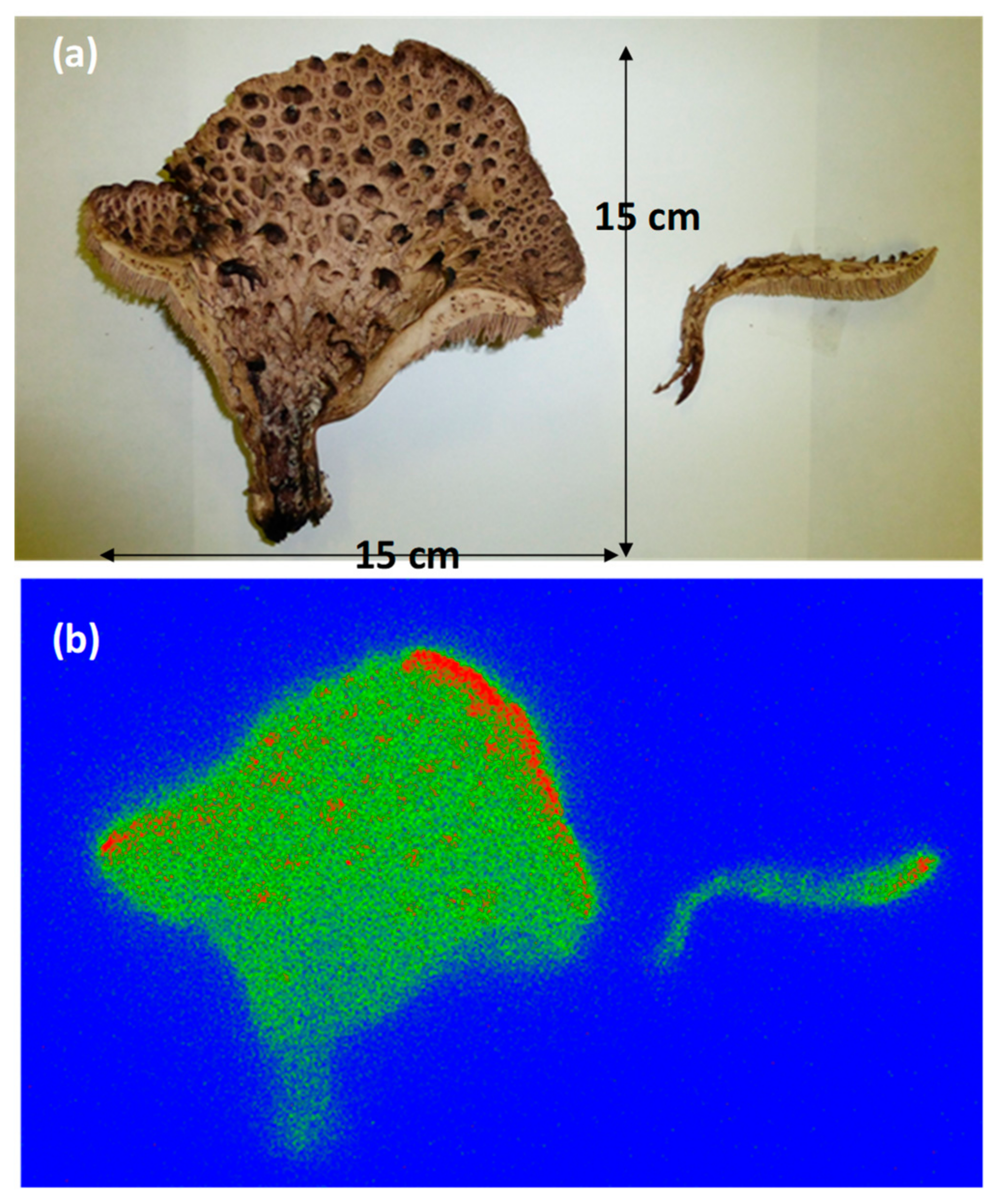
4.3. Mushroom
5. Conclusions
Author Contributions
Funding
Acknowledgments
Conflicts of Interest
References
- Amemiya, Y.; Matsushita, T.; Nakagawa, A.; Satow, Y.; Miyahara, J.; Chikawa, J. Design and performance of an imaging plate system for X-ray diffraction study. Nucl. Instrum. Meth. Phys. Res. 1988, 266, 645–653. [Google Scholar] [CrossRef]
- Takahashi, K.; Kohda, K.; Miyahara, J.; Kanemitsu, Y.; Amitami, A.; Shionoya, A. Mechanism of photostimulated luminescence in BaFX:Eu2+ (X=Cl, Br) phosphors. J. Lumin. 1984, 31, 266–268. [Google Scholar] [CrossRef]
- Iwabuchi, Y.; Mori, N.; Takahashi, K.; Matsuda, T.; Shionoya, S. Mechanism of photostimulated luminescence process in BaFBr.Eu2+ phosphors. Jpn. J. Appl. Phys. 1994, 33, 178–185. [Google Scholar] [CrossRef]
- Miyahara, J. Autoradiography and radiography-Imaging plate and its applications. Radioisotopes 1988, 47, 143–154. (In Japanese) [Google Scholar] [CrossRef]
- Sonoda, M.; Takano, M.; Miyahara, J.; Kato, H. Computed radiography utilizing scanning laser stimulated luminescence. Radiology 1983, 148, 833–838. [Google Scholar] [CrossRef] [PubMed]
- Simpson, S.J.; La Niece, S. New light on old swords from Iran. Br. Mus. Technol. Res. Bull. 2010, 4, 95–101. [Google Scholar]
- Shizuma, K.; Kajimoto, T.; Endo, S.; Matsugi, K.; Arimatsu, Y.; Nojima, H. Non-destructive analysis of ancient bimetal swords from western Asia by γ-ray radiography and X-ray fluorescence. Nucl. Instrum. Meth. Phys. Res. 2017, 407, 244–255. [Google Scholar] [CrossRef]
- Sugiura, Y.; Shibata, M.; Ogata, Y.; Ozawa, H.; Kanasashi, K.; Takenaka, C. Evaluation of radiocesium concentrations in new leaves of wild plants two years after the Fukushima Dai-ichi Nuclear Power Plant accident. J. Environ. Radioact. 2016, 160, 8–24. [Google Scholar] [CrossRef] [PubMed]
- Nakanishi, T.M. An overview of our research. In Agricultural Implications of the Fukushima Nuclear Accident; Nakanishi, T.M., Tanoi, L., Eds.; Springer Open: Tokyo, Japan, 2016; pp. 1–9. ISBN 978-4-431-55826-2. [Google Scholar] [CrossRef]
- Sugita, R.; Hirose, A.; Kobayashi, N.K.; Tanoi, K.; Nakanishi, T.M. Imaging techniques for radiocesium in soil and plants. In Agricultural Implications of the Fukushima Nuclear Accident; Nakanishi, T.M., Tanoi, L., Eds.; Springer Open: Tokyo, Japan, 2016; pp. 247–263. ISBN 978-4-431-55826-2. [Google Scholar] [CrossRef]
- Hirose, A.; Kobayashi, N.I.; Tanoi, K.; Nakanishi, T.M. A microautoradiographic methods for fresh-frozen sections to reveal the distribution of radionuclides at the cellular level in plants. Plant Cell Physiol. 2014, 55, 1194–1202. [Google Scholar] [CrossRef] [PubMed]
- Tsoulfanidis, N. Measurement and Detection of Radiation (Japanese translation by Eiji Sakai); Gendai Kogakusha: Tokyo, Japan, 1986; ISBN 4-87472-128-1. [Google Scholar]
- Brodzinski, R.L.; Brown, D.P.; Evance, J.C., Jr.; Hensley, W.K.; Reeves, J.H.; Wogman, N.A. An ultra background germanium gamma-ray spectrometer. Nucl. Instrum. Meth. Phys. Res. 1985, 239, 207–213. [Google Scholar] [CrossRef]
- Shizuma, K.; Fukami, K.; Iwatani, K.; Hasai, H. Low-background shielding of Ge detectors for the measurement of residual 152Eu radioactivity induced by neutrons from the Hiroshima atomic bomb. Nucl. Instrum. Meth. Phys. Res. 1992, 66, 459–464. [Google Scholar] [CrossRef]
- IAEA. Decay of the Transactinium Nuclides; Technical Report Series No.261; IAEA: Vienna, Austria, 1986. [Google Scholar]
- Shizuma, K.; Nursal, W.I.; Sakurai, Y. Long-term monitoring of radiocesium concentration in sediments and river water along five rivers in Minami-Soma City during 2012–2016 following the Fukushima Dai-ichi Nuclear Power Plant accident. Appl. Sci. 2018, 8, 1319. [Google Scholar] [CrossRef]
- Shizuma, K.; Fujikawa, Y.; Kurihara, M.; Sakurai, Y. Identification and temporal decrease of 137Cs and 134Cs in ground water in Minami-Soma City following the accident at the Fukushima Dai-ichi Nuclear Power Plant. Environ. Pollut. 2017, 234, 1–8. [Google Scholar] [CrossRef] [PubMed]
- Oba, Y. Radiocesium Contamination in Japanese fir after Fukushima Daiichi Nuclear Power Plant Accident. Ph.D. Thesis, Graduate School of Integrated Arts and Sciences, Hiroshima University, Hiroshima, Japan, 2017. [Google Scholar]


| Nuclide | Halflife | Radioctivity(Bq) | Radiation | Energy (MeV) | Emission Probability a (%) |
|---|---|---|---|---|---|
| 241Am | 432.2 y | 370 k | α | 5.443 | 13.1 |
| α | 5.486 | 85.2 | |||
| γ | 0.0263 | 2.4 | |||
| γ | 0.0595 | 35.7 | |||
| 147Pm | 2.62 y | – | β | 0.224 b | 100 |
| 90Sr | 28.74 y | 20 k | β | 0.546 b | 100 |
| (90Y) | 64.1 h | β | 2.2280 b | 100 | |
| 137Cs | 30.04 y | 370 k | β | 0.514 b | 94.6 |
| β | 1.17 b | 5.4 | |||
| γ | 0.662 | 85 | |||
| 60Co | 5.271 y | 370 k | β | 0.318 b | 99.92 |
| γ | 1.173 | 99.99 | |||
| γ | 1.333 | 99.98 | |||
| 40K | 1.277 × 10⁹ y | – | β | 1.312 b | 89.3 |
| γ | 1.461 | 10.7 |
 |
| Nuclide | Eβ (MeV) | μm (m2 kg−1) | μAl (cm−1) |
|---|---|---|---|
| 147Pm | 0.224 | 9.35 | 252 |
| 60Co | 0.318 | 6.30 | 170 |
| 137Cs | 0.514 | 3.65 | 98.5 |
| 40K | 1.312 | 1.25 | 33.6 |
| 90Sr | 0.546 | 3.38 | 91.2 |
| 90Y | 2.28 | 0.66 | 17.9 |
© 2019 by the authors. Licensee MDPI, Basel, Switzerland. This article is an open access article distributed under the terms and conditions of the Creative Commons Attribution (CC BY) license (http://creativecommons.org/licenses/by/4.0/).
Share and Cite
Shizuma, K.; Oba, Y. Low-Background Shielding Box for Autoradiography of Environmental Samples and the α-, β-, and γ-ray Sensitivities of the Imaging Plates. Appl. Sci. 2019, 9, 5209. https://doi.org/10.3390/app9235209
Shizuma K, Oba Y. Low-Background Shielding Box for Autoradiography of Environmental Samples and the α-, β-, and γ-ray Sensitivities of the Imaging Plates. Applied Sciences. 2019; 9(23):5209. https://doi.org/10.3390/app9235209
Chicago/Turabian StyleShizuma, Kiyoshi, and Yurika Oba. 2019. "Low-Background Shielding Box for Autoradiography of Environmental Samples and the α-, β-, and γ-ray Sensitivities of the Imaging Plates" Applied Sciences 9, no. 23: 5209. https://doi.org/10.3390/app9235209
APA StyleShizuma, K., & Oba, Y. (2019). Low-Background Shielding Box for Autoradiography of Environmental Samples and the α-, β-, and γ-ray Sensitivities of the Imaging Plates. Applied Sciences, 9(23), 5209. https://doi.org/10.3390/app9235209