Review

Surface Alterations Induced on Endodontic Instruments by Sterilization Processes, Analyzed with Atomic Force Microscopy: A Systematic Review

1
Department of Clinical and Experimental Medicine, University of Foggia, Via Rovelli 50, 71122 Foggia, Italy
2
Department of Basic Medical Sciences, Neurosciences and Sensory Organs, Division of Complex Operating Unit of Dentistry, “Aldo Moro” University of Bari, Piazza G. Cesare 11, 70124 Bari, Italy
3
Multidisciplinary Department of Medical-Surgical and Odontostomatological Specialties, University of Campania “Luigi Vanvitelli”, 80121 Naples, Italy
4
Department of Surgical Sciences, Dental School, University of Turin, 10124 Turin, Italy
*
Author to whom correspondence should be addressed.
Appl. Sci. 2019, 9(22), 4948; https://doi.org/10.3390/app9224948
Submission received: 29 October 2019 / Revised: 13 November 2019 / Accepted: 14 November 2019 / Published: 17 November 2019

Abstract

:
Endodontic canal disinfection procedures that use sodium hypochlorite, and subsequently, heat sterilization procedures can alter the surface of endodontic instruments, described as corrosion and micropitting. These phenomena can be visualized on the surface of the instruments by SEM and atomic force microscopy analyses. The endodontic instruments used in probing, pre-enlargement, and shaping phases are made of steel alloy or nickel-titanium alloy (NiTi) and are subject to torsional, flexor, and cyclic fatigue; indeed, reuse of these instruments must be done with the knowledge that these instruments are subject to fracture following stress caused during their use. Fracture of the instrument within the canal is an eventuality that can lead to failure of the treatment, and therefore it is important to try to reduce situations that can contribute to the fracture. This review was performed based on the PRISMA protocol. Studies were identified through bibliographic research using electronic databases. A total of 1036 records were identified on the PubMed and Scopus databases. After screening the articles, restricted by year of publication (1979 to 2019), there were 946 records. With the application of the eligibility criteria (all the articles pertaining to the issue of sterilization in endodontics), there were 228 articles. There were 104 articles after eliminating overlaps. There were 50 articles that discussed the influence of sterilization procedures on the surface characteristics of endodontic instruments, and 26 articles that measured parameters on surface alteration. Applying the inclusion and exclusion criteria resulted in a total of eleven articles for quantitative analysis. Four articles were in reference to the primary outcome, eight articles to secondary outcome, and five articles to tertiary outcome. The meta-analysis showed a statistically significant surface alteration effect after five autoclaves and after immersion in the canal irrigants after 10 min.

1. Introduction

Endodontic instruments are commonly used in dental practice to perform endodontic treatments of vital and necrotic teeth, endodontic retreatments, pulpotomies, pulpectomy and specification procedures. Depending on the phase of treatment, endodontic instruments are divided into instruments for probing the endodontic canal for the pre-enlargement, and glidepath for shaping the canal or instruments for the closure and three-dimensional sealing of endodontic canals [1,2]. Many of these tools are reusable after performing cleaning, disinfection, and sterilization procedures by autoclaving [3].
Endodontic canal disinfection procedures that use sodium hypochlorite [4], and subsequently, heat sterilization procedures, can alter the surface of endodontic instruments, described as corrosion and micropitting, phenomena [5] that can be visualized by SEM and atomic force microscopy analyses.
The endodontic instruments used in the probing, pre-enlargement, and shaping phases are made of steel alloy or nickel-titanium alloy (NiTi) and are subject to torsional, flexor, and cyclic fatigue, indeed the reuse of these instruments must be done with the knowledge that these tools are subject to fracture following stress caused during their use [6]. Fracture of the instrument within the canal is an eventuality that can lead to failure of the treatment, and therefore it is important to try to reduce situations that can contribute the fracture.
On the surface subject to fatigue, surface alterations can give rise to microcrack which can lead to fracture of the instrument, and also reduce the cutting capacity of the blades on the endodontic files [7]. Therefore, in order to maintain the same cutting efficiency, the endodontist has to exert greater pressure on the instrument with an increase in torsional fatigue stress [6].
An atomic force microscope is an instrument capable of analyzing the surface of instruments. It consists of a cantilever with a pointed tip (tip) mounted on the end, typically composed of silicon or silicon nitride and having a radius of curvature of the “order of nanometers”. The sample to be scanned, through the Vand Der Waals forces, interacts with the tip of the detector by flexing it. There are several methods to detect any cantilever movement. The majority of atomic force microscopy (AFM) systems use laser beam detection, which is an optical system with position sensitive detectors called photodiodes. The laser light is reflected by the cantilever on the position-sensitive photodiode. Very small forces are produced between the probe and the surface to be scanned, and these are the forces that allow the AFM system to record the deflection of the cantilever. The cantilever deflection is called “cantilever rigidity”. This rigidity can be measured by Hooke’s law. Rigidity is recorded visually and can be viewed on the computer in real time. The surface scan of endodontic instruments is used both in non-contact mode and in contact mode and, in general, the scanned surfaces start at 3 mm from the tip of the instrument up to 6 mm. The parameters which are considered with AFM for the analysis of a surface are the arithmetic mean roughness(AMR) of the maximum height (MH) and root mean square (RMS). The atomic force microscopy, therefore, provides detailed information with measurable parameters of possible alterations and irregularities present on the surface of an instrument [8].
Surface alterations can represent a problem in the use of endodontic instruments. A study by Ylmaz, in 2018, identified surface alterations described as surface roughness with statistically significant results for instruments constructed with new M-wire and EDM alloys [9].
One problem of reusing endodontic instruments that are subject to fatigue is the deterioration they suffer that results from their use in the dental canal for the removal of dentin, as well as the corrosive action by the root canal irrigants such as sodium hypochlorite, and subsequently, the action of the temperature and steam induced by the autoclave sterilization process. The surface alterations are well described in a study by Inan, in 2007, on the universal ProTaper, after clinical use and sterilization [10].
Fayyad and Mahran, in a 2013 study, demonstrated by AFM analysis that the alterations on Twisted Files [11], Hero Shaper, RaCe, and GTX instruments were statistically significant after immersion in 5% sodium hypochlorite, however, the alterations were not statistically significant after EDTA immersion. In contrast, Ametrano et al., in 2010, reported significant results for instruments immersed in EDTA [12]. Other studies have report conflicting data, such as the study conducted by Casella, in 2011, in which there was no variation in corrosion resistance for some instruments (K FILE and GT-rotary) unlike the K3 knife immersed in 5% sodium hypochlorite [13]. In addition, studies conducted at the Sem da Razavianet, in 2015, reported an increase in roughness directly related to the number of sterilization cycles performed on endodontic instruments [14].
In contrast, there is debate within the scientific community regarding whether there are statistically significant surface alterations induced by the autoclave or the canal irrigants. This review aims to try to clarify this aspect by investigating the literature to extrapolate the data on surface alterations in endodontic instruments in order to statistically analyze them in a meta-analysis.
Previous systematic reviews on this topic have not included the effect of surface alterations of endodontic instruments subjected to heat sterilization. There is only one systematic review that analyzes the variations in torsional properties subjected to autoclave sterilization.
This review could help endodontists who perform endodontic therapy and reuse endodontic instruments daily. Awareness of the greater or lesser risk of potential fracture triggered by surface variations due to heat or use of canal irrigants on the instruments could be helpful.

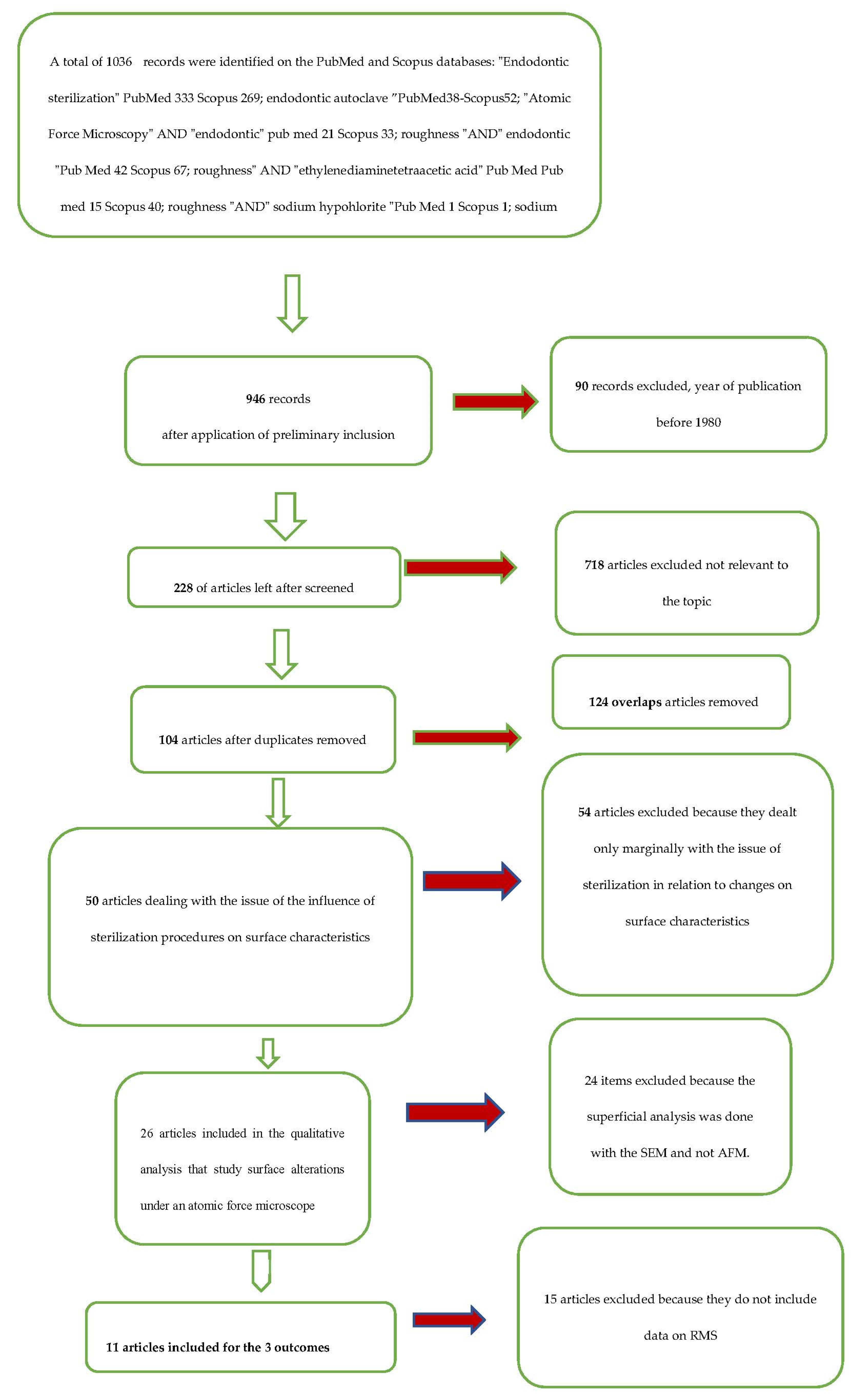
2. Materials and Methods

This systematic review was conducted based on the Prisma protocol.
The study was constructed using the following PICO elements for questions: Population (endodontic instruments); intervention (surface alterations induced by sterilization processes and root canal irrigation); control (new endodontic instruments not subject to sterilization; and outcome (surface alterations induced by the sterilization process by autoclave, and by root canal irrigants such as sodium hypochlorite and EDTA).
The following PICO question was formulated: To what extent, statistically significant, the sterilization processes and the used canal irrigants alter the surface of the rotating endodontic instruments with respect to the control?
After an initial selection phase of article identification in the databases, the potentially eligible articles were qualitatively evaluated in order to investigate the surface alterations of endodontic instruments resulting from the sterilization of instruments and disinfection of endodontic canals.

2.1. Eligibility Criteria

This literature review took into consideration in vitro and clinical studies that concerned the subject of sterilization and the influence of the latter on the physical and chemical properties of endodontic instruments. In particular, articles that dealt with the corrosive phenomena and surface alterations considered by microscopy methods (atomic force microscopy), conducted in recent years, and published with abstracts in English, were considered potentially eligible.
Articles from the last 40 years were chosen, because disinfection and sterilization procedures have changed in light of new discovered infectious contaminants, such as HIV and HCV viruses and the prong of spongiform encephalopathy. Furthermore, the methods used to manufacture the instruments have changed with the introduction of new alloys and new instruments. Therefore, in summary, potentially eligible articles included studies that investigated the influence of sterilization and disinfection procedures on endodontic canals, as well as on the physical and chemical characteristics of endodontic instruments, however, articles published more than 40 years ago and those that did not present an abstract in English were excluded.
Finally, the articles that were potentially eligible were subjected to a full text analysis to verify their use for a qualitative and quantitative analysis.
The inclusion and exclusion criteria applied in the full text analysis are the following:
  • Include all those studies that describe the alterations induced by the sterilization methods of the endodontic instruments, analyzed using atomic force microscopy;
  • Include all the articles that describe the alterations induced by root canal irrigants (sodium hypochlorite and EDTA), analyzed using atomic force microscopy;
  • The exclusion criteria are to exclude all those studies that do not report data (average and standard deviation) on surface irregularities (AMR, MH, and RMS).

2.2. Research Methodology

The studies were identified through a bibliographic research on electronic databases.
The literature search was conducted using the search engines “PubMed” and “Scopus”. The search on the providers was conducted between 12 September 2019 and 18 September 2019 and the last search for a partial update of the literature was conducted on 1 October 2019.
The following search terms were used on PubMed and Scopus: “Endodontic sterilization” PubMed 333 and Scopus 269; “endodontic autoclave” PubMed 38 and Scopus 52; “atomic force microscopy” AND “endodontic” PubMed 21 and Scopus 33; “roughness” AND “endodontic“ Pub Med 42 and Scopus 67; “roughness” AND “ethylenediaminetetraacetic acid” PubMed 15 and Scopus 40; “roughness“ AND ”sodium hypochlorite“ PubMed 1 and Scopus 1; “sodium hypochlorite” AND “atomic force microscopy” PubMed 40 and Scopus 80; “atomic force microscopy” AND “NiTi rotary instruments” PubMed 1 and Scopus 2 (Table 1).

2.3. Screening Methodology

The records obtained were, subsequently, examined by two independent reviewers (M.D. and S.D), and a third reviewer (E.L.) acted as a decision maker in situations of doubt. The screening included the analysis of the title and the abstract to eliminate the recordings not related to the topics of the review. After the screening phase, the overlaps were removed and the complete texts of the articles were analyzed, from which the ones eligible for the qualitative analysis and the inclusion in the meta-analysis for the three results were identified. The results sought by the two reviewers were:
(1)
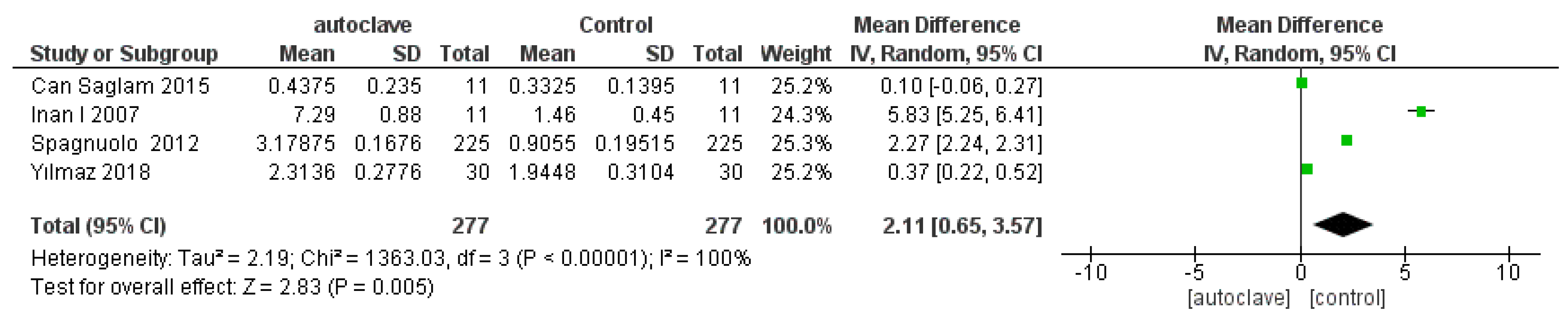
Primary outcome, variations of the root mean square root (RMS) of endodontic instruments subjected to five autoclave cycles as compared with non-autoclaved control;
(2)
Secondary outcome, variations of the root mean square (RMS) of endodontic instruments exposed to sodium hypochlorite 5% as compared with the control group;
(3)
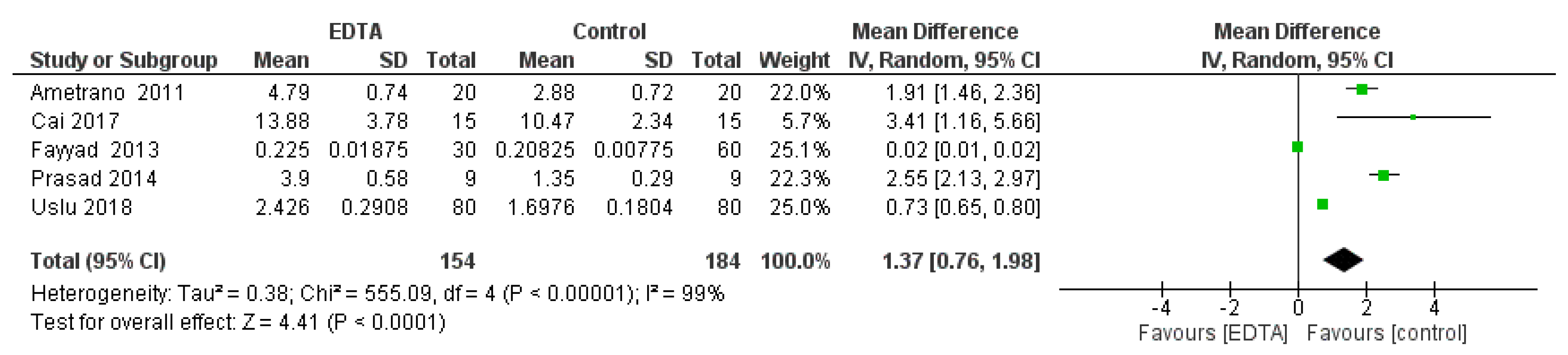
Tertiary outcome, variations of the root mean square (RMS) of the endodontic instruments described at EDTA 10% as compared with the control group.
The fourth reviewer, with supervisory duties, was L.Lo.M. The K agreement between the two screening reviewers was 0.8464 (Table 2). The K agreement was based on the formulas of the Cochrane Handbook for Systematic Reviews [15].
The Newcastle–Ottawa scale for case-control studies was used to assess the risk of bias in the included studies. The quantitative analysis was performed with the Rev Manager software 5.3 (Cochrane Collaboration, Copenhagen, Denmark [16].

3. Results

A total of 1036 records were identified on the PubMed and Scopus databases (Table 1). After screening the articles, with the restriction by year of publication (1979 to 2019), there were 946 records. With the application of the eligibility criteria (all the articles pertaining to the issue of sterilization in endodontics), there were 228 articles. There were 104 articles after eliminating overlaps. There were 50 articles that discussed the influence of sterilization procedures on the surface characteristics of endodontic instruments, and 26 that measured parameters on surface alteration.
Applying the inclusion and exclusion criteria resulted in a total of eleven articles for quantitative analysis.
Four articles were in reference to the primary outcome, eight to the secondary outcome, and five to the tertiary outcome. The entire selection and screening procedures are described in the flow chart (Figure 1).

3.1. Study Characteristics and Data Extraction
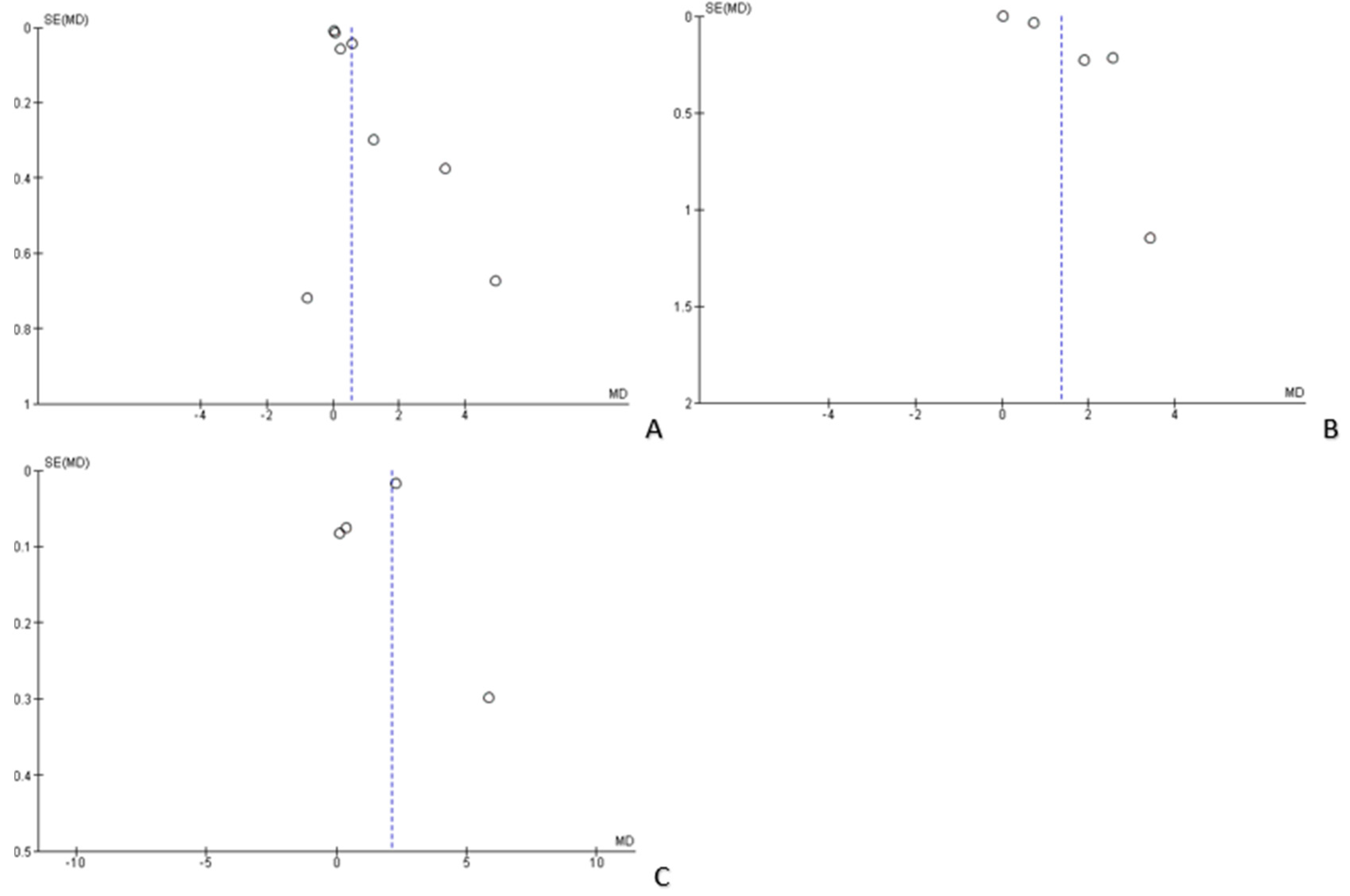
The studies included for quantitative analysis were:
  • First outcome: Yılmaz et al., 2017 [9]; Spagnuolo et al., 2012 [17]; Inan et al., 2007 [10]; and Can Saglam et al., 2015 [18];
  • Second outcome: Uslu et al., 2018 [19]; Can Saglam et al., 2015 [18]; Fayyad et al., 2013 [11]; Ametrano et al., 2010 [12]; Topuz et al., 2008 [20]; Cai et al., 2017 [21]; Saglam et al., 2012 [22]; and Prasad et al., 2014 [23];
  • Third outcome: Uslu et al., 2018 [19]; Fayyad et al., 2013 [11]; Ametrano et al., 2010 [12]; Cai et al., 2017 [21]; and Prasad et al., 2014 [23].
The extracted data included the magazine (author, data, and journal); the endodontic instrumentation object of measurement (name, taper, and diameter at tip); the method of sterilization by heat (temperature, pressure, and time); the number autoclave cycles or irrigants; the number of instruments (control and experimental); the number of surfaces scanned by the instrument; the number of total scans; the size of the scanning surface; and the data concerning the root mean square (RMS) ± standard deviation.
The data extracted for the tree outcomes are shown in Table 3 and Table 4.

3.2. Risk of Bias

The risk of bias was assessed through the Newcastle–Ottawa case-control scale. The results are reported in detail in Table 5. For each category, a value of one to three was assigned (one = low and three = high).
The risk of bias within the individual studies was low enough that the methods of investigation adopted for the controls were identical to the cases included in the meta-analysis. The Prasad study [23] was the only study of the exposure time of endodontic instruments to canal irrigants that was not well defined, exposing the study to a bias.
The risk of bias between the various studies was considered high, and therefore partly limited the importance of the results. The heterogeneity of the studies depended mainly on the diversity of the instruments, which were similar, in some cases, only in terms of tip diameter, taper, and type of metal alloy.
The heterogeneity of the studies was represented by funnel plots of the four outcomes, as shown in Figure 2.

3.3. Data Analysis

The statistical analysis of the data was performed using the Rev Manager 5.3 software (Copenhagen, 153 Denmark, The Nordic Cochrane Centre, The Nordic Cochrane Collaboration, 2014) and the results were represented by forest plots for each of the outcomes.
For the primary outcome, variations of the root mean square root (RMS) of endodontic instruments subjected to five autoclave cycles as compared with the non-autoclaved control, the comparison showed high heterogeneity of the studies, with an I2 equal to 100%. For this reason, a random effects model was used. Overall, for the primary outcome, meta-analysis was favorable for the control group. The studies that present data with a statistically significant difference are Inan et al., 2007 [10] and Spagnuolo et al., 20012 [17]. The studies by Ylmaz et al., 2018 [9] and Can Saglam et al., 2015 [22] are exactly at the center of the line of no effect. The studies by Ylzam and Can Saglam are exactly at the center of the line of no effect, however, the remaining two studies are favorable for the group subjected to control, their confidence intervals do not intercept the line of no effect (Figure 3).
For the secondary outcome, variations of the root mean square (RMS) of endodontic instruments exposed to sodium hypochlorite 5% as compared with the control group, the comparison showed high heterogeneity among the studies, with an I2 equal to 98%. For this reason, for the second outcome, a random effects model was applied to avoid minimizing the roles of smaller-dimension studies. For the second outcome, the forest plot is in favor of the subject group control.
The studies that reported statistically significant data in favor of the control group are Ametrano, 2011; Prasad, 2014; Topuz, 2008; and Uslu, 2018. The Cai’s study was the only study that was in favor of the group subjected to sodium hypochlorite, even though its confidence interval crosses the line of no effect. The other studies report statistically insignificant data (Figure 4).
For the tertiary outcome, variations of the root mean square (RMS) of the endodontic instruments exposed to EDTA 10% as compared with the control group, the comparison showed high heterogeneity between the studies, with an I2 of 99%, and therefore a random effects model was applied. For the tertiary outcome, the forest plot is in favor of the control group except for the study by Fayyad which is positioned in the line of no effect (Figure 5).

4. Discussion

The results of the meta-analysis for the three outcomes are in agreement in establishing that the superficial alterations induced by autoclave, from the sodium hypochlorite and from the EDTA, are statistically significant surface alterations that represent points where instrument fractures can be triggered. In addition, the alterations induced on the surface analyzed by SEM and AFM show that the alterations can also be expressed on the cutting surface, altering, in a pejorative sense, the cutting efficacy.
For the first outcome, the studies, in the literature, that supported a statistically significant alteration are:
  • In 2007, Inan reported statistically significant data for all the instruments of the ProTaper series (S1, S2, F1, F2), and reported that the superficial deterioration induced by the autoclave is greater for ProTaper finished than for ProTaper shaping;
  • In 2012, Spagnuolo confirmed, in agreement with Inan’s data, that multiple cycles (autoclave sterilization) modified the surface topography and chemical composition of conventional NiTi (F2 ProTaper) and TiN-coated (alpha kit) instruments, in a statistically significant way (after five autoclave cycles).
Sodium hypochlorite certainly alters the surface of NiTi instruments and innumerable studies are in agreement such as Uslu et al., 2018 [19]; Ametrano et al., 2010 [12]; Topuz et al., 2008 [20]; and Prasad et al., 2014 [23]. Furthermore, a study conducted by Yokoyama et al., 2004 [24] stated that the action of sodium hypochlorite causes a worsening of the surface in endodontic instruments that facilitates their rupture following flexor and torsional stress.
The statistical analysis, in a similar way but with fewer studies, also confirms that the EDTA determines an increase in surface irregularities in a statistically significant way, and studies that confirm it after 10 min of exposure are well highlighted in the forest plot (Figure 5). Studies that are in contrast to the present meta-analysis report conflicting data regarding the action of EDTA on the surface. It seems that for exposures less than 5 min they do not alter the surface, however, according to Bonaccorsa et al. [25], a passivation phenomenon could lead to the creation of a complex between the metallic ions and the EDTA at a PH lower than four which renders the instrument resistant.

5. Conclusions

In conclusion, based on the present systematic analysis we affirm that autoclave induces a statistically significant corrosive phenomena, called micropitting, after five cycles of autoclave and determined by the heat, and comparatively, hypochlorite determines corrosion after only 5 min of exposure and EDTA after 10 min of exposure.
Superficial alterations, which are widely discussed in the literature, can determine the triggering of fractures in instruments subjected to cyclic fatigue and torsional fatigue. Therefore, it is important for endodontist to have knowledge of such corrosive phenomena, induced by irrigants such as sodium hypochlorite and EDTA, on instruments that can be reused and autoclaved.

Author Contributions

Conceptualization, M.D., D.S., and B.R.; methodology, K.Z. and M.D.; software, M.A. validation, M.D., and L.L.M.; formal analysis, D.S., M.D., and V.C.; investigation, L.L. and M.D.; resources, L.L.M.; data curation, E.L. and M.D.; writing—original draft preparation, M.D.; writing—review and editing, M.D., L.L.M., L.L.R., D.C., and F.M.

Funding

This research received no external funding.

Acknowledgments

All the acknowledgements go to Lorenzo Lo Muzio, Director of the Dental Clinic and President of the Department of Clinical and Experimental Medicine of the University of Foggia, who gave fundamental technical support in the writing of this article.

Conflicts of Interest

The authors declare no conflict of interest.

References

  1. Inan, U.; Keskin, C. Torsional Resistance of ProGlider, Hyflex EDM, and One G Glide Path Instruments. J. Endod. 2019. [Google Scholar] [CrossRef]
  2. Troiano, G.; Dioguardi, M.; Cocco, A.; Zhurakivska, K.; Ciavarella, D.; Muzio, L.L. Increase in [corrected] the glyde path diameter improves the centering ability of F6 Skytaper. Eur. J. Dent. 2018, 12, 89–93. [Google Scholar] [CrossRef] [PubMed]
  3. Laneve, E.; Raddato, B.; Dioguardi, M.; Di Gioia, G.; Troiano, G.; Lo Muzio, L. Sterilisation in Dentistry: A Review of the Literature. Int. J. Dent. 2019, 2019, 6507286. [Google Scholar] [CrossRef] [PubMed]
  4. Dioguardi, M.; Di Gioia, G.; Illuzzi, G.; Laneve, E.; Cocco, A.; Troiano, G. Endodontic irrigants: Different methods to improve efficacy and related problems. Eur. J. Dent. 2018, 12, 459–466. [Google Scholar] [CrossRef] [PubMed]
  5. Berutti, E.; Angelini, E.; Rigolone, M.; Migliaretti, G.; Pasqualini, D. Influence of sodium hypochlorite on fracture properties and corrosion of ProTaper Rotary instruments. Int. Endod. J. 2006, 39, 693–699. [Google Scholar] [CrossRef] [PubMed]
  6. Dioguardi, M.; Sovereto, D.; Aiuto, R.; Laino, L.; Illuzzi, G.; Laneve, E.; Raddato, B.; Caponio, V.C.A.; Dioguardi, A.; Zhurakivska, K.; et al. Effects of Hot Sterilization on Torsional Properties of Endodontic Instruments: Systematic Review with Meta-Analysis. Materials 2019, 12, 2190. [Google Scholar] [CrossRef] [PubMed]
  7. Shen, Y.; Coil, J.M.; McLean, A.G.; Hemerling, D.L.; Haapasalo, M. Defects in nickel-titanium instruments after clinical use. Part 5: Single use from endodontic specialty practices. J. Endod. 2009, 35, 1363–1367. [Google Scholar] [CrossRef]
  8. Ozyurek, T.; Yilmaz, K.; Uslu, G.; Plotino, G. The effect of root canal preparation on the surface roughness of WaveOne and WaveOne Gold files: Atomic force microscopy study. Restor. Dent. Endod. 2018, 43, e10. [Google Scholar] [CrossRef]
  9. Yilmaz, K.; Uslu, G.; Ozyurek, T. Effect of multiple autoclave cycles on the surface roughness of HyFlex CM and HyFlex EDM files: An atomic force microscopy study. Clin. Oral Investig. 2018, 22, 2975–2980. [Google Scholar] [CrossRef]
  10. Inan, U.; Aydin, C.; Uzun, O.; Topuz, O.; Alacam, T. Evaluation of the surface characteristics of used and new ProTaper Instruments: An atomic force microscopy study. J. Endod. 2007, 33, 1334–1337. [Google Scholar] [CrossRef]
  11. Fayyad, D.M.; Mahran, A.H. Atomic force microscopic evaluation of nanostructure alterations of rotary NiTi instruments after immersion in irrigating solutions. Int. Endod. J. 2014, 47, 567–573. [Google Scholar] [CrossRef] [PubMed]
  12. Ametrano, G.; D’Anto, V.; Di Caprio, M.P.; Simeone, M.; Rengo, S.; Spagnuolo, G. Effects of sodium hypochlorite and ethylenediaminetetraacetic acid on rotary nickel-titanium instruments evaluated using atomic force microscopy. Int. Endod. J. 2011, 44, 203–209. [Google Scholar] [CrossRef] [PubMed]
  13. Casella, G.; Rosalbino, F. Corrosion behaviour of NiTi endodontic instrument. Corros. Eng. Sci. Technol. 2011, 46, 521–523. [Google Scholar] [CrossRef]
  14. Razavian, H.; Iranmanesh, P.; Mojtahedi, H.; Nazeri, R. Effect of Autoclave Cycles on Surface Characteristics of S-File Evaluated by Scanning Electron Microscopy. Iran. Endod. J. 2016, 11, 29–32. [Google Scholar] [CrossRef]
  15. Higgins, J.P.T.; Green, S. Cochrane Collaboration. Cochrane Handbook for Systematic Reviews of Interventions; Wiley-Blackwell: Chichester, UK; Hoboken, NJ, USA, 2008; 649p. [Google Scholar]
  16. Lo, C.K.; Mertz, D.; Loeb, M. Newcastle-Ottawa Scale: Comparing reviewers’ to authors’ assessments. BMC Med. Res. Methodol. 2014, 14, 45. [Google Scholar] [CrossRef] [PubMed]
  17. Spagnuolo, G.; Ametrano, G.; D’Anto, V.; Rengo, C.; Simeone, M.; Riccitiello, F.; Amato, M. Effect of autoclaving on the surfaces of TiN -coated and conventional nickel-titanium rotary instruments. Int. Endod. J. 2012, 45, 1148–1155. [Google Scholar] [CrossRef] [PubMed]
  18. Can Saglam, B.; Gorgul, G. Evaluation of surface alterations in different retreatment nickel-titanium files: AFM and SEM study. Microsc. Res. Tech. 2015, 78, 356–362. [Google Scholar] [CrossRef]
  19. Uslu, G.; Ozyurek, T.; Yilmaz, K. Effect of Sodium Hypochlorite and EDTA on Surface Roughness of HyFlex CM and HyFlex EDM Files. Microsc. Res. Tech. 2018, 81, 1406–1411. [Google Scholar] [CrossRef]
  20. Topuz, O.; Aydin, C.; Uzun, O.; Inan, U.; Alacam, T.; Tunca, Y.M. Structural effects of sodium hypochlorite solution on RaCe rotary nickel-titanium instruments: An atomic force microscopy study. Oral Surg. Oral Med. Oral Pathol. Oral Radiol. Endod. 2008, 105, 661–665. [Google Scholar] [CrossRef]
  21. Cai, J.J.; Tang, X.N.; Ge, J.Y. Effect of irrigation on surface roughness and fatigue resistance of controlled memory wire nickel-titanium instruments. Int. Endod. J. 2017, 50, 718–724. [Google Scholar] [CrossRef]
  22. Saglam, B.C.; Kocak, S.; Kocak, M.M.; Topuz, O. Effects of irrigation solutions on the surface of ProTaper instruments: A microscopy study. Microsc. Res. Tech. 2012, 75, 1534–1538. [Google Scholar] [CrossRef] [PubMed]
  23. Prasad, P.S.; Sam, J.E.; Arvind Kumar, K. The effect of 5% sodium hypochlorite, 17% EDTA and triphala on two different rotary Ni-Ti instruments: An AFM and EDS analysis. J. Conserv. Dent. 2014, 17, 462–466. [Google Scholar] [CrossRef] [PubMed]
  24. Yokoyama, K.; Kancko, K.; Yabuta, E.; Asaoka, K.; Sakai, J. Fracture of nickel-titanium superelastic alloy in sodium hypochlorite solution. Mater. Sci. Eng. A 2004, 369, 43–48. [Google Scholar] [CrossRef]
  25. Bonaccorso, A.; Tripi, T.R.; Rondelli, G.; Condorelli, G.G.; Cantatore, G.; Schafer, E. Pitting corrosion resistance of nickel-titanium rotary instruments with different surface treatments in seventeen percent ethylenediaminetetraacetic Acid and sodium chloride solutions. J. Endod. 2008, 34, 208–211. [Google Scholar] [CrossRef]
Figure 1. Flow chart of the different phases of the systematic review.
Applsci 09 04948 g001
Figure 2. Funnel plots of the evaluation of heterogeneity for the (A) first, (B) second, (C) third outcomes.
Applsci 09 04948 g002
Figure 3. Forest plot of the random effects model of the meta-analysis of the primary outcome.
Applsci 09 04948 g003
Figure 4. Forest plot of the random effects model of the meta-analysis of the secondary outcome.
Applsci 09 04948 g004
Figure 5. Forest plot of the random effects model of the meta-analysis of the tertiary outcome.
Applsci 09 04948 g005
Table 1. Complete overview of the search methodology.
Database-ProviderKey WordsSearch DetailsNumber of RecordsNumber of Records
after Restriction by Year of Publication (Last 40 Years)
Number of Articles Remaining after the Elimination of Records not Related to the Issue of Sterilization Influence on Endodontic InstrumentsArticles After Remove Overlaps ArticlesNumber of Articles Dealing with the Problem of the Influence of Sterilization Procedures on the Surface Characteristics of Endodontic InstrumentsNumber of Articles that Have Analyzed the Surface Alterations with Methods Different from the Atomic Force MicroscopyNumber of Articles that Analyzed Surface Alterations with Atomic Force MicroscopyNumbers of Articles Included in the Quantitative Analysis for the 3 Outcomes
PubMed“endodontic sterilization”“endodontic” [All Fields] AND (“sterilization”[All Fields] OR “sterilization”, reproductive”[MeSH Terms] OR (“sterilization”[All Fields] AND “reproductive”[All Fields]) OR “reproductive sterilization”[All Fields] OR “sterilization”[All Fields] OR “sterilization”[MeSH Terms])33329146
PubMed“endodontic autoclave”“endodontic” [All Fields] AND “autoclave” [All Fields]383825
PubMed“atomic force microscopy” AND “endodontic”“atomic force microscopy” [All Fields] AND “endodontic” [All Fields]21219
PubMed“roughness” AND “endodontic”“roughness” [All Fields] AND “endodontic” [All Fields]424111
PubMed“roughness” AND “ethylenediaminetetraacetic acid”“roughness” [All Fields] AND (“ethylenediaminetetraacetic” [All Fields] AND “acid” [All Fields])15152
PubMed“roughness” AND “sodium hypochlorite”“roughness” [All Fields] AND (“sodium” [All Fields] AND “hypochlorite” [All Fields])111
PubMed“sodium” “hypochlorite” AND “atomic force microscopy”“sodium hypochlorite” [All Fields] AND “atomic force microscopy” [All Fields]404013
PubMed“atomic force microscopy” AND “NiTi rotary instruments”“atomic force microscopy” [All Fields] AND “NiTi rotary instruments” [All Fields]111
Scopus“endodontic sterilization”TITLE-ABS-KEY (endodontic AND sterilization)26922556
Scopus“endodontic autoclave”TITLE-ABS-KEY (endodontic AND autoclave)525225
Scopus“atomic force microscopy” AND “endodontic”TITLE-ABS-KEY (“atomic force microscopy” AND “endodontic”)333313
Scopus“roughness” AND “endodontic”TITLE-ABS-KEY (“roughness” AND “endodontic”)676512
Scopus“roughness” AND sodium “hypochlorite”TITLE-ABS-KEY (“roughness” AND “sodium” AND “hypochlorite”)111
Scopus“roughness” AND “ethylenediaminetetraacetic acid”TITLE-ABS-KEY (“roughness” AND “ethylenediaminetetraacetic acid”)40402
Scopus“sodium hypochlorite” AND “atomic force microscopy”TITLE-ABS-KEY (“sodium hypochlorite” AND “atomic force microscopy”)80809
Scopus“atomic force microscopy” AND “NiTi rotary instruments”TITLE-ABS-KEY (“atomic force microscopy” AND “NiTi rotary instruments”)222
Total records 103694622810450242611
Table 2. K agreement calculation, Po = 0.94 (proportion of agreement), Pe = 0.6092 (agreement expected), K agreement = 0.8464 (<0 no agreement, 0.0 to 0.20 slight agreement, 0.21 to 0.40 fair agreement, 0.41 to 0.60 moderate agreement, 0.61 to 0.80 substantial agreement, and 0.81 to 1.00 almost perfect agreement). The K agreement was calculated from the 50 articles and included eleven articles with the application of the inclusion and exclusion criteria.
//Reviewer 2Reviewer 2Reviewer 2
IncludeExcludeUnsureTotal
Reviewer 1include110011
Reviewer 1exclude236038
Reviewer 1unsure1001
total1436050
Table 3. Primary outcome (extraction of data relating to the root mean square detected on the surface of the endodontic instruments subjected to atomic force microscopy examination, with respect to control and after 5 cycles of autoclaving).
Autor, Data, JournalSterilization Method (Autoclave Temperature, Pressure Exposure Time)Endodontic Instruments (Diameter and Taper at the Tip)Autoclave CyclesNumber of InstrumentsSurfaces Scanned by InstrumentNumber of Total ScansScanning SurfaceRoot Mean Square (RMS) ± Standard Deviation
Yılmaz et al., 2017, Clin. Oral. Investig. [9]Autoclave 134 °C, 30 psi for 20 min HyFlex EDM (25/08)0215305 × 5 μm48.62 ± 7.76 nm
5215305 × 5 μm57.84 ± 6.94 nm
Spagnuolo et al., 2012, Int. Endod. J. [17]Autoclave 121 °C, 15 psi, for 15 minProTaper
F2 (25/08)
0151522515 × 15 μm203.75 ± 35.81 nm
5151522515 × 15 μm715.22 nm ± 37.71
Can Saglam et al., 2015, Microsc. Res. Tech. [18]Autoclave 121 °C for 20 minProTaper retreatment D1 (30/09)0111112 × 2 μm1.33 ± 0.558 nm
5111112 × 2 μm1.75 ± 0.940 nm
Inan et al., 2007, J. Endod. [10]Autoclave 134 °C for 18 minProTaper
F2 (25/08)
0111111 × 1 μm1.46 ± 0.45 nm
1111111 × 1 μm7.29 ± 0.88 nm
Table 4. Extracted data relating to the secondary outcome and tertiary outcome, the data extracted are the root mean square (RMS), the total number of surfaces scanned, the endodontic instruments being scanned by the atomic force microscopy, and the irrigants used with the relative concentrations.
Autor, Data, JournalEndodontic Instruments (Diameter and Taper at the Tip)Irrigant Used (Concentration and Exposure Time)Number of InstrumentsSurfaces Scanned by InstrumentNumber of Total ScansScanning SurfaceRoot Mean Square (RMS) ± Standard Deviation
Uslu et al., 2018, Microsc. Res. Tech. [19]HyFlex EDM (25/.08)control420805 × 5 μm42.44 ± 4.51 nm
NaOCl 5.25% for 5 min420805 × 5 μm57.05 ± 8.55 nm
EDTA 17% for 5 min420805 × 5 μm60.65 ± 7.27 nm
Can Saglam et al., 2015, Microsc. Res. Tech. [18]ProTaper retreatment D1 (30/09) Control111112 × 2 μm1.33 ± 0.558 nm
NaOCl 2% for 5 min111112 × 2 μm2.24 ± 0.555 nm
Fayyad et al., 2013, Int. Endod. J. [11]RaCecontrol4156020 × 20 μm83.3 ± 3.1 nm
NaOCl 5.25% for 5 min2153020 × 20 μm92.3 ± 23.5 nm
EDTA 17% for 5 min2153020 × 20 μm90 ± 7.5 nm
Ametrano et al., 2011, Int. Endod. J. [12]ProTaper
F2 (25/08)
control120201 × 1 μm2.88 ± 0.72 nm
NaOCl 5.25% for 5 min120201 × 1 μm4.10 ± 1.13 nm
EDTA 17% for 5 min120201 × 1 μm4.79 ± 0.74 nm
Topuz et al., 2008, Oral. Surg. Oral. Med. Oral. Pathol. Oral. Radiol. Endod. [20]RaCe rotary NiTi files (30.06)control111111 × 1 μm2.06 ± 0.49 nm
NaOCl 5.25% for 5 min111111 × 1 μm6.99 ± 2.18 nm
Saglam et al. Microsc. Res. Tech. 2012 [22]ProTapar f3 (30.08)control112125 × 5 μm1.31 ± 0.558 nm
NaOCl 5% for 10 min112125 × 5 μm3.20 ± 1.280 nm
Prasad et al., 2014, J. Conserv. Dent. [23] iRaCe-R3control1991 × 1 μm1.35 ± 0.29 nm
NaOCL 5%1991 × 1 μm4.74 ± 1.09 nm
EDTA 17%1991 × 1 μm3.90 ± 0.58 nm
Cai et al., 2017, Int. Endod. J. [21]HyFlex (25.06)control115151 × 1 μm10.12 ± 1.88 nm
NaOCl 5.25% for 10 min115151 × 1 μm9.35 ± 2.05 nm
control115151 × 1 μm10.47 ± 2.34 nm
EDTA 17% for 10 min115151 × 1 μm13.88 ± 3.78 nm
Table 5. Assessment of risk of bias within the studies (Newcastle–Ottawa scale) with scores 7 to 12 = low quality, 13 to 20 = intermediate quality, and 21 to 24 = high quality.
Selection Comparability Exposure Score
ReferenceDefinition of CasesRepresentativeness of CasesSelection of ControlsDefinition of ControlsComparability of Cases and Controls on the Basis of the Design or AnalysisAscertainment of ExposureSame Method of Ascertainment for Cases and ControlsNon-Response Rate
Cai et al., 2017 [21]3333333021
Prasad et al., 2014 [23]2222223015
Saglam et al., 2012 [22]3233333020
Topuz et al., 2008 [20]3233333020
Ametrano et al., 2011 [12]2222333017
Fayyad et al., 2013 [11]2222233016
Can Saglam et al., 2015 [18]3233333020
Uslu et al., 2018 [19]3333333021
Inan et al., 2007 [10]3233333020
Yılmaz et al., 2017 [9]3333333021
Spagnuolo et al., 2012 [17]3333333021

