Article

Epigallocatechin-3-Gallate (EGCG), An Active Constituent of Green Tea: Implications in the Prevention of Liver Injury Induced by Diethylnitrosamine (DEN) in Rats

by
Saleh A. Almatroodi
,
Mohammed A. Alsahli
,
Hanan Marzoq Alharbi
,
Amjad Ali Khan
and
Arshad Husain Rahmani
*
Department of Medical Laboratories, College of Applied Medical Sciences, Qassim University, Buraidah 52571, Saudi Arabia
*
Author to whom correspondence should be addressed.
Appl. Sci. 2019, 9(22), 4821; https://doi.org/10.3390/app9224821
Submission received: 14 October 2019 / Revised: 3 November 2019 / Accepted: 5 November 2019 / Published: 11 November 2019
(This article belongs to the Special Issue Immunohistochemical Expression)

Abstract

:

Featured Application

EGCG, an active constituent of green tea acts as a hepatoprotectant by reducing the serum levels of liver functional enzymes, increasing total anti-oxidative capacity, reducing pathological changes and apoptosis. Moreover, EGCG displayed a powerful hepatoprotective additive as it considerably mitigates the liver toxicity and apoptosis induced by DEN.

Abstract

Liver diseases are one of the most detrimental conditions that may cause inflammation, leading to tissue damage and perturbations in functions. Several drugs are conventionally available for the treatment of such diseases, but the emergence of resistance and drug-induced liver injury remains pervasive. Hence, alternative therapeutic strategies have to be looked upon. Epigallocatechin-3-gallate (EGCG) is a naturally occurring polyphenol in green tea that has been known for its disease-curing properties. In this study, we aimed to evaluate its anti-oxidative potential and protective role against diethylnitrosamine (DEN)-induced liver injury. Four different groups of rats were used for this study. The first group received normal saline and served as the control group. The second group received DEN (50 mg/kg body wt) alone and third group received DEN plus EGCG (40 mg/kg body wt) only. The fourth group were treated with EGCG only. The liver protective effect of EGCG against DEN toxicity through monitoring the alterations in aspartate transaminase (AST), and alanine transaminase (ALT) and alkaline phosphatase (ALP) activities, serum level of pro-inflammatory mediators and anti-oxidant enzymes, histopathological alterations, measurement of cellular apoptosis, and cell cycle analysis was examined. The rats that were given DEN only had a highly significantly elevated levels of liver enzymes and pro-inflammatory cytokines, highly decreased anti-oxidative enzymes, and histological changes. In addition, a significant elevation in the percentage of apoptotic nuclei and cell cycle arrest in the sub- G1 phase was detected. EGCG acts as a hepatoprotectant on DENs by reducing the serum levels of liver functional enzymes, increasing total anti-oxidative capacity, reducing pathological changes and apoptosis, as well as causing the movement of cells from the sub G1 to S or G2/M phase of the cell cycle. In conclusion, EGCG displayed a powerful hepatoprotective additive as it considerably mitigates the liver toxicity and apoptosis induced by DEN.

1. Introduction

The liver is one of the important organs of our body, having a vital role in the processes of metabolism and detoxification [1]. Around 10% of the world population is oppressed by liver disease [2]. Diseases, such as non-alcoholic fatty liver disease, are actively correlated with obesity, alcoholic steatosis, fibrosis, cirrhosis, and diabetes mellitus. Metabolic syndrome, chronic hepatitis, and hepatocellular carcinoma are some of the most comprehensive and colloquial liver diseases, drawing considerable attention from medical professionals and scientists [3,4]. In chronic liver injury, a large number of pro-inflammatory cytokines are released from injured cells, which stimulate cells, such as Kupffer cells, to deliver more inflammatory mediators and various types of free radicals. This signal is further amplified by the recruitment of neutrophils to the site of injury. Free radicals’ release is known to induce cell/tissue damage via lipid peroxidation. As a result, the wound healing process is also induced to maintain homeostasis, wherein hepatic stellate cells are activated to release fibrogenic mediators accountable for the degradation of damaged cells and construction of new cells. However, Liver fibrosis results from chronic damage to the liver in conjunction with the accumulation of ECM proteins, which is a characteristic of most types of chronic liver diseases during persistent injury, wound healing is disturbed, and the degradation process is constrained. Subsequently, a large amount of collagen accumulates and causes fibrosis or cirrhosis [5].
Treatment methods involve the use of drugs, such as corticosteroids, anti- tumor necrosis factor (TNF) antibodies, and other antioxidants [6]. Several drugs are directly or indirectly linked to drug-induced liver injury and exhibit trivial adverse drug reactions, such as cell swelling, degeneration, necrosis, inflammation, hemorrhages, and fatty changes. Drug toxicity is a major issue of the available therapeutic drugs against liver disease. Despite these adverse reactions, a large mass of drugs shows stunted incidences of detrimental hepatic reactions. Consequently, liver injury is predominantly diagnosed only after extensive clinical applications of drugs. Antibiotics, statins, non-steroidal anti-inflammatory drugs, antiepileptics, and tuberculostatics are the most common causes of drug-induced liver injury [7]. Similarly, N-nitroso compounds, such as diethylnitrosamine (DEN) and dimethylnitrosamine (DMN), bear hepatotoxic and carcinogenic effects. These compounds are biotransformed to alkylating metabolites that causes DNA adduct formation. This biotransformation is mediated by a cyto-chrome P450 enzyme-dependent pathway, mainly including the enzyme CYP2E1. Parenteral or oral administration of modest quantities of DEN or DMN may cause extensive liver damage, including fibrosis, intense neutrophilic infiltration, extensive centrilobular hemorrhagic necrosis, and bridging necrosis, ultimately resulting in hepatocarcinogenesis. The tenacity of hepatic alterations induced via DEN have allowed it to be used in the establishment of a provocative experimental model for studies in anticipation of pathogenic alterations in hepatocarcinogenesis [8,9,10].
Although prerogative methods of treatment exist for maximum liver diseases, many types still remain immedicable and the emergence of drug resistance is most prevalent [2]. Also, drug-induced liver injury remains a challenge in diagnosis. Therefore, other safe, inexpensive, effective, and more reliable strategies for treatment are needed to control liver-associated diseases. Alternative medicines based on natural products are being increasingly used in the treatment of various diseases without any adverse effects on normal physiological processes. Natural products and their related products are being used as alternatives to conventional drugs in the management of various diseases. They are normally associated with secondary metabolites produced by an organism, which are actively involved in stimulating defense mechanisms against microorganisms, insects, and competing plants. A large number of plant products, including flowers, bark, leaves, and stem, are still commonly used in healthcare management worldwide. They have engendered a rich origin of structurally diverse substances with an expanded range of biological activities that may lead to the development of alternative therapies [11,12,13,14].
Green tea is one type of tea that is made from Camellia sinensis leaves and is frequently used as a beverage worldwide, including in Saudi Arabia. Green tea is mixture of various compounds and is also rich in polyphenols, which are powerful antioxidants [15,16]. Among the various polyphenolic substances, epigallocatechin-gallate (EGCG) is usually measured as a primary antioxidant in green tea extract [17], which has been proposed as being responsible for many of the potential promoting effects of tea [18,19]. EGCG is supposed to mitigate inflammatory processes and oxidative stress, thereby reducing liver injury [16,20,21,22]. Considering the importance and essentiality of the pathogenesis of liver diseases and its increasing incidence worldwide, we have made this study. Henceforth, an experimental study was performed to evaluate the liver protective effect of EGCG against nitrosodimethylamine-induced liver injury in rats.

2. Materials and Methods

2.1. Animal Model and Sample Collection

Rats (male Wistar 200 ± 25 g), aged 6 weeks, were collected from King Saud University, Saudi Arabia and were acclimatized for one week. The animal house was suitably ventilated with a 12-h cycle of day as well as night light conditions and the temperature was maintained at around 25 °C. The animals were fed a standard rodent pellet diet and had ad libitum access to water. All protocols concerning current study were in compliance with the ethical guidelines of the institute. The animals were categorized into four groups (a total number of eight rats in each group): Group 1: control; group 2: DEN (50 mg/kg bw in vehicle solution, oral gavage); group 3: DEN + EGCG (40 mg/kg bw in vehicle solution, oral gavage); and group 4: EGCG only, Table 1. All animals were sacrificed after 10 weeks of treatment and samples, such as blood and tissue, were collected for further analysis to achieve the objectives of the study. The serum was collected from the blood by centrifugation for 10 min at 1500× g and stored at a low temperature of −80 °C until further biochemical analysis. The aim of the study was achieved through the measurement of liver enzyme levels, levels of pro-inflammatory markers, expression of Phosphatase and tensin homolog (PTEN) protein through immunohistochemistry, and identification of apoptosis by Terminal deoxynucleotidyl transferase dUTP nick end labeling (TUNEL) staining. To provide greater insight into the subject, flow cytometry and transmission electron microscopy were done to evaluate the cell cycle and ultra-structural changes of the tissue. The research methodologies were directed towards the following landmark in a stepwise manner.

2.2. Determination of Liver Function (ALT, ALP, and AST) Enzymes, SOD, CAT, and GPx Antioxidant Enzymes and Total Antioxidant Capacity

Aspartate transaminase (AST), alanine transaminase (ALT), and alkaline phosphatase (ALP) levels were measured in serum. Total antioxidant capacity (TAC), superoxide dismutase (SOD), Glutathione peroxidase (GPx) and catalase (CAT) were assayed to measure the levels of hepatic injury, according to the instructions provided by the manufacturer (Abcam, UK).

2.3. Determination of C-Reactive Protein, Interleukin-6 (IL-6), and Tumor necrosis factor-α (TNF-α)

Serological analysis was performed to measure the serum levels of C-reactive protein (CRP), IL-6, and TNF-α. Specific rat ELISA kits (Abcam, UK) were used for the measurement of serum C- reactive protein (CRP), IL-6, and TNF-α according to the instructions provided by the manufacturer.

2.4. Histopathological Analysis

Liver tissues were collected and fixed in 10% formalin solution (neutral buffered saline) and processed for histopathological analysis as per the standardized procedure [23]. A paraffin block was sectioned using a rotary microtome, sections were placed onto glass slides, and then dried overnight. Thin paraffin sections (5 μm) were prepared and then stained with hematoxylin and eosin (H&E) dyes and observed under a light microscope, and photomicrographs were taken by pathologists who were blinded to the control and treatment groups.

2.5. Immunohistochemical Analysis

Expression of PTEN protein was analyzed through immunochemistry as per the method previously described [24,25]. Briefly, tissue sections were deparaffinized in xylene, and treated with 3% hydrogen peroxide for 15 to 20 min to block endogenous peroxidase activity. Antigen retrieval was made in citrate buffer, pH 6.0, for 25 min and then tissues were blocked in 5% normal serum for 30 min. Slides were incubated with primary antibody as PTEN monoclonal mouse antihuman antibody followed by secondary biotinylated antibody. Sections were then washed in phosphate buffer and incubated with streptavidin peroxidase for 20 to 25 min. Lastly, diaminobenzidine (DAB) was used as chromogen and then sections were counterstained with hematoxylin stain, a photograph was captured, and the results were interpreted accordingly.

Scoring Method

PTEN protein showing less than or equal to 10% of cells showing positivity was considered a negative case. If more than 10% of cells were positive for PTEN, this was considered as a positive case. The expression positivity was applied, +1 for 10% to 30% expression was considered as weak expression, +2 for 31% to 70% expression was moderate positivity, and +3 for more than 71% was considered strong expression. A total of 5 fields from each tissue were selected, and 100 cells from each field were counted and the result was interpreted

2.6. TUNEL Assay

Apoptosis in liver tissues was detected by the terminal deoxynucleotidyl transferase-mediated dUTP nick end-labelling (TUNEL) kit (Abcam, UK) following the manufacturer’s protocol. Concisely, terminal deoxynucleotidyl transferase (TdT) attached to 3′-OH ends of DNA fragments generated and catalyzed the addition of biotin-labeled deoxynucleotides. Moreover, biotinylated nucleotides were bound with a streptavidin-horseradish peroxidase conjugate. Finally, diaminobenzidine (DAB) reacted with the horseradish peroxidase-labelled sample to generate a colored (brown) substrate at the site of DNA fragmentation. Apoptotic activity was quantified by the apoptotic index, which represented the percentage of apoptotic epithelial cells in each tissue. Apoptosis was measured by counting the percentage of positive cells and a photograph was taken, and the result was interpreted.

2.7. Transmission Electron Microscopy (TEM)

Transmission electron microscopy was performed using the method previously described [16] with little modifications. Briefly, around a 3 to 5 mm piece of liver tissue was fixed in freshly prepared 3% glutaraldehyde at 4 °C for 24 h. The samples were washed in phosphate buffer and stored at 4 °C for further processing. After fixation, the samples were washed in 1% osmium tetroxide, dehydrated via ethanol series and cleared using propylene oxide, and then embedded in resin. Round 50-nm thick sections were cut using ultramicrotome and sections were stained through uranyl acetate and lead citrate, photographed with transmission electron microscopy, and the results were interpreted accordingly.

2.8. Cell Cycle Analysis

Flow cytometry using propidium iodide (PI) staining was used to identify the cell distribution during the various phases of the cell cycle according to the manufacturer’s instructions. Concisely, cells were harvested and washed in phosphate buffer saline, and fixed in cold ethanol for 25 to 30 min at a low temperature of 4 °C. In addition, cells were washed two times in phosphate buffer saline and centrifuged and the RNase was added. Finally, 150–200 µL of propidium iodide was added and forward scatter and side scatter were measured to identify single cells. This is usually determined by their frequency histogram, which offers information about the relative frequency of cells in the phases of the cell cycle.

2.9. Statistical Analysis

Data from each treated group are expressed as means ± SEM. Statistical comparison between groups was made using SPSS software by matching analysis of variance. A p-value < 0.05 was considered as statistically significant.

3. Results

3.1. EGCG Reduces the Serum Level of Biochemical Enzymes

In liver injury, alterations occur in the transportation function of hepatocytes, thus triggering leakage in the plasma membrane and henceforth causing an increase of serum levels of liver enzymes. The levels of liver function enzymes, including alanine transaminase (ALT), alkaline phosphatase (ALP), and aspartate transaminase (AST), were measured in all experimental groups. The levels of enzymes, including ALT, ALP, and AST, were significantly higher in the DEN only-treated group (disease control) than the normal control group (p < 0.05). Moreover, DEN with EGCG-treated group demonstrated significantly lower levels of all tested enzymes in comparison to the DEN-only group (p < 0.05) (Figure 1).

3.2. Antioxidant Activity of EGCG

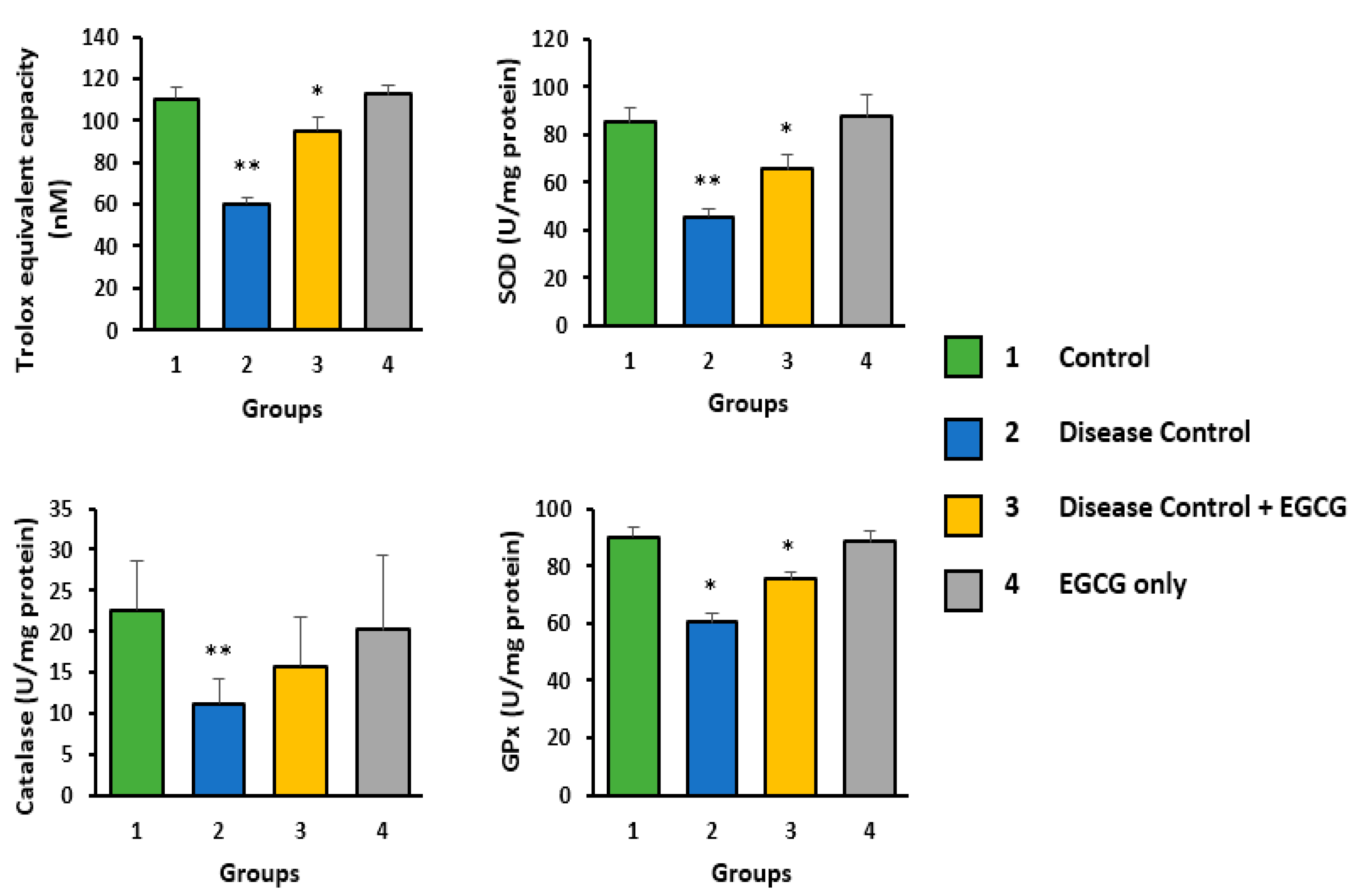
The antioxidant activity of EGCG was analyzed by measuring the serum levels of hepatic antioxidant enzymes, including SOD, CAT, and GPx. The activities of antioxidant enzymes, such as SOD, CAT, and GPx, were found to be significantly decreased in the DEN only-treated group compared to the control group (Figure 2). Furthermore, the activities of SOD, CAT, and GPx meaningfully increased in the DEN + EGCG group (p < 0.005). Moreover, the total antioxidant capacity was significantly decreased in the DEN only-treated group as compared to the control group. However, the total antioxidant capacity was found to be significantly increased in the DEN + EGCG group (p < 0.005) (Figure 2). This result clearly demonstrated that EGCG plays a vital role in liver damage protection by improving antioxidant enzyme levels.

3.3. EGCG Reduces the Serum Levels of CRP and Pro-Inflammatory Mediators—TNF-α and IL-6

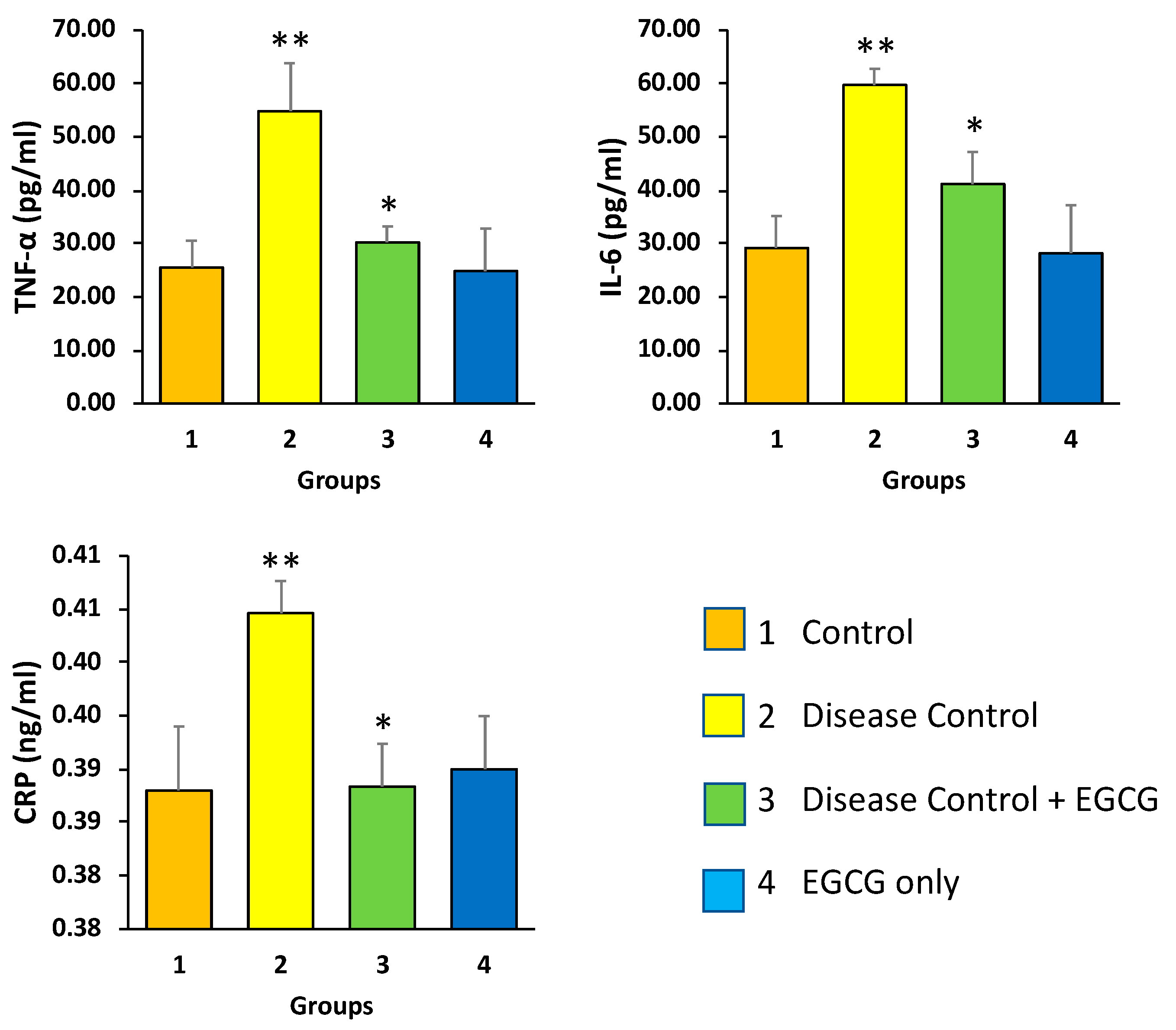
DEN treatment increases the levels of pro-inflammatory cytokines TNF-α and IL-6 in the serum of animals, as compared to the serum of animals without any treatment (control) (p < 0.05). On the other hand, treatment with EGCG significantly decreased their levels (p < 0.05). The level of CRP was high in the serum of animals of the DEN only-treated group, as compared to the serum of animals without any treatment, and treatment with EGCG significantly decreased the level of CRP (Figure 3).

3.4. EGCG Reduces Hepatic Histological Alterations

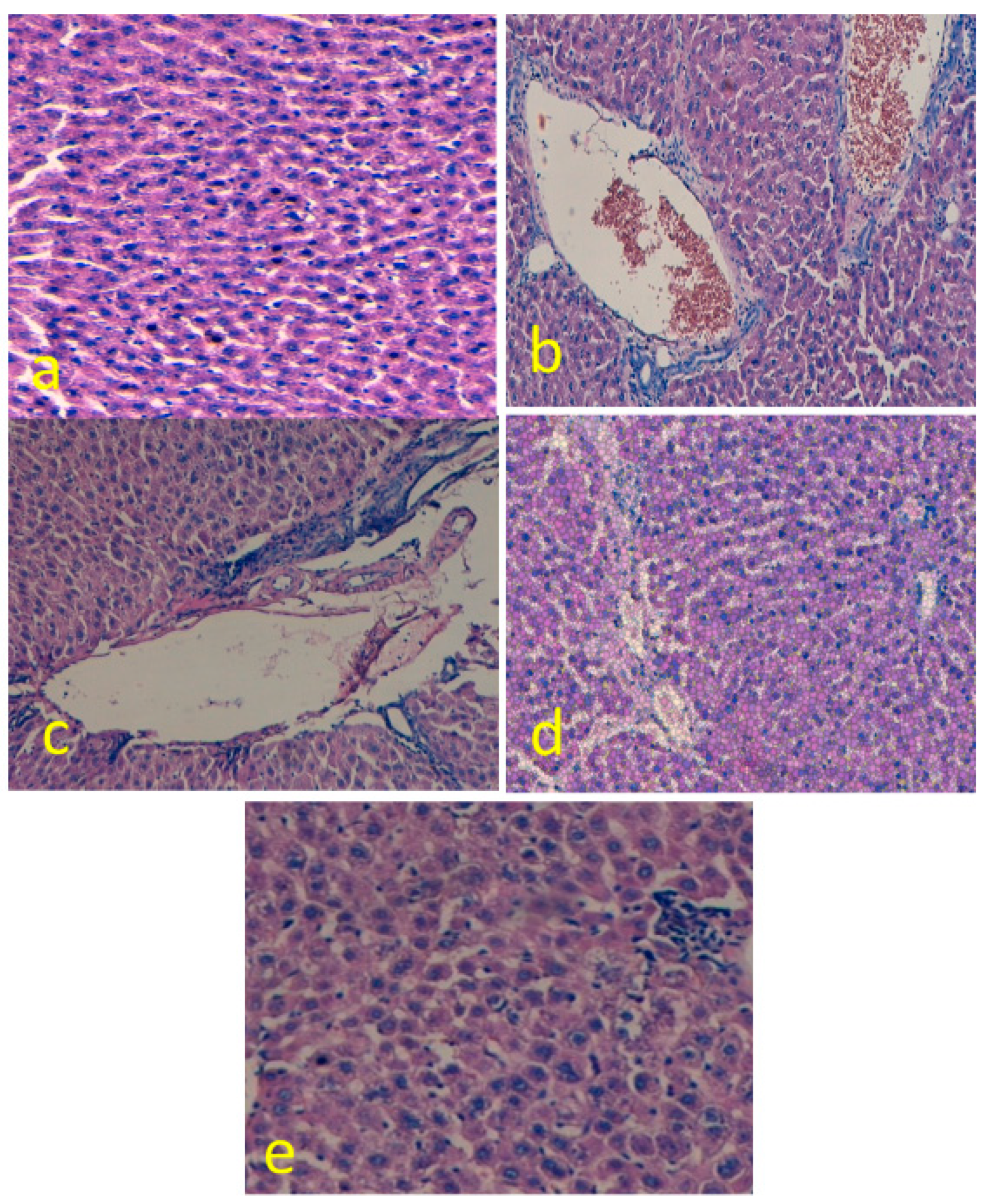
Liver tissues from all the experimental groups were analyzed through H&E staining and histological findings were compared accordingly (Figure 4a–e). The DEN only-treated group displayed altered liver tissues, including dilate and congested central vein, hemorrhage and increased inflammatory cells in the portal tract, hemorrhage, and necrosis. However, these alterations were found to be significantly mild/lower in the EGCG-treated group (p < 0.05).

3.5. Immunohistochemical Analysis

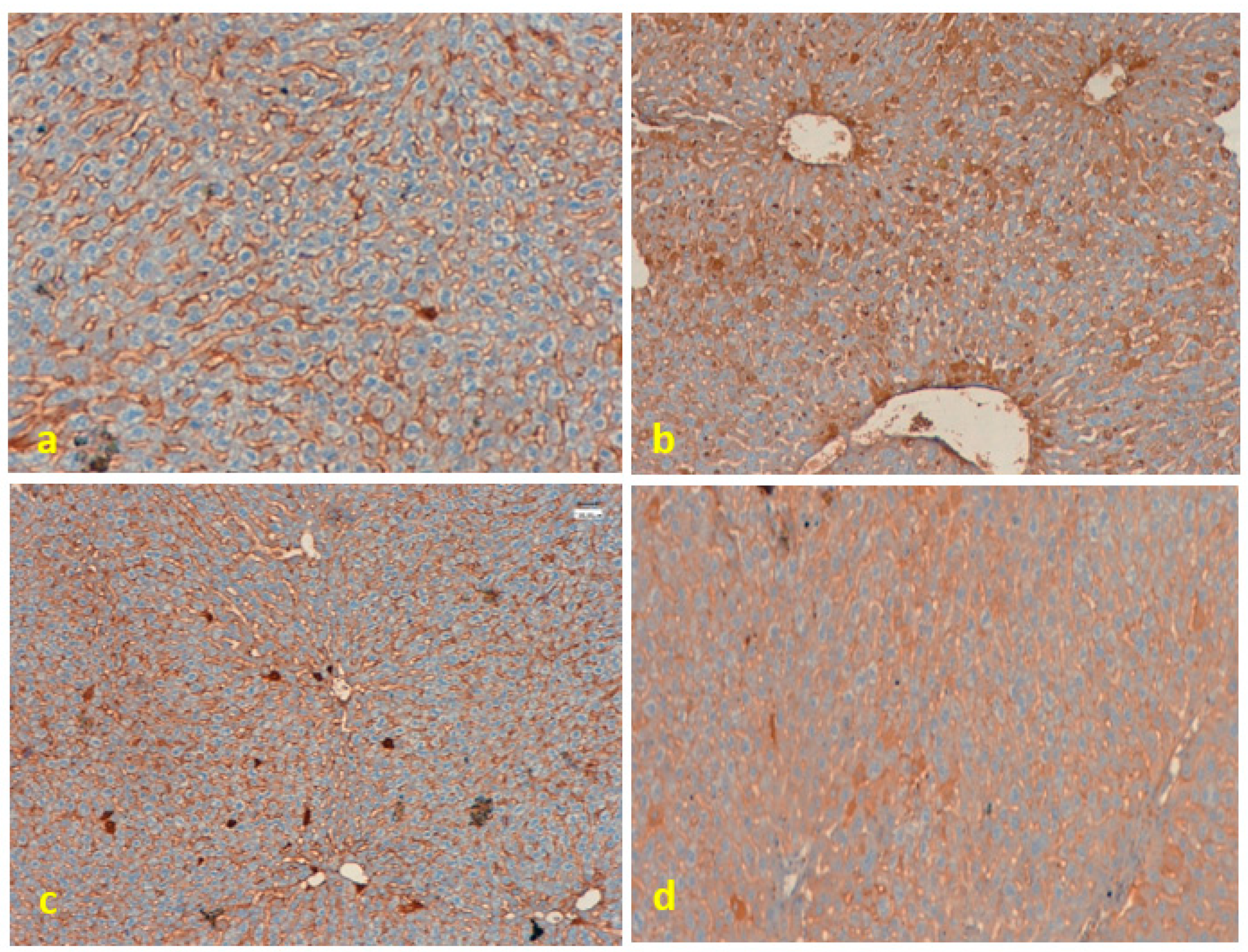
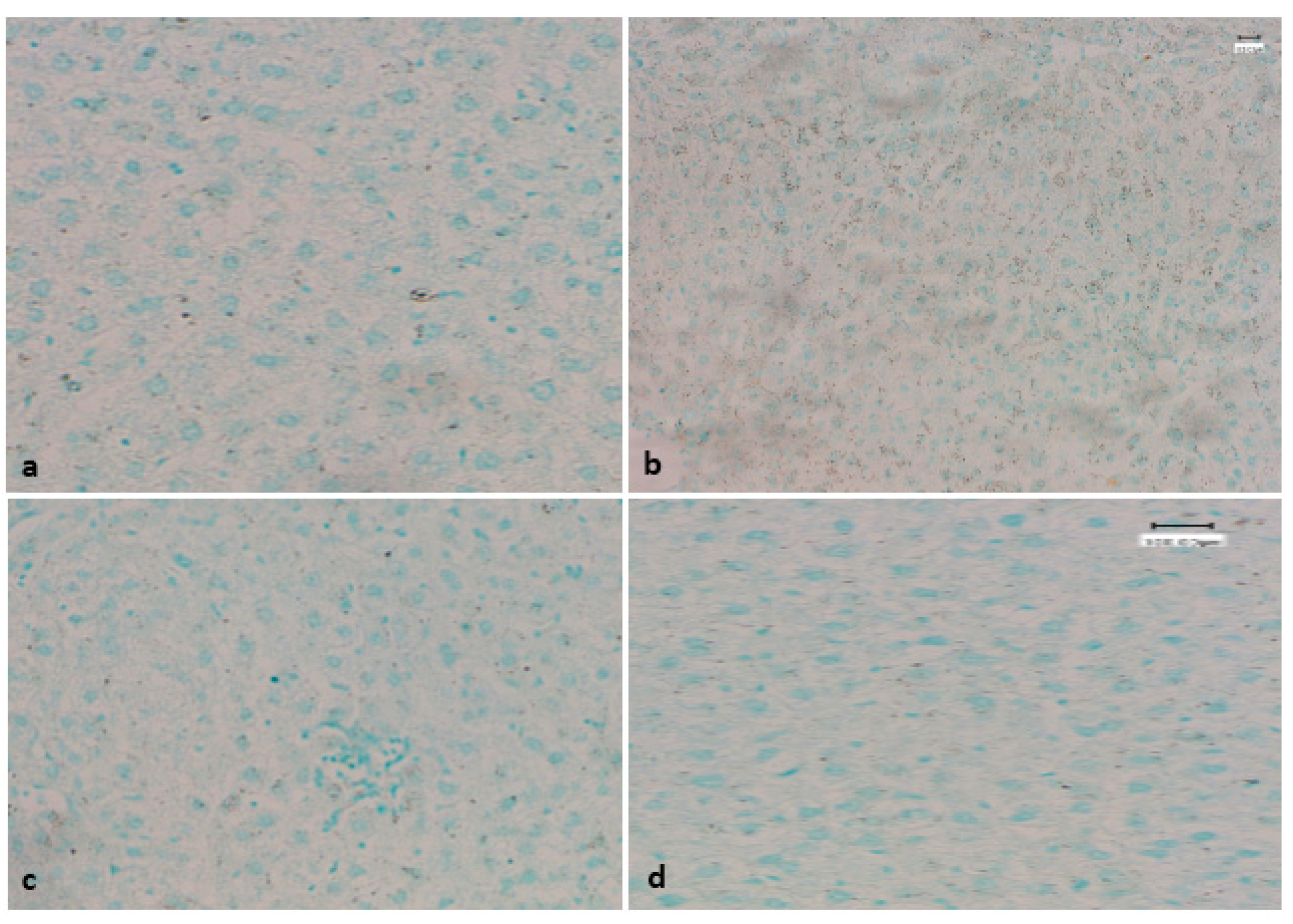
Expression of PTEN protein was evaluated in the tissues of different groups via immunohistochemistry staining. PTEN protein expression was noticed in all groups (Figure 5a–d) and loss of PTEN protein expression was not observed in any of the groups. The expression pattern of PTEN protein among different groups was found to be statistically insignificant (p > 0.05).

3.6. Evaluation of Apoptotic Bodies Via TUNEL Assay

Control tissue, EGCG-treated group, and DEN + EGCG group tissue did not possess apoptotic nuclei as indicated by the TUNEL assay. However, some positive TUNEL cells were observed in the DEN only-treated group (Figure 6a–d).

3.7. Effect of EGCG on Ultrastructure Changes of Liver

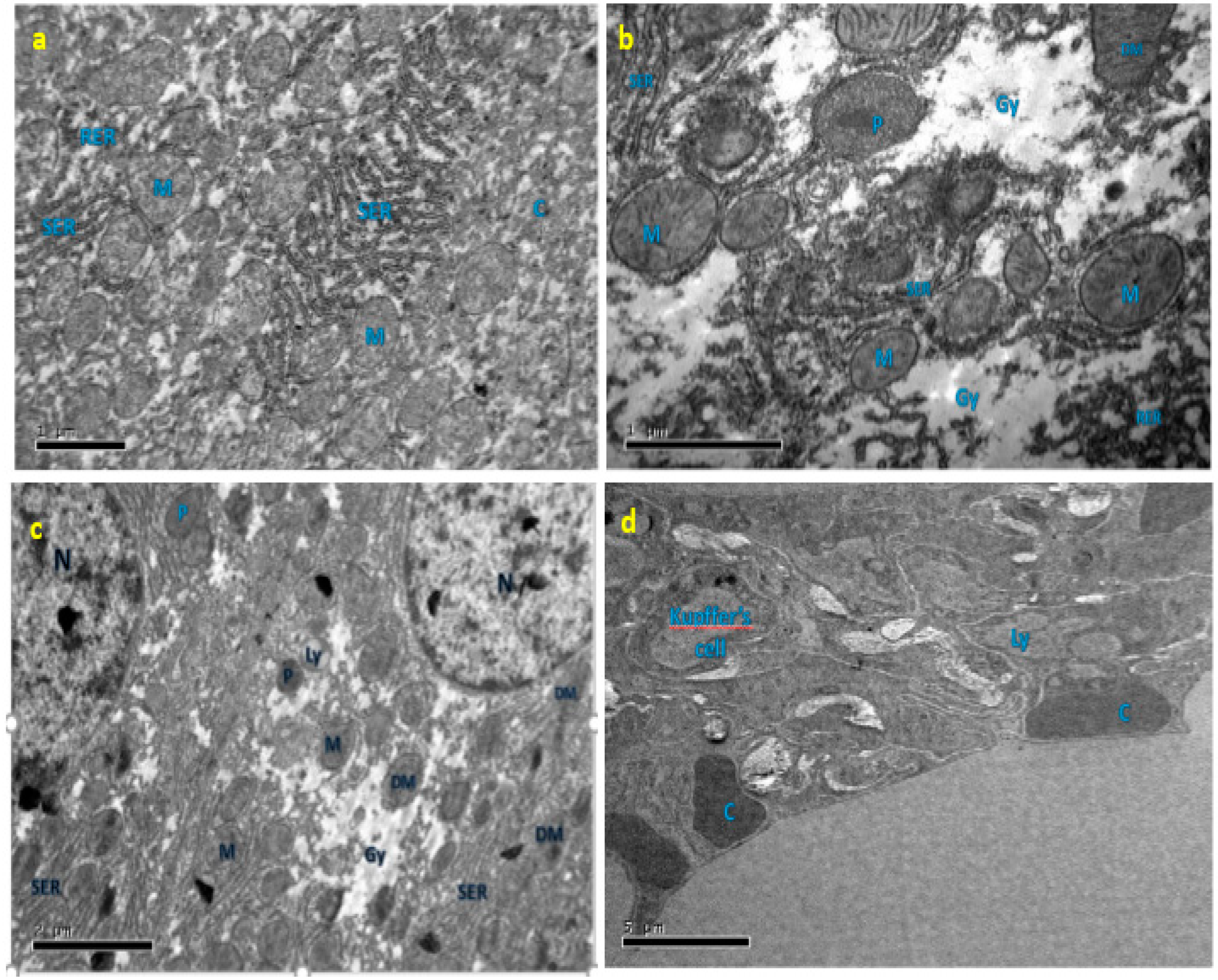
An electron microscopy-based experiment was performed to analyze the ultrastructure of liver tissue. The hepatocytes of the liver tissue of the normal control group demonstrated normal mitochondria and most of the mitochondria were spherical or round-shaped, dense granulated cytoplasm, smooth endoplasmic reticulum tubules with cristae normally present and normal rough endoplasmic reticulum whereas the observation of hepatocytes in the DEN-treated group showed extensive cellular damage. It was seen that a swollen and reduced number of mitochondria, distorted shape mitochondria, large clumps of glycogen surrounding cell organelles, broken SER tubules, and dilation of the rough endoplasmic reticulum (p < 0.05). A number of damaged hepatocytes were found in the DEN-only treatment group, and the majority of the cytoplasmic organelles in these cells were degraded. The hepatocytes of liver tissue of the DEN plus EGCG-treated group showed a reduction of the damage in hepatocytes. Mitochondria are spherical or round-shaped but the presence of some dilated mitochondria, presence of glycogen clump, well-developed SER-ER tubule, and few lysosomes. The normal hepatocytes were seen in the EGCG only-treated group (Figure 7a–d).

3.8. Effect of EGCG on the Cell Cycle

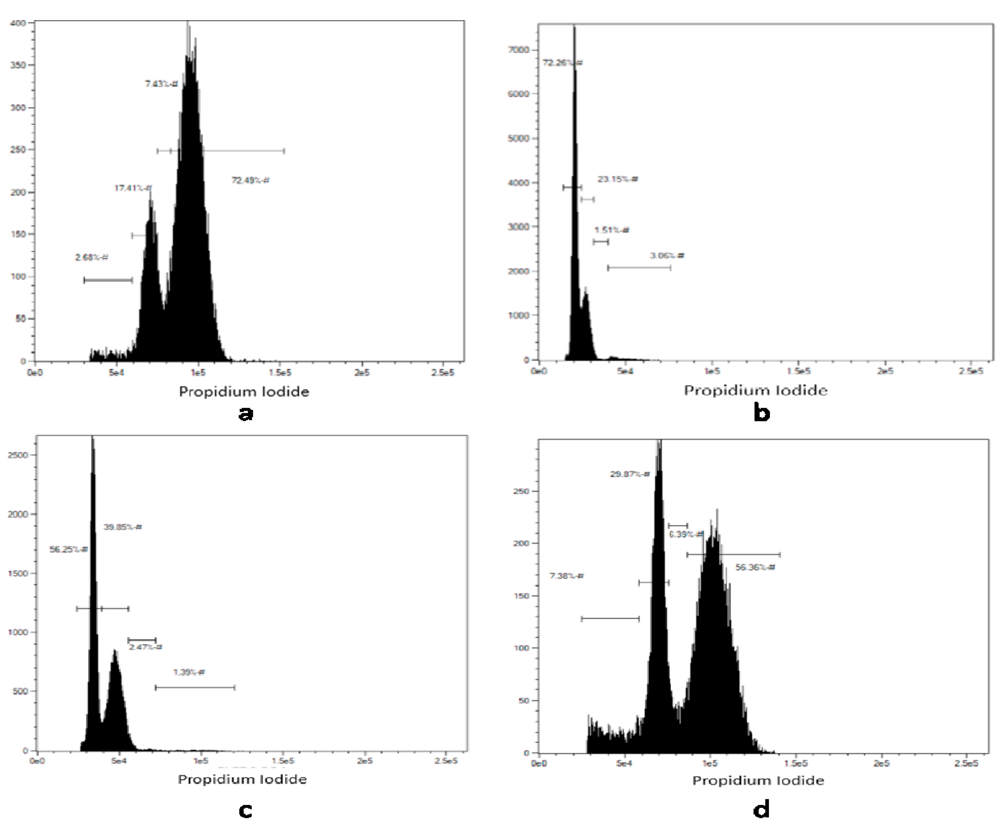
Cell cycle analysis was done using flow cytometry. A relative frequency of cells in different phases of the cell cycle were calculated. The control group (a) contained around 80% of cells in the S and G2/M phase. However, cells were arrested in the sub-G1 phase in tissues treated with DEN only (b). On the other hand, the EGCG plus DEN (c) treatment allowed the cells to move from the sub-G1 phase to the G1 and S phase as indicated by an increased frequency of cells in the S and G2/M phase (~93%). (d) The EGCG only-treated group (Figure 8a–d and Figure 9a–d).

4. Discussion

The current study results provide evidence of the role of EGCG in the regulation of apoptosis, antioxidant production, and inflammatory cytokines in rat liver damage with the concept of mitochondrial homeostasis. Significantly, the current study results also advocate for an argument for further analysis of EGCG or structurally related molecules as inhibitors of TNF-α and IL-6 synthesis and possibly other cytokine-regulated diseases. The anti-inflammatory, tumor-suppressing, and anti-oxidative effects of EGCG have been studied formerly. Howbeit, its antioxidant effect is the most imperative one [26,27]. During liver injury, the generation of free radicals and reactive oxygen species plays a critical role in the augmentation of tissue damage. As the injury proceeds, inflammatory cascades are activated, leading to the release of pro-inflammatory cytokines and free radicals, which finally causes activation of inflammatory Kupffer cells and recruitment of neutrophils at the site of injury. Stimulated inflammatory cells are accountable for the generation of more free radicals and provoked secretion of pro-inflammatory mediators. A large amount of released free radicals induce lipid peroxidation in the cellular phospholipid bilayer, which can damage the cytosol and other cellular organelles [5]. Thoughtfully, the anti-oxidative effects of EGCG come into action through activation of anti-oxidation protective signaling, thus reducing injury [28,29]. Similarly, other studies have shown anti-oxidative and preventive effects of EGCG on different hepatic injury animal models [30,31].
In the current study, DEN-induced liver injury in a rat model was characterized by an increase in the secretion of pro-inflammatory cytokines, including IL-6 and TNF-α. This study shows that EGCG ameliorated liver injury along with a decrease of IL-6 and TNF-α levels in serum. These results are supported by previous studies in which IL-6 deficiency or IL-6R blockade ameliorated hepatocyte damage, indicating that the control of IL-6 production may produce a valuable outcome in liver injury [32,33]. A previous study by Jamal et al. [34] showed that administration of green tea polyphenols showed a role in the prevention of acute liver injury in mice by reducing CRP, IL-6, and TNF-α. However, this study specifically provides insight into one of the potential mechanisms of EGCG’s liver protective activity.
Moreover, EGCG treatment also increases the levels of anti-oxidative enzymes SOD, CAT, and GPx, suggesting the activation of anti-oxidation protective signaling pathways to provide protection against oxidative damage. The upregulation of serum levels of AST, ALP, and ALT is a strong indication of the manifestation of oxidative stress in DEN-induced liver injury. The current study result demonstrates that EGCG reduces oxidative stress by reducing the serum levels of these liver functionality enzymes. It has been reported that oxidative stress regulates the apoptotic signaling pathway in cells. This result agrees with a previous finding that treatment with EGCG plays a significant role in the reduction of liver injury, oxidative stress, and the inflammatory response [5]. Moreover, EGCG also significantly attenuated the severity of CCl (4)-induced liver injury and the progression of liver fibrosis. Another study reported that in vitro model of oxidative stress induced by ethanol provided evidence that EGCG prevented some aspect of liver cell injury caused by ethanol [35].
PTEN is one of the tumor suppressor genes and it is located in the 10q23 region encoding for a 403-amino acid multifunctional protein that comprises lipid and protein phosphatase activities [36]. The overexpression/altered expression of PTEN protein has been noticed in tumors [37]. Several studies have suggested a role for PTEN in hepatic injury [38,39], but the expression pattern of PTEN protein among different groups was statistically insignificant in the current study.
Mitochondria are the major source of endogenous reactive oxygen species, and altering mitochondrial homeostasis is a known mechanism for toxic compounds, including DEN [40,41]. EGCG treatment significantly improves mitochondrial health and number as determined by our TEM study; several studies [42,43,44] with EGCG are in accordance with our study. Further studies are needed to fully investigate this mechanism.
Previous study have suggested that PARP is a family of proteins involved in a number of cellular processes including DNA repair and apoptosis [45]. This has been evidently established by an increase in apoptotic nuclei upon DEN-induced liver injury. However, EGCG decreases the presence of apoptotic nuclei as demonstrated by the TUNEL assay. Therefore, the present data advocate that administration of EGCG plays a role in the reduction of liver injury-related cell apoptosis, possibly by decreasing cleaved caspase-3 and cleaved caspase-9 levels. Thus, the accumulation of DEN in the liver caused several damaging effects, including marked pathological changes in liver enzyme activities, generation of reactive oxygen species, and DNA damage, as well as apoptosis. These effects were reversed by administration of EGCG. Additionally, EGCG administration displayed a clear effect on the cell cycle, accompanied by a decrease in the sub-G1 population (indicating apoptosis) and a significant increase in S or G2/M (indicating cell proliferation), the mechanism of which needs to be further elucidated. A previous finding was in accordance with the current finding and the study reported negative regulators of the protein kinases and cyclins, thus arresting the cell cycle at the eG0/G1 phase [46], and EGCG, a chief compound of green tea, inhibited DNA synthesis and arrested the cell cycle at the G0/G1 phase [47].
In conclusion, the result of the current study demonstrates that EGCG treatment reduced the amount of DEN-induced hepatic injury by decreasing mitochondrial oxidative stress and cellular apoptosis. Furthermore, it was also revealed that EGCG inhibits CRP, TNF-α, and IL-6 production and suppresses the inflammation process. Hence, EGCG can be used as a plausible therapeutic tool against liver injury and other liver-associated diseases.

Author Contributions

Conceptualization, S.A.A. and A.H.R.; Formal analysis, M.A.A.; Methodology, H.M.A.; Supervision, H.M.A.; Validation, A.A.K.; Writing—original draft, S.A.A. and A.H.R.; Writing—review & editing, M.A.A., A.A.K. and A.H.R.

Funding

This work was funded and supported by Qassim University Research Deanship Grants # cams1-2018-1-14-S-3360.

Conflicts of Interest

There are no conflict of interest regarding this study.

Data Availability

The data used to support the findings of the current study are included within the article.

References

  1. Samuel, A.J.S.J.; Mohan, S.; Chellappan, D.K.; Kalusalingam, A.; Ariamuthu, S. Hibiscus vitifolius (Linn.) root extracts shows potent protective action against anti-tubercular drug induced hepatotoxicity. J. Ethnopharmacol. 2012, 141, 396–402. [Google Scholar] [CrossRef] [PubMed]
  2. Mishra, B.B.; Tiwari, V.K. Natural products: An evolving role in future drug discovery. Eur. J. Med. Chem. 2011, 46, 4769–4807. [Google Scholar] [CrossRef] [PubMed]
  3. Gao, M.; Nettles, R.E.; Belema, M.; Snyder, L.B.; Nguyen, V.N.; Fridell, R.A.; Serrano-Wu, M.H.; Langley, D.R.; Sun, J.H.; O’Boyle, D.R., II; et al. Chemical genetics strategy identifies an HCV NS5A inhibitor with a potent clinical effect. Nature 2010, 465, 96. [Google Scholar] [CrossRef] [PubMed]
  4. Ghosh, N.; Ghosh, R.; Mandal, V.; Mandal, S.C. Recent advances in herbal medicine for treatment of liver diseases. Pharm. Biol. 2011, 49, 970–988. [Google Scholar] [CrossRef] [PubMed]
  5. Tipoe, G.L.; Leung, T.M.; Liong, E.C.; Lau, T.Y.H.; Fung, M.L.; Nanji, A.A. Epigallocatechin-3-gallate (EGCG) reduces liver inflammation, oxidative stress and fibrosis in carbon tetrachloride (CCl4)-induced liver injury in mice. Toxicology 2010, 273, 45–52. [Google Scholar] [CrossRef] [PubMed]
  6. Louvet, A.; Mathurin, P. Alcoholic liver disease: Mechanisms of injury and targeted treatment. Nat. Rev. Gastroenterol. Hepatol. 2015, 12, 231. [Google Scholar] [CrossRef] [PubMed]
  7. Björnsson, E. Drug-induced liver injury in clinical practice. Aliment. Pharmacol. Ther. 2010, 32, 3–13. [Google Scholar] [CrossRef] [PubMed]
  8. Park, D.H.; Shin, J.W.; Park, S.K.; Seo, J.N.; Li, L.; Jang, J.J.; Lee, M.J. Diethylnitrosamine (DEN) induces irreversible hepatocellular carcinogenesis through overexpression of G1/S-phase regulatory proteins in rat. Toxicol. Lett. 2009, 191, 321–326. [Google Scholar] [CrossRef] [PubMed]
  9. Magee, P.N.; Barnes, J. The production of malignant primary hepatic tumours in the rat by feeding dimethylnitrosamine. Br. J. Cancer 1956, 10, 114. [Google Scholar] [CrossRef] [PubMed]
  10. Verna, L.; Whysner, J.; Williams, G.M. N-nitrosodiethylamine mechanistic data and risk assessment: Bioactivation, DNA-adduct formation, mutagenicity, and tumor initiation. Pharmacol. Ther. 1996, 71, 57–81. [Google Scholar] [CrossRef]
  11. Paterson, I.; Anderson, E.A. The renaissance of natural products as drug candidates. Science 2005, 310, 451–453. [Google Scholar] [CrossRef] [PubMed]
  12. Teuten, E.L.; Xu, L.; Reddy, C.M. Two abundant bioaccumulated halogenated compounds are natural products. Science 2005, 307, 917–920. [Google Scholar] [CrossRef] [PubMed]
  13. Rollinger, J.; Langer, T.; Stuppner, H. Strategies for efficient lead structure discovery from natural products. Curr. Med. Chem. 2006, 13, 1491–1507. [Google Scholar] [CrossRef] [PubMed]
  14. Majeed, R.; Reddy, M.V.; Chinthakindi, P.K.; Sangwan, P.L.; Hamid, A.; Chashoo, G.; Saxena, A.K.; Koul, S. Bakuchiol derivatives as novel and potent cytotoxic agents: A report. Eur. J. Med. Chem. 2012, 49, 55–67. [Google Scholar] [CrossRef] [PubMed]
  15. Beecher, G.R.; Warden, B.A.; Merken, H. Analysis of tea polyphenols. Proc. Soc. Exp. Biol. Med. 1999, 220, 267–270. [Google Scholar] [PubMed]
  16. Salah, N.; Miller, N.J.; Paganga, G.; Tijburg, L.; Bolwell, G.P.; Riceevans, C. Polyphenolic flavanols as scavengers of aqueous phase radicals and as chain-breaking antioxidants. Arch. Biochem. Biophys. 1995, 322, 339–346. [Google Scholar] [CrossRef] [PubMed]
  17. Isbrucker, R.A.; Bausch, J.; Edwards, J.A.; Wolz, E. Safety studies on epigallocatechin gallate (EGCG) preparations. Part 1: Genotoxicity. Food Chem. Toxicol. 2006, 44, 626–635. [Google Scholar] [CrossRef] [PubMed]
  18. Lambert, J.D.; Yang, C.S. Mechanisms of cancer prevention by tea constituents. J. Nutr. 2003, 133, 3262S–3267S. [Google Scholar] [CrossRef] [PubMed]
  19. Wolfram, S.; Wang, Y.; Thielecke, F. Anti-obesity effects of green tea: From bedside to bench. Mol. Nutr. Food Res. 2006, 50, 176–187. [Google Scholar] [CrossRef] [PubMed]
  20. Frei, B.; Higdon, J.V. Antioxidant activity of tea polyphenols in vivo: Evidence from animal studies. J. Nutr. 2003, 133, 3275S–3284S. [Google Scholar] [CrossRef] [PubMed]
  21. Kundu, J.K.; Na, H.K.; Chun, K.S.; Kim, Y.K.; Lee, S.J.; Lee, S.S.; Lee, O.S.; Sim, Y.C.; Surh, Y.J. Inhibition of Phorbol Ester–Induced COX-2 Expression by Epigallocatechin Gallate in Mouse Skin and Cultured Human Mammary Epithelial Cells. J. Nutr. 2003, 133, 3805S–3810S. [Google Scholar] [CrossRef] [PubMed]
  22. Yang, F.; Oz, H.S.; Barve, S.; De Villiers, W.J.; McClain, C.J.; Varilek, G.W. The green tea polyphenol (−)-epigallocatechin-3-gallate blocks nuclear factor-κB activation by inhibiting IκB kinase activity in the intestinal epithelial cell line IEC-6. Mol. Pharmacol. 2001, 60, 528–533. [Google Scholar] [PubMed]
  23. Bancroft, J.D.; Gamble, M. Theory and Practice of Histological Techniques, 6th ed.; Churchill Livingstone, Elsevier: London, UK, 2008. [Google Scholar]
  24. Rahmani, A.; Alzohairy, M.; Khadri, H.; Mandal, A.K.; Rizvi, M.A. Expressional evaluation of vascular endothelial growth factor (VEGF) protein in urinary bladder carcinoma patients exposed to cigarette smoke. Int. J. Clin. Exp. Pathol. 2012, 5, 195–200. [Google Scholar] [PubMed]
  25. Rahmani, A.H.; Babiker, A.Y.; AlWanian, W.M.; Elsiddig, S.A.; Faragalla, H.E.; Aly, S.M. Association of cytokeratin and vimentin protein in the genesis of transitional cell carcinoma of urinary bladder patients. Dis. Mark. 2015, 2015, 204759. [Google Scholar] [CrossRef] [PubMed]
  26. Singh, B.N.; Shankar, S.; Srivastava, R.K. Green tea catechin, epigallocatechin-3-gallate (EGCG): Mechanisms, perspectives and clinical applications. Biochem. Pharmacol. 2011, 82, 1807–1821. [Google Scholar] [CrossRef] [PubMed]
  27. Yang, C.S.; Wang, X. Green tea and cancer prevention. Nutr. Cancer 2010, 62, 931–937. [Google Scholar] [CrossRef] [PubMed]
  28. Thangapandiyan, S.; Miltonprabu, S. Epigallocatechin gallate effectively ameliorates fluoride-induced oxidative stress and DNA damage in the liver of rats. Can. J. Physiol. Pharmacol. 2013, 91, 528–537. [Google Scholar] [CrossRef] [PubMed]
  29. Moravcová, A.; Cervinkova, Z.; Kucera, O.; Mezera, V.; Lotková, H. Antioxidative effect of epigallocatechin gallate against D-galactosamine-induced injury in primary culture of rat hepatocytes. Acta Med. 2014, 57, 3–8. [Google Scholar]
  30. Yao, H.T.; Yang, Y.C.; Chang, C.H.; Yang, H.T.; Yin, M.C. Protective effects of (-)-epigallocatechin-3-gallate against acetaminophen-induced liver injury in rats. Biomedicine 2015, 5, 15. [Google Scholar] [CrossRef] [PubMed]
  31. Tak, E.; Park, G.C.; Kim, S.H.; Jun, D.Y.; Lee, J.; Hwang, S.; Song, G.W.; Lee, S.G. Epigallocatechin-3-gallate protects against hepatic ischaemia-reperfusion injury by reducing oxidative stress and apoptotic cell death. J. Int. Med. Res. 2016, 44, 1248–1262. [Google Scholar] [CrossRef] [PubMed]
  32. Karatayli, E.; Hall, R.A.; Weber, S.N.; Dooley, S.; Lammert, F. Effect of alcohol on the interleukin 6-mediated inflammatory response in a new mouse model of acute-on-chronic liver injury. Biochim. Biophys. Acta Mol. Basis Dis. 2019, 1865, 298–307. [Google Scholar] [CrossRef] [PubMed]
  33. Farouk, S.; Sabet, S.; Zahra, F.A.A.; El-Ghor, A.A. Bone marrow derived-mesenchymal stem cells downregulate IL17A dependent IL6/STAT3 signaling pathway in CCl4-induced rat liver fibrosis. PLoS ONE 2018, 13, e0206130. [Google Scholar] [CrossRef] [PubMed] [Green Version]
  34. Jamal, M.H.; Ali, H.; Dashti, A.; Al-Abbad, J.; Dashti, H.; Mathew, C.; Al-Ali, W.; Asfar, S. Effect of epigallocatechin gallate on uncoupling protein 2 in acute liver injury. Int. J. Clin. Exp. Pathol. 2015, 8, 649–654. [Google Scholar] [PubMed]
  35. Oliva, J.; Bardag-Gorce, F.; Tillman, B.; French, S.W. Protective effect of quercetin, EGCG, catechin and betaine against oxidative stress induced by ethanol in vitro. Exp. Mol. Pathol. 2011, 90, 295–299. [Google Scholar] [CrossRef] [PubMed] [Green Version]
  36. Milella, M.; Falcone, I.; Conciatori, F.; Cesta Incani, U.; Del Curatolo, A.; Inzerilli, N.; Nuzzo, C.; Vaccaro, V.; Vari, S.; Cognetti, F.; et al. PTEN: Multiple Functions in Human Malignant Tumors. Front. Oncol. 2015, 5, 24. [Google Scholar] [CrossRef] [PubMed] [Green Version]
  37. Rahmani, A.; Alzohairy, M.; Babiker, A.Y.; Rizvi, M.A.; Elkarimahmad, H.G. Clinicopathological significance of PTEN and bcl2 expressions in oral squamous cell carcinoma. Int. J. Clin. Exp. Pathol. 2012, 5, 965–971. [Google Scholar] [PubMed]
  38. Su, S.; Luo, D.; Liu, X.; Liu, J.; Peng, F.; Fang, C.; Li, B. miR-494 up-regulates the PI3K/Akt pathway via targetting PTEN and attenuates hepatic ischemia/reperfusion injury in a rat model. Biosci. Rep. 2017, 37. [Google Scholar] [CrossRef] [PubMed] [Green Version]
  39. Cheng, Y.; Tian, Y.; Xia, J.; Wu, X.; Yang, Y.; Li, X.; Huang, C.; Meng, X.; Ma, T.; Li, J. The role of PTEN in regulation of hepatic macrophages activation and function in progression and reversal of liver fibrosis. Toxicol. Appl. Pharmacol. 2017, 317, 51–62. [Google Scholar] [CrossRef] [PubMed]
  40. Chang, W.; He, W.; Li, P.P.; Song, S.S.; Yuan, P.F.; Lu, J.T.; Wei, W. Protective effects of Celastrol on diethylnitrosamine-induced hepatocellular carcinoma in rats and its mechanisms. Eur. J. Pharmacol. 2016, 784, 173–180. [Google Scholar] [CrossRef] [PubMed]
  41. Sun, Q.; Long, Z.; Wu, H.; Liu, Y.; Wang, L.; Zhang, X.; Wang, X.; Hai, C. Effect of alcohol on diethylnitrosamine-induced hepatic toxicity: Critical role of ROS, lipid accumulation, and mitochondrial dysfunction. Exp. Toxicol. Pathol. 2015, 67, 491–498. [Google Scholar] [CrossRef] [PubMed]
  42. James, K.D.; Kennett, M.J.; Lambert, J.D. Potential role of the mitochondria as a target for the hepatotoxic effects of (-)-epigallocatechin-3-gallate in mice. Food Chem. Toxicol. 2018, 111, 302–309. [Google Scholar] [CrossRef] [PubMed]
  43. Shen, K.; Feng, X.; Su, R.; Xie, H.; Zhou, L.; Zheng, S. Epigallocatechin 3-gallate ameliorates bile duct ligation induced liver injury in mice by modulation of mitochondrial oxidative stress and inflammation. PLoS ONE 2015, 10, e0126278. [Google Scholar] [CrossRef] [PubMed]
  44. Mezera, V.; Endlicher, R.; Kucera, O.; Sobotka, O.; Drahota, Z.; Cervinkova, Z. Effects of Epigallocatechin Gallate on Tert-Butyl Hydroperoxide-Induced Mitochondrial Dysfunction in Rat Liver Mitochondria and Hepatocytes. Oxid. Med. Cell. Longev. 2016, 2016, 7573131. [Google Scholar] [CrossRef] [PubMed] [Green Version]
  45. Luo, X.; Kraus, W.L. On PAR with PARP: Cellular stress signaling through poly (ADP-ribose) and PARP-1. Genes Dev. 2012, 26, 417–432. [Google Scholar] [CrossRef] [PubMed] [Green Version]
  46. Jiang, F.; Jiang, R.; Zhu, X.; Zhang, X.; Zhan, Z. Genipin inhibits TNF-alpha-induced vascular smooth muscle cell proliferation and migration via induction of HO-1. PLoS ONE 2013, 8, e74826. [Google Scholar]
  47. Liu, P.L.; Liu, J.T.; Kuo, H.F.; Chong, I.W.; Hsieh, C.C. Epigallocatechin gallate attenuates proliferation and oxidative stress in human vascular smooth muscle cells induced by interleukin-1beta via heme oxygenase-1. Mediat. Inflamm. 2014, 2014, 523684. [Google Scholar] [CrossRef] [PubMed] [Green Version]
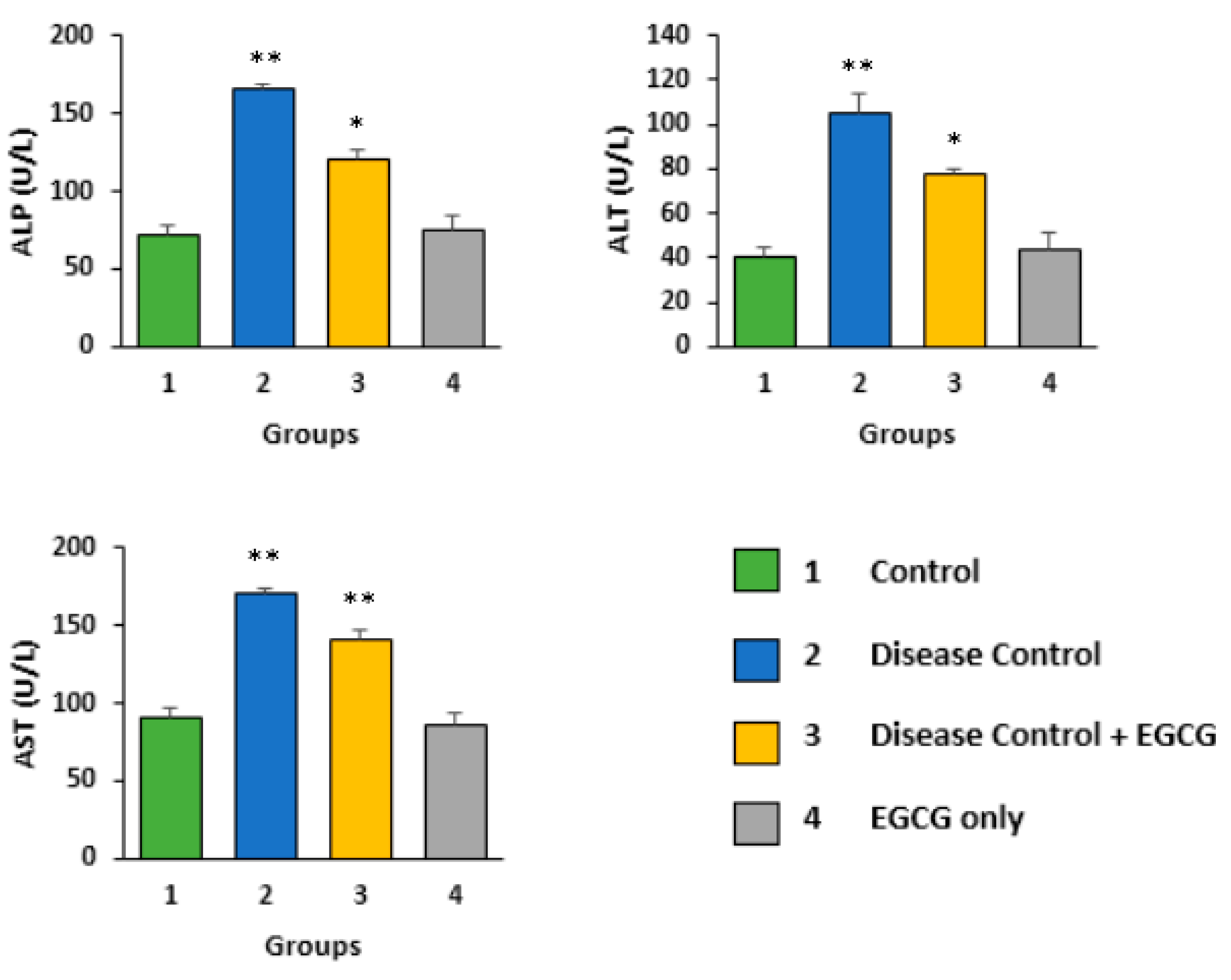
Figure 1. Effect of EGCG on Alanine aminotransferase (ALT), Alkaline phosphatase (ALP), and Aspartate transaminase (AST) activity in DEN-induced liver injury. Enzyme levels, including ALT, ALP, and AST, were significantly higher in the DEN-treated group than the normal control group. Moreover, the DEN + EGCG group revealed considerably lower ALT, ALP, and AST values than the DEN-only group. Statistical significances are compared between control versus DEN-treated groups only (p < 0.01), and DEN-treated versus DEN plus EGCG (p < 0.05).
Applsci 09 04821 g001
Figure 2. Effect of EGCG on antioxidant enzymes Superoxide Dismutase (SOD), Catalase (CAT), and Glutathione peroxidase (GPx) and total antioxidant capacity in DEN-induced liver injury. The levels of antioxidant enzymes were significantly lower in the DEN only-treated group than the control group. The DEN + EGCG group displayed significantly higher antioxidant enzyme levels than the DEN-only group (p < 0.01). Total antioxidant capacity was also found to be significantly increased in the DEN + EGCG group (p < 0.05).
Applsci 09 04821 g002
Figure 3. Effect of EGCG on the serum levels of TNF-α, CRP, and IL-6. EGCG treatment significantly decreases the levels of Tumour Necrosis Factor alpha (TNF-α), c-reactive protein (CRP), and Interleukin 6 (IL-6) in the DEN + EGCG group (p < 0.05) as compared to the DEN-treated group (p < 0.01).
Applsci 09 04821 g003
Figure 4. Histopathological analysis. H&E (X40) stained photomicrographs of liver tissues showing (a) the normal architecture of hepatocytes—control group. (bd) DEN only-treated tissues displaying dilate and congested central vein, hemorrhage, and increased inflammatory cells in the portal tract, hemorrhage, and necrosis. (e) EGCG-treated tissue, where the liver tissue alteration was significantly lowered compared to the disease control group (DEN only-treated group).
Applsci 09 04821 g004
Figure 5. Immunohistochemical analysis. Phosphatase and tensin homolog (PTEN) protein expression was evaluated in different treatment groups (ad). (a) The control group showed high PTEN expression; (b) the DEN-treated group also displayed a high expression of PTEN; (c). PTEN expression was also noted in the DEN + EGCG treated group and (d) EGCG-only group.
Applsci 09 04821 g005
Figure 6. TUNEL Assay. Apoptotic nuclei were absent in (a) the liver tissue of the control group; (c) tissue from the DEN + EGCG group and (d) tissue from the EGCG only-treated group. However, (b) the liver tissue of DEN only-treated group indicated the presence of some positive apoptotic nuclei.
Applsci 09 04821 g006
Figure 7. Ultrastructural changes of liver. Electron microscopy was used to observe the ultrastructure of hepatic cells. (a) Hepatocytes of the liver tissue of the control group revealed normal mitochondria and most of the mitochondria are spherical or round-shaped, smooth endoplasmic reticulum tubules with cristae normally present, and normal rough endoplasmic reticulum; (b) DEN only-treated tissues displaying extensive cellular damage. Swollen and reduced number of mitochondria, distorted shape mitochondria, large clumps of glycogen surrounding cell organelles, broken SER tubules, and dilation of the rough endoplasmic reticulum was seen. (c) EGCG plus DEN-treated tissue—where the liver tissue alteration was significantly reduced as compared to the disease control group (p < 0.05) (DEN only-treated group). (d) EGCG only-treated group exhibited normal hepatocytes.
Applsci 09 04821 g007
Figure 8. Cell cycle analysis. Relative frequencies of cells in different phases of the cell cycle in The control group (a) contained around 80% of cells in the S and G2/M phase. However, cells were arrested in the sub-G1 phase in tissues treated with DEN only (b). On the other hand, the EGCG plus DEN (c) treatment allowed the cells to move from the sub-G1 phase to the G1 and S phase as indicated by an increased frequency of cells in the S and G2/M phase. (d) The EGCG only-treated group.
Applsci 09 04821 g008
Figure 9. Cell cycle significance analysis between the groups using a bar diagram. Relative frequencies of cells in different phases of the cell cycle in (a) control, (b) DEN only-treated, (c) DEN + EGCG-treated, and (d) EGCG only-treated groups.
Applsci 09 04821 g009
Table 1. Animal grouping.
Experimental GroupGroup NumberTreatmentNumber of Animals
Per Group (n)
Negative control1Normal rats administered with vehicle solution08
Disease control2DEN administered via oral gavage08
Treatment3DEN and EGCG08
Treatment4EGCG with vehicle solution08