Review

3D Soft-Tissue Prediction Methodologies for Orthognathic Surgery—A Literature Review

by
Elena Carlotta Olivetti
1,
Sara Nicotera
1,
Federica Marcolin
1,*,
Enrico Vezzetti
1,
Jacqueline P. A. Sotong
2,
Emanuele Zavattero
2 and
Guglielmo Ramieri
2
1
Department of Management and Production Engineering, Politecnico di Torino, corso Duca degli Abruzzi 24, 10129 Torino, Italy
2
Department of Maxillofacial unit, Città della Salute e della Scienza HOSPITAL, 10129 Torino, Italy
*
Author to whom correspondence should be addressed.
Appl. Sci. 2019, 9(21), 4550; https://doi.org/10.3390/app9214550
Submission received: 2 July 2019 / Revised: 11 October 2019 / Accepted: 22 October 2019 / Published: 26 October 2019
(This article belongs to the Special Issue Engineering for Surgery)

Abstract

:
Three-dimensional technologies have had a wide diffusion in several fields of application throughout the last decades; medicine is no exception and the interest in their introduction in clinical applications has grown with the refinement of such technologies. We focus on the application of 3D methodologies in maxillofacial surgery, where they can give concrete support in surgical planning and in the prediction of involuntary facial soft-tissue changes after planned bony repositioning. The purpose of this literature review is to offer a panorama of the existing prediction methods and software with a comparison of their reliability and to propose a series of still pending issues. Various software are available for surgical planning and for the prediction of tissue displacements, but their reliability is still an unknown variable in respect of the accuracy needed by surgeons. Maxilim, Dolphin and other common planning software provide a realistic result, but with some inaccuracies in specific areas of the face; it also is not totally clear how the prediction is obtained by the software and what is the theoretical model they are based on.

1. Introduction

Patients presenting dentofacial deformities are commonly subject to combined orthodontic and surgical treatment such as Le Fort I osteotomy (LFI), bilateral sagittal split osteotomy (BSSO), intraoral ramus vertical osteotomy (IVRO), sagittal split ramus osteotomy (SSRO), bimaxillary surgery and genioplasty. These interventions have been commonly planned with two-dimensional methodologies; today, the last challenge is the three-dimensional (3D) surgery planning. The refining of 3D graphics and imaging tools gives the chance to explore surgical planning and prediction of the effects of different clinical approaches; these techniques are based on images acquired with computed tomography (CT), cone bean computed tomography (CBCT) and multi-slice computed tomography (MSCT), which provide volumetric images of facial anatomical structure. Additionally, the surface of the face can be scanned and mapped to underline the effect of changes in facial appearance using 3D laser technology [1] that gives a great contribution to surgeons to decide on the type of surgeries as well as on the magnitude and direction of surgical movements to correct facial dysmorphology. Moreover, a deep interest in the prediction of soft-tissue response to hard tissue movements has been growing and two-dimensional conventional methodologies seem to be insufficient for this aim as they do not take into account the third dimension.
Indeed, the possibility to know the soft-tissue response to surgical operations helps surgeons to plan surgical movements and it gives surgeons more information about the need of orthodontic decompensation. Furthermore, the purpose of these interventions is not only to correct the facial dysmorphology from a functional point of view, but also to obtain an aesthetic enhancement of patients’ facial aspect. Therefore, an accurate treatment planning is very important to obtain a good aesthetic and occlusal result [2]. For the aforementioned reasons, having a preview of the soft-tissue arrangement is extremely important. In future, this opportunity could bring to have a surgical planning methodology able to best match patients’ aesthetic expectations, additionally to the functional corrective aspect.
Several methods have been considered to forecast soft tissue responses; the most common are the mass-spring model (MSM), finite element model (FEM) and mass tensor model (MTM). Most of the software packages currently adopted in clinical practice are based on these models. Generally, those softwares seem to reach an acceptable overall accuracy, but with inaccuracies for specific areas of the face, for example around the lips.
Studies have been conducted to develop 2D prevision models based on the ratio between facial and bony movements, particularly focusing on face sub regions [3]; by comparing predictive models constructed on this ratio and patients’ post-operative conditions, it is stated that traditional approaches show limits in forecasting the outcome of large and complex movements. Moreover, a large number of variables must be selected [4].
Although there are several studies on the soft tissue changes after maxillary osteotomies, few of them report a systematic analysis.
The target of this systematic review is to gather information on the existing prediction methods and software of soft tissue displacements after dysmorphism corrective surgery, and to draw conclusions on the accuracy and reliability of these software in the preview of surgical outcome. Additionally, some problems related to the level of accuracy needed by surgeons, to the predictive imprecision reported in the studies and to the magnitude of the acceptable error are presented.
This work is structured as follows. The section Material and Methods describes the methods that have been adopted for the literature review. The section Results includes the clinical details of patients involved and the details of the articles considered; the articles have been divided in subsections according to the software used by the authors. Finally, the section Discussion and Conclusions discusses the current prediction methods and concludes the work.

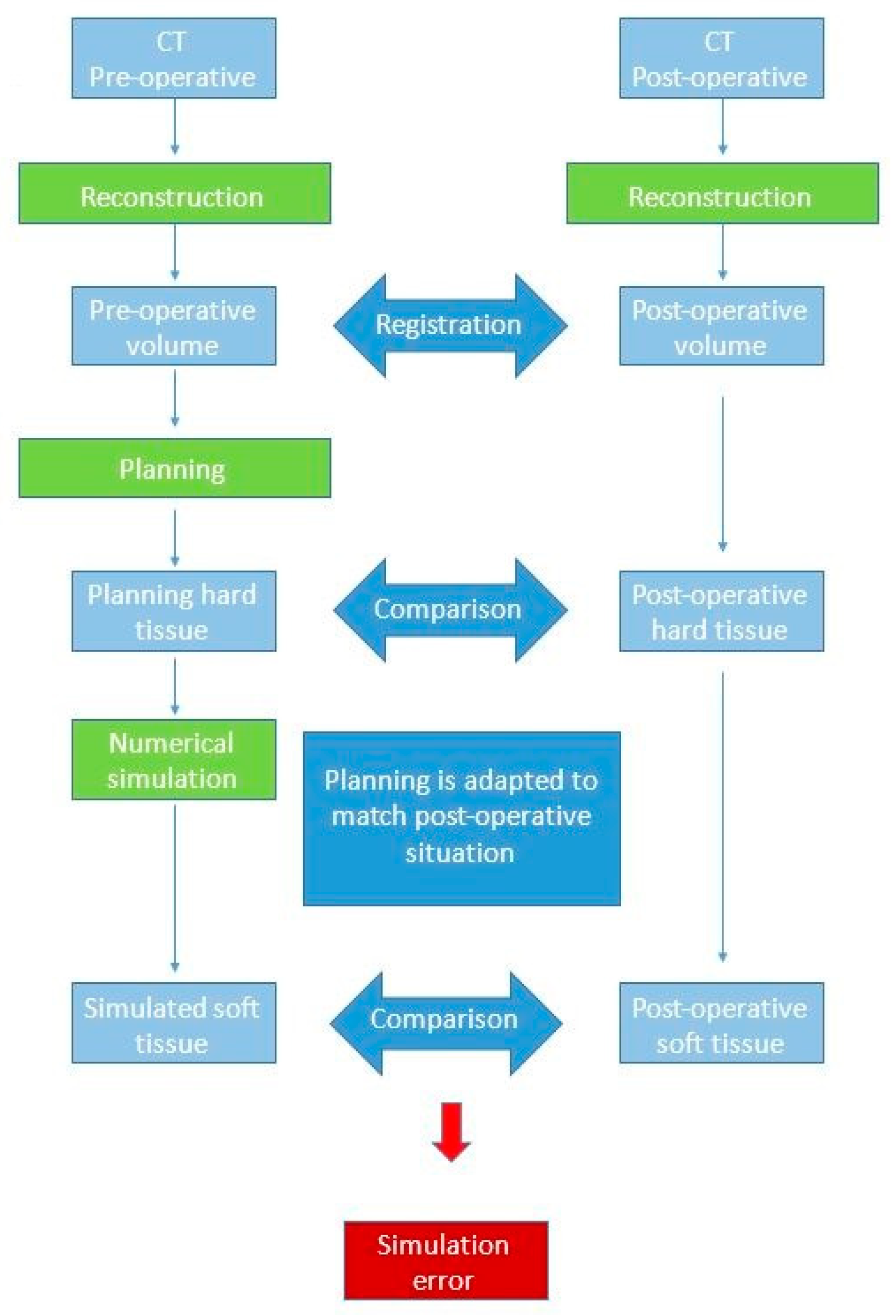
2. Materials and Methods

The research of this systematic review is based on the Population Intervention Control Outcome Study design (PICOS) format (Table 1). PubMed and Scopus are the databases adopted for our research. The considered keywords are: orthognathic surgery, facial dysmorphism, surgical planning, 3D, 3D face analysis, soft tissue, BSSO, bilateral sagittal split osteotomy, Le Fort I, prediction methods.
The research has been set on different combinations of keywords; at first, we focused generically on the orthognathic surgery, then the type of interventions has been specified: BSSO, IVRO, SSRO and LFI. Finally, the research has been limited to soft tissue simulation and prediction.
All the articles found were assessed by three authors and classified in: prospective study (PS), retrospective study (RS), case series study (CS).
Single cases reports have been excluded from our analysis because they have been considered not clinically significant.
Articles published before 2000 have not been considered.
The following data have been recorded for each eligible study: first author and year of publication, journal, study design, sample size, gender, mean age, diagnosis, imaging technology, typologies of surgery, software used for the prediction, soft and hard tissue landmarks considered, time interval from surgery to post-surgical imaging, results and conclusions of the authors.

3. Results

Different responses have been obtained varying the insertion order of the keywords. After having combined the outcomes and removed the duplicates, the remaining articles have been evaluated on the basis of their relevance to the topic. In the end, 24 articles to be deepened and a number of interesting articles to be referenced for a better comprehension of the topic have been selected.

3.1. Clinincal Details

A total of 24 articles have been compared in this work; 12 are retrospective studies, two are case studies and five are prospective studies. Remaining articles have not declared categorization. The sample of patients involved in each study vary from seven to 100 subjects. The soft tissue prediction has been assessed for Le Fort I osteotomy, BSSO, BSSRO and genioplasty in the correction of different types of facial dysmorphism.
Table 2 and Table 3 report a brief summary of the referenced papers we focused on. In particular, the demographic details of the patients are summarized in Table 2. A brief overview of methodologies and results of each article is reported in Table 3.
CBCT has been used for the assessment of soft tissues changes in twelve of the twenty-four studies, with the addition of 3D photographs for one article, while cephalometric radiographs have been used in the remaining eight works, with the addition of CT and MSCT for two of them.
The timing of post-surgical imaging has been stated in all articles.

3.2. Prediction Methodologies

Several approaches have been considered to make a mathematical three-dimensional prediction of soft tissue changes after orthognathic surgery. MSM, FEM [5,6] and MTM are the most common. These have been developed into software packages which are currently used in clinical practice. The functioning of these software is generally acceptable if we consider the creation of a plausible facial outcome, but this prediction does not necessarily match with the real outcome. Moreover, studies show that the prediction accuracy decreases for specific facial areas, especially around the lip, and with the increasing complexity of the surgery (larger bony repositioning often results in a higher inaccuracy of the prediction). The studies presented in this literature review evaluate soft-tissue predictions obtained with different software packages.

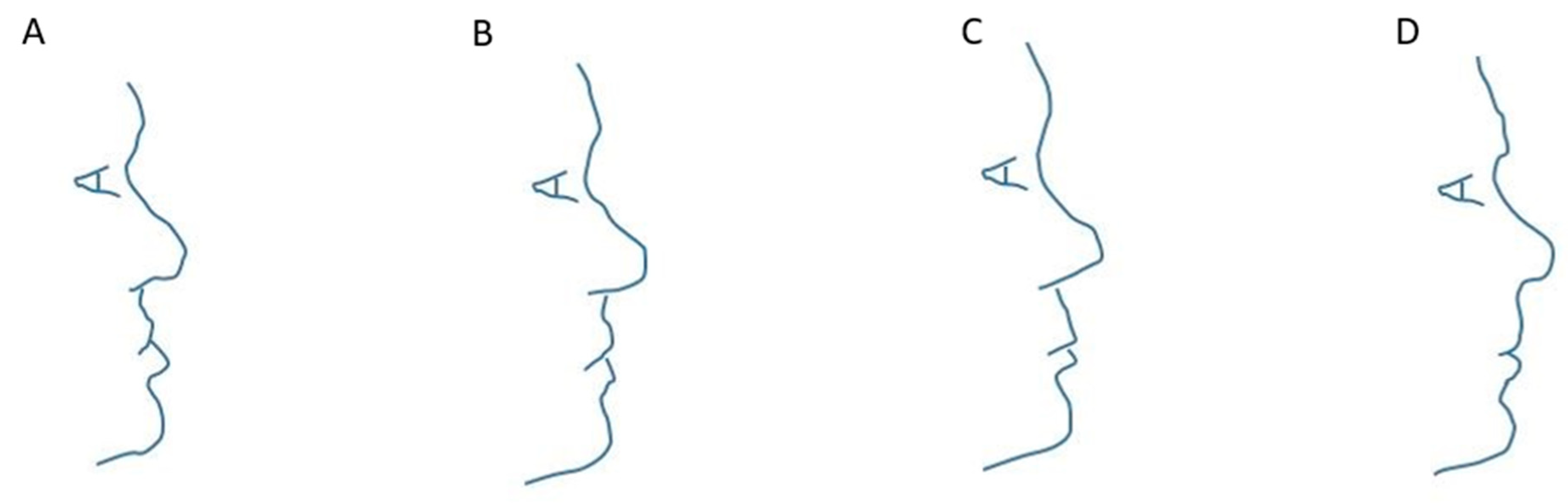
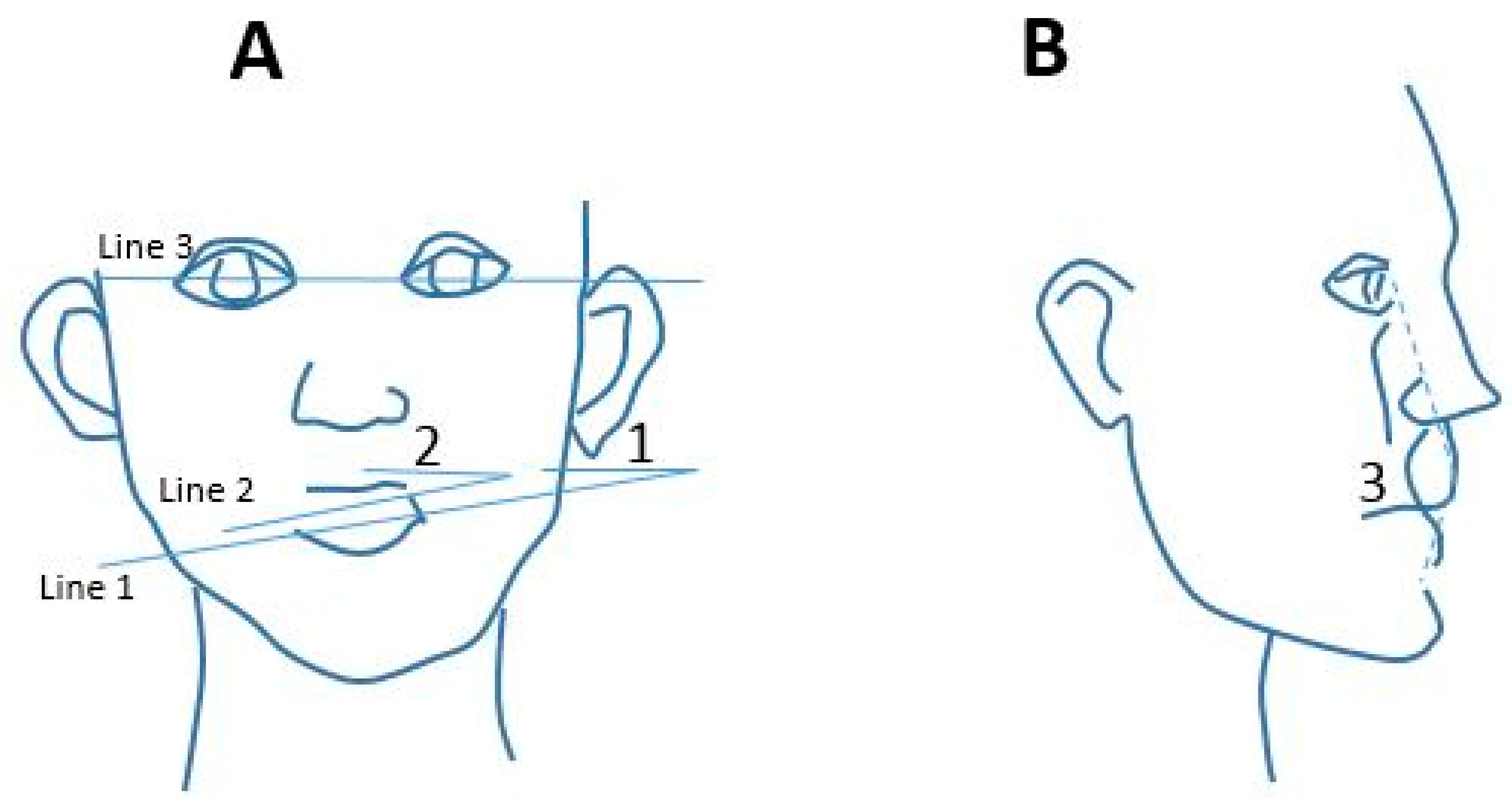
3.2.1. Works Based on Traditional (2D) Methodologies

Since a certain number of 3D software currently available for clinical practice are based on algorithm developed to work on 2D and then adapted to 3D representation of data, a section dedicated to traditional soft-tissue prediction is introduced. Moreover, since many surgeons currently rely more on traditional techniques than on three-dimensional methods, it is reasonable to focus on the limitations of this technology; 2D analysis based on lateral cephalograms is subject to errors caused by the impossibility to accurately locate points on both hard and soft tissue relying only on 2D anatomic structures [7]. Analysis performed on cephalograms reported that reasonable prediction can be made on soft-tissue landmarks positions, but with low accuracy concerning the lips and particularly in the vertical prediction of displacements [8]. Moreover, the inaccuracy increases with a higher level of complexity of intervention, which leads to errors both in hard- and soft-tissue simulation [9].
In this section, we particularly focused on study assessing the accuracy of Dolphin Imaging, but we also reported studies that took into consideration other software.
Peterman et al. worked on the quantification of the accuracy of Dolphin VTO (visual treatment objective) in predicting soft tissue changes in patients with a class III deformity and on the validation of its efficacy. In their study, authors considered fourteen patients receiving comprehensive orthodontic treatment and orthognathic surgery including both maxillary advancement and/or mandibular setback. Cephalometric tracing and analysis were performed with Dolphin Imaging software; pre- and post-operative and traced cephalometrics were superimposed using the cranial base as reference. The maxillary movements were recorded at the anterior nasal spine (ANS) and A point, while the mandibular changes were recorded at B point and pogonion (Pg) in both x and y axis. Subsequently, the pre-treatment profile pictures were superimposed to digitally-traced soft-tissue landmarks of pre-treatment cephalometric radiograph to initiate software VTO simulation (Figure 1). Finally, a prediction profile photograph generated by the software was compared to actual post-operative patient’s profile photograph; coordinates of nine soft-tissue landmarks were used to quantify on each axis the differences between the predicted and the actual of patients. The results reported in this study are consistent with the prevision error calculated in previous works; Dolphin Imaging software was found to have a much larger standard deviation in the Y direction than in the X, with various degrees of accuracy in both directions for all soft tissue landmarks. The accuracy was 79% and 61% for X-axis and Y-axis respectively, with an acceptable error set at 2.00 mm. Furthermore, for these surgical cases, the least accurate prediction was the one concerning the lower lip region. In conclusion, authors affirmed that VTO program can be useful for rough movement previsions during surgical planning, but surgeons cannot rely on it for precise surgical movements (measurement range lower than 1.00 mm) [10].
In the study of Pektas et al., the treatment planning for each patient was constituted on the base of clinical and cephalometric evaluations, and pre-operative study models. All patients were treated with pre- and post-surgical fixed orthodontic appliances. Scanned images of cephalometric radiographs were processed using Dolphin Imaging software; the Ricketts and Steiner analysis was selected to start the digitization. To compare actual outcome and software simulation, firstly the superimposition of pre- and post-operative cephalograms was performed on the sella-nasion plane registered at sella. Landmarks located on structures not involved in the surgery were directly transposed from pre-operative to post-operative cephalogram to refine the superimposition process. The objective of an accurate superimposition of pre- and post-operative cephalograms was to construct a spreadsheet for landmarks movements to quantify changes of each point after operation; the position of points was defined in form of x and y. In accordance with the spreadsheet, a simulation of the treatment was produced, and the prediction tracings were obtained from actual post-operative changes amount. Actual post treatment tracings and prediction tracings were superimposed and differences have been measured again according to an x, y coordinate system.
The mean differences between the predicted and the actual result were less than 1 mm in four of seven soft tissue measurements; predictions turn out to be more accurate for the sagittal plane than for the vertical one. The largest errors were reported for the lower lip region, in accordance with other studies considered by authors and investigating other software [11].
Ana de Lourdes sá de Líra et al. studied the accuracy of digital prediction with respect to the actual post-operative outcome with Dolphin program. Patients with class II malocclusion were involved. Eighty Caucasian subjects treated with combined surgical and orthodontic treatment were considered. Cephalometric radiographs of pre- and post-operative were digitized; a time of at least one year from surgical to post-operative was required to rule out any effects of post-operative swelling. Digital tracings were performed with Dolphin imaging software and digital lateral cephalometric radiographs were traced at the same time to minimize the error variance; before tracing, Dolphin program was used to carry out the compensation for the effect of radiographic magnification. An x y coordinates system was constructed with the x axes corresponding to the horizontal Frankfort plane and the y-axes passing through Nasion and perpendicular to x-axes. On x-axes, vertical changes have been measured, while on y-axes authors evaluated horizontal changes. Dalberg formula was used in evaluating the reproducibility of measurements; with regard to the method error, it did not exceed 0.37° for angular measurements and 0.29 mm for linear measurements. Results of the statistical analysis were based on mean values found in each studied group. The analysis of the authors underlined that there were no significant differences between soft-tissue values at pre-operative (t1), post-operative (t2) and predicted (t3) stages. In both groups, surgeries have been more extensive than planned; facial convexity and the distance between the lips and the cranial base presented similar values between t2 and t3. The conclusion of the authors was that statistical differences in the considered measurements did not invalidate surgical prediction performed with the software, because forecasted changes were sufficiently close to of the surgical outcomes [12].
Lira et al. followed the same approach of the previous article on two groups of patients; the former underwent Le Fort I without segmental surgery, while on the latter, in addition to LFI surgery, mandibular setback was performed with an SSRO. What came out from this study was that 12 months after the operation surgical reductions in mandibular length and angle were substantially greater than indicated by predictive cephalometric tracings. The analysis of dental and skeletal mandibular changes in anteroposterior and vertical directions revealed that the effect on the profile was greater than those of the maxilla; this is probably due to the fact that the mandible is a moving structure and the upper lip leans upon the lower lip. Maxilla advancements were performed for both groups according to the planning provided by Dolphin imaging software; in group two, mandibular setback was statistically overestimated by the software planning. Nevertheless, authors concluded that statistically the differences in the considered measurements do not invalidate the surgical prediction generated with the software, because the predicted result was close to the surgical outcome. Moreover, they noticed that concerning the mandible, dentoskeletal measurements in horizontal and vertical directions showed a greater correlation to the profile than the maxillary measurements [13].
Magro-Filho et al. worked on a subjective comparison of the soft-tissue surgical simulation of two software: Dentofacial Planner Plus (Dentofacial Software, Toronto, Ontario, Canada) and Dolphin Imaging (version 9.0). Surgeries considered in the study involved retro-positioning of the mandible and advancement of the maxilla, in which linear movements were at least 4 mm for one bone segment or in the sum of both mandibular and maxillary movements, without, or least possible, postsurgical orthodontic movements. Profile predictions were made six months after the surgery; real movements, obtained from the superimposition of the cephalometries of pre- and post-operative of each subject, were used as values of the prediction. Soft-tissue images of pre-operative, real predictive images of Dentofacial Planner Plus (DFP) and Dolphin Imaging (DI) and post-operative images were compared (Figure 2). With Photoshop program (version 6.0, Adobe, San Jose, Calif), pre- and post-operative profile images were digitized and standardized; then, images were imported in DFP and DI. Movements of maxilla and mandible were made using the real values extracted from the comparison between pre- and post-operative lateral cephalograms of the surgical displacements along the vertical and horizontal directions for molars and incisors of the maxilla and for incisors of the mandible. The tip of the nose, the nasolabial angle, upper and lower lips, menton region, the base of the mandible and the complete profile were used to compare simulated and real images. A group of orthodontists, maxillofacial surgeons and general dentists evaluated the similarity among the programs simulated images and the real post-operative images with a scale ranging from “very similar” to “different”. For the comparison of soft-tissue cephalometric points, to compare the two programs and to judge the criteria of similarity, chi-square test was used. From their analysis, the authors reported that for nasal tip, chin area and mandibular base DI gave better results, while for nasolabial angle, upper lip and lower lip DFP produced a more accurate prediction; authors also reported that there was no difference in the evaluation of the complete profile. Moreover, in terms of time, working with DI was longer than working with DFP. In conclusion, it was stated that in treating subjects affected by dentofacial deformities, it would be better if orthodontists and oral maxillofacial surgeons were to base on their clinical experience and to use software just as a co adjuvant. Considering the methodology of the study, there were not many differences in obtaining a two-dimensional prediction of patient’s profile [14].
Other software are available for the prediction of soft tissue changes. OrthoForecast is a data-based prediction software; its database contains information of 400 patients (100 pts with asymmetry, 100 pts with skeletal class II jaw relationship and 200 pts with skeletal class III jaw relationship). All patients underwent SSRO or IVRO, with or without LFI osteotomy (respectively, 352 and 48 cases). It was evaluated by Abe et al.; the study included 15 patients with facial asymmetry, 15 with skeletal class II jaw relationship and 15 with skeletal class III jaw relationship. Patients underwent LFI osteotomy, SSRO and IVRO. Twenty-four landmarks were chosen and digitized. Some angles were also compared between the following lines: line 1, the line connecting the bilateral mandibular angles; line 3, the line connecting the bilateral eyespots; line 2, the line connecting the bilateral oral angles. In the asymmetry group, they measured angle 1, identified by line 1 and line 3 and angle 2, identified by line 2 and line 3 on the frontal facial photograph to assess the asymmetry of the face. In the class II and class III groups, the value of angle 3, identified by the angle formed by lines connecting eyespot, upper lip, and soft tissue B-point on the lateral facial photograph was measured. Angle 3 was the reference for evaluating the convexity of the face, which was originally defined as soft-tissue convexity (Figure 3). The distances measured between landmarks on the actual and on the predicted images were analyzed, and the similarity between real and software-generated images was assessed by 39 evaluators. The mean difference measured between the positions of landmarks is less than 3.4 mm and less than 1.0° in the evaluated angles. Moreover, more than half of the evaluators stated that, in all groups, the predicted images were very ‘similar’ or ‘similar’ to the actual situation of the patients in the post-operative condition. Less than 6% of the evaluators ranked the predicted images as ‘different’ from the actual outcome. In conclusion, OrthoForecast is considered by the authors to be a software with high levels of accuracy, reliability and usefulness [15].
Donatsky et al. evaluated another prediction software, TIOPS. This is a computerized, cephalometric, orthognathic surgical planning system previously used in studies on hard tissue stability and accuracy. As other soft tissue prediction systems, TIOPS is based on predefined ratios of hard to soft tissue movements; its present algorithms partly rely on cephalometric observation of the post-operative situation. The study included 52 patients. Clinical photographs, study models mounted on an articulator (SAM) and standardized lateral cephalometric radiographs of the pre-operative situation were performed. Standardized lateral cephalometric radiographs were performed 5–6 weeks after surgery. The mean accuracy of the planned and predicted results of both hard and soft tissue varied from 0.0 mm to 0.5 mm from one cephalometric reference point to another. In the locus of cephalometric reference points, where it was shown that there were significant differences between planned/predicted hard and soft tissue changing in terms of position, these significant inaccuracies were moderately small and varied from 0.2 mm to 1.1 mm, with the exception of the horizontal position of the lower lip. However, the variability of the previewed hard and soft tissue individual result was evaluated to be relatively high. The study demonstrated moderately high predictability of the immediate post-surgical hard and soft tissue outcome. However, due to the significant variability of simulated individual results, it would be better to be cautious in presenting the planned and predicted hard and soft-tissue results to the individual patient in the pre-operative [16].
Finally, Chai Hui Koh et al. evaluated the accuracy of soft tissue predictions generated by the CASSOS (Computer-Assisted Simulation System for Orthognathic Surgery) software in Chinese skeletal class III patients underwent bimaxillary surgery. The digitization of pre-surgical and post-surgical lateral cephalograms of 35 patients was performed with the CASSOS program. A simulation of the surgery was performed on the pre-surgical tracing. An analysis was performed on thirty-two linear measurements on the cephalograms superimposition to assess the differences in the soft tissue profile between the post treatment results and the predicted result. It showed differences on 16 linear measurements with the most prediction errors on the upper and lower lip having a mean difference relatively small, with a greatest mean difference of 2 mm in the vertical position of stomion inferius. The authors concluded that CASSOS 2001 produced a clinically meaningful forecast of soft-tissue profile changes following bimaxillary surgery [17].
The same CASSOS software was evaluated by Jones et al. for 33 patients affected by class III skeletal deformities and underwent maxillary advancement (17 patients) or bimaxillary surgery (16 patients) [18]. The post-operative cephalograms were used to determine surgical bony movements to produce the CASSOS profile simulation. Linear differences between the predicted profile and the real outcome were measured at 12 soft tissue landmarks. The authors’ conclusion was that the profile predictions obtained with CASSOS could be considered useful for both type of surgeries, even if substantial variations were found. As previously [17], the most inaccuracies were found in the lip region.

3.2.2. Works Based on Maxilim Software (3D)

In this section a collection of studies is presented considering patients’ soft tissue prediction obtained with Maxilim software (Medicim—Medical Image Computing, Mechelen, Belgium). This software, based on the mass tensor model algorithm (MTM), allows surgeons to determine bony movements and to see the effect of the procedure [19]. MTM has been introduced by Cotin et al.; the geometry of the anatomical structure is discretized into a tetrahedral mesh in which the displacement vector at a generic internal point is defined as a function of vertices displacement vectors. The elastic force is written as a function of these same four vectors. The displacement for a set of mass points is set fixed and other model points will move as consequence of elastic forces due to the displacements of fixed points; finally, the new rest position of the free points is computed by integrating the Newtonian motion equation [20].
Shafi et al. [21] investigated the accuracy of Maxilim using cone-beam computed tomography (CBCT) scans in pre- and post-surgery phases (6–12 months after surgery) for 13 patients subjected to Le Fort I surgery. A 3D mesh was generated from skeletal movements (predicted model). Subsequently, the mesh generated from the post-operative of the patient and the predicted model were compared to evaluate the accuracy of the prediction. The soft tissue was divided in different areas: nose, right and left nares, right and left paranasal regions upper and lower lip and chin. The absolute distance was calculated between the meshes for each region. For each facial region, the absolute distance was calculated between the meshes.
In almost all the facial regions, the accuracy was significantly less than 3.00 mm (3.00 mm is clinically acceptable). The upper lip area was the exception; in fact, for this region, the accuracy was greater than 3.00 mm. In all cases, Maxilim produced an over prediction of the new position of the upper lip. A possible reason could be a non-linear response of the upper lip to some hard-tissue movements and, consequently, the modeling algorithm used (MTM) hardly previewed this response.
The conclusion of the authors was that the 3D soft tissue prediction for Le Fort I advancements produced by Maxilim was in general clinically satisfactory, but it was associated with marked errors around the region of the upper lip.
Liebregts et al. evaluated the accuracy of Maxilim for 3D simulation of soft tissue changes after bimaxillary osteotomy. The 3D rendered pre- and post-operative scans were matched. Segmented maxilla and mandible were aligned to the post-operative position. In order to calculate the error between the simulation and the actual post-operative condition, authors used 3D distance maps and cephalometric analysis. Concerning the facial profile, the mean absolute error between the 3D simulation and the actual post-operative facial profile was 0.81 ± 0.22 mm for the face as a whole. In this study, the accuracies of the simulation (average absolute error ≤2 mm) for the whole face and for the upper lip, lower lip and chin sub regions were 100%, 93%, 90% and 95%, respectively.
Authors affirmed that the MTM based soft tissue simulation is an accurate model for the prediction of soft tissue changes following bimaxillary surgery. The magnitude of skeletal movements influences the accuracy of the prediction; moreover, the age of the patient and the use of V–Y closure affect the precision of the predicted model. Low predictability on the upper and lower lip regions is registered once again [22].
The same authors evaluated Maxilim performances for soft-tissue simulation in 100 patients underwent BSSO for mandibular advancement [23]. As in the previous case, the accuracy of the simulation was assessed with two methods, a 3D cephalometric analysis and a 3D distance map for the entire face and for specific regions of interest. Their analysis showed that for the entire face the mean absolute 90th percentile error was less than 2 mm (clinically acceptable); the least accuracy resulted in the region of the lower lip, while the most accurate prediction involved the sub nasal region. A possible explanation suggested by authors for labial inaccuracies was the difficulty to replicate labial position during different image acquisition.
The conclusion drawn by authors on the basis of this analysis was that the soft-tissue prediction produced by Maxilim software was clinically acceptable both for the whole face and for restricted regions, but the limitation was the fact that patients considered in the study underwent a single type of surgery instead of combined operations.
In the analysis by Mundluru et al., 13 non-syndromatic adults with a midline deviation of the chin point not less than 2.0 mm underwent Le Fort I or bimaxillary osteotomies (BSSO) to correct facial asymmetry. The accuracy of an innovative concept for the soft-tissue changes prediction was evaluated. The segmentation of the 3D model was performed to identify the following regions: upper lip, lower lip, chin area, right and left paranasal regions, nose and right and left cheeks. To evaluate the prediction reliability, the surface distances between predicted and actual post-operative positions were measured. Particularly, mean (signed) and mean absolute distances were calculated on 3D meshes for each region. Through a directional analysis, the accuracy of the prediction of soft-tissue changes was evaluated. The results showed that the distances between the predicted and the actual post-operative soft tissue models were less than 2.0 mm in all regions. In details, a general tendency to the under prediction was found in the area of the cheek and of the chin (medio-laterally). An over-prediction of changes was found at the inferior border of the mandible (bilaterally) [24].
De Riu, et al. realized a retrospective study with the purpose of giving an objective quantification of accuracy of the Maxilim virtual planning method, through the comparison between planned and actual movements in jaw osteotomy. They started from the assumption that a simple superimposition of simulation and cephalometry results did not allow to define a correlation between positional error and surgical movements. Indeed, a slight positional error could be insignificant talking about large movements, but could be unacceptable dealing with small displacements. A 3D planning was performed, and virtual frontal and lateral cephalometries of pre-operative and simulated surgery were extracted; those were compared to define the predicted movements of the jaw in reference to cranial bones. All surgeries have been planned by the same surgeons using Maxilim software, with digital intermediate splints to guide osteotomies. Cephalometric analysis has been performed with Dolphin. To evaluate the accuracy, the mean linear difference between planned and actual movements was considered. Measurement of the differences between planned and actual movements were deemed accurate. As reported by authors, most of the clinical studies validating the virtual surgical planning set to 2 mm the criterion for the prediction success, with a success rate slightly lower than 100%. In this study, some significant differences with other works were found; first, differences between planned and achieved anterior facial heights were found (p = 0.033). This discrepancy was relevant (p = 0.042) in patients that did not undergo genioplasty, while it was not significant in patients who underwent this surgery (0.235). This error is probably due to the approximation of the soft tissue model, which does not allow the management of the vertical dimension. Presumably, genioplasty compensate the error on facial eight. The second error reported by authors was in the differences between actual and planned measurements for SNA (angle between Sella, Nasion and A point) and for SNB (angle between Sella, Nasion and B point), with p = 0.008 and p = 0.006 respectively. In the opinion of the authors, virtual planning inaccuracies were principally caused by the difficulty of simulate the soft tissue changes. Moreover, they concluded that virtual planning could not relieve surgeons of the necessity of monitoring jaws movements intraoperatively and of a real time supervision of planned and actual outcome, despite a high level of accuracy for most of the analyzed parameters [25].
The double blind prospective study performed by Van Hemelen et al. had the aim of providing a comparison between a traditional planning method and a 3D planning performed with Maxilim, both for hard and soft tissue [26]. In their analysis, authors considered 66 patients affected by class II or class III angle malocclusion underwent bimaxillary osteotomy (46 patients), BSSO (17) patients and LeFort I osteotomy (3 patients). Genioplasty was performed on 28 patients. For the 2D planning, clinical facial examination, lateral and frontal cephalograms were taken; 17 and 16 cephalometric landmarks were considered for 3D and for 2D respectively, to perform the validation of both 2D and 3D planning. The traditional planning was performed with Onyx Ceph Version 3.1.111 and the analyses of accuracy was performed by measuring projections on x and y axis of distances between the 16 selected points on the planned and on the actual post-operative. In the case of the 3D planning, the landmarks were defined in three dimensions and the planned and post-operative data were aligned. Distances between the 17 cephalometric points defined on planned and actual models were evaluated in depth, height and distance in the sagittal plane.
As a result of the analysis, authors reported that, for hard tissue planning, the mean differences occurring between actual and planned outcome evaluated at cephalometric points were 1.71 mm and 1.42 mm in the horizontal direction and 1.69 mm and 1.44 mm in the vertical direction, for 2D and 3D respectively. Concerning soft tissue displacements, the mean values computed in the horizontal direction were 2.29 mm for traditional planning and 1.48 for Maxilim prediction; in the vertical direction, authors noted mean values of 2.07 mm and 1.46 mm for 2D and 3D techniques respectively. These evaluations led authors to conclude that there was a statistically significant difference in the soft-tissue prediction between the two considered methods (chi-squared test and independent t-test were used to determine statistical significance, with p < 0.005); particularly, the 3D approach seemed to be more predictive than the traditional one. No statistically significant differences were reported for hard tissue planning.
The conclusion reached by authors was that, concerning the soft tissue prediction, the 3D planning approach here performed with Maxilim software led to a more accurate planning than the traditional method, even with disadvantages such as the cost of the software and of the CBCT scans. Moreover, a higher learning time must be considered when planning with the 3D method. In the authors’ opinion, 3D planning will not replace the traditional prediction if those negative aspects are not addressed.
The study by Nadjmi et al. focused on the accuracy of soft-tissue profile changes simulated by Maxilim with respect to the accuracy obtained with Dolphin. Particularly, the goal of the comparison was to assess if the 3D prevision was more accurate than the 2D one. A “natural head distance” was defined as distance between suprasternal notch and soft tissue pogonion to replicate patients’ head position. On lateral cephalogram, soft tissue and hard tissue landmarks (15 and 25 points respectively) were located; soft tissue predictions were generated with Dolphin and Maxilim. The two predictive results were superimposed with patient’s post-operative profile photograph and differences in terms of linear measurements of landmarks in x and y directions were measured. All comparison steps were performed by the same operator. The movements of bony structures in the simulation must be the same as performed by the surgeon, to correctly compare the two predictions. Since this study made a comparison between 2D and 3D simulations, even if Maxilim gave in output 3D models, only profile (2D) images were considered. From the analysis of the results, the authors found that both Dolphin and Maxilim predictions were accurate, but the simulation obtained with Maxilim made possible a quantification of volumetric changes (lips aspect) and a prevision of changes in the transverse plane. Even if the study focused on the comparison of accuracies (and for this reason only the lateral prediction was considered) between the two software, authors stated that 3D simulation would be helpful in complicated surgeries thanks to the third-dimensional information [27].

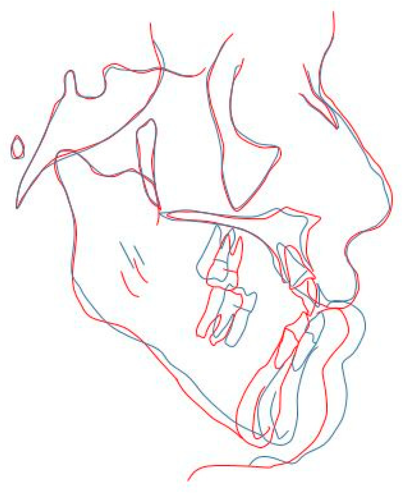
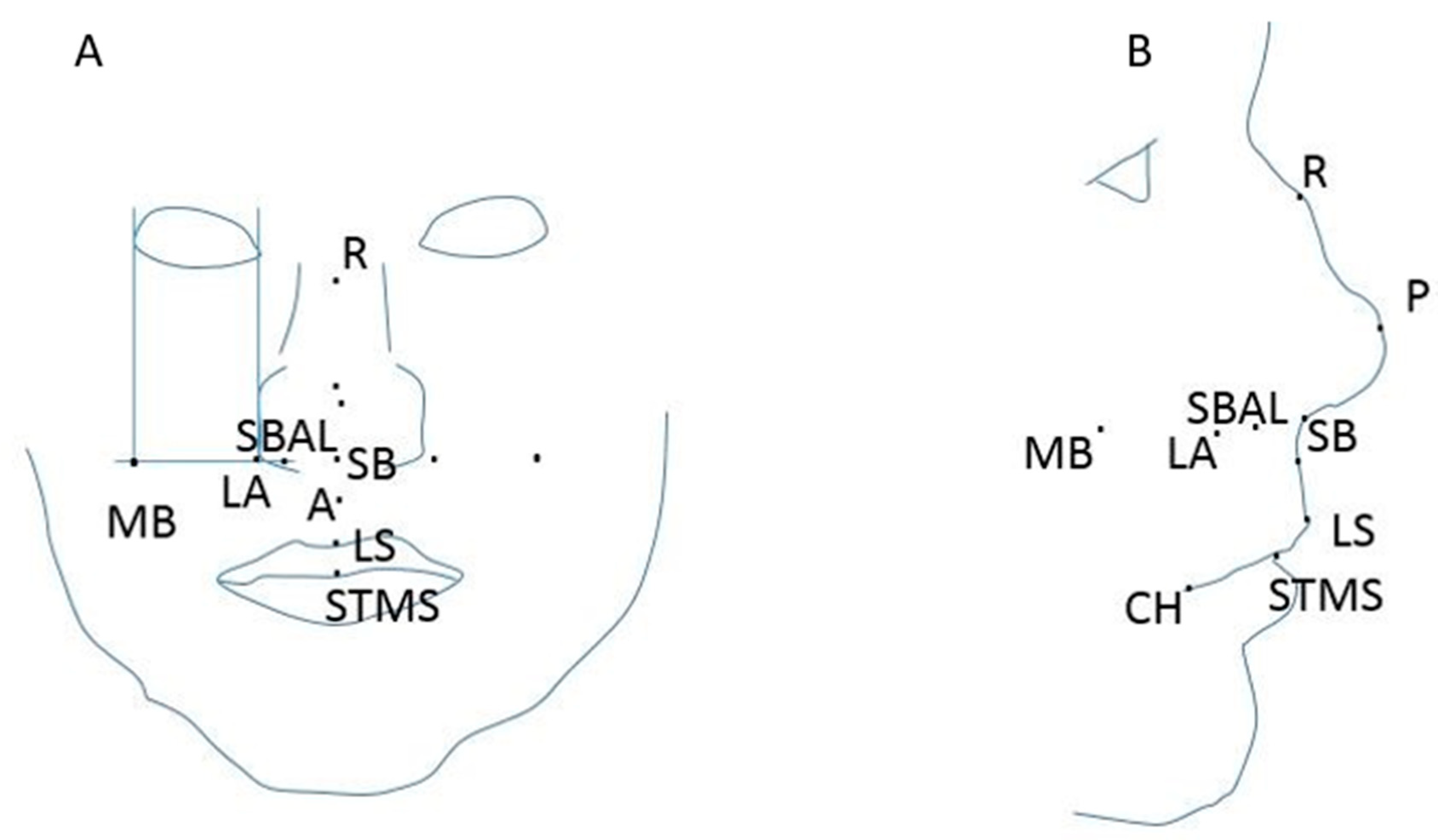
3.2.3. Works Based on Other 3D Software

The work of Resnick et al. investigates the accuracy of Dolphin 3D soft tissue prediction using seven patients who had a single-segment Le Fort I osteotomy. Dolphin 3D Imaging (Dolphin Imaging and Management Solutions, Chatsworth, CA, USA) is based on a landmark photographic morphing algorithm developed for 2D prediction and adapted to 3D. The user must locate 79 landmarks, 47 on the hard tissue and 37 on the soft tissue, on the CT volume; the system generates adaptable curves between the landmarks, similarly to the tracing of a lateral cephalometry.
On the pre-operative segments LF I osteotomy was simulated by Dolphin, then hard and soft tissue landmarks were assigned, morphing curves were adjusted and finally prediction image (Tp) and post-operative image (T1) were aligned and registered for the measurement of the differences. For each patient, the error was calculated as the difference occurring between Tp and T1 at 14 points, six on the midline and eight laterally and at the nasolabial angle (Figure 4). Twelve of these points are standard anthropometric or cephalometric landmarks. Two new points were introduced for the study: lateral ala (LA), obtained from the intersection of the line tangent to endocanthion (EN) and the one tangent to subalare (SBAL), and maxillary buttress (MB), traced at the intersection of lines tangent to exocanthion (EX) and SBAL (Figure 4A).
In closing, their analysis showed that the capability to predict the soft-tissue changes in the three dimensions after LF I surgery using Dolphin software had some limitations. For linear changes, its accuracy was acceptable, while it was not acceptable for lateral points of the face. Concerning the midline, changes at nasal base were more subject to prediction errors [28].
Bianchi et al. [29] and Marchetti et al. [30] worked on SurgiCase_CMF using CBCT and MSCT respectively of 10 patients with facial deformities. In the study presented by Bianchi et al., patients underwent LFI, BSSO and genioplasty. The pre- and post-operative CT images were aligned on the top of each other. Firstly, a registration point was made, with the location of two corresponding landmarks on pre- and post-operative conditions. After the manual registration, automatic surface registration was carried out using an iterative closest point algorithm. A crucial point in surfaces registration was to indicate those that did not change after the surgery; authors used the eyes area and the forehead as fixed regions. Finally, the post-operative soft tissue surface was compared with the software virtual simulation. A comparison algorithm was used to measure the distance of every triangle corner of the post-operative surface against the preoperative one. The percentage of error was <2.0 mm for the 86.80% of patients, even if there were important errors in the top lip and chin regions. The results in the group of patients studied with CBCT (which had a reliability of 86.8%) stated that the use of SurgiCase_CMF 1.2 software combined with CBCT data allowed to achieve a realistic preview with low X-ray exposure [29].
Marchetti et al. divided virtual surgical planning in four steps (Figure 5): (1) CT data reconstruction; (2) generation of 3D models of hard and soft tissues; (3) various virtual surgical planning and simulation mode; (4) different pre-operative previews of the soft tissues. Skeletal model surgical planning and simulation were performed on the base of the 3D CT; the soft tissue model was made through a physical model. According to clinical options, the software generated a set of simulations and models of the soft tissues; in order to prevent temporomandibular functional problems, an orthodontist assessed the pre-operative plans. From the comparison of simulation and CT surgical outcomes, it was found that the prediction of soft tissue situation had a reliability greater than 91%, with a percentage of error lower than 2 mm for 76%–99%. According to the obtained results, SurgiCase_CMF was able to give a realistic and accurate preview of the face of the subjects undergoing surgery, but there were significant errors in the lip and chin regions, as in the previous studies [30].
3dMDvultus is a software package in which the rendering function is based on Mass Spring Model. MSM assumes a discretization of a deformable object into n mass points and a set of m connections between each n mass point. In a tetrahedral discretization, for each mesh node a point is allocated, and it is defined a linear spring for all the edges of the mesh. This linear spring follows Hook’s law. This model has the advantage of being simple and computationally efficient, but the disadvantage is that the elastic behavior of the model is determined by the spring constant; the value of this constant is an approximation and it has no true bio-mechanical relevance [19].
Khambay et al. used 3dMDvultus to predict the soft tissue changes after surgery. The work included 10 patients who underwent Le Fort I osteotomy. Ten landmarks were used to measure the distances between the pre- and post- operative meshes. Moreover, the percentage of mesh points minor or equal to 2.0 mm were calculated for the full face and for specific anatomical regions. The results demonstrated that the percentage of mesh with errors minor or equal to 2.0 mm for the full face was 85.2%–94.5% and 31.3%–100% for anatomical regions. The range of root mean square (RMS) error was from 2.49 mm to 0.94 mm. The most of mean linear distances computed between the surfaces was equal or less than 0.8 mm, but it increased for the mean absolute distances. From this analysis, it was stated that the use of specific anatomical regions was more significant in clinical practice than the use of full face [31].
As in the previous article, the software considered by Terzic et al. is 3dMDvultus. Three-dimensional photographs of patients’ head in natural position (lips and muscles at rest, open eyes, neutral facial expression) of pre- and post-operative were taken; CT and/or CBCT images were imported to the software platform. Pre-operative 3D photograph was fused with pre-operative CT/CBCT images. In the same manner, post-operative bony skull data was fused with pre-operatives by matching bone areas not involved in surgeries. According to actual post-surgical CT, 3D bone segments were reproduced in the pre-operative skull; the software rendering function (mass spring model) was activated. Osteotomy segments were moved to real post-operative position and the soft-tissue rendering generated the textured facial soft tissue prediction. After the fusion of 3D photographs of predicted and real post-operative soft tissue, a horizontal plane was positioned arbitrarily to divide the face in an upper part not involved in surgery, and a lower part including the overlap of simulated and real post-surgery result. Averaged distribution of absolute error showed more discrepancies between predicted and real post-operative outcome in the lower part of the face: in 29.8% of lower halves authors found errors exceeding 3.00 mm. This preliminary study concluded that the software platform had an insufficient accuracy in the forecast of 3D soft-tissue displacements [32].
Ullah et al. carried out a retrospective study in which they focused on the ability of 3dMDvultus to give a prediction of the face appearance of patients undergoing LFI maxillary advancement. The null hypothesis they started from was that the mean difference in absolute distance between the predicted surface generated by the software and the real 3D facial surface measured at eight anatomical regions did not exceed 3 mm. Pre- and post-operative CBCT for each patients were imported into 3dMDvultus and hard tissue and soft tissue were separately segmented and saved in STL (standard triangulation language) format. A template of actual surgical changes was produced using a CAD/CAM software (VRMesh, VirtualGrid, Seattle, WA, USA). Post-operative hard and soft tissue linked together were aligned to the anterior cranial base of pre-operative hard tissue of the same patient. Regions of maxilla and mandible of post-operative models were saved as STL file and imported into 3dMDvultus to generate a template of the actual maxillo-mandibular complex aligned in the same 3D space as the pre-operative image. Using the same software, a LFI osteotomy was simulated for the pre-operative hard tissue. The resulting soft-tissue prediction was exported as an STL file for the analysis. The predicted model and the real surgical result were imported as mesh in VRMesh; the differences between the two models were rendered as a color map, which showed the overall accuracy of the software. After the analysis on the overall surface, the accuracy was evaluated subdividing the mesh into anatomical regions: chin, lower lip, upper lip, nose, right and left nares, right and left paranasal areas. From the analysis of the results, the authors concluded that since the distances between software-generated surfaces and real facial surfaces were lower than 3 mm, 3dMDvultus 3D soft-tissue predictions were acceptable for clinical use in LFI osteotomy. MSM seemed to correctly predict the positions of lip and chin, while improvements should be made for nasal and paranasal regions [33].
In the prospective study by Holzinger et al., 16 patients with open bite dentofacial-dysmorphosis and underwent orthognathic surgery (surgery first) were analyzed. The surgery was planned using conventional sketches as a new software developed by the authors, SOTIRIOS planning software. A conventional pre-surgical planning was carried out using three sides X-rays and, in addition, a CT scan was performed before and 6 months after surgery. A constrained fitting approach using statistical shape modeling (SSM) technique to generate the patient-specific data was used in combination with the anatomical landmarks to compute the patient specific model. A quantitative comparison of the soft tissue prediction and post-operative data was made showing a mean error of 1.46 mm ± 1.53 mm. Authors concluded that the SOTIRIOS planning software was quite accurate and enabled the surgeon to predict the soft tissue outcome [34].
A comparison between Dolphin 3D, ProPlan CMF and an in-house probabilistic finite element (PFEM) [6] was carried out by Knoops et al. [35]. The retrospective study included seven patients who underwent LFI maxillary advancement. From CBCT taken preoperatively, 3D models of bone and soft tissue were constructed. On the base of the advancement and rotation movements measured on the post-operative images, a virtual Le Fort I osteotomy was performed and three different soft tissue predictions were realized with the three softwares. Firstly, the three predictions were compared with the pre-operative model to evaluate differences. After that, the three predictions were compared with the real post-operative outcome, to assess which was the most predictive model. As the patients involved underwent a LFI maxillary advancement, the areas of interest for the comparison with post-operative situation were the upper lip and the paranasal regions. From the outcomes analysis, it resulted that the prediction generated by Dolphin 3D was affected by a general under-prediction of paranasal displacements, while both ProPlan CMF and the PFEM model were characterized by an over-prediction of displacements involving cheilion regions. Average root mean square distances and average percentage of points less than 2 mm between real post-operative images and predictions were computed (Table 3); statistically significant differences resulted from the Friedman test. In particular, the post hoc Wilcoxon signed-rank test proved that RMS for PFEM and RMS for ProPlan had statistically significant lower values with respect to RMS for Dolphin 3D (p = 0.016). On the opposite, the differences between ProPlan and PFEM could not be considered statistically significant (p = 0.219).
The conclusions of the authors was that ProPlan CMF and PFEM gave soft-tissue predictions with significant accuracies, particularly when using the correct post-operative maxillary position; because of the limitations introduced by a landmark-based algorithm and a sparse architecture, Dolphin 3D resulted to be inaccurate when surgery involved large maxillary advancements, particularly on lateral points.
Nam et al. [36] tried to assess if a 3D virtual surgery could accurately predict the soft tissue outcome of 29 patients who underwent bimaxillary orthognatic surgery (LFI, BSSO and genioplasty) performed by the same surgeon. The predicted outcome was generated using the Simplant Pro program. All simulations were performed by the same operator. The produced simulation of the soft tissue result was superimposed on the post-operative of the patients, and 10 landmarks were designed and positioned on the two models; the simulation error was determined by measuring distances (x, y, z) between the same landmarks on predicted and real images. From this analysis, an accuracy of 52.8% resulted, disagreeing with previous study reporting accuracies around 80%. Most of the errors were found for landmarks of lower lips, mouth corner and chin area (pogonion and menton), with a general larger error in points located on the mandible. Authors suggested as a possible cause of this trend the complexity of the surgery, involving more movements in the mandible rather than in the maxilla; it could determine lower errors in the maxilla but higher inaccuracy in the mandible. Inaccuracies reported for 18 of the 30 soft-tissue landmarks were considered statistically significant (p < 0.05), with errors concentrated at mouth corners. Possible explanations addressed by the authors were the ethnic differences between the data used for developing the software (Caucasian population) and the data used in this study (Asian population); moreover, it was not negligible that errors up to 2 mm could be due to the positioning of soft tissue landmarks. In conclusion, authors stated that 3D software in soft tissue prediction could have great potential, but the accuracy must be improved.
Even if it is out of the scope of the present work, it is important to state that not only the surgical planning plays a crucial role in the optimal process of care of the patients. It is not negligible that maxillofacial surgery, as well as other branches of surgeries, could lead to medical complications such as venous thromboembolism, which can have fatal consequences when moving to the lung (DVT/PE) [37]. Moreover, surgeons must manage patients with rare disorders of hemostasis, which need particular care in the perioperative phase. The works by Simurda et al. [38,39], and Ghadimi et al. [40] have been referred to in order to deepen understanding of this topic.

4. Discussion and Conclusions

In this systematic review we have summarized twenty-four studies focused on the evaluation of soft-tissue prediction software for maxillofacial surgery. The treated software are commonly used in clinical procedures to have a preview of the patients’ outcome after corrective surgery; the most common methodologies on which these software are based are the mass spring model, the finite element model and the mass tensor model, each of them with its advantages and disadvantages. MSM presents an easy architecture and a low memory usage but it has no real biomechanical foundations; FEM is more relevant from a biomechanical point of view, but it has a high computational cost and a rather long simulation time; MTM tries to combine the advantages of MSM and of FEM [18]. In particular, the lip region is the most difficult to predict, since lips do not rely on bony structures; moreover, it is difficult to replicate the same lip position from one acquisition to another. Due to this particularity, lips are supposed to be particularly involved in involuntary displacements caused by surgeries performed on different facial areas. The difficulty in accurately predicting the appearance of this sub region is a crucial point in the development of a prediction method, since it is particularly related to aesthetic self-perception and satisfaction of the patient. With respect to the traditional algorithms based on hard- to soft-tissue ratio, methodologies involving biomechanical models of viscoelasticity of the soft tissue to mimic its elastic deformation seems to be able to give a more realistic simulation. Despite the number of computer programs dedicated to three-dimensional prediction and surgical planning, maxillofacial practitioners often prefer to rely on conventional two-dimensional methods. This fact could be explained with the overall results given by these methodologies, which seem to be only partially relevant and/or reliable in the practice of daily clinical activity. Concerning the works based on Maxilim, Shafi et al. found that this software makes a clinically satisfactory prediction of soft tissue changes, but with an over-prediction of the position of the upper lip [21]; according to Liebregts et al., its accuracy is influenced by the magnitude of maxillary and mandibular movements, by the age of the patient and by the usage of V-Y closure, and these authors further reported a low predictability of lower and upper lips [22]. Moreover, a general tendency was reported towards an under-prediction in the area around the cheek and chin and towards an over-prediction at the mandibular inferior border [24]. It was suggested that Maxilim inaccuracy in the prediction of the lip region might be due to the modelling algorithm, i.e., mass tensor, and might indicate a non-linear response of the upper lip [21]. The study by De Riu et al., although not directly focused on soft tissue prediction, reports that inaccuracies in virtual planning are primarily due to the soft-tissue virtual model [25].
With regard to Dolphin software, Resnick et al. reported that the ability to predict 3D soft tissue changes was limited, with an accuracy that may be acceptable for linear changes but not for lateral facial points [28]. Peterman et al. reported various degrees of accuracy at each soft-tissue landmark in the horizontal and vertical axes; lower lip prediction was the least accurate. A similar result of low prediction accuracy for the lower lip region is reported by Pektas et al. [11]. Conversely, two articles affirmed that the statistical differences between Dolphin soft tissue prediction and the real outcome of the patient did not invalidate the software prediction, as it was close to real surgical results [12,13]. Dolphin 3D imaging uses a landmark-based photographic morphing algorithm that was developed for two-dimensional prediction and then adapted to three-dimensional prediction; this could be an explanation for its imprecisions. Moreover, it requires plotting 79 landmarks on the CT volume (42 for the hard tissue and 37 for the soft tissue); the localization of such a large number of points seems to require too much time for a stable usage in clinical daily practice.
Among the other considered software, SurgiCase_CMF was analyzed by Bianchi et al. [29] and Marchetti et al. [30]; they reported that the percentage of error was less than 2 mm for the majority of patients, but in both studies an important error in the areas of lips and chin was reported. Khambay et al., Terzic et al. and Ullah et al. used 3dMDvultus to predict soft tissue changes; in general, they reported that its use on specific anatomical regions was more meaningful than on the full face [31,32,33]. Moreover, they reported an insufficient prediction accuracy particularly for the lower part of the face with errors exceeding 3 mm [32], and for nasal and paranasal regions [33]. Surprisingly, prediction results seemed to be reliable for lips and chin.
The results reported for TIOPS showed significant inaccuracies in the predicted horizontal position of the lower lips and a relatively high variability of the predicted individual surgical outcome. The authors suggest care is needed when presenting the predicted image to patients before the operation [16,41]. The SOTIRIOS planning software and the CASSOS program were found to be quite accurate [34] and able to give a clinically useful prediction of the soft-tissue profile change [17]. OrthoForecast software resulted as being in general an accurate tool for soft-tissue prediction; indeed, more than half of the involved evaluators assessed the predicted images to be very similar or similar to the actual post-operative images [15].
As a result of this review, it seems that Dolphin, CASSOS and OrthoForecast are the most accurate software in soft-tissue prediction. However, their reliability is not sufficient when further surgery is carried out and the complexity of the operation increases; moreover, they provide only partial information on the prediction, due to the lack of the third dimension. From this viewpoint, all articles agree on the potentiality of 3D methodologies. Maxilim produced clinically meaningful results, but its inaccuracy in specific facial areas (particularly in the lip region) led to the conclusion that further steps are needed to give surgeons a tool for predicting facial movements (including involuntary movements) that significantly supports the planning of the intervention in terms of aesthetic success. The comparison between 2D and 3D methodologies shows that the first reach better results in terms of accuracy and surgeon satisfaction. Consequently, the latter are still not able to meet medical needs.
The analysis of these studies has led us to consider some issues that are still pending. Almost all of them report problems of reliability of the software or inaccuracy in specific areas; this shows that an ameliorative method would be necessary to improve prediction results. For this reason, it would be useful to clearly understand the way the existing software compute the tissue displacements. Moreover, it is important to notice that some of the considered software (Dolphin 3D) are not based purely on 3D methods, but they are an adaptation of a 2D algorithm to 3D data; the transfer of 2D data to a 3D representation may cause errors. This aspect affects both hard- and soft-tissue prediction, and it is more evident in soft tissue simulations, due to the complexity of its behavior (involuntary displacements). The tendency of surgeons to prefer conventional methods suggests that, despite the general positive opinion emerging from the studies, the reliability of automatic systems is not sufficient, since it has not significantly increased above the two-dimensional methods commonly used in clinical routine. Indeed, it seems to lack consistency between the results obtained with prediction software and practitioner satisfaction. As previously reported, if the statistical differences in the evaluated measurements do not invalidate surgical predictability software, which parameters can be used to evaluate the precision of the prediction? It may be that the evaluation of automatic output is qualitative more than quantitative. Surgeon opinion on prediction software is that, even if the soft-tissue simulation outputs are plausible with a possible result of the interventions, these do not correspond to the effective post-operative outcome; moreover, this discrepancy is even more evident when the complexity of surgery increases. Neither hard- to soft-tissue ratio-based algorithms nor those based on viscoelastic models seem to meet surgeon demand. Another aspect is that currently soft tissue prediction seems to be focused on specific intervention effects more than on involuntary displacements; presently, these displacements affect, in an unpredictable manner, the facial outcome of patients. An accurate and reliable prevision method should consider these displacements to give surgeons an effective and helpful tool.
To improve prediction, a clear technical scientific evaluation approach should be established, which seems to be lacking in certain studies. Moreover, it would be better to clarify the level of precision of the predicted outcome needed by surgeons. These aspects are also an incentive for researchers to investigate the correlation between the extent of soft tissue changes with respect to STTs (soft-tissue thickness) and BMI (body mass index).

Author Contributions

Conceptualization, F.M. and E.Z.; methodology, J.P.A.S., S.N., F.M.; software, E.C.O. and S.N.; validation, S.N., E.C.O., and J.P.A.S.; formal analysis, F.M.; investigation, S.N.; resources, J.P.A.S. and E.Z.; data curation, E.C.O. and S.N.; writing—original draft preparation, S.N., E.C.O., and J.P.A.S.; writing—review and editing, F.M. and E.Z.; visualization, F.M.; supervision, F.M., E.Z., E.V., G.R.; project administration, E.V. and G.R.; funding acquisition, E.V. and G.R.

Funding

This research received no external funding.

Conflicts of Interest

The authors declare no conflict of interest.

References

  1. Bruce, V. Applied Research in face Processing. In The Oxford Handbook of Face Perception; Oxford University Press: New York, NY, USA, 2011; pp. 131–146. [Google Scholar]
  2. Eckardt, C.; Cunningham, S. How predictable is orthognathic surgery? Eur. J. Orthod. 2004, 26, 303–309. [Google Scholar] [CrossRef] [PubMed] [Green Version]
  3. Jo, L.-J.; Weng, J.-L.; Ho, C.-T.; Lin, H.-H. Three-dimensional region-based study on the relationship between soft and hard tissue changes after orthognatic surgery in patients with prognathism. PLoS ONE 2018, 13, e0200589. [Google Scholar]
  4. Suh, H.-Y.; Lee, H.-J.; Lee, Y.-S.; Eo, S.-H.; Donatelli, R.; Lee, S.-J. Predicting soft tissue changes after orthognatic surgery: The sparse partial least squares method. Ang. Orthod. 2019, 89, 910–916. [Google Scholar] [CrossRef] [PubMed]
  5. Kim, D.; Chun-Yu Ho, D.; Mai, H.; Zhang, X.; Shen, S.G.F.; Shen, S.; Yuan, P.; Liu, S.; Zhang, G.; Zhou, X.; et al. A clinically validated simulation method for facial soft tissue change prediction following double jaw orthognatic surgery. Med. Phys. 2017, 44, 4252–4261. [Google Scholar] [CrossRef]
  6. Knoops, P.; Borghi, A.; Ruggiero, F.; Badiali, G.; Bianchi, A.; Marchetti, C.; Rodriguez-Flores, N.; Breakey, R.; Jeelani, O.; Dunaway, D.; et al. A novel soft tissue prediction methodology for orthognatic surgery based on probabilistic finite alament modelling. PLoS ONE 2018, 13, e0197209. [Google Scholar] [CrossRef]
  7. Swennen, G.; Schutyser, F.; Barth, E.; de Groeve, P.; de Mey, A. A new method of 3D cephalometery part I: The anatomic cartesian 3D reference system. J. Craniofac. Surg. 2006, 17, 314–325. [Google Scholar] [CrossRef]
  8. Van Twisk, P.-H.; Tenhagen, M.; Gul, A.W.E.; Kuonstaal, M. How accurate is the soft tissue prediction of Dolphin Imaging for orthognatic surgery? Int. Orthod. 2019, 17, 488–496. [Google Scholar] [CrossRef]
  9. Rustemeyer, J.; Groddeck, A.; Zwerger, S.; Bremerich, A. The accuracy of two-dimensional planning for routine orthognatic surgery. Br. J. Oral Maxillofac. Surg. 2010, 48, 271–275. [Google Scholar] [CrossRef]
  10. Peterman, R.; Jiang, S.; Johe, R.; Mukherjee, P. Accuracy of Dolphin visual treatment objective (VTO) patients treated with maxillary advancement and mandibular set back. Prog. Orthod. 2016, 17, 17–19. [Google Scholar] [CrossRef]
  11. Pektas, Z.; Kircelli, B.; Cilasun, U.; Uckan, S. The accuracy of computer assisted surgical planning in soft tissue prediction following orthognathic surgery. Int. J. Med. Robot. Comput. Assist. Surg. 2007, 3, 64–71. [Google Scholar] [CrossRef]
  12. De Lira, A.D.L.S.; de Moura, W.L.; Artese, F.; Bittencourt, M.A.V.; Nojima, L.I. Surgical prediction of skeletal and soft tissue changes in treatment of Class II. J. Cranio-Maxillo-Fac. Surg. 2013, 41, 198–203. [Google Scholar] [CrossRef] [PubMed]
  13. De Lira, A.D.L.S.; de Moura, W.L.; de Barros Vieira, J.M.; Nojima, M.G.; Nojima, L.I. Surgical prediction of skeletal and soft tissue changes in Class III treatment. J. Oral Maxillofac. Surg. 2012, 70, 290–297. [Google Scholar] [CrossRef] [PubMed]
  14. Magro-Filho, O.; Magro-Hernica, N.; Queiroz, T.P.; Aranega, A.M.; Garcia, I.R., Jr. Comparative study of 2 software programs for predicting profile changes in Class III patients having double-jaw orthognathic surgery. Am. J. Orthod. Dentofac. Orthop. 2010, 137, 452.e1–452.e5. [Google Scholar] [CrossRef] [PubMed]
  15. Abe, N.; Kuroda, S.; Furutani, M.; Tanaka, E. Data-based prediction of soft tissue changes after orthognathic surgery: Clinical assessment of new simulation software. Int. J. Oral Maxillofac. Surg. 2015, 3, 90–96. [Google Scholar] [CrossRef] [PubMed]
  16. Donatsky, O.; Hillerup, S.; Bjorn-Jorgensen, J.; Jacobsen, P. Computerized cephalometric orthognathic surgical simulation, prediction and postoperative evaluation of precision. Int. J. Oral Maxillofac. Surg. 1992, 21, 199–203. [Google Scholar] [CrossRef]
  17. Koh, C.H.; Chew, M.T. Predictability of soft tissue profile changes following bimaxillary surgery in skeletal class III chinese patients. J. Oral Maxillofac. Surg. 2004, 62, 1505–1509. [Google Scholar] [CrossRef] [PubMed]
  18. Jones, R.; Khambay, B.; McHugh, S.; Ayoub, A. The validity of a computer-assisted simulation system for orthognatic surgery (CASSOS) for planning the surgical correction of class III skeletal deformities: Single jaw vs. bimaxillary surgery. Oral Maxillofac. Surg. 2007, 36, 900–908. [Google Scholar] [CrossRef]
  19. Mollemans, W.; Schutyser, F.; Nadjmi, N.; Maes, F.; Suetens, P. Predicting soft tissue deformations for a maxillofacial surgery planning system: From computational strategies to a complete clinical validation. Med. Image Anal. 2007, 11, 282–301. [Google Scholar] [CrossRef]
  20. Cotin, S.; Dekingette, H.; Ayache, N. A hybrid elastic model allowing real-time cutting, deformations and force-feedback for surgery training and simulation. Vis. Comput. 2000, 16, 437–452. [Google Scholar] [CrossRef]
  21. Shafi, M.; Ayoub, A.; Ju, X.; Khambay, B. The accuracy of three-dimensional prediction planning for the surgical correction of facial deformities using Maxilim. Int. J. Oral Maxillofac. Surg. 2013, 42, 801–806. [Google Scholar] [CrossRef]
  22. Liebregts, J.; Tong, X.; Timmermans, M.; de Konig, M.; Bergè, S.; Hoppenreijs, T.; Maal, T. Accuracy of three-dimensional soft tissue simulation in bimaxillary osteotomies. J. Cranio-Maxillo-Fac. Surg. 2015, 43, 329–335. [Google Scholar] [CrossRef] [PubMed]
  23. Liebregts, J.; Timmermans, M.; de koning, M.; Bergè, S.; Maal, T. Three-dimensional facial simulation in bilateral sagittal split osteotomy: A validation study of 100 patients. J. Oral Maxillo Fac. Surg. 2015, 73, 961–970. [Google Scholar] [CrossRef] [PubMed]
  24. Mundluru, T.; Almukhtar, A.; Ju, X.; Ayoub, A. The accuracy of three-dimensional prediction of soft tissue changes following the surgical correction of facial asymmetry: An innovative concept. Int. J. Oral Maxillofac. Surg. 2017, 46, 1517–1524. [Google Scholar] [CrossRef] [PubMed] [Green Version]
  25. De Riu, G.; Virdis, P.; Meloni, S.; Lumbau, A.; Vaira, L. Accuracy of computer-assisted orthognathic surgery. J. Cranio-Maxillofac. Surg. 2018, 46, 293–298. [Google Scholar] [CrossRef]
  26. Van Hemelen, G.; van Genechten, M.; Renier, L.; Desmedt, M.; Verbruggen, E.; Nadjimi, N. Three-dimensional virtual planning in orthognatic surgery enhances the accuracy of soft tissue prediction. J. Cranio-Maxillo-Fac. Surg. 2015, 43, 918–925. [Google Scholar] [CrossRef]
  27. Nadjmi, N.; Tehranchi, A.; Azami, N.; Saedi, B.; Mollemans, W. Comparison of soft tissue profiles in Le Fort I osteotomy patients with Dolphin and Maxilim softwares. Am. J. Orthod. Dentofac. Orthoprothes. 2013, 144, 654–662. [Google Scholar] [CrossRef]
  28. Resnick, C.; Dang, R.; Glick, S.; Padwa, B. Accuracy of three-dimensional soft tissue prediction for Le Fort I osteotomy using Dolphin 3D software: A pilot study. Int. J. Oral Maxillofac. Surg. 2017, 46, 289–295. [Google Scholar] [CrossRef]
  29. Bianchi, A.; Muyldermans, L.; di Martino, M.; Lancellotti, L.; Amadori, S.; Sarti, A.; Marchetti, C. Facial Soft Tissue Esthetic Predictions: Validation in Craniomaxillofacial Surgery with Cone Beam Computed Tomography Data. J. Oral Maxillofac. Surg. 2010, 68, 1471–1479. [Google Scholar] [CrossRef]
  30. Marchetti, C.; Bianchi, A.; di Martino, M.; Lancellotti, L.; Sarti, A. Validation of new soft tissue software in orthognathic surgery planning. Int. J. Oral Maxillofac. Surg. 2011, 40, 26–32. [Google Scholar] [CrossRef]
  31. Khambay, B.; Ullah, R. Current methods of assessing the accuracy of three-dimensional soft tissue facial prediction: Technical and clinical considerations. Oral Maxillofac. Surg. 2015, 44, 132–138. [Google Scholar] [CrossRef]
  32. Terzic, A.; Combescure, C.; Scolozzi, P. Accuracy of computational soft tissue predictions in orthognathic surgery from three-dimensional photographs 6 months after completion of surgery: A preliminary study of 13 patients. Int. Soc. Aesthet. Plast. Surg. 2013, 38, 184–191. [Google Scholar] [CrossRef] [PubMed]
  33. Ullah, R.; Turner, P.; Khambay, P. Accuracy of three-dimensional soft tissue predictions in orthognathic surgery after Le Fort I advancement osteotomies. Br. J. Oral Maxillofac. Surg. 2014, 53, 153–157. [Google Scholar] [CrossRef] [PubMed]
  34. Holzinger, D.; Juergens, P.; Shahim, K.; Reyes, M.; Schicho, K.; Millesi, G.; Perisanidis, C.; Zeilhofer, H.; Seemann, R. Accuracy of soft tissue prediction in surgery-first treatment concept in orthognathic surgery: A prospective study. J. Cranio-Maxillofac. Surg. 2018, 46, 1455–1460. [Google Scholar] [CrossRef] [PubMed]
  35. Knoops, P.; Borghi, A.; Breakey, R.; Ong, J.; Jeelani, N.; Bruun, R.; Schievano, S.; Dunaway, D.J.; Padwa, B.L. Three-dimensional soft tissue prediction in orthognatic surgery: A clinical comparison of Dolphin, ProPlan CMF and probabilistic finite element modelling. Int. J. Oral Maxillofac. Surg. 2019, 48, 511–518. [Google Scholar] [CrossRef]
  36. Nam, K.-U.; Hong, J. Is three-dimensional soft tissue prediction by software accurate? J. Craniofac. Surg. 2015, 26, e729–e733. [Google Scholar] [CrossRef]
  37. Brice, W.; Indresano, A.; O’Rian, F. Venous tromboembolism in oral maxillofacial surgery: A literature review. J. Oral Maxillo Fac. Surg. 2011, 69, 840–844. [Google Scholar]
  38. Simurda, T.; Kubisz, P.; Necas, L.; Stasko, J. Perioperative coagulation management in in a patient with congenital afibrinogemia during revision total hip arthroplasty. Semin. Thromb. Emost. 2016, 42, 689–692. [Google Scholar]
  39. Simurda, T.; Stanciakova, L.; Stasko, J.; Dobrotova, M.; Kubisz, P. Yes or no for secondary prophilaxis in afibrinogenemia? Blood Coagul. Fibrinolysis 2015, 26, 978–980. [Google Scholar]
  40. Ghadimi, K.; Levy, J.; Welsby, I. Perioperative management of the bleeding patient. Br. J. Anaesth. 2016, 117, iii18–iii30. [Google Scholar] [CrossRef] [Green Version]
  41. Donatsky, O.; Bjorn-Jorgensen, J.; Hermund, U.; Nielsen, H.; Holmqvist-Larsen, M.; Nerder, P. Accuracy of combined maxillary and mandibular repositioning and of soft tissue prediction in relation to maxillary antero-superior repositioning combined with mandibular set-back. J. Cranio-Maxillofac. Surg. 2009, 37, 279–284. [Google Scholar] [CrossRef]
Figure 1. Super-imposition of pre- and post- and traced cephalometric tracing showing advancement of the maxilla and setback of the mandible [10].
Applsci 09 04550 g001
Figure 2. Profile images of a patient with Class III malocclusion, treated with orthognathic surgery: (A) pre-treatment; (B) simulation from Dentofacial Planner Plus (DFP); (C) simulation from Dolphin Imaging (DI); (D) actual post-operative photograph [14].
Applsci 09 04550 g002
Figure 3. Construction of feature points and lines (A) 1, Angle between line 1 (connecting the bilateral mandibular angles) and line 3 (connecting the bilateral eyespots); 2, angle between line 2 (connecting the bilateral oral angles) and line 3; (B) 3, angle formed with lines connecting the eyespot point, upper lip point, and soft tissue B-point [15].
Applsci 09 04550 g003
Figure 4. Points for measurement by Resnick et al. (A) Frontal view. The two points derived for this study are shown: lateral ala point (LA), defined as the intersection of lines tangent to endocanthion (EN) and subnasale (SBAL), and maxillary buttress (MB) point, and defined as the intersection of lines tangent to exocanthion (EX) and SBAL. (B) Lateral view showing all points [28].
Applsci 09 04550 g004
Figure 5. Virtual surgical planning proposed by Marchetti et al. [30].
Applsci 09 04550 g005
Table 1. PICOS (Population Intervention Control Outcome Study) criteria for the systematic review.
PopulationPatients with angle class II, III dentoskeletal deformities, indicated for a maxillary osteotomy to correct the malocclusion
InterventionLe Fort I osteotomy, bimaxillary osteotomy, BSSO (bilateral sagittal split osteotomy), IVRO (intraoral ramus vertical osteotomy), SSRO (sagittal split ramus osteotomy), genioplasty
Comparison3D orthognathic surgery planning and prediction method
OutcomeSoft tissue post-operative change, prediction and accuracy of the technique
Study designClinical trials, retrospective and prospective studies (CT, RS and PS, respectively) with the aim of assessing the methodologies of soft and hard tissue prediction.
Table 2. Demographic details of patients (acronyms: PS is prospective study, RS is retrospective study, CS is case series study, NA is not available/not reported).
ReferencesJournalStudy DesignSample SizeMale Pts.Female Pts.Mean AgeDiagnosisSurgery
N. Abe et al. 2015Int J Oral and maxillofacial surgeryRS45NANANAAsymmetry,
Class I,
Class II
LFI, SSRO, IVRO
C.M. Resnick et al. 2016J oral maxillofacial surgeryRS72518.1Maxilla hypoplasiaA single segment LFI osteotomy
A. Bianchi et al. 2010J oral maxillofacial surgeryNA106424NALFI, BSSO, genioplasty
B. Khambay et al. 2015Int J Oral and maxillofacial surgeryNA10NANANANALFI
M.I. Shafi et al. 2013Int J Oral and maxillofacial surgeryNA1385NANALFI
J. Liebregts et al. 2014J of cranio-maxillo-facial surgeryNA60154526NALFI, BSSO
A. Marchetti et al. 2011Int J Oral and maxillofacial surgeryNA1055NANALFI, BSSO, genioplasty
T. Mundluru et al. 2017Int J Oral and maxillofacial surgeryRS13NANANAAsymmetryBimaxillary, BSSO
Ana de Lourdes sa de lira et al. 2012J Oral Maxillofacial SurgeryCS, RS8019(group1)
20(group2)
21(group1)
20(group2)
22.8(group1)
22.4(group2)
Class IIgroup1: BSSO
group2: bimaxillary
Ana de Lourdes sa de lira et al. 2012J Oral Maxillofacial SurgeryCS, RS7610(group1)
26(group2)
26(group1)
14(group2)
25.4(group1)
25.6(group2)
Class IIILFI, bimaxillary
A. Terzic et al. 2013Aesthetic Plastic SurgeryRS135825.2NABimaxillary (10 pts.)
BSSO (3 pts.)
R.J. Peterman et al. 2016Progress in OrthodonticRS1431122.5Class IIILFI, BSSO
R. Ullah et al. 2014British J of Oral and Maxillofacial SurgeryRS135823Class IIILFI
Osvaldo Magro Filho et al. 2010Am J Orthod Dentofacial OrthopNA10NANANAClass IIILFI, BSSO
D. Holzinger et al. 2018J of cranio-maxillo-facial surgeryPS168826Class III,II,INA
Chay Hui Koh et al. 2004J Oral Maxillofacial SurgeryNA35142122.8Class IIIBimaxillary
O. Donatsky et al. 2009J of cranio-maxillo-facial surgeryPS52213120Class IIILFI, SSRO
Z.Ö. Pektas et al. 2007Int J Med and Robotics Comput Assist SurgRS114723.5NALFI, BSSO
De Riu et al. 2018J of cranio-maxillo-facial surgeryRS49193026.4NABimaxillary
Van Hemelen et al. 2015J of cranio-maxillo-facial surgeryPS662937NAAngle class II/class III malocclusionBimaxillary,
BSSO,
LFI,
genioplasty
Knoops et al. 2019Int J Oral and maxillofacial surgeryRS72518Maxillary sagittal hypoplasiaLFI maxillary advancement
Liebregts et al. 2015J Oral Maxillofacial SurgeryPS100356532Nonsyndromic mandibular hypoplasiaBSSO
Nam et al. 2015J of craniofacial surgeryPS291613NANALFI, BSSRO, genioplasry
Nadjmi et al. 2013Am J Orthod Dent OrthopRS13211NANALFI, BSSO
Table 3. Imaging technology, software used for prediction, number of landmarks for soft tissue and hard tissue, time interval from surgery to post-operative imaging and results.
ReferenceImaging TechnologySoftware Adopted for PredictionAdoption of Land-MarksSoft Tissue Land-MarksHard Tissue Land-MarksTime Interval from Surgery to Post-Surgery CTResultsConclusions
N. Abe et al. 2015CBCTOrthoForecastyes11 on frontal image, 13 on lateral image, 3 linesNone12 monthsMean difference in the location of points on predicted and actual postoperative <3.4 mm and <10° in considered anglesUseful, accurate, reliable software to predict soft tissue changes after surgery
C.M. Resnick et al. 2016CBCT, 3D photographsDolphin 3Dyes374214 monthsMean linear prediction error of 2.91 ± 2.16 mm; mean error at nasolabial angle of 8.1° ± 5.6°. Poorest agreement at subnasale. Most accurate lateral point: maxillary buttress (mean error 3.31 ± 1.81 mm). Least accurate lateral point: lateral ala (mean error 4.54 ± 1.97 mm)Prediction accuracy acceptable for linear changes in the midline, not for lateral points. In the midline changes at nasal base most prone to prediction error (mean error 2.84 ± 2.55 mm)
B. Khambay et al. 2015CBCT3dMDvul-tusyes10None6 months85.2%–94.4% points with error <2 mm; RMS error 0.94–2.49 mmThe use of specific anatomic regions is more meaningful than full face
M.I. Shafi et al. 2013CBCTMaxilimyesNANA6–12 monthsAccuracy <3 mm except for upper lip regionClinically satisfactory for assessed regions but associated with marked errors in upper lip region
J. Liebregts et al. 2014CBCTMaxilimyes11None12 monthsMean absolute error for whole face:
0.81 ± 0.22 mm. Accuracy for whole face, upper lip, lower lip, chin subregion: 100%, 93%, 90%, 95% respectively.
The prediction accuracy is influenced by the magnitude of maxillary/mandibular advancement, age of patient, usage of V-Y closure. Low predictability on upper/lower lip regions.
A. Marchetti et al. 2011MSCT, cephalo-metric radio-graphsSurgiCase_CMFyesNANA6 monthsMean abs error: 0.50–1.15 mm; percentage of simulation with error ≤ 2 mm: 76–99%Reliability of soft tissue preview > 91%
Realistic, accurate forecast of pt’s face after surgery
D. Holzinger et al. 2018CT, cephalo-metric radio-graphsSotirios planning softwareyesNA156 monthsMean error of 1.46 mm ± 1.53 mmQuite accurate software; clinically satisfactory 3D soft tissue prediction
A. Bianchi et al. 2010CBCTSurgiCase_CMFyesNANA6 monthsAverage abs error: 0.94 mm. St.dev: 0.90 mm Percentage of error <2 mm for 86.8%Reliability results shows that the software makes a realistic preview with low X-ray exposure
T. Mundluru et al. 2017CBCTMaxilimyesNANA6–12 monthsMean abs distance < 2 mm for all regions.Prediction of soft tissue clinically acceptable; prediction error < 2 mm; under-prediction around the cheek and chin medio-laterally with opposite effect at inferior border of mandible bilaterally
O. Donatsky et al. 2009Cephalometric radiographsTIOPSyes885–6 weeksMean accuracy: 0.0–0.5 mm. Inaccuracies: 0.2–1.1 mmUseful in surgical simulation, planning and prediction. High variability of predicted individual outcome for hard and soft tissue
Z.Ö. Pektas et al. 2007Cephalometric radiographsDolphin imaging version 10.0yes7512 monthsLeast accurate site: lower lip. Most accurate sites: tip of nose, subnasale. Mean difference < 1 mm (4 pts on 7)Satisfactory in predicting soft tissue outcome
R.J. Peterman et al. 2016Lateral cephalometric radiographsDolphin imaging version 11.0.03.37yes9146 monthsError range: ±2 mm (X axis).Not useful for precise treatment planning
Ana de Lourdes sa de Lira et al. 2012Lateral cephalogramDolphin imagingnoNANA12 monthsSurgeries more extensive than planned. Similar values between predicted and actual results for facial convexity and distance from lips to cranial baseStatistical differences do not invalidate surgical predictability because predicted modifications close to surgical results
Ana de Lourdes sa de Lira et al. 2012Lateral cephalogramDolphin imaging version 11.0noNANA12 monthsPredicted surgical reduction in mandibular length and angle significantly greater than actual post- operativeStatistical differences do not invalidate surgical predictability because predicted modifications close to surgical results
R. Ullah et al. 2014CBCT3dMDvultusyesNANA6–12 monthsMean abs distance between actual and predicted soft tissue < 3 mm and ranged from 0.65 mm (chin) to 1.17 (upper lip)3D soft tissue prediction is acceptable for LFI advancement osteotomy
Osvaldo Magro Filho et al. 2010Lateral cephalometric radiographsDentofacial planner plus (DFP), Dolphin imaging (DI)noNANA6 monthsNo statistical differences between the two software (p = 0.7945)DFP better in predicting nasolabial angle, upper/lower lips. DI better in predicting nasal tip, chin, sub mandibular area
A. Terzic et al. 2013CT, CBCT3dMDvultus version 2.2.0.8yesNANA6 monthsMean difference between surfaces:
−0.64 mm (surgically treated lower half). Errors >3 mm: 4% for upper half and 29.8% for lower half
Accuracy in soft tissue prediction is insufficient
Chay Hui Koh et al. 2004Lateral cephalogramsCASSOSyes16NA6 monthsSignificant differences between 16 of 32 soft tissue measurements (p < 0.05)
Under estimation of vertical position for upper/lower lips, over estimation of horizontal position of lower lip
Clinically useful prediction of soft tissue profile change in Chinese patients
De Riu et al. 2018Frontal/lateral cephalometric radiograph, CBCTMaxilim, DolphinyesNANA3–5 daysDifferences among 12 of the 15 parameters were considered not significant. Significant differences reported for SNA (p = 0.008), SNB (p = 0.006), anterior facial height (p = 0.033). Average error: 1.98 mm (linear measures), 1.19° (angle measures)Good degree of accuracy for most of parameters assessed. Tendency towards under-projection of jaws. 3D simulation of soft tissue currently does not allow an accurate management of facial height and chin position
Van Hemelen et al. 2015Frontal/lateral cephalometric radiograph, CBCTOnyx Ceph,
Maxilim
yes17 (3D)
16 (2D)
17 (3D)
16 (2D)
4 monthshorizontal direction: 2.29 mm (2D) vs. 1.48 mm (3D)
vertical direction: 2.07 mm (2D) vs. 1.46 mm (3D)
3D method more predictive, but more time consuming and more difficult to use.
Knoops et al. 2019CBCTDolphin 3D, ProPlan CMF, PFEMYes (Dolphin 3D)NANA1 yearDolphin 3D: RMS = 1.8 ± 0.8 mm
ProPlan: RMS = 1.2 ± 0.4 mm
PFEM: RMS = 1.3 ± 0.4 mm
Dolphin 3D: limited accuracy
ProPlan/PFEM: better 3D prediction, mismatch in maxillary position (predicted/real)
Liebregts et al. 2015CBCTMaxilimyes556 monthsMean abs. err:
whole face: 0.9 ± 0.3 mm
upper lip: 1.2 ± 0.5 mm
chin area:
0.8 ± 0.5 mm
Accurate soft tissue simulation for single surgery
Nam et al. 2015CTSimplant ProyesNA306 monthsMean differences:
0.73 mm (x axis), 1.39 mm (y axis), 0.85 mm (z axis).
Accuracy: 52.8%
It is needed to improve soft tissue prediction accuracy
Nadjmi et al. 2013CBCT, lateral cephalograms, profile photographsMaxilim, Dolphinyes25154 monthsError range (horizontal plane): −1.41 to 1.20 mm (Dolphin)
−1.60 to 1.50 mm (Maxilim)
Error range (vertical plane): −1.85 to 1.55 mm (Dolphin)
−4.25 to 2.42 mm (Maxilim)
Errors with Dolphin are higher than with Maxilim, but not statistically significant (p > 0.05). Both showed inaccuracies (chin, lips) for complicate surgeries

