Article

Composite Xenohybrid Bovine Bone-Derived Scaffold as Bone Substitute for the Treatment of Tibial Plateau Fractures

by
Riccardo Ferracini
1,2,
Alessandro Bistolfi
3,
Riccardo Garibaldi
1,
Vanessa Furfaro
3,
Agnese Battista
3 and
Giuseppe Perale
4,5,*
1
Department of Surgical Sciences and Integrated Diagnostics, University of Genova, Largo R. Benzi 10, 16132 Genova, Italy
2
IRCCS Ospedale Policlinico San Martino, Largo R. Benzi 10, 16132 Genova, Italy
3
Department of Traumatology and Rehabilitation, C.T.O. Hospital-A.O.U. Città della Salute e della Scienza, Via Zuretti 29, 10126 Turin, Italy
4
Ludwig Boltzmann Institute for Experimental and Clinical Traumatology, Donaueschingenstrasse 13, 1200 Vienna, Austria
5
Industrie Biomediche Insubri SA, Via Cantonale 67, 6805 Mezzovico-Vira, Switzerland
*
Author to whom correspondence should be addressed.
Appl. Sci. 2019, 9(13), 2675; https://doi.org/10.3390/app9132675
Submission received: 30 April 2019 / Revised: 20 June 2019 / Accepted: 26 June 2019 / Published: 30 June 2019
(This article belongs to the Section Nanotechnology and Applied Nanosciences)

Abstract

:

Featured Application

1. Fractures of the tibial plateau are common, representing 1% of all human fractures. Conservative and surgical treatments have been described for their management. 2. Comminuted or displaced fractures of the plateau can be challenging for orthopaedic surgeons, requiring often the use of bone grafts. 3. Autologous iliac bone graft (AIBG) is the clinical gold standard for bone transplantation. Still, this procedure exposes patients to some relevant risks. 4. Autologous iliac bone graft is the clinical gold standard for bone transplantation. Still, this procedure exposes patients to some relevant risks. 3. In order to overcome risks and limitation of AIBG, biomaterials are valuable bone substitutes when bone transplantation is required. 5. We treated our patients with a composite xenohybrid bovine bone-derived matrix, enriched in poly(L-lactide-co-caprolactone) (PLCL) and RGD-containing collagen fragments (obtained from gelatin).

Abstract

Introduction: Tibial plateau fractures represent a common challenge for orthopaedic surgeons, sometimes representing complex cases to manage, where augmentation using bone grafts is required for stabilisation. Autologous iliac bone graft (AIBG) is the current gold standard for bone grafting. In order to overcome limitations related to the procedure, alternative strategies, like allogenic and xenogeneic bone substitutes have been investigated. Here, within the framework of an observational clinical study, we report clinical and radiological outcomes of patients treated for tibial plateau fractures with a composite xenohybrid bone graft, aiming at assessing clinical and radiological outcomes. Materials and Methods: We performed a cohort retrospective study of patients treated for tibial plateau fractures from May 2017 to January 2018. Thirty-four patients, i.e. 100% of those having received the bone graft under investigation for tibial plateaux fracture treatment, met the inclusion criteria and were enrolled in the study. Patients were assessed at 2 weeks, and then at a 1-, 3-, and 6-months, and 1-year follow-up. At each evaluation patients filled a visual analogue scale (VAS) for the level of pain during the day life activities and underwent physical exam and anteroposterior and lateral projection radiographs of the knee. At 1 year the Tegner Lysholm Scoring Scale, International Knee Document Committee 2000 (IKDC 2000), and Short Form (36) Health Survey (SF-36) were administered. Results: At 1-year, mean VAS decreased from 6.33 ± 1.40 to 1 ± 0.79 (P < 0.0001); Tegner Lysholm Scoring Scale was 89 ± 4.10 and mean IKDC 2000 was 78.67 ± 3.31. No infections, neurovascular complications or adverse effects related to implants were reported during the clinical exams at follow-up. Mean ROM was 124 ± 6°. Radiographs did not show defects of consolidation or progressive post-surgical subsidence and demonstrated a good grade of integration of the implant. Conclusions: Clinical and radiological outcomes, and scores of questionnaires, were good. The xenograft has demonstrated to be a safe biomaterial, with satisfactory mechanical and biological performances in the mid-term period. It also showed a high grade of osteointegration and remodelling.

1. Introduction

Tibial plateau fractures represent a common orthopaedic care problem accounting for 1–2% of all human fractures and are particularly frequent in patients over 50 years old [1], most commonly due to high-energy traumas. A stable, congruent, well-aligned, and painless knee joint along with a restored range of motion are all goals of treatment of such fractures, in order to reduce the risk of potentially severe complications, for example, post-traumatic osteoarthritis [2].
Several surgical and non-surgical approaches for the management of tibial plateau fractures have been described, but open reduction and plating still remain the treatment of choice in most centres [3,4].
In some cases, due to comminuted fractures or excessive bone substance loss, it may be necessary to integrate surgery with bone grafting. Many bone grafting strategies have been investigated so far, each displaying advantages and limitations [5,6]. As a general paradigm, an ideal bone substitute should be both osteoconductive, meaning that it should allow vascular ingrowth and bone remodelling, and osteoinductive, stimulating mesenchymal stem cells (MSCs) to differentiate into osteoblast progenitors [7]. Autologous iliac bone graft (AIBG) is the current clinical gold standard for bone transplantation, however donor site morbidity, the need for an additional surgery incision and limited available material to be harvested are still not negligible restrictions to this approach [8,9,10]. Bone allograft, from live donor or cadaver, have also been proposed, though risk of disease transmission and biocompatibility of such scaffolds are still challenging issues for clinical use [11]. Hence, alternative strategies, such as synthetic bone grafts, have gained increasing attention since they are readily available, easily tailored in different shapes, osteoconductive, and display a predictable absorption rate [12]. Nonetheless, biocompatibility and biomechanical stability requirements of bone substitutes are well met by the use of calcium phosphates (CP) materials; in particular, it has been shown in animal studies that CP-containing grafts are completely remodelled and reabsorbed in 12 to 26 weeks [13,14,15]. Bovine xenografts are also valid scaffolds for bone grafting, resembling very closely the structure of human cancellous bone [16]. Still, the need for sterilization of raw animal derived material could alter its biological and mechanical properties; hence, more recently, the application of low temperature processing and composite technologies to xenograft in order to improve biological and mechanical performances is giving promising results, both in investigational and clinical settings [17,18].
SmartBone® (SB) (Industrie Biomediche Insubri SA, Switzerland) is a composite xenohybrid scaffold (CXS), obtained from a bovine bone-derived mineral matrix, reinforced with aliphatic polyester poly(L-lactide-co-ε-caprolactone) (PLCL) and RGD-containing collagen fragments (obtained from animal-derived gelatin, where RGD is the tripeptide Arg-Gly-Asp consisting of Arginine, Glycine, and Aspartate), which improve elasticity, blood affinity and cell attachment respectively [18,19,20].
This presents the clinical and radiological findings of a cohort of patients suffering tibial plateau fracture treated with percutaneous or open reduction and internal fixation (ORIF) augmented with the CXS described above. Primary objectives of the study were: to assess both clinical and radiological mid-term outcomes of patients included in the study (primary endpoint); to evaluate the mid-term safety of implants on patients, recording any adverse effect or complication related to the procedure. Secondary endpoint was to evaluate the ability of the xenograft to integrate with the surrounding bone tissue and promote new bone matrix deposition.

2. Materials and Methods

2.1. Clinical Data

The present study was reported according to the STrengthening the Reporting of OBservational studies in Epidemiology (STROBE) guidelines (www.strobe-statement.org). All procedures were performed in strict accordance with the recommendations of the Declaration of Helsinki as revised in Fortaleza (2013) for investigations with human subjects and followed good clinical practice (GCP). The study protocol was approved by the United Ethical Committee of the “Città della Scienza e della Salute”, Turin, Italy (approval n. 0004336), which comprehends also the hospital were all patients were treated. All patients signed an informed consent form to document that they understood the aims of the study and authorized the use of their data for research purposes. Patients were allowed to ask questions pertaining to this study and were thoroughly informed consequently.

2.2. Study Design, Patients’ Selection, and Endpoints

We performed a retrospective analysis of our prospectively collected database of patients treated for tibial plateau fracture, in order to assess the mid-term safety and biomechanical performances of SB, which we used as bone substitute for the surgical treatment of our patients.
The study is based on clinical data and radiological images prospectively harvested before, during, and after the procedure. We considered patients treated for tibial plateau fracture at our institution from May 2017 to January 2018. None of the patients included in the study suffered major soft tissue damages other than local haematoma. Inclusion criteria to participate this study were: age > 18; patients who expressed their informed consent to participate the study; diagnosis of tibial plateau fracture; bone augmentation with SB; availability of complete clinical and radiological data from follow-up. Exclusion criteria were: age < 18 and the presence of comorbidities, like metabolic bone diseases (such as e.g. hyperparathyroidism and dialysis), diabetes, or malignancies. Differences in gender or tobacco use were not exclusion criteria. Primary osteoporosis was not considered in the patient selection.

2.3. Surgical Technique and Rehabilitation

All patients had surgery within 48 hours from injury, after administration of spinal or general anaesthesia. A tourniquet was routinely positioned before starting surgical procedure; all patients were operated in a supine position, with injured leg in semi-flexion. Fractures were reduced through a percutaneous or open approach, depending on the cases; SB blocks, which size and shape were customised during surgery considering the features of bone defect for each singular patient, were positioned through a minimally invasive approach via the bony window exploited for reduction; finally, fixation of fracture line and implant were performed with screws and plates (see exemplificative Figure 1, Figure 2 and Figure 3). In one case fixation of the plateau fracture occurred through an arthroscopic approach. All the patients included in the study had identical post-operative regimen. Active knee mobilization and static quadriceps exercises were encouraged from the third day after surgery. Partial weight bearing was allowed at 4–6 weeks after surgery, and progressively increased to achieve full weight bearing at 12 weeks. Prophylaxis for thromboembolic events was obtained through administration of 4000 IU/day of low molecular weight heparin (LMWH) (enoxaparin), until full weight bearing was allowed.

2.4. Statistical Analisis

Categorical variables are expressed as frequencies (percentages); continuous variables are expressed as mean ± standard deviation (SD). Student t-test was used to compare independent means. Significance was set at p < 0.05.

2.5. Follow-Up

Patients were clinically and radiologically assessed before surgery, at 2 weeks, and then at 1, 2, and 6 months, and 1 year after surgery. Same radiologist carried out the imaging, while evaluations were pooled by different clinicians. Clinical assessment consisted of a visual analogue scale (VAS) for the evaluation of pain, where patients were asked to report the level of pain during the day life activities; measurement of the range of motion (ROM) of the knee joint; and objective examination, intended to highlight possible alterations of surgical scar, vascular and nervous deficits, as well as infectious complications. Moreover, at 1-year follow-up Tegner Lysholm Knee Scoring Scale [21] and International Knee Document Committee 2000 (IKDC 2000) [22] questionnaires were administered during control visits, in order to evaluate patients’ subjective perception of knee function. One year after surgery, patients answered to the Short Form (36) Health Survey (SF-36) questionnaire, which scores the general physical and mental health on the basis of eight scales each ranging from 0 to 100 [23]. Radiological assessment contemplated the acquisition of anteroposterior and lateral projections radiographs at each follow-up. Images were examined in order to evaluate various features of healing process, like callus formation and maintenance of fracture reduction; as well as to detect the presence of possible malalignment, pseudo-arthrosis, bone non-unions, and articular surface depression or widening (see exemplificative Figure 1, Figure 2 and Figure 3).
Patients, where timing allowed, where further controlled at 2 years with general clinical follow-up.

3. Results

3.1. Demographics and Type of Fracture

Thirty-four patients met the inclusion criteria and were included in the study: 18 males (53%) and 16 females (47%). Average age of this group of patients was 57.5 (std. 12.72) years, ranging from 32 to 81 years. Schatzker type II fractures were identified in 18 patients (52.9%), Schatzker type III in 6 patients (17.6%), and Schatzker type VI in 10 patients (29.4%). Right lower limb was involved in 21 cases; in the remaining 13 cases left side was affected. Weight, height, and Body Mass Index (BMI) of each patient were also recorded and are synthesised in Table 1.

3.2. Clinical Assessment

The average values recorded from VAS for pain evaluation decreased from 6.33 ± 1.40 points at the first 2 weeks follow-up, to 1 ± 0.79 at 1 year (P < 0.0001). Detailed data about average values from VAS in Table 2. At 1-year post-surgery mean ROM was 124 ± 6°. Overall, all patients showed a symmetrical performances recovery.
None of the 34 patients who participated the study reported vascular or nervous deficits. Moreover, we did not observe any clinically evident infectious complication.
At 1 year, the average score obtained from Tegner Lysholm Knee Scoring Scale was 89 ± 4.10; the average score derived from IKDC 2000 questionnaire was 78.67 ± 3.31.
The average values of each scale of the SF-36 are reported in Table 2.

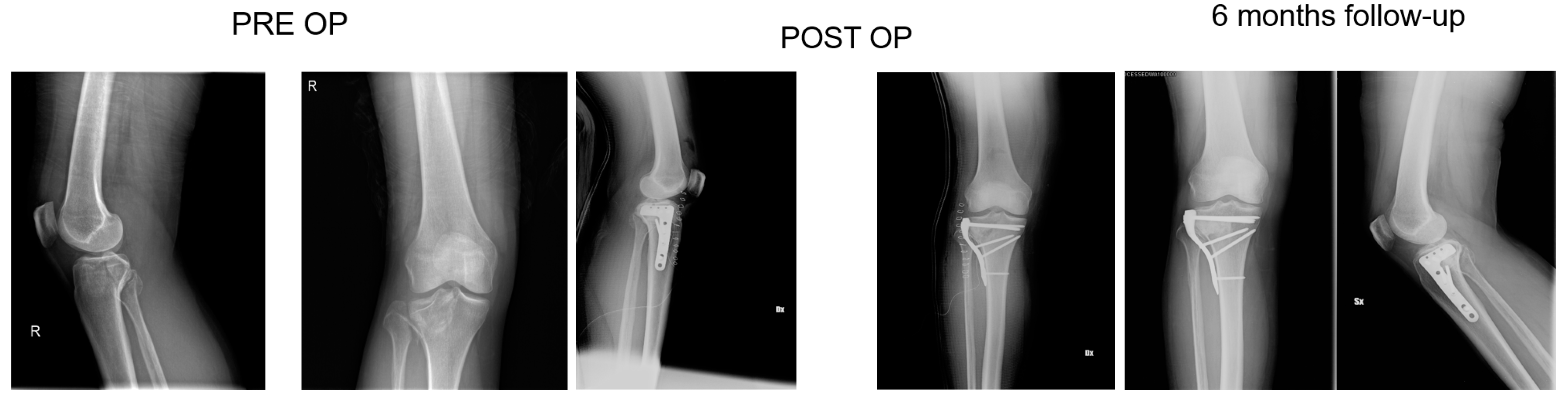
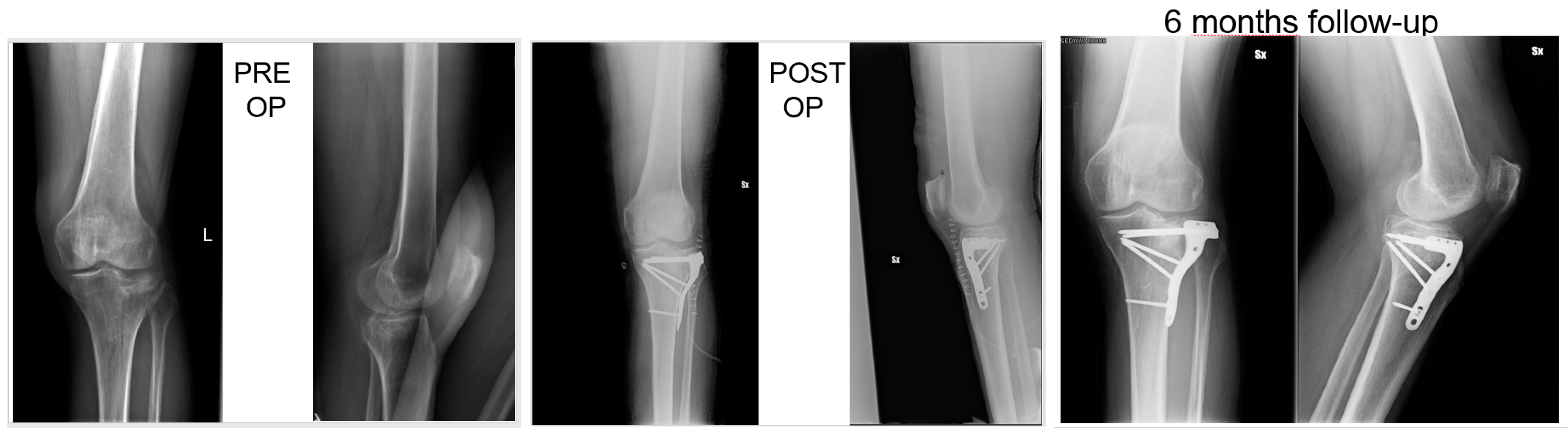
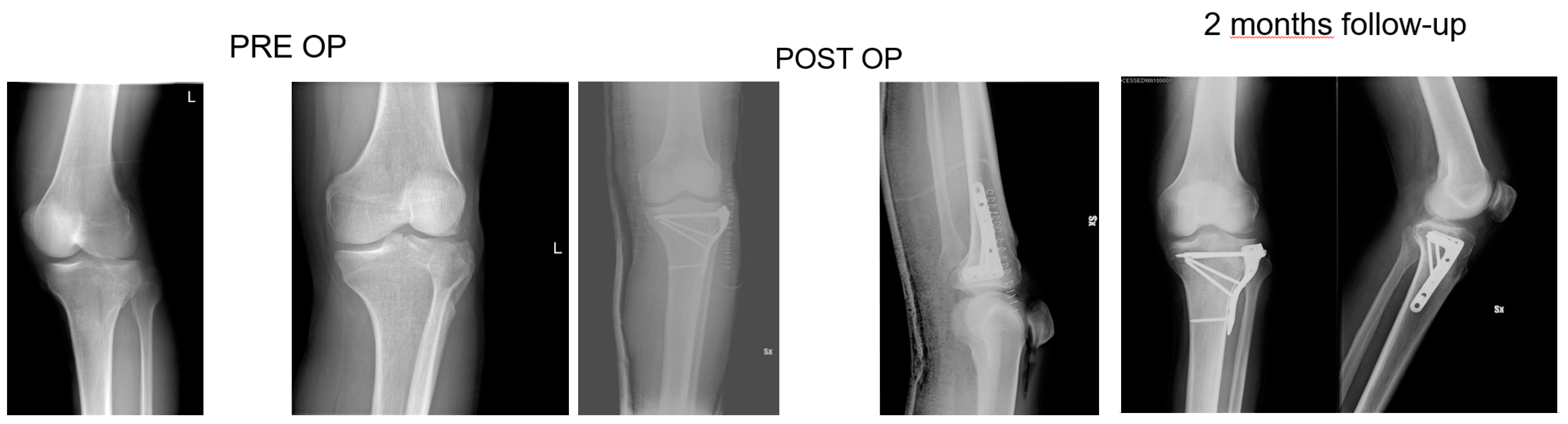
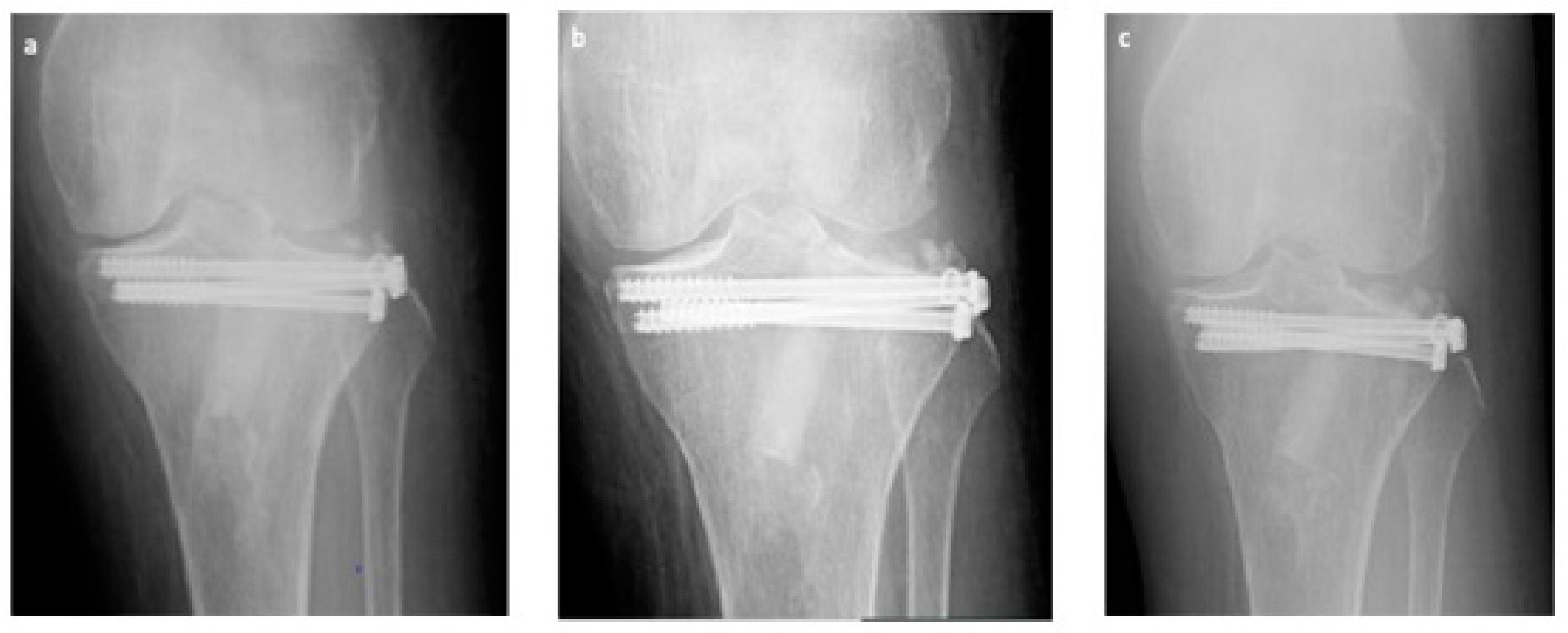
3.3. Radiological Assessment

At each follow-up, we did not observe neither fracture recurrences nor radiographically noticeable subsidence or diastasis of the articular surface at follow up.
We did not detect pseudo-arthrosis, nor bone non-unions nor delayed consolidations [24]. Articular alignment was considered acceptable in all 34 cases at each evaluation (see also Figure 4).
At six months, X-rays images showed that all 34 fractures displayed a good consolidation rate, as confirmed by radiological reports. In particular, radiolucency of the xenografts was assessed [20], which became progressively more similar to that of the surrounding healthy bone tissue (see exemplificative Figure 1, Figure 2 and Figure 3 and comparative images in Figure 4).

3.4. Statistical Analysis

The results are reported in Table 1 and Table 2.

3.5. Long-Term Follow-Up

All 34 patients where fully examined at 1-year follow-up, while, due to timing reasons, 26 patients (76.5%) were also clinically examined at 2 years follow-up, and 5 patients (14.7%) at 1.5 years. Only 3 patients had only a 1-year follow-up. All these 31 patients presented clinically stable conditions and stable performances of the affected limb. Hence, none of the patients observed either at 2 years and 1.5 years follow-up required any further imaging investigations. None of the patients treated with this technique has been lost at this follow up.

4. Discussion

Various strategies for the treatment of tibial plateau fractures have been described in the medical literature, so far. Conservative treatment, such as traction and cast bracing, are viable approaches only in selected cases [25,26]. However, in general, conservative treatment displays poorer outcomes in comparison to surgery, due to the prolonged immobilisation in a cast brace or hospitalization required, which can eventually lead to joint stiffness, and further risks like pulmonary embolism and ulcers [3,27].
Allowing a more rapid return to mobilisation and weight bearing, surgery is preferable in most cases and closed reduction and minimally invasive fixation, ORIF, and arthroplasty are all valid techniques for the treatment of tibial plateau fractures [28,29,30]. Moreover, in some circumstances like challenging comminuted, displaced fractures, excessive loss of bone matrix or severe depressions of articular surface, bone graft substitutes to augment metaphyseal defects are needed. Historically, cancellous bone harvested from iliac crest has been considered the gold standard for bone grafting. A comprehensive review of the literature published in 2013 by Goff et al. [31] showed that AIBG was the most commonly performed procedure for bone augmentation in the management of tibial plateau fracture. However, this approach involves an additional incision to harvest the graft, which may eventually lead to infection, haematoma, neurovascular complications and fractures, overall representing a further risk exposure for the patients [9,10,31]. Furthermore, some variables like quantitative limitations to the bone that can be safely harvested, biological quality and structural strength of the graft are still concerning factors which led to the development of bone substitutes [16]. A vast variety of bone substitutes are available on market and currently used in clinical settings, each displaying some limitations [6,32]. Recently, some authors have focused on investigating xenohybrid materials, which combine biological qualities of auto- and allo-grafts along with mechanical performances of synthetic scaffolds. The CXS we studied as bone graft in the treatment of tibial plateau fractures is a bovine bone-derived matrix enriched in poly(L-lactide-co-ε-caprolactone) (PLCL), in order to improve compressive strength and elasticity, and collagen fragments exposing the RGD sequence, which improve hydrophilicity, so favouring blood affinity, and enhance cells attachment [18,19,20]. It has been proved that its particular affinity to blood facilitates attachment by MSCs, which then differentiate in osteoblasts, leading to deposition of new bone matrix that eventually degrades and substitutes the CXS [7,18].
This CXS has already been safely used in oral and maxillary surgery with promising results, and its biological and mechanical characteristics make it an interesting tool for orthopaedic surgery as well [18,33].
Tegner Lysholm Knee Scoring Scale is a scoring system based on ten questions regarding function, pain and stability of the knee joint [20]. Scores <65 are considered poor; fair if comprised between 65 and 83; good when ranging from 84 to 90; excellent if >90 [34]. The mean Tegner Lysholm Knee Scoring Scale we obtained from our patients was 89 ± 4.10, which is considered good. IKDC 2000 is a form composed of ten sections, each one containing several questions, in which patient is questioned about subjective perception of knee function [35]. The final score is expressed in a scale from 0 to 100; mean IKDC 2000 of our patients was 78.67 ± 3.31, which indicates that 1 year after surgery patients were satisfied about the grade of their knee function. SF-36 is a questionnaire that assesses general health status through 36 questions [23]. The results of the questions are then transformed in eight scales, each one ranging from 0 to 100. The higher the score the higher the level of healthiness related to the parameter assessed by that specific scale. The mean values of SF-36 are reported in Table 2. For each scale values are excellent and similar to those of the general population. Mean VAS decreased from 6.33 ± 1.40 to 1 ± 0.79, difference that we considered significant (p < 0.0001). None of the 34 patients reported any local complication related to the implant, nor neurovascular or infectious adverse effect, which points out the safety of the graft. We used literature data within our study as a referenced historical control group.
The comprehensive review of the literature published in 2013 by Goff et al. [31] analysed data from 672 patients from different studies, with different biomaterials used: fracture healing was uneventful in over 90% of the cases over a variant period of time; secondary collapse of the knee joint surface was reported in 8.6% in the biological substitutes, 5.4% in the hydroxyapatite, 3.7% in the calcium phosphate cement, and 11.1% in the calcium sulphate cases. The recorded incidence of primary surgical site and donor site infection (3.6%) was not statistically significant different, however donor site-related pain was reported up to 12 months following autologous iliac bone graft (AIBG) harvest, i.e. in the most commonly performed procedure for bone augmentation in the management of tibial plateau fracture. Bucholz et al. [36] reported good clinical outcomes in a series of patient with tibial plateau fractures treated with a hydroxyapatite bone substitute, however highlighting some complications like infections and collapses. Moreover, they considered the biodegradation process of the graft as slow. Keating et al. [37] described a series of 49 patients whom bone augmentation was performed with a CaP bone cement, reporting good results with regard to return to activity and clinical outcomes; however, 7% of patients presented complications. Similar results have been obtained by other authors, using the same type of synthetic biomaterial [38,39,40]. Moreover, Simpson et al. [38] reported a significant reduction in mean duration of surgery in patients treated with the CaP bone cement if compared with patients who underwent AIBG (55 vs 101 min, p < 0.0001), which have implications on overall costs of procedures [41]. We quantitatively assessed the grade of knee joint mobilisation, which is crucial to restore weight bearing, through measurement of the ROM. Our series of patients had a mean ROM of 124 ± 6° at 1-year follow-up. Russel et al. [42] in a prospective, randomised, multi-centric study comparing CaP cement with AIBG, registered that 69% of patients of the study group had ROM > 120° at average 12 months follow-up. Newmann et al. [43] studied a group of patients treated with allograft, that reported mean ROM of 118° at 12 months; however, one patient presented osteomyelitis and in four cases subsidence of the joint.
Comparison with the current literature is challenging, due to the lack of standardised follow-up protocols and differences in quantitative assessment of outcomes among the studies. However, the implant of SB in our series resulted in a standard return to knee mobilisation, as highlighted by the mean ROM at last follow-up and the return to partial weight bearing at 4–6 weeks. We also described no complications, like infections and joint subsidence, if compared to similar studies present in the medical literature [31]. Radiological follow-up did not show diastasis or depressions of tibial articular surface excluding the incomplete subchondral reductions documented at the first post op X-ray, which means that mechanical properties of SB were adequate for high and complex forces which the plateau commonly undergoes. At each follow-up, the radiolucency of implants was progressively more similar to radiolucency of the surrounding bone (see exemplificative Figure 1 and Figure 2). We interpreted this evidence as supporting the thesis that SB would have been able to integrate with autologous bone tissue, favouring deposition of new bone matrix within an ongoing remodelling process: indeed, there is a clear morphological pattern on the evolution of the standard X-Ray imaging series over time which shows the substitution of the grafted material with a more homogeneous signal in the area of graft implant. As already demonstrated, the progressive remodelling together with an increase of the mineral signal cannot be dependent on the active remodelling of the graft per se given it is a decellularized matrix. Therefore, the increase in the density over time depends on novel mineral matrix apposition likely induced by the graft, as previously shown both in vivo and in vitro [7,20].
The literature on the use of xenografts in the setting of tibial plateau fractures is still limited. In 2009, Basel et al. [27] described clinical and radiological findings of a series of patient treated for tibial plateau fractures with bone xenograft, but their study did not include any quantitative assessment of outcomes. The majority of studies reported in the literature, about bone augmentation for the treatment of such fractures, involve AIBG; controlled studies comparing different types of bone graft are scarce [31]. However, innovative biomaterials for bone transplantation have demonstrated to be safe, reliable, and biocompatible tools for bone transplantation [16]. Their use resulted in most cases in reduced rate of complications, operative times, operative costs, and hospital length of stay, along with comparable or better results in term of clinical and radiological outcomes [8,38,39,40,41]. Furthermore, xenohybrid materials demonstrated that can be successfully used as carriers to locally administer drugs or MSCs, making them a useful resource for orthopaedic surgeons, in a variety of conditions [7,20,44,45]. Hence, more clinical studies to better understand the role of biomaterials in orthopaedic surgery are encouraged. This study shows some limitations, like the relatively small number of patients having been chosen within the framework of an observational study, the retrospective nature of the analysis, which includes different subtypes of fractures and the lack of a pre-existing quantitative scale defining radiological outcomes; at this last regard, we had hence to develop a new protocol [20]. Also, the ROM of the contralateral (healthy) joint was not reported as it was considered as physiological, while the aim was to test the improvement in the operated joint. Finally, it would have been useful to evaluate a control group a that has been treated in the same hospital with the same clinical procedures in order to evaluate the influence of the bone substitute on the healing. Nevertheless, here we have collected strong clinical evidences already from the 1-year follow-up, being confirmed by all follow-ups (91.2%) recorded after 1.5 and 2 years, including the lack of long-term complications. Overall, collected data offers a confirmation to investigated clinical endpoints and confirms material properties too. A further study will address bone density and graft density on a selected group of patients using a novel TC protocol.

5. Conclusions

Clinical and radiological outcomes of our series of 34 patients treated for fractures of the tibial plateau were good and comparable to literature data [45]. At 1-year follow-up, the xenohybrid biomaterial we studied has demonstrated to be a safe bone substitute and to possess adequate mechanical and biological properties to sustain healing and consolidation of tibial plateau fractures. SB has also showed good biocompatibility and capacity of osteointegration. This evidence makes SB a valuable biomaterial for orthopaedic surgery when bone graft is required.

Author Contributions

Conceptualization, G.P., A.B. (Alessandro Bistolfi) and R.F.; Methodology, G.P., R.F.; Formal Analysis, A.B. (Alessandro Bistolfi); Investigation, R.G.; Resources, V.F. A.B. (Agnese Battista); Data Curation, R.G.; Writing—Original Draft Preparation, A.B. (Alessandro Bistolfi); Writing—Review and Editing, R.G.; Visualization, R.F.; Supervision, R.F.; Project Administration, A.B. (Alessandro Bistolfi); Funding Acquisition, G.P.

Funding

This research received no external funding.

Conflicts of Interest

Prof. Dr. Giuseppe Perale is among shareholders of I.B.I. sa, the Swiss Company owning intellectual property rights on SmartBone®, manufacturing and commercializing it. Prof. Dr. Riccardo Ferracini is an external clinical advisor to the same company. Dr. Riccardo Garibaldi was involved as a guest master student, from University of Genova (Italy), by the same company at the time of this study.

Clinical Data

This study protocol was approved by the United Ethical Committee of the “Città della Scienza e della Salute”, Turin, Italy (approval n. 0004336), which comprehends also the hospital were all patients were treated. All patients signed an informed consent form to document that they understood the aims of the study and authorized the use of their data for research purposes. Patients were allowed to ask questions pertaining to this study and were thoroughly informed consequently. All procedures were performed in strict accordance with the recommendations of the Declaration of Helsinki as revised in Fortaleza (2013) for investigations with human subjects and followed good clinical practice and ISO14155 prescriptions. The study is sponsored by Industrie Biomediche Insubri SA (Switzerland), coordinated by Dr. A. Bistolfi, co-author in this work, and monitored by Prof. Dr. G. Perale, co-author in this work.

References

  1. Court-Brown, C.M.; Caesar, B. Epidemiology of Adult Fractures: A Review. Injury 2006, 37, 691–697. [Google Scholar] [CrossRef] [PubMed]
  2. Koval, K.J.; Helfet, D.L. Tibial Plateau Fractures: Evaluation and Treatment. J. Am. Acad. Orthop. Surg. 1995, 3, 86–94. [Google Scholar] [CrossRef] [PubMed]
  3. Schatzker, J.; McBroom, R.; Bruce, D. The Tibial Plateau Fracture. The Toronto Experience 1968–1975. Clin. Orthop. Relat. Res. 1979, 108, 94–104. [Google Scholar]
  4. Kandemir, U.; Maclean, J. Surgical Approaches for Tibial Plateau Fractures. J. Knee Surg. 2014, 27, 21–29. [Google Scholar] [PubMed]
  5. Athanasiou, V.T.; Papachristou, D.J.; Panagopoulos, A.; Saridis, A.; Scopa, C.D.; Megas, P. Histological Comparison of Autograft, Allograft-Dbm, Xenograft, and Synthetic Grafts in a Trabecular Bone Defect: An Experimental Study in Rabbits. Med. Sci. Monit. 2010, 16, BR24–BR31. [Google Scholar] [PubMed]
  6. Ferracini, R.; Martinez Herreros, I.; Russo, A.; Casalini, T.; Rossi, F.; Perale, G. Scaffolds as Structural Tools for Bone-Targeted Drug Delivery. Pharmaceutics 2018, 10, 122. [Google Scholar] [CrossRef]
  7. Roato, I.; Belisario, D.C.; Compagno, M.; Verderio, L.; Sighinolfi, A.; Mussano, F.; Genova, T.; Veneziano, F.; Pertici, G.; Perale, G.; et al. Adipose-Derived Stromal Vascular Fraction/Xenohybrid Bone Scaffold: An Alternative Source for Bone Regeneration. Stem Cells Int. 2018, 2018, 4126379. [Google Scholar] [CrossRef]
  8. Almaiman, M.; Al-Bargi, H.H.; Manson, P. Complication of Anterior Iliac Bone Graft Harvesting in 372 Adult Patients from May 2006 to May 2011 and a Literature Review. Craniomaxillofac. Trauma Reconstr. 2013, 6, 257–266. [Google Scholar] [CrossRef]
  9. Arrington, E.D.; Smith, W.J.; Chambers, H.G.; Bucknell, A.L.; Davino, N.A. Complications of Iliac Crest Bone Graft Harvesting. Clin. Orthop. Relat. Res. 1996, 300–309. [Google Scholar] [CrossRef]
  10. Dimitriou, R.; Mataliotakis, G.I.; Angoules, A.G.; Kanakaris, N.K.; Giannoudis, P.V. Complications Following Autologous Bone Graft Harvesting from the Iliac Crest and Using the Ria: A Systematic Review. Injury 2011, 42 (Suppl. 2), S3–S15. [Google Scholar] [CrossRef]
  11. Bauer, T.W.; Muschler, G.F. Bone Graft Materials. An Overview of the Basic Science. Clin. Orthop. Relat. Res. 2000, 377, 10–27. [Google Scholar] [CrossRef]
  12. Ali Akbari Ghavimi, S.; Ebrahimzadeh, M.H.; Solati-Hashjin, M.; Abu Osman, N.A. Polycaprolactone/Starch Composite: Fabrication, Structure, Properties, and Applications. J. Biomed. Mater. Res. A 2015, 103, 2482–2498. [Google Scholar] [CrossRef] [PubMed]
  13. Knaack, D.; Goad, M.E.; Aiolova, M.; Rey, C.; Tofighi, A.; Chakravarthy, P.; Lee, D.D. Resorbable Calcium Phosphate Bone Substitute. J. Biomed. Mater. Res. 1998, 43, 399–409. [Google Scholar] [CrossRef]
  14. Lange, T.; Schilling, A.F.; Peters, F.; Mujas, J.; Wicklein, D.; Amling, M. Size Dependent Induction of Proinflammatory Cytokines and Cytotoxicity of Particulate Beta-Tricalciumphosphate in Vitro. Biomaterials 2011, 32, 4067–4075. [Google Scholar] [CrossRef]
  15. Niedhart, C.; Maus, U.; Piroth, W.; Miltner, O.; Schmidt-Rohlfing, B.; Siebert, C.H. Evaluation of a Resorbable, in Situ Setting Bone Substitute in a Sheep Model. J. Biomed. Mater. Res. B Appl. Biomater. 2004, 71, 123–129. [Google Scholar] [CrossRef]
  16. Campana, V.; Milano, G.; Pagano, E.; Barba, M.; Cicione, C.; Salonna, G.; Lattanzi, W.; Logroscino, G. Bone Substitutes in Orthopaedic Surgery: From Basic Science to Clinical Practice. J. Mater. Sci. Mater. Med. 2014, 25, 2445–2461. [Google Scholar] [CrossRef] [PubMed]
  17. Ceccarelli, G.; Presta, R.; Benedetti, L.; de Angelis, M.G.C.; Lupi, S.M.; Rodriguez, Y.; Baena, R. Emerging Perspectives in Scaffold for Tissue Engineering in Oral Surgery. Stem Cells Int. 2017, 2017, 4585401. [Google Scholar] [CrossRef]
  18. D’Alessandro, D.; Perale, G.; Milazzo, M.; Moscato, S.; Stefanini, C.; Pertici, G.; Danti, S. Bovine Bone Matrix/Poly(L-Lactic-Co-Epsilon-Caprolactone)/Gelatin Hybrid Scaffold (Smartbone((R))) for Maxillary Sinus Augmentation: A Histologic Study on Bone Regeneration. Int. J. Pharm. 2017, 523, 534–544. [Google Scholar] [CrossRef]
  19. Pertici, G.; Carinci, F.; Carusi, G.; Epistatus, D.; Villa, T.; Crivelli, F.; Rossi, F.; Perale, G. Composite Polymer-Coated Mineral Scaffolds for Bone Regeneration: From Material Characterization to Human Studies. J. Biol. Regul. Homeost. Agents 2015, 29 (Suppl. 1), 136–148. [Google Scholar]
  20. Salerno, M.; Cenni, E.; Fotia, C.; Avnet, S.; Granchi, D.; Castelli, F.; Micieli, D.; Pignatello, R.; Capulli, M.; Rucci, N.; et al. Bone-Targeted Doxorubicin-Loaded Nanoparticles as a Tool for the Treatment of Skeletal Metastases. Curr. Cancer Drug Targets 2010, 10, 649–659. [Google Scholar] [CrossRef]
  21. Tegner, Y.; Lysholm, J. Rating Systems in the Evaluation of Knee Ligament Injuries. Clin. Orthop. Relat. Res. 1985, 198, 43–49. [Google Scholar] [CrossRef]
  22. Irrgang, J.J.; Ho, H.; Harner, C.D.; Fu, F.H. Use of the International Knee Documentation Committee Guidelines to Assess Outcome Following Anterior Cruciate Ligament Reconstruction. Knee Surg. Sports Traumatol. Arthrosc. 1998, 6, 107–114. [Google Scholar] [CrossRef] [PubMed]
  23. Ware, J.E., Jr.; Sherbourne, C.D. The Mos 36-Item Short-Form Health Survey (Sf-36). I. Conceptual Framework and Item Selection. Med. Care 1992, 30, 473–483. [Google Scholar] [CrossRef] [PubMed]
  24. Grottoli, C.F.; Ferracini, R.; Compagno, M.; Tombolesi, A.; Rampado, O.; Pilone, L.; Bistolfi, A.; Borrè, A.; Cingolani, A.; Perale, G. A Radiological Approach to Evaluate Bone Graft Integration in Reconstructive Surgeries. Appl. Sci. 2019, 9, 1469. [Google Scholar] [CrossRef]
  25. DeCoster, T.A.; Nepola, J.V.; El-Khoury, G.Y. Cast Brace Treatment of Proximal Tibia Fractures. A Ten-Year Follow-up Study. Clin. Orthop. Relat. Res. 1988, 231, 196–204. [Google Scholar]
  26. Jensen, D.B.; Rude, C.; Duus, B.; Bjerg-Nielsen, A. Tibial Plateau Fractures. A Comparison of Conservative and Surgical Treatment. J. Bone Jt. Surg. Br. 1990, 72, 49–52. [Google Scholar] [CrossRef]
  27. Bansal, M.R.; Bhagat, S.B.; Shukla, D.D. Bovine Cancellous Xenograft in the Treatment of Tibial Plateau Fractures in Elderly Patients. Int. Orthop. 2009, 33, 779–784. [Google Scholar] [CrossRef]
  28. Koval, K.J.; Sanders, R.; Borrelli, J.; Helfet, D.; DiPasquale, T.; Mast, J.W. Indirect Reduction and Percutaneous Screw Fixation of Displaced Tibial Plateau Fractures. J. Orthop. Trauma 1992, 6, 340–346. [Google Scholar] [CrossRef] [PubMed]
  29. Canadian Orthopaedic Trauma Society. Open Reduction and Internal Fixation Compared with Circular Fixator Application for Bicondylar Tibial Plateau Fractures. Results of a Multicenter, Prospective, Randomized Clinical Trial. J. Bone Jt. Surg. Am. 2006, 88, 2613–2623. [Google Scholar] [CrossRef]
  30. Malviya, A.; Reed, M.R.; Partington, P.F. Acute Primary Total Knee Arthroplasty for Peri-Articular Knee Fractures in Patients over 65 Years of Age. Injury 2011, 42, 1368–1371. [Google Scholar] [CrossRef]
  31. Goff, T.; Kanakaris, N.K.; Giannoudis, P.V. Use of Bone Graft Substitutes in the Management of Tibial Plateau Fractures. Injury 2013, 44, S86–S94. [Google Scholar] [CrossRef]
  32. Cancedda, R.; Dozin, B.; Giannoni, P.; Quarto, R. Tissue Engineering and Cell Therapy of Cartilage and Bone. Matrix Biol. 2003, 22, 81–91. [Google Scholar] [CrossRef]
  33. Secondo, F.; Grottoli, C.F.; Zollino, I.; Perale, G.; Lauritano, D. Positioning of a Contextual Implant Along with a Sinus Lift Anchored with a Block of Heterologous Bone. Oral. Implantol. (Rome) 2017, 10, 457–467. [Google Scholar] [CrossRef] [PubMed]
  34. Mitsou, A.; Vallianatos, P.; Piskopakis, N.; Maheras, S. Anterior Cruciate Ligament Reconstruction by over-the-Top Repair Combined with Popliteus Tendon Plasty. J. Bone Jt. Surg. Br. 1990, 72, 398–404. [Google Scholar] [CrossRef]
  35. Irrgang, J.J.; Anderson, A.F.; Boland, A.L.; Harner, C.D.; Kurosaka, M.; Neyret, P.; Richmond, J.C.; Shelborne, K.D. Development and Validation of the International Knee Documentation Committee Subjective Knee Form. Am. J. Sports. Med. 2001, 29, 600–613. [Google Scholar] [CrossRef] [PubMed]
  36. Bucholz, R.W.; Carlton, A.; Holmes, R. Interporous Hydroxyapatite as a Bone Graft Substitute in Tibial Plateau Fractures. Clin. Orthop. Relat. Res. 1989, 240, 53–62. [Google Scholar] [CrossRef]
  37. Keating, J.F.; Hajducka, C.L.; Harper, J. Minimal Internal Fixation and Calcium-Phosphate Cement in the Treatment of Fractures of the Tibial Plateau. A Pilot Study. J. Bone Jt. Surg. Br. 2003, 85, 68–73. [Google Scholar] [CrossRef]
  38. Simpson, D.; Keating, J.F. Outcome of Tibial Plateau Fractures Managed with Calcium Phosphate Cement. Injury 2004, 35, 913–918. [Google Scholar] [CrossRef]
  39. Lobenhoffer, P.; Gerich, T.; Witte, F.; Tscherne, H. Use of an Injectable Calcium Phosphate Bone Cement in the Treatment of Tibial Plateau Fractures: A Prospective Study of Twenty-Six Cases with Twenty-Month Mean Follow-Up. J. Orthop. Trauma. 2002, 16, 143–149. [Google Scholar] [CrossRef]
  40. Horstmann, W.G.; Verheyen, C.C.; Leemans, R. An Injectable Calcium Phosphate Cement as a Bone-Graft Substitute in the Treatment of Displaced Lateral Tibial Plateau Fractures. Injury 2003, 34, 141–144. [Google Scholar] [CrossRef]
  41. Mehta, S.; Blagg, R.; Willcockson, J.; Gociman, B.; Yamashiro, D.; Siddiqi, F. Cost-Effectiveness Analysis of Demineralized Bone Matrix and Rhbmp-2 Versus Autologous Iliac Crest Bone Grafting in Alveolar Cleft Patients. Plast. Reconstr. Surg. 2018, 142, 737–743. [Google Scholar] [CrossRef] [PubMed]
  42. Russell, T.A.; Leighton, R.K.; Alpha-BSM Tibial Plateau Fracture Study Group. Comparison of Autogenous Bone Graft and Endothermic Calcium Phosphate Cement for Defect Augmentation in Tibial Plateau Fractures. A Multicenter, Prospective, Randomized Study. J. Bone Jt. Surg. Am. 2008, 90, 2057–2061. [Google Scholar] [CrossRef] [PubMed]
  43. Newman, J.T.; Smith, W.R.; Ziran, B.H.; Hasenboehler, E.A.; Stahel, P.F.; Morgan, S.J. Efficacy of Composite Allograft and Demineralized Bone Matrix Graft in Treating Tibial Plateau Fractures with Bone Loss. Orthopedics 2008, 31, 649. [Google Scholar] [CrossRef] [PubMed]
  44. Cheng, H.; Chawla, A.; Yang, Y.; Li, Y.; Zhang, J.; Jang, H.L.; Khademhosseini, A. Development of Nanomaterials for Bone-Targeted Drug Delivery. Drug Discov. Today 2017, 22, 1336–1350. [Google Scholar] [CrossRef] [PubMed]
  45. Manidakis, N.; Dosani, A.; Dimitriou, R.; Stengel, D.; Matthews, S.; Giannoudis, P. Tibial plateau fractures: Functional outcome and incidence of osteoarthritis in 125 cases. Int. Orthop. 2010, 34, 565–570. [Google Scholar] [CrossRef] [PubMed]
Figure 1. Fifty-seven-year-old patient, X-rays: preoperative, postoperative and control at 6 months follow up of a Shatzker type 2 fracture, AO 41 B1.3.
Applsci 09 02675 g001
Figure 2. Sixty-year-old patient, X-rays: preoperative, postoperative and control at 6 months follow up of a Shatzker type 3 fracture, AO 41 B 1.3.
Applsci 09 02675 g002
Figure 3. 68 years old patient, X-rays: preoperative, postoperative and control at 2 months follow up of a Shatzker type 2 fracture, AO 41 B1.3.
Applsci 09 02675 g003
Figure 4. Exemplificative case, in anteroposterior RX, at 1 (a), 3 (b), and 6 (c) months follow-up respectively, well evidencing good consolidation, absence of non-union or retarded consolidation and graft good integration confirmed by progressive loss of radiolucency of the graft itself.
Applsci 09 02675 g004
Table 1. Demographics.
SexMale18 (53%)
Female16 (47%)
AgeMeans and SD57.5 ± 12.782
Height (m)1.71 ± 0.10
Weight (kg)71.17 ± 7.78
BMI (kg/m2)24.23 ± 1.97
Schatzker Classification [3]Type II18 (52.9%)
Type III6 (17.6%)
Type VI10 (29.4%)
Table 2. Mean visual analogue scale (VAS) and Short Form (36) Health Survey (SF-36) values.
VAS2 weeks post-op6.33 ± 1.40
1 month post-op5.2 ± 0.89
3 months post-op3.1 ± 0.71
6 months post-op1.43 ± 0.69
1 year post-op1 ± 0.79
Percentage decrease84.20%
SF-36 [23]Limitations in physical activities92.5 ± 11.65
Limitations in social activities96.67 ± 6.23
Limitations in usual role activities because of physical health96.83 ± 3.34
Bodily pain91.8 ± 9.31
General mental health86.93 ± 3.78
Limitations in usual role activities because of emotional problems94.33 ± 12.89
Vitality87.5 ± 6.79
General health perception93.6 ± 4.90

