Article

Intrant ELISA: A Novel Approach to Fabrication of Electrospun Fiber Mat-Assisted Biosensor Platforms and Their Integration within Standard Analytical Well Plates

by
Samira Hosseini
1,
Pedram Azari
2,
Mohammad M. Aeinehvand
1,
Hussin A. Rothan
3,
Ivan Djordjevic
4,*,
Sergio O. Martinez-Chapa
1,* and
Marc J. Madou
5,6
1
Department of Electrical Engineering, School of Engineering and Sciences, Tecnologico de Monterrey, Monterrey 2501, Mexico
2
Department of Biomedical Engineering, Faculty of Engineering, University of Malaya, Kuala Lumpur 50603, Malaysia
3
Department of Molecular Medicine, Faculty of Medicine, University of Malaya, Kuala Lumpur 50603, Malaysia
4
School of Material Science and Engineering, Nanyang Technological University, Singapore 639798, Singapore
5
Department of Biomedical Engineering, University of California, Irvine, CA 92697, USA
6
Department of Mechanical and Aerospace Engineering, University of California, Irvine, CA 92697, USA
*
Authors to whom correspondence should be addressed.
Appl. Sci. 2016, 6(11), 336; https://doi.org/10.3390/app6110336
Submission received: 8 September 2016 / Revised: 24 October 2016 / Accepted: 24 October 2016 / Published: 5 November 2016
(This article belongs to the Special Issue Nanomanufacturing of Biomedical Systems and Devices)

Abstract

:
A combination of far-field electrospinning (FFES) and free-radical polymerization has been used to fabricate coated electrospun polymer fiber mats as a new type of biosensor platform. Poly (3-hydroxybutyrate-co-3-hydroxyvalerate) (PHBV) electrospun fibers were dip-coated with different compositions of poly methyl methacrylate-co-methacrylic acid (poly(MMA-co-MAA)). This synergistic approach utilizes large specific surface area of PHBV fibers and co-polymer coatings that feature an optimum concentration of surface carboxyl (–COOH) groups. The platform surface morphology, porosity and tunable hydrophobicity enhance biomolecular interactions via plurality of molecular forces. These customized fiber mats have been integrated into a newly designed 96-well plate called an “intrant enzyme-linked immunosorbent assay” or i-ELISA. I-ELISA allows colorimetric sandwich assay to be carried out without any modifications or additional steps in ELISA methodology. By introducing the fiber mats in fabrication of i-ELISA via extensions on the lid, we address some of the limitations of the previous designs while demonstrating an enhanced signal intensity up to 12 times higher than that of conventional assays. With improved sensitivity, specificity and accuracy in the detection of dengue virus, i-ELISA has proven to be a reliable platform for biomolecular recognition. The proposed fiber mat-assisted well plate in this study holds great potential as a universal approach for integration of different types of fiber mats with pre-designed specific properties in order to enhance the detection sensitivity of the assay.

1. Introduction

Application of papers in analytical platforms was known as early as the 17th century with the use of cellulose fibrous structure for chromatography [1]. Recent advances in specific healthcare areas such as visual bio-diagnostics and hand-held detection devices have opened a new demand for the application of fiber-based materials [2,3,4]. Various paper-based bio-analytical platforms were developed in the last decades such as dot-immunoglobin assays (DIAs), microfluidic paper-based analytical devices (μPADs), laminated paper-based analytical devices (LPADs), immunospot, nitrocellulose pads (NC-PADs) and paper-based ELISA (enzyme-linked immunosorbent assay) (P-ELISA) well plates [2]. Fabrication of these platforms mainly involves techniques such as printing, waxing, patterning and heating [2]. Those techniques are promising as potential healthcare solutions, yet for addressing point-of-care issues, better control over the paper source itself is imperative [5]. Although application of papers has made an obvious shift in the healthcare paradigm and opened a wide outlook into the bio-analytical domain, developed paper-based platforms are still suffering from a lack of reproducibility and limited detection options [2,6]. In particular, paper-based analytical devices suffer from one inherent limitation common in majority of these platforms: insufficient level of sensitivity and accuracy [2,7].
Fiber segments integrated in existing paper platforms are commercially available nitrocellulose (NC) with hydroxyl groups (–OH) as a part of the chemical structure. Different modification techniques such as oxidation, etherification, esterification, etc. have been used to convert the fairly unreactive –OH functional groups to more reactive surface carboxyl (–COOH) groups [8,9,10,11]. Apart from the intricacy of such modification techniques, they often compromise the NC characteristics during the modification procedure. Most importantly, the properties of NC materials such as porosity and permeability vary from one manufacturer to another and it is difficult to maintain consistency over those parameters when dealing with commercial paper products. In order to overcome those limitations there is necessity of developing a customized material with a higher degree of control over improved material characteristics.
In a novel approach presented in this paper, we have developed a polymer-coated fiber mat that can be a suitable substitution for the papers in the existing paper-based analytical devices (Figure 1). Furthermore, we fabricated a new class of fiber (or paper)-assisted analytical well plate that, while being simple and cost effective, addresses some of the major drawbacks of the previously developed fiber-based platforms. The proposed strategy introduces fiber mats into the assay by incorporating them into the lid of the well plate using pillar extensions, as presented in Figure 2. This new design allows the fiber mats to physically penetrate the assay. For that reason, the developed platform is named intrant ELISA (abbreviated as i-ELISA; the word “intrant” is taken from the Latin root for the “entering object”). In this new strategy, protein immobilization and subsequent detection of the target analyte takes place on both sides of the platform (inside the well and on the fiber mat). This arrangement enhances the chance for bio-recognition as the specific surface area is significantly increased, which in turn enables early detection of diseases such as dengue virus.
Prevalent in tropical and subtropical areas, dengue fever (DF) is considered as one of the deadliest diseases that can be a universal threat by individuals who contract the infection while traveling abroad [12,13,14]. Dengue starts with simple dengue fever (DF) but can foster severe manifestations such as dengue hemorrhagic fever (DHF) and dengue shock syndrome (DSS) [12,14,15,16,17]. With a yearly 400 million infection cases and 21,000 fatalities, effective early dengue detection remains a major challenge. The disease can result in death of the patient in a relatively short time (seven days) if the illness enters the acute phases [18]. According to statistics, dengue is one of the major threats to public health in 112 countries worldwide [19]. Highly sensitive and selective dengue detection is expected to diminish the mortality rates of this fatal infection from 20% to below 1% [20,21,22]. Due to their high specific surface area, fibrous polymer platforms seem to be materials of choice for early virus detection through drastically increased analytical sensitivity.
While fabrication technology of i-ELISA can be accepted as a general strategy to integrate different types of fibers/papers into this analytical platform, we introduce a new type of customized material, which enables us to tailor the physical and chemical properties more efficiently than with paper strips. Taking the advantage of far-field electrospinning (FFES) and dip-coating techniques, polymer-coated electrospun fibers were produced for integration into an i-ELISA. Electrospun fibers were fabricated from poly (3-hydroxybutyrate-co-3-hydroxyvalerate) (PHBV) and were coated by different compositions of poly methyl methacrylate-co-methacrylic acid (poly(MMA-co-MAA)) synthesized in free-radical polymerization reaction. In our previous work, we demonstrated the advantage of poly(MMA-co-MAA) coatings that increased dengue detection limits when applied in various shapes and sizes [4,23,24]. The combination of FFES and dip-coating results in a paper-like fiber material with controlled chemistry, surface morphology, hydrophobicity and pore size. The fiber mats obtained in this work are advantaged with an optimum density of permanent -COOH functional groups that not only obviate surface modification but also facilitate biomolecular interaction through several molecular mechanisms when applied in our novel i-ELISA platform for dengue virus (DENV) detection.

2. Materials and Methods

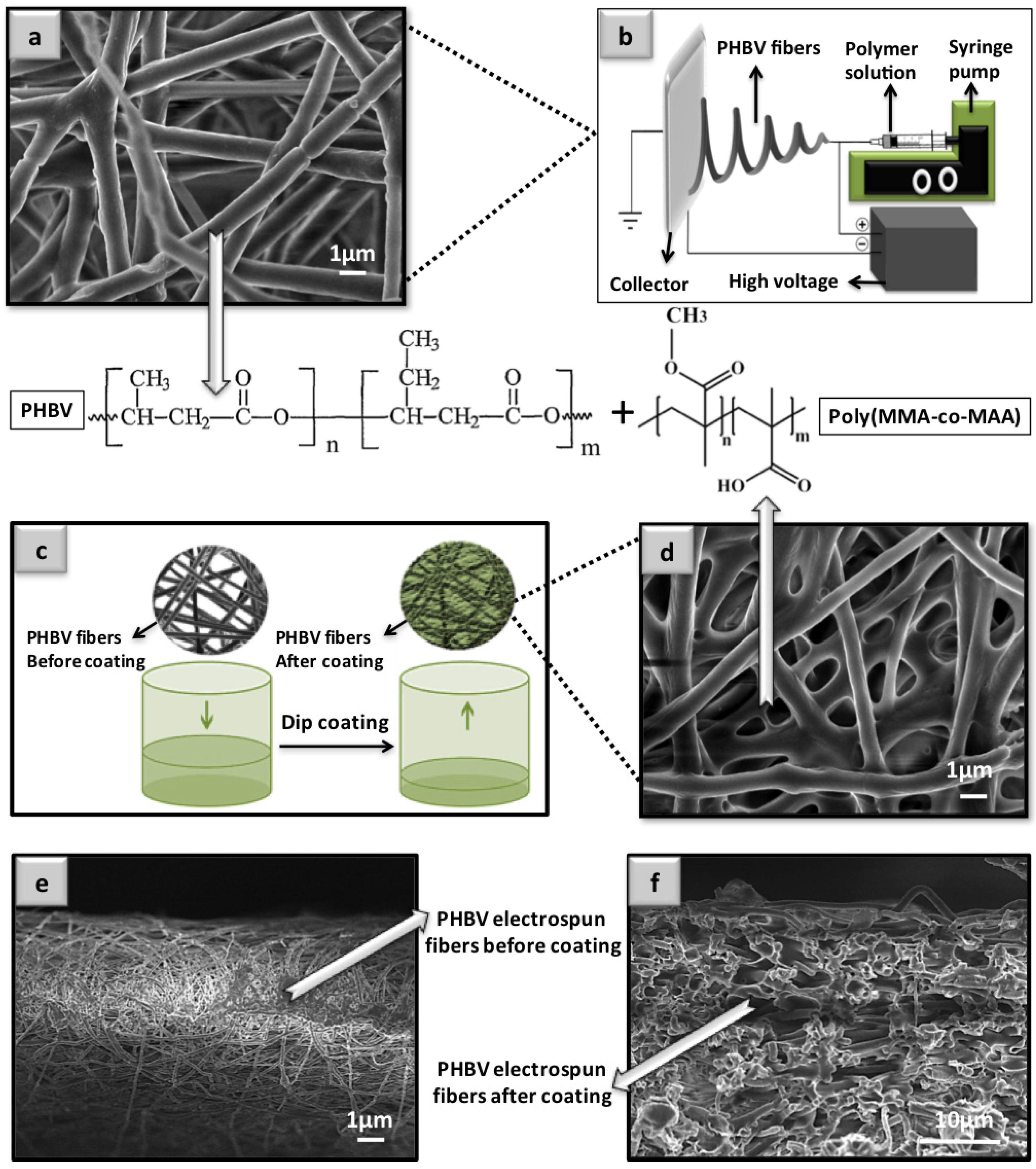
2.1. Fabrication of the Polymer-Coated Electrospun Fibers

Step 1. Electrospun fibers were produced from a PHBV solution (Sigma, San Antonio, TX, USA) by using FFES as shown in Figure 1a,b. Dissolved PHBV in chloroform (CHCl3, Sigma, San Antonio, TX, USA) and dimethylformamide (DMF, Thermo Fisher Scientific, Waltham, MA, USA) was injected from a needle, drawn into fibers and deposited on an aluminum collector plate (Reynolds Wrap, Lincolnshire, IL, USA). Electrospun PHBV fiber mats were peeled from the collector plate and stored for further experiments. The electrospinning setup, except for high voltage power supply, was placed inside a clear acrylic box to have a careful control over the experimental environment. Molecular sieves and silica gel (200 g) were placed inside the perforated box that allowed simultaneous humidity absorption and solvent evaporation. A hygrometer (Hanna Instruments HI 8564, Dallas, TX, USA) was placed inside the box to monitor relative humidity and temperature during the electrospinning procedure. Relative humidity and temperature during the electrospinning time were kept in the range of 28 ± 2 °C and 61%–65%, respectively. The duration of electrospinning was approximately 7 h while 20 mL of polymer solution was spun for each sample. Detailed information related to electrospinning procedure can be found in the supplementary section.
Step 2. Different compositions of poly(MMA-co-MAA) have been synthesized by a previously reported method [25]. Four different compositions were synthesized as follows: poly(MMA-co-MAA-9:1), poly(MMA-co-MAA-7:3) and poly(MMA-co-MAA-5:5). The numbers in the brackets mark the molar ratios of the monomers involved in the polymerization reaction. For ease of discussion, these compositions are referred to as Comp.(9:1), Comp.(7:3) and Comp.(5:5) further on in the text. Detailed description of synthesis reaction setup can be found in the supplementary section.
Step 3. The electrospun PHBV fibers were coated with PMMA and different compositions of poly(MMA-co-MAA) through simple and straightforward methods of dip-coating (Figure 1c) [24]. Fiber sheets were immersed in the polymer solutions (5%) for 3 s, dried at ambient temperature, and cut into circles of 6 mm diameter (Figure 1c). The round-shaped coated electrospun fiber mats were used in the fabrication of i-ELISA. Further technical details of the polymer synthesis, fiber production and coating procedure can be found in the supplementary section.

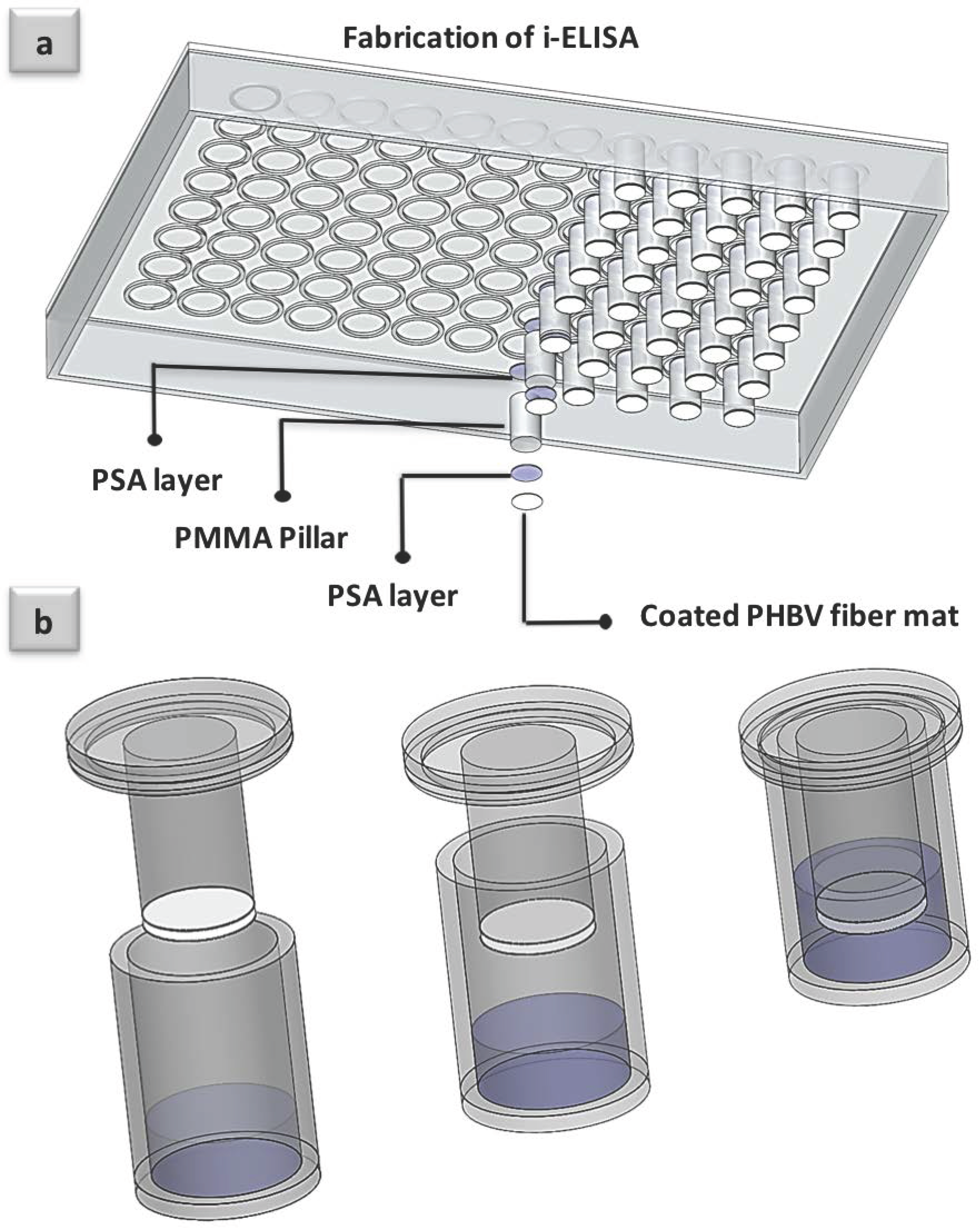
2.2. Fabrication of i-ELISA

The polymer-coated PHBV fiber mats were integrated into a novel design of 96-well plate. Figure 2a shows a schematic representation of the prototype’s segments. Each lid has 96 attached PMMA pillars (6 mm diameter × 8.7 mm length) which were fabricated through a layer-by-layer assembly of PMMA pillars and two pressure sensor adhesive layers (PSA; ~100 µm thickness, model FLEXmount DFM-200-Clear) (FLEXcon Company, Spencer, MA, USA) for each individual PMMA pillar. One PSA layer was used to affix the pillar section to the lid and the other one was used to secure the round-shaped polymer-coated PHBV electrospun fiber mat (~500 µm thick), as shown in Figure 2a. A computer numerical controlled (CNC) precision tool (model Vision 2525, Vision Engraving and Routing Systems, Phoenix, AZ, USA) and a vinyl cutting/plotter machine (Model PUMA II, GCC, Taipei, China) were used for cutting out the PMMA pillars and fabricating the PSA layers from large PMMA and PSA sheets, respectively.

2.3. Morphology Analysis by Scanning Electron Microscopy (SEM) and Water-in-Air Contact (WCA) Angle Analysis

Surface morphology of the coated and uncoated PHBV fibers was analyzed with scanning electron microscopy equipped with a field emission gun (JSM7600F, JEOL, Santa Barbara, CA, USA) following the previously reported experimental method [24]. Frontal and cross-section images of the coated and uncoated samples were analyzed in order to study the effect of coating on the morphology of the PHBV fibers.
Water-in-air contact angle (WCA) was measured for the droplets of distilled water deposited on the coated and uncoated surfaces of PHBV fiber mats. The average contact angle for each sample was calculated from the measurements performed on five separate droplets of distilled water on three samples from each composition [24]. Contact angles were measured 1 min after deposition of the droplets at the center and on the four corners of the samples. Standard deviations, in these experiments, were negligible as a minor spreading (±0.3°) was observed for almost all of the measurements (n = 15). Contact angle measurement was also performed on the 96-well plate made from polystyrene (PS) for a clear comparison between wettability of the commercially available ELISA well plates and the customized platforms.

2.4. Colorimetric Sandwich ELISA for DENV Detection

Sandwich ELISA, vaunted as the most specific and reliable biorecognition protocol, was selected as the method of interest for minimizing the risk of non-specific bindings and obtaining accurate results [26]. In this protocol, the virus was immobilized between two antibodies (Abs): capture Ab (rabbit anti-dengue virus 2 antibody, ab155042, Abcam, San Francisco, CA, USA) and primary Ab (mouse IgG2a anti-DV, ab155863, Abcam, San Francisco, CA, USA). Since these two Abs are propagated in the bodies of different hosts, they are incapable of binding each other. However, a secondary labeled Ab (anti-mouse igG2a alkaline phosphatase, ab97242, Abcam, San Francisco, CA, USA) can further bind to the primary Ab and generate the colorimetric detection signal. The chance of cross-reactivity or analytes’ interface for the antibodies was reported to be minimal (less than 2%) according to the data sheets received from the manufacturer. Along every single set of the experiments, conventional ELISA in untreated PS 96-well plate (Nunc, Roskilde, Denmark) was also conducted as a control (the sandwich ELISA protocol is described in the supplementary section). To reduce experimental inconsistency, all conducted assays were performed under the same conditions using the same reagents. No major intraday variability was observed for the performed assays in this study. Enveloped dengue virus was prepared in our laboratories via a clinical isolation and processing of dengue serotype 2 obtained from a patient’s serum sample (DV2-isolate Malaysia M2, Gen Bank Toxonomy No.: 11062).
Depending on the purpose of the assay, a variety of DENV concentrations was prepared by serial dilutions. In order to calibrate the assay, serial diluted virus solutions in the concentration range of 3.5 × 10⁻⁵ p.f.u/mL to 3.5 × 10⁻² p.f.u/mL were applied in the assay. Calibration curves were plotted after conversion of the data to the logarithm values. The detection range was studied by running the assay in a wide concentration range between 3.5 × 10⁻⁵ p.f.u/mL and 3.5 × 10⁵ p.f.u/mL. Detection comparison, however, was selected from the results obtained only for DENV concentration = 3.5 × 10⁻² p.f.u/mL. Negative controls were calculated from the assays conducted in the absence of virus (n = 24). Cut-off values for each individual platform were considered as twice of the mean values of the negative controls, which is in accordance with the approved methodologies [15]. Only those replicates that have shown optical density (OD) greater than corresponding cut-off values were interpreted as positives readings. Detection results have been plotted after subtracting the cut-off values from the original data. Positive detection results were accompanied by their negative controls for additional clarification. A total number of 96 samples including 72 positive and 24 negative replicates were examined for each individual platform in sandwich assay to study the sensitivity and specificity of the proposed methodology in contrast to the conventional ELISA. Limit of detection (LoD) for each separate platform was determined from the standard deviations in the case of minimum DENV concentration and slopes of the corresponding calibration plot [26,27,28,29,30,31,32]. Calculations are presented in the supplementary section.

3. Results and Discussion

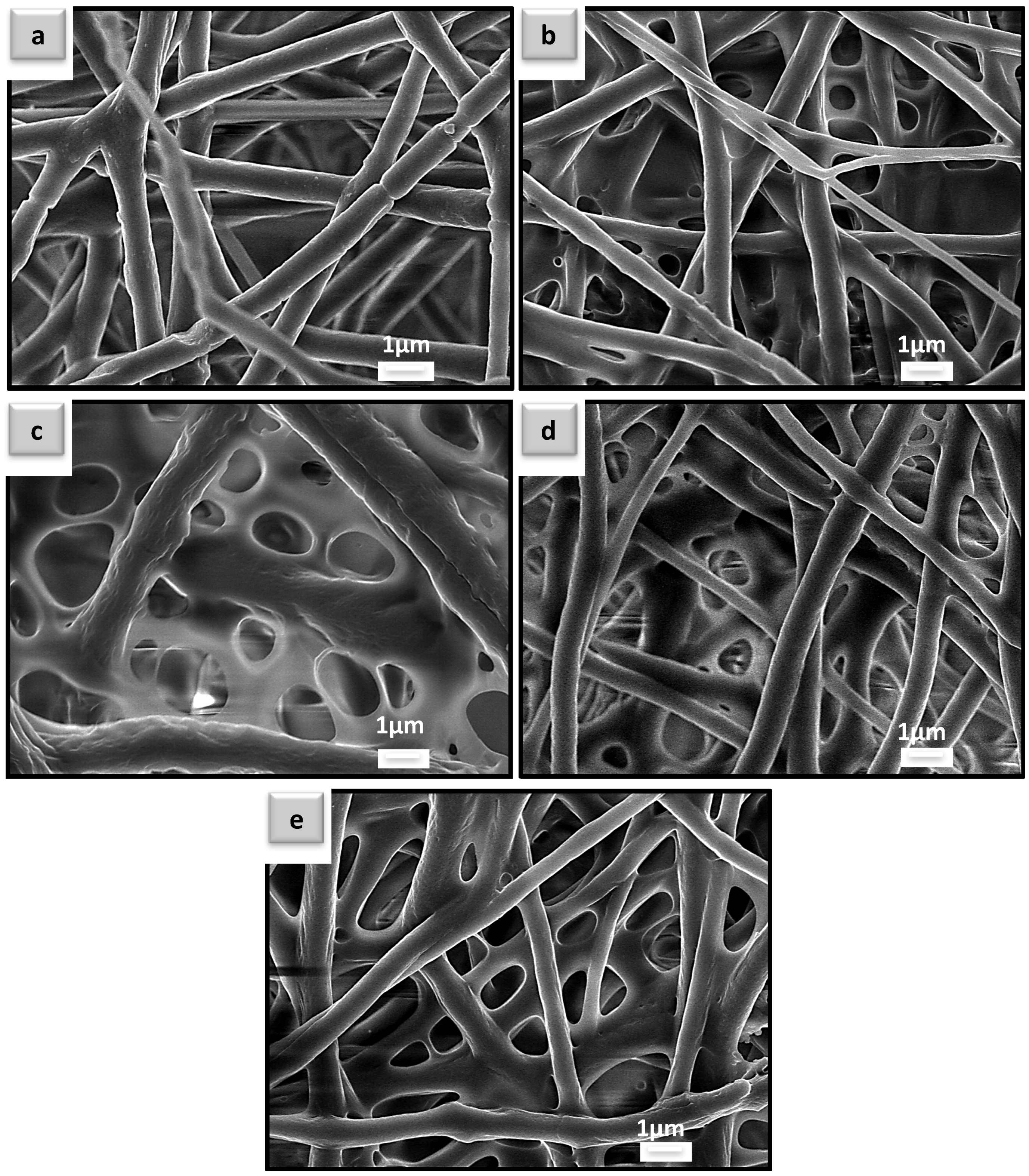
3.1. Morphology Analysis of the Coated and Uncoated Poly(3-hydroxybutyrate-co-3-hydroxyvalerate) (PHBV) Electrospun Fiber Mats

Surface morphologies of the uncoated and polymer-coated PHBV electrospun fibers, recorded by SEM, are presented in Figure 1. A very clear morphology alteration by PMMA-co-MAA coating of the fibers can be observed by SEM image comparison between pristine fibers and polymer-coated PHBV mats (Figure 1a,d). In particular, Figure 1d depicts the highly porous structure of the polymer-coated fibers with the average pore size of ~3 μm (measurements performed for 100 random pores in five different coated samples). General pore size reduction of the coated samples is essential as it prevents the entrapment of the biomolecules in the network of the fibers (this phenomenon is further discussed in Section 3.4). Note that the images in Figure 1a,d were chosen as representative from SEM analysis (complete SEM analysis can be found in supplementary section; Figure A1). Representative cross-section images of the uncoated and polymer-coated PHBV fibers are also shown in Figure 1e,f, respectively. Coverage of the interstitial spaces between the electrospun fibers by coated segments can be recognized by comparing these images as well (Figure 1e,d). Overall, our method enables full cover of 3D fibrous structures used in this experiment. Similar fibrous structure was observed for almost all the polymer-coated PHBV fibers. This result is consistent with our previous findings where the similar coatings were fabricated on different type of polymer fibers [24]. In this study, however, a different approach was utilized by integrating the fiber onto the specially designed ELISA plate lids (Figure 2). With this fabrication strategy of well plates, i-ELISA introduces paper segments or polymer fiber mats from the top of the well plate into the assay.

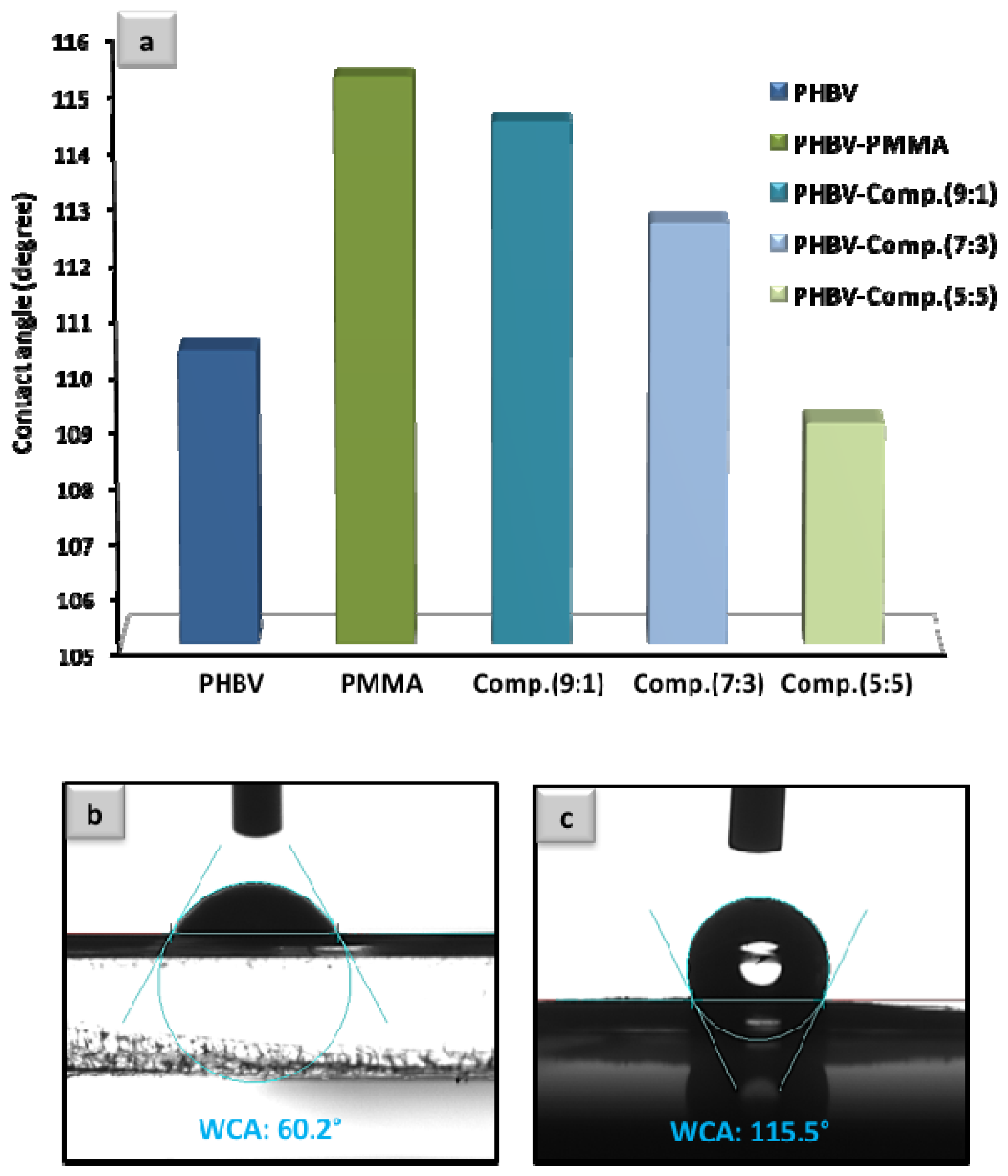
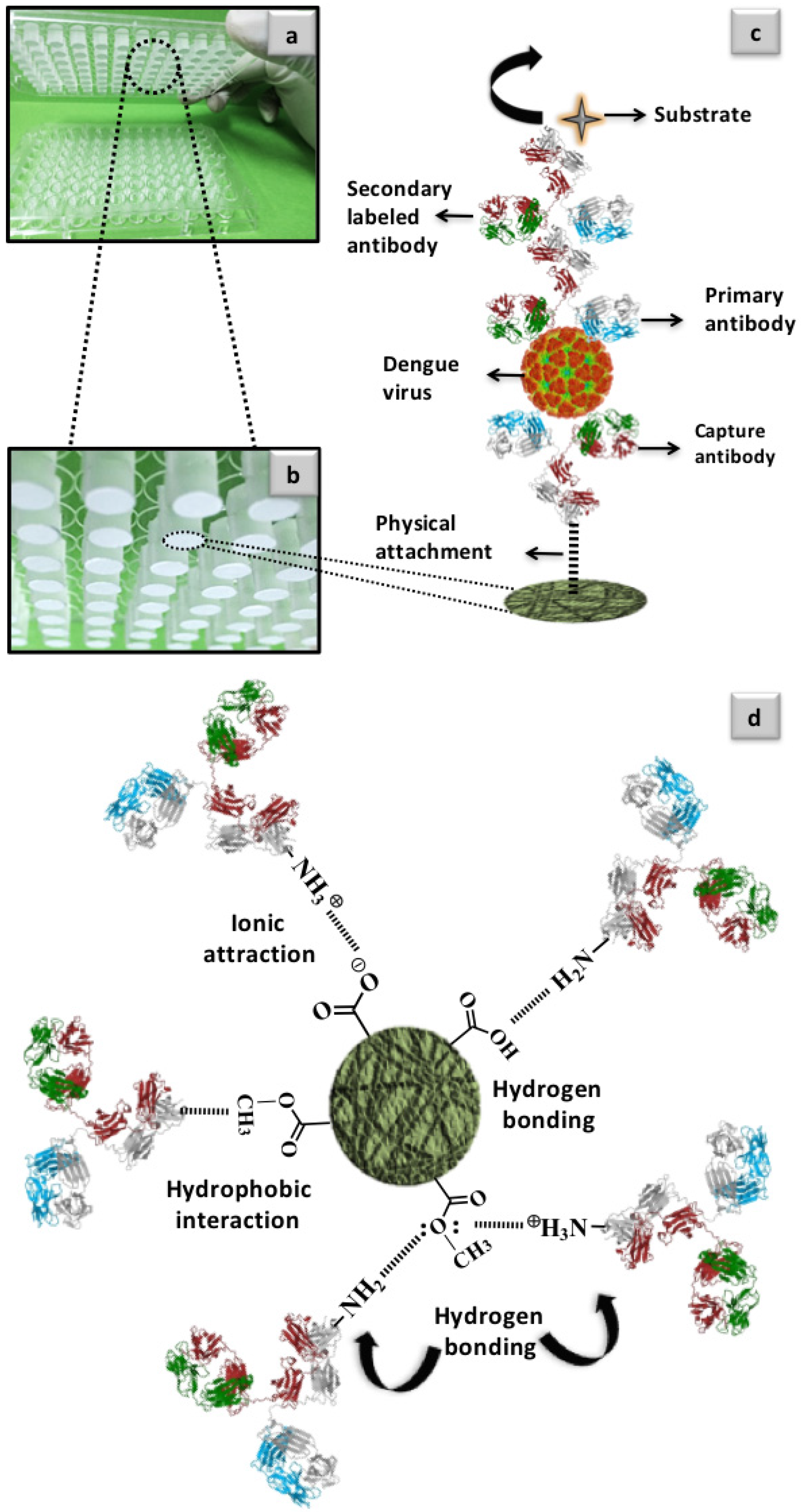
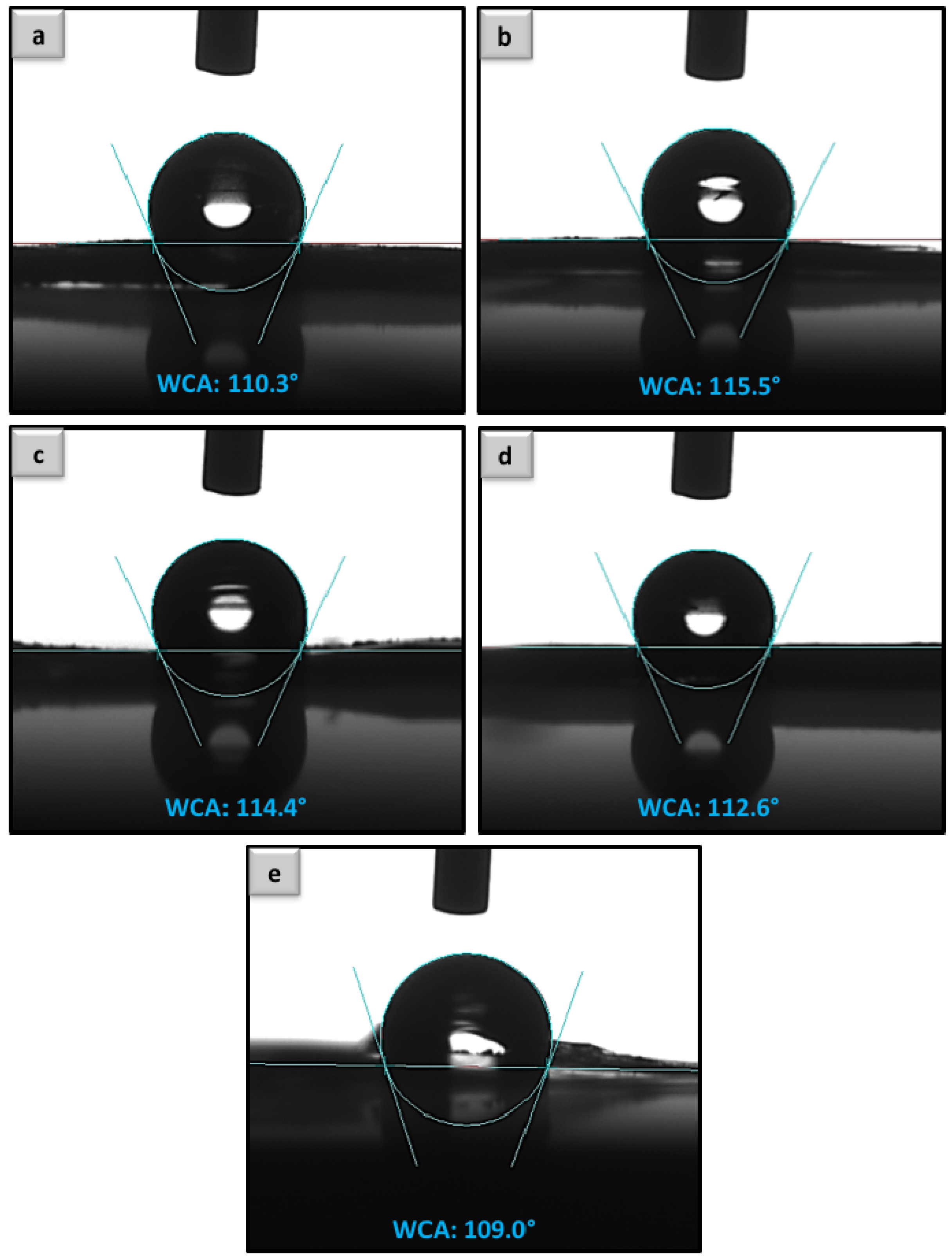
3.2. Contact Angle Analysis and the Fundamental Aspect of Physical Interaction between Protein Molecules and Poly Methyl Methacrylate-Co-Methacrylic (poly(MMA-co-MAA))-Coated Fibers

Surface wettability of the developed platforms was analyzed by WCA measurement and results are presented in Figure 3. The uncoated PHBV fiber mat has shown the average contact angle of ~110°, which is in agreement with the previously reported data [33]. PMMA coating has increased the hydrophobicity of electrospun PHBV fibers up to the contact angle of ~115°. As expected, WCA for the coated fibers has shown a gradual decrease in WCA as the ratio of MAA monomer in polymer compositions increased from Comp.(9:1) to Comp.(5:5). The deliberate alteration of MMA/MAA ratio in polymer backbone (Figure 1) was used to control surface concentration of –COOH groups, which in turn resulted in WCA presented in Figure 3a. It is important to note that we measured the WCA of PS (commercial ELISA well plate) to be ~60°, which is in agreement with the reported results [34]. This drastic difference between two platforms (i-ELISA and PS conventional well plate used as control) is expected to have a strong influence on detection sensitivity. General hydrophobicity of the developed fiber mats, in this study, has drawn a great deal of importance when analyte-surface interaction is concerned. However, the complexity of such heterogenic interactions involves both surface chemistry and morphology that must be taken under consideration [35].
Among several different forces that have an impact on biomolecular interfaces, three main interactions are known as the most influential driving forces in analyte-surface binding. The forces that have frequently been reported in the literature for their considerable impact on protein immobilization are as follows: electrostatic (ionic) attraction, hydrophobic interaction and hydrogen (H) bonding [24,36,37]. The ionic attraction is sensitive to pH changes and is relatively weak in comparison to other two forces while H-bonding facilitates the most substantial protein attachment [36]. In the present case, polymer-coated PHBV fibers create the platform for the mentioned physical forces to play their role in protein immobilization (Figure 4a,c). Figure 4d outlines the possible interactions that might occur between an Ab and a PHBV fiber mat coated with poly(MMA-co-MAA). While –COO groups can electrostatically bind –NH3+ groups (within appropriate pH range), the hydrophobic –COOCH3 groups of the surface promote the eminent hydrophobic force that bind Ab molecules to the fibrous surface [36]. At the same time, though in a completely different manner, available –COOH groups of the platforms can bind with –NH2 groups of the Ab molecules via H-bonding and provide an additional mean of interaction [36]. Abundant oxygen from methacrylate –COOCH3 groups might also contribute interfacial binding with amine groups of the Ab’s (Figure 4d). Looking into the chemical structures depicted in Figure 1, it can be considered that there is a major competition between hydrophobic interaction and H-bonding in attracting Abs to the surface. PMMA-coated PHBV platform has shown the highest WCA among all the developed platforms that, in turn, facilitates biomolecule immobilization via hydrophobic interaction. On the other hand, PHBV coated with PMMA is privileged with the abundance of oxygen atoms in its structure. This gives an overall electronegativity to the surface that highly promotes electrostatic interaction even though this force contributes in protein immobilization in a weaker manner [36].
In addition to hydrophobic character of all the coated samples (WCA~109°–115° for Comp.(5:5)-(9:1); Figure 4a) poly(MMA-co-MAA) macromolecules also contain pendant –COOH groups that can trap Ab molecules through H-bonding, which could strongly affect the sensitivity of the detection platform. The complexity of heterogeneous (or interfacial) macromolecular binding is extended further by taking under consideration the surface concentration of –COOH groups. For instance, overly functionalized surfaces (such as the case of Comp. (5:5) composition with the highest concentration of –COOH groups) could often cause relatively inefficient protein immobilization due to the steric repulsion or multiple surface binding [24,38]. In opposite cases when the concentration of surface functional groups is insufficient, the immobilized proteins could lose their activity due to close proximity to the solid platform. Overall, it is difficult to predict such phenomena and therefore any proposed method for biorecognition through heterogenic macromolecular interactions must be experimentally investigated and optimized in detail [23,37,39,40].
The application of cross-linkers for covalent immobilization of the proteins to the surface is often recommended; however, driven by our previous findings, we have selected the method of physical immobilization for this study [37]. Covalent protein immobilization is often performed by carbodiimide chemistry as the covalent attachment of the proteins to the surface is expected to be stronger than physical immobilization [41,42]. Nevertheless, it has been frequently reported that the covalent immobilization of the proteins is not always reproducible in comparison to the direct physical immobilization [24,37,41,43,44]. For instance, instead of producing highly reactive NHS-ester groups, under particular circumstances, carbodiimide treatment might result in formation of unreactive functionalities such as anhydride groups [41]. Apart from complexity, this particular method can also cause an early cross-linking inside the individual biomolecules that consequently makes the immobilization troublesome [44]. In our previous experiences, application of carbodiimide treatment resulted in repetitive and consistent drop in the analytical sensitivity, which is most likely caused by low selectivity [23,37,45]. Considering our experience with similar polymer systems, we have refined the straight-forward method of physical surface immobilization and subsequent detection of DENV.

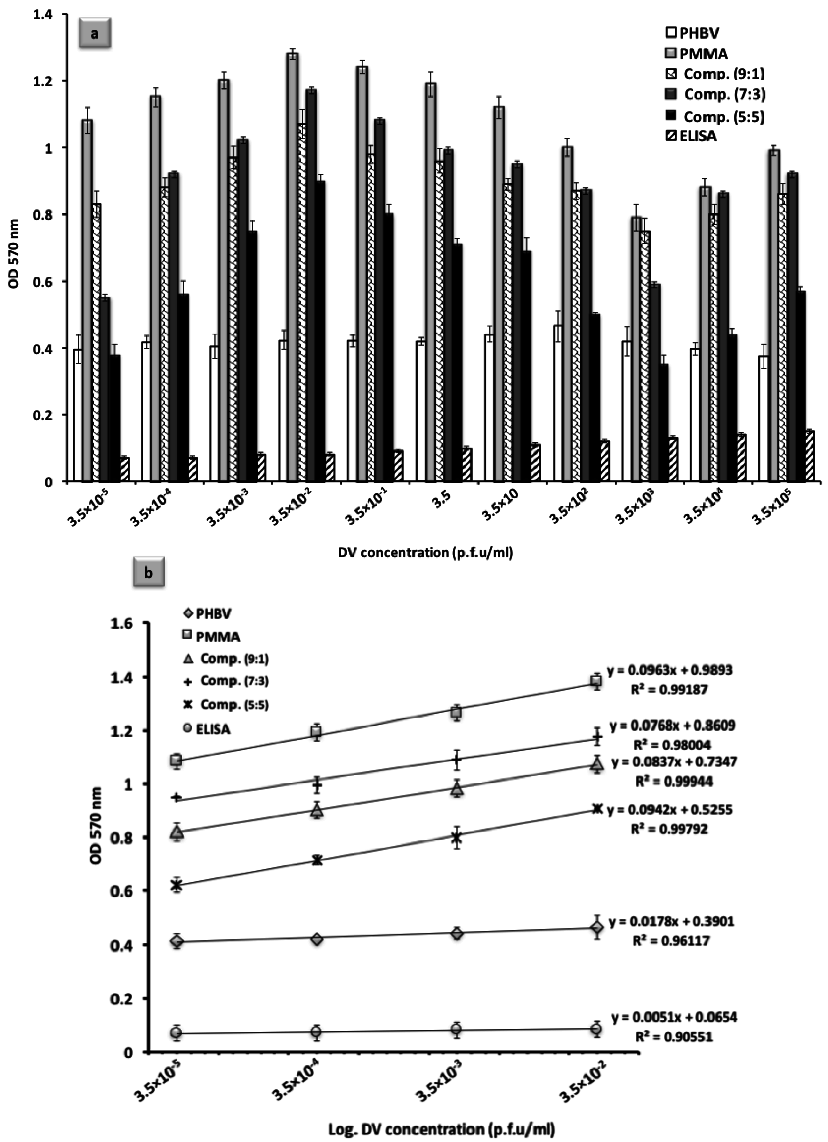
3.3. Analytical Method Optimization: Calibration and Evaluation of the Assay

A sandwich ELISA was carried out for a wide range of DENV concentration (3.5 × 10⁻⁵ p.f.u/mL to 3.5 × 10⁵ p.f.u/mL) in order to assess the performance of the developed platforms as a function of DENV concentration. Figure 5a, shows an initial increase of OD (optical density) values as a function of increasing concentrations of DENV. This increase in detection signal was limited to the certain threshold (3.5 × 10⁻² p.f.u/mL) from which the signal intensity decreased in the higher range of DENV concentrations. This phenomenon is most likely the result of the large size of biomolecules used in the assay as such macromolecules might lose their activity by denaturation caused by the steric repulsion [46]. Therefore, lower concentrations of biomolecules facilitate biomolecular interaction in a more efficient manner. Nonetheless, developed platforms have continuously generated the positive detection signals that are considerably greater than that of conventional ELISA assay.
Sandwich ELISA in different concentrations of DENV (3.5 × 10⁻⁵ p.f.u/mL to 3.5 × 10⁻² p.f.u/mL) was performed in order to carefully calibrate the assay. In a general observation from the plotted calibration curves in Figure 5b, it is evident that the higher level of confidence was achieved for the generated detection signals by coated electrospun fibers in comparison to the uncoated fibers or the conventional ELISA. Among coated PHBV fibers with different compositions of the polymer, the highest correlation coefficient value (R2 = 0.9994) was obtained for the coated fibers with Comp.(9:1). While coated fibers with PMMA and Comp.(5:5) have also shown comparative linearity, both compositions refer to the higher slopes for their respective calibrations plots than the rest. Coated fibers, in general, have proven to have a higher level of performance compared to the pure PHBV fibers and the actual ELISA, regardless of the polymer composition used for the coating. Even uncoated PHBV fibers have shown greater R2 value (0.9612) than ELISA, which confirms that the conventional analytical assay requires considerable improvements when it operates in lower concentrations of the target analyte.
When the assay is conducted in the absence of target analyte, accurate analytical systems are likely to generate minor or no positive detection signal, as the biomolecular chain that leads to generation of the final signal is intentionally broken. As one of the evaluation methods, non-specific bindings at deliberate negative and positive replicates can be quantitatively measured and compared to the true negative and positive results thus establishing sensitivity and specificity of the assay [22]. In our investigation, a total number of 72 positive and 24 negative controls from an overall of 96 replicates were examined to assess the performance of each individual uncoated/coated fiber mats in the sandwich assay. Table 1 represents the evaluation parameters for the assays conducted with coated and uncoated PHBV fibers. Proposed fiber-based platforms in this study exhibited an average sensitivity of 97.49%, which was greatly improved in contrast to the clinical method, ELISA (76.39%, Table 1). Specificity of the proposed platforms was also found to be in a higher range than the clinical assay (Table 1, average 90.83%). Additional data analysis demonstrated significantly higher level of accuracy (average 95.83%) for the developed platforms in comparison to the conventional ELISA (77%). Table 1 also provides the calculated LoD values for proposed fiber mats. Developed fiber platforms have proven to be capable of detecting only few plaque-forming units of DENV in blood serum. In contrast, minimum DENV concentration detectable by conventional ELISA was found to be 5 × 10³ p.f.u/mL, which is classified as a relatively late phase in DF (day 4–6 from the onset of the fever) [15,24,37,47,48]. The ability of the proposed platforms in detecting only few plaque-forming units of the target analyte is of a crucial importance as future developments of such methodology can shift the detection time towards considerably earlier stages of DF for improved dengue surveillance.

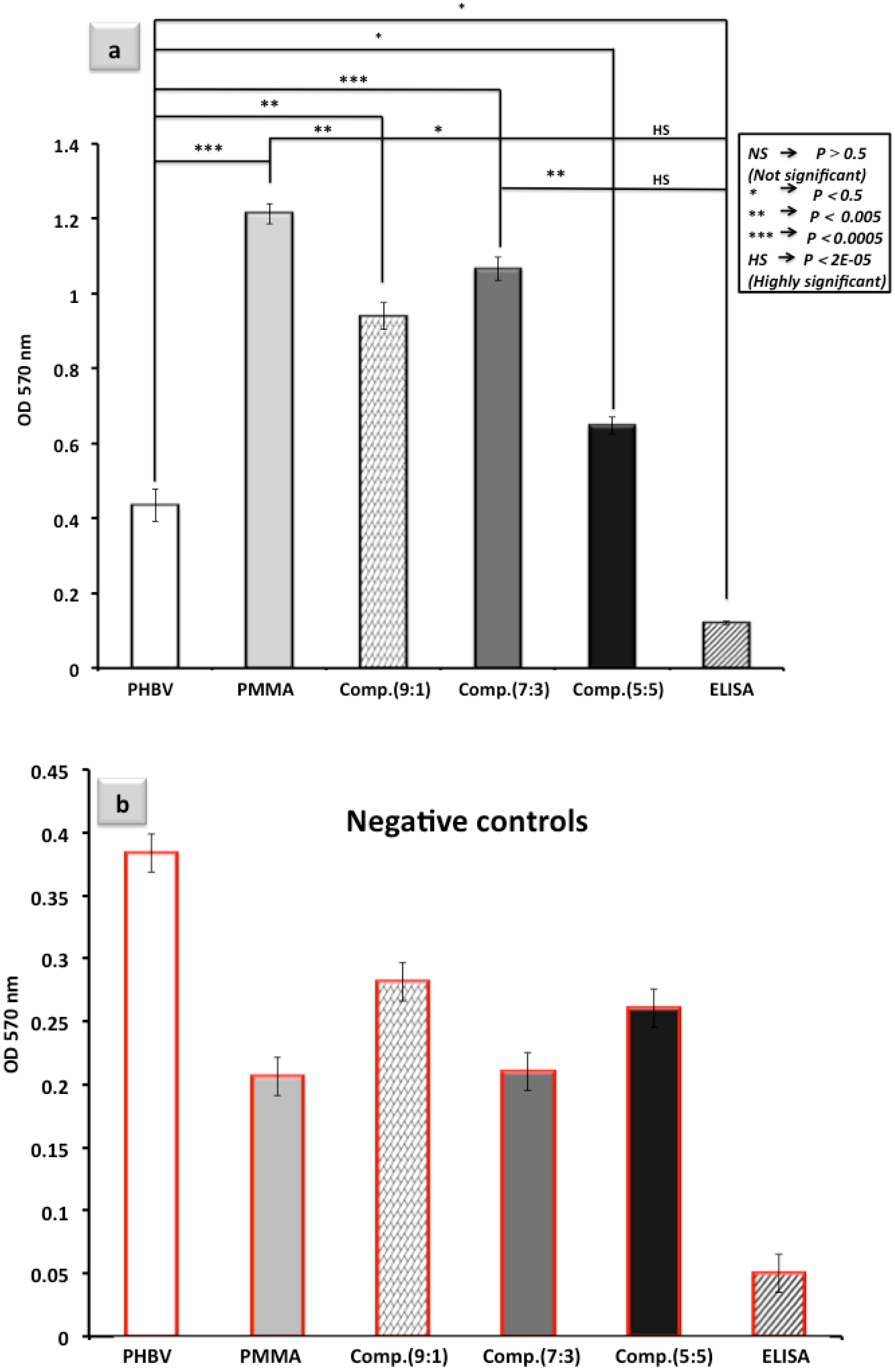
3.4. Detection Performance as a Function of Poly(MMA-co-MAA) Polymer Coating Composition

In order to compare the performance of uncoated and coated PHBV fiber mats in the actual virus detection, colorimetric sandwich ELISA assay was performed to detect DENV as the target analyte. Figure 6 summarizes the detection results into two plots: detection signal intensities and negative controls (Figure 6a,b respectively). For clear comparison with the clinical standard method, ELISA was performed (in PS as a standard analytical well plate) as control. In order to perform substantial number of trials, the standard deviations in each set of the experiments are the results of 12 successive replicates. The fiber segments of i-ELISA were observed to be highly stable throughout several steps of incubation. As can be observed from Figure 6a, resultant optical density (OD) values are in the range of 0.4–1.2, which is generally in a greater range than previously reported values for DENV detection via conventional ELISA [14,15,17].
Although pure PHBV fibers have generated slightly higher detection signals than a conventional assay, it was observed that PHBV fibers (without polymer coating) resulted in considerable false positive signals when the assay was performed in the absence of DENV, as presented in Figure 6b. This result can be considered as direct evidence which proves that hydrophobicity of the PHBV fibers does not necessarily protect the surface from the risk of buffer penetration. In a sandwich assay, if primary Abs are entrapped inside the structure of the material, they would not be thoroughly eliminated in the following washing steps of the assay. Entangled primary Abs could couple with the secondary labeled Abs in the next step, thus leading to the false positive signal as can be seen in Figure 6b. Therefore, tuning the pore size, distribution and hydrophobicity of the bioreceptor surface highly influences the performance of the platforms in the assay. PMMA-coated PHBV fibers resulted in the greatest level of detection among all the coated samples. The next in line was coated fiber with Comp.(7:3) which has generated OD values comparable to that of PMMA-coated surfaces. Statistical analysis presented in Figure 6a shows an overall higher performances of the developed platforms in comparison to the conventional ELISA. In particular, from the presented results it can be observed that the coated PHBV fibers with PMMA and Comp.(7:3) offered significant detection sensitivity in comparison to the rest of the coating polymer compositions and the conventional assay (Figure 6a). Considering the major forces that influence the analyte-surface attraction (discussed in Section 3.2), the experimental results in Figure 6 indicate that the predominant force is hydrophobic interaction. However, relatively high OD intensities recorded for poly(MMA-co-MAA) coatings with moderate concentrations of –COOH groups (Comp.(9:1) and (7:3); Figure 6) could be related to H-binding between coatings and Ab molecules. In summary, the results presented here do not only justify this novel and highly effective analytical method for early detection of DENV but they also provide the insight into physical protein-surface, nano-scale interactions, an important aspect for future development of sensitive biosensor platforms. In addition, electrospinning technique could be optimized to produce sub-micron polymer fibers [49], which could increase the level of sensitivity of fibrous platform.
Although there are several different techniques for regeneration of the analytical platforms such as treatment with strong oxidizer, acid/base components and/or concentrated salts [50,51], reusability of the developed platforms is not debated. Since i-ELISA can be fabricated through an inexpensive and straightforward method of molding, the device can be considered as a disposable analytical platform. Therefore, it seems highly possible that i-ELISA could pave the way to the future of the electrospun fiber mat-assisted analytical well plates [50]. The fact that our novel design can be applicable for integration of several different types of paper and/or fiber products into the assay endorses such a platform as a fairly unique development. One of the most important aspects in development of novel bio-sensing platforms is the shelf life of the bioreceptor surfaces. In that perspective, stability of the surface functionalities is highly important. Conventionally modified bioreceptor surfaces often lose their activities after a specific period of time due to the aging effect or reorientation phenomenon. Generated functional groups on the surface tend to reorganize themselves in order to occupy lower levels of energy, thereby resulting in deactivation of the modified surfaces. In the present case, available surface functionalities were formed through free-radical polymerization reaction. For that reason, the surface functional groups (–COOH) are an inherent part of the chemical structure and are not expected to rearrange with time [37]. Additionally, polymer-coated electrospun PHBV fibers have shown great stability throughout the time-consuming and tedious sandwich ELISA, which involves a number of steps with several incubations.
In our previous attempt at fabrication of electrospun fiber mat-based platforms, we used PHB electrospun fibers as the supporting material for dip coating [24]. The same strategy was applied for fabrication of coated fiber mats by using PHBV as the supporting material in this study. The reason for this alteration was due to the specific characteristics of the PHBV material itself. PHBV was found to possess a level of robustness in the time-consuming and laborious sandwich ELISA experiments. Additionally, PHBV is more electronegative than PHB due to the presence of oxygen atoms in its chemical structure. This specific characteristic of PHBV promotes biomolecular interaction through known forces of electrostatic interaction as well as H-bonding. For all the mentioned reasons, the performance of PHBV-coated fiber mats was superior as the intensity of detection signal was measured to be six times greater in comparison to PHB [24].

3.5. Advantages of i-ELISA in Comparison to the Competing Technologies

Although a variety of different techniques such as printing, patterning, waxing, heating, etc. have been used in fabrication of paper-based analytical devices, such platforms suffer from lack of control over the properties of the paper itself [2]. Paper segments in most of these platforms are chosen from commercially available cellulose products. Known as a hydrophilic material, NC possess WCA of 21° to 67°, depending on the type of the paper and the specific manufacturer [52]. This level of hydrophilicity is not favorable for protein immobilization as it may induce protein entrapment and subsequently lead to the false positive signal. This phenomenon, to a large extent, is one of the responsible factors for low sensitivity of the paper-based analytical devices. Moreover, NC contains –OH functional groups in its structure. Different treatment techniques have been employed to modify the surface of NC by converting less reactive –OH groups to the more reactive –COOH groups [8,9,10,11]. Nonetheless, as it was discussed before, such surface modification techniques tend to compromise the characteristics of the NC papers while generated –COOH functionalities are not guaranteed to last long. Furthermore, current paper-based platforms mainly rely on the visual readout as opposed to the analytical signal from the optical density or fluorescence measurement that corresponds to the exact concentration of the target analyte in blood serum. Semi-quantitative analysis with such devices is often carried out using tablets, smart phones and/or digital cameras [6]. Therefore, almost none of these paper-based techniques are comparable to the “gold standard” method (ELISA) that has been used for more than three decades in laboratories and hospitals. One of the justifications to accept those semi-quantitative methods is that such platforms have shown reliability in remote/rural areas and resource-limited environments that are in major need of such extreme point-of-care (EPOC) devices. Our strategy is to provide more sensitive analytical results even for EPOC applications by replacing the paper strips with more controllable polymer fiber mats. I-ELISA can successfully address a number of drawbacks from the previous paper-based platforms:
  • I-ELISA is the first developed electrospun fiber mat-assisted analytical well plate that strictly follows the principles of the standard ELISA method.
  • I-ELISA well plates can be fabricated by a slight modification in the current well plate by the injection molding process. Therefore, our platform can be highly cost-effective in comparison to the vying products.
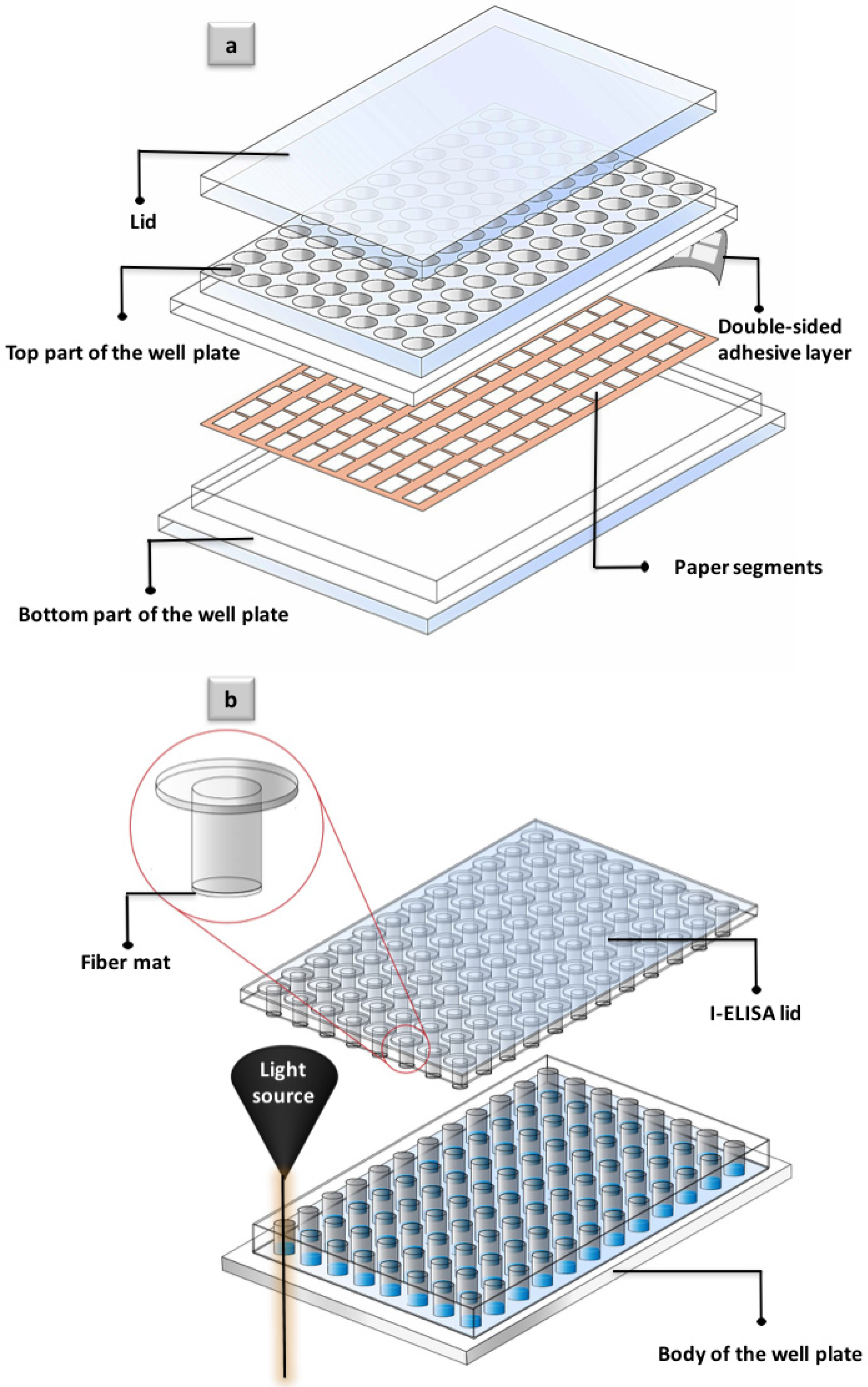
  • Unlike previously developed paper-based well plates, which are often composed of several different layers that need to be assembled before the application (Figure 7a), i-ELISA consists of only two parts (lid and body of the well plate, Figure 7b), in the same fashion as commercially available analytical well plates. This fact to a considerable extent makes i-ELISA practical as the proper installation of the multilayer platforms is often beyond expertise of the lab technicians.
  • I-ELISA can be used as a universal approach through which a wide variety of fiber/paper products with their special properties and functionalities can be introduced to the assay.
  • As a unique platform, i-ELISA can offer the visual detection judgment as the white color of the paper makes the perfect background for colorimetric detection. More importantly, i-ELISA enables routine quantification of the exact signal intensity, as the bottom part of the well plate stays intact for a regular colorimetric readout (Figure 7b).
  • I-ELISA is privileged with a great stability of the fiber parts as well as the well plate itself. In most of the paper-based well plates, the paper segments are printed or sprayed at the bottom of the well plate [2]. Printed sections are highly sensitive and are normally damaged through the numerous pipetting steps that are performed in a typical assay. In the present prototype, however, the paper part is barely touched throughout the pipetting procedure and thus remains whole (Figure 7b).
  • Unlike previous nitrocellulose NC-based analytical platforms, polymer-coated electrospun PHBV is a customized fiber material that can possess tailored surface properties, tuned wettability and specific morphological characteristics. Fiber segments of i-ELISA not only offer desirable surface characteristics but also are benefited with inherent –COOH functional groups that make the application of surface modification techniques needless.

4. Conclusions

This study presents fabrication and processing of a novel electrospun fiber mat-assisted analytical well plate, i-ELISA. Associated fiber segments in this platform are a novel class of material made from electrospun poly(3-hydroxybutyrate-co-3-hydroxyvalerate) (PHBV) fibers coated with different compositions of poly methyl methacrylate-co-methacrylic (poly(MMA-co-MAA)) synthesized in free-radical polymerization reaction. The developed well plate has successfully addressed some of the major drawbacks of the previous paper-based analytical platforms. I-ELISA was tested for diagnosis of dengue virus via physical immobilization of the proteins by using a sandwich colorimetric assay. Proposed technology has proven to greatly enhance the detection signal intensity up to 12 times greater than the conventional assay, while the limit of detection (LoD) values reveal a significant improvement (~500 times in the case of PMMA-coated electrospun PHBV fibers) in comparison to the conventional clinical assay. I-ELISA has also shown a considerably higher level of sensitivity, specificity and accuracy in comparison to the clinically approved ELISA. In particular, i-ELISA was capable of detecting only few plaque-forming units of dengue virus in blood serum, which corresponds to the primary stage of dengue infection. Development of i-ELISA can open new opportunities to the future production of cost-effective, disposable and reliable diagnostic tools that can be utilized for enhanced detection of viruses, not only limited to dengue virus as presented in this study. The proposed strategy described in this paper can be applied for fabrication of a new generation of analytical well plates that can incorporate a wide variety of customized or commercial fiber/paper products into the actual analytical assay. Developed fiber segments of i-ELISA, in turn, have shown great potential for integration into the microfluidic devices or other fiber-based analytical platforms.

Acknowledgments

The authors would like to acknowledge the financial support of Tecnologico de Monterrey, Mexico, for the special grant (grant number: 002EICII01) awarded to the Sensors and Devices Focus Group, School of Engineering and Sciences, Tecnologico de Monterrey, Monterrey, Mexico. This research has been partly supported by University Malaya Research Grant (grant number: TR001D-2014B). The authors also acknowledge the partial support of High Impact Research Grant (grant number: UM.C/625/1/HIR/MOE/SC/33/1) provided by Ministry of Higher Education, Malaysia. The authors would like to thank Amir Hossein Hosseini for his contribution in the graphical designs.

Author Contributions

P.A. fabricated the electrospun fibers while S.H. analyzed the fiber materials and conducted the immunoassay for detection of dengue virus. M.M.A contributed to fabrication of intrant ELISA while H.A.R. isolated and propagated dengue virus for further experiments. I.D. supervised the project in the case of sample analysis and detection. S.O.M.C. guided researchers through the experimental and writing processes while M.J.M. provided the groups with the scientific insights regarding the fundamentals and application of the new strategy. All the authors have read and corrected the manuscript.

Conflicts of Interest

The authors declare no conflict of interest.

Appendix A. Supplementary Section

Fabrication of Electrospun Poly(3-hydroxybutyrate-co-3-hydroxyvalerate) (PHBV) Fibers

The PHBV fiber mats were fabricated by far-field electrospinning (FFES) technique in the following steps:
  • 20 mg of PHBV was dissolved (10 wt %) in chloroform/ dimethylformamide (DMF) (9:1) and ejected from a 20 G needle (inner diameter = 0.9 mm) at the speed of 3 mL/h.
  • A voltage of 10 kV was loaded between the needle and an aluminum plate (40 cm × 40 cm), which was the target for fiber deposition. The distance between the needle and the aluminum plate was set to be 18 cm.
  • The polymer solutions were drawn into the PHBV fibers and were deposited on the aluminum plate. The sheet of PHBV electrospun fiber was then peeled off from the aluminum plate.

Synthesis and Processing of Poly Methyl Methacrylate-Co-Methacrylic (poly(MMA-co-MAA)) Compositions

Four different compositions of poly(MMA-co-MAA) have been synthesized via free-radical polymerization reaction with azobisisobutyronitrile (AIBN, Friedemann Chemical, Cologne, Germany) as an initiator and in tetrahydrofuran THF (Thermo Fisher Scientific, Waltham, MA, USA) as the solvent. Methyl methacrylate (MMA) and methacrylic acid (MAA) monomers were purchased from Sigma, San Antonio, TX, USA. MMA monomer was distilled prior to application in polymerization reaction. The abbreviation for each composition indicates the initial molar ratio of the monomers. For example, poly(MMA-co-MAA-9:1) refers to 90% of MMA and 10% of MAA in polymerization reaction mixture. Other copolymer compositions are: poly(MMA-co-MAA-7:3) and poly(MMA-co-MAA-5:5). For ease of discussion, the mentioned copolymer compositions are referred to as Comp.(9:1), Comp.(7:3) and Comp.(5:5). When MMA is the sole monomer in the polymerization reaction, the result of the synthesis is PMMA (100% of MMA and 0% of MAA). The pure PMMA was also polymerized under the exact same reaction condition. A two-neck round-bottom flask was fixed with a condenser, sealed inlet and used for reactants feeding. The round-bottom flask was charged with a pre-calculated amount of MMA monomer in 50 mL of THF under stirring condition for 5 min. A mixture of MAA monomer and initiator (AIBN, 0.164 g) was slowly added to the reaction mixture. Polymerization reaction was carried out for 8 h at 90 °C and was eventually stopped by adding reaction mixture into 1 L of distilled water. Immediate precipitation of the white color polymer was observed at this stage that was a clear confirmation on the formation of copolymer compositions. White-colored precipitations were filtered, thoroughly washed with distilled water and dried in vacuum oven at 40 °C for further experiments.

Dip-Coating Procedure

The electrospun PHBV fiber mats were coated by the straightforward method of dip-coating. Substrates (PHBV fibers) were coated by immersing fiber sheets into the different polymer solutions (5% in THF). Fiber sheets were taken out from the polymer solutions after 3 s of dipping and allowed to dry at room temperature. The paper-like polymer-coated PHBV sheets with the thickness of ~500 µm were cut into the circle-shaped pieces (diameter = 6 mm) for fixing onto the intrant ELISA system.

Dengue Virus Propagation in Mosquito Cells and Titration

A clinical isolate of dengue serotype 2 from a patient’s serum sample (DV2-isolate Malaysia M2, Gen Bank Toxonomy No.: 11062) was used for virus propagation by a single passage on C6/36 mosquito cells. The dengue-infected cells with obvious cytopathic effects (CPE) were lysed by freeze and thaw cycle. The culture medium was centrifuged at 1800 rpm for 10 mins to remove cell debris, filtered (0.2 µm), portioned into aliquots, and stored at −80 °C until used. The viral titer of the dengue suspension was established by serial dilutions on Vero cells using plaque assay. In brief, a 10-fold serial dilution of medium supernatant was added to new Vero cells grown in a 24-well plate (1.5 × 10⁵ cells) and incubated for 1 h at 37 °C. The cells were then overlaid with Dulbecco’s modified Eagle’s medium containing 1.1% methylcellulose. Viral plaques were stained with naphthol blue-black dye after 5 days of incubation. Virus titers were calculated according to the following formula: titer (p.f.u/mL) = number of plaques/volume of diluted virus added to the well × dilution factor of the virus used to infect the well in which plaques were enumerated. The final dengue virus titer that was used in serial dilutions had the concentration of 3.5 × 10¹⁰ p.f.u/mL.

Sandwich ELISA Procedure

Step 1. Each well of i-ELISA was filled with 100 µL of capture antibody (Ab), rabbit anti-dengue virus 2 antibody (ab155042, Abcam, San Francisco, CA, USA) diluted (1:500) in coating buffer. Coating buffer was prepared by mixing 0.85 g of sodium chloride (NaCl, Sigma, San Antonio, TX, USA), 0.14 g of disodium phosphate (Na2HPO4, Sigma, San Antonio, TX, USA), and 0.02 g of monosodium phosphate (NaH2PO4, Sigma, San Antonio, TX, USA) in 100 mL of phosphate-buffered saline (PBS, Fisher Scientific, Waltham, MA, USA) with the pH of 7.4. Incubation was carried out for 2 h at 37 °C.
Step 2. Washing step was performed by charging each well with 200 µL of washing buffer which consists of 0.05% Tween 20 (Sigma, San Antonio, TX, USA) in PBS solution (pH = 7.4). This step was performed at room temperature three times (each time 5 min) by using a shaker with the shaking speed of 1000 rpm. The exact same process was performed between each two steps of the sandwich assay. It is important to note that the lid was placed on top of well plate not only during the washing steps but also throughout all different steps of the sandwich ELISA. Therefore, paper segments of i-ELISA have been directly in contact with the respective buffers in the conducted incubations.
Step 3. The blocking step was performed in order to achieve high selectivity and to avoid non-specific bindings. This step was conducted by charging each well with 100 µL of blocking buffer and incubating well plates at 37 °C for 1 h. Blocking buffer was prepared by adding 1 g of bovine serum albumin (BSA, Sigma, San Antonio, TX, USA) to 100 mL of washing buffer. Blocking buffer used in the assay had a controlled pH of 7.4.
Step 4. Each well of i-ELISA was charged with 100 µL of the diluted virus solution and incubation lasted for 2 h at 37 °C. Virus solution was prepared by the serial dilution method in a variety of different concentrations depending on the application of the assay.
Step 5. Primary Ab solution was prepared by diluting mouse IgG2a anti DV (ab155863, Abcam, San Francisco, CA, USA) in diluting buffer (DB) with the ratio of 1:200 (Ab:DB). Diluting buffer was made by adding 0.4 g of BSA, 4 mL of PBS buffer and 120 µL of Trintonx-100 into 36 mL of distilled water at the pH of 7.4. Each well received 100 µL of primary Ab solution and well plates were placed in the incubator of 37 °C for 2 h.
Step 6. The last incubation was carried out by adding 100 µL of secondary antibody solution that was also diluted in diluting buffer (1:500). Anti-mouse igG2a alkaline phosphatase (ab97242, Abcam, San Francisco, CA, USA) was used as the secondary Ab. This step was performed at the temperature of 37 °C for 30 min.
Step 7. In the final step, wells were thoroughly washed and charged with 100 µL of mixed substrate (alkaline phosphatase blue microwell substrate, components A and B). This reaction was conducted at ambient temperature and stopped after 15 min by adding 100 µL of alkaline phosphatase stopping solution (A585, Sigma, San Antonio, TX, USA). Signal intensity was recorded by using Bio-Rad ELISA reader (model 680) at the specific wavelength of 570 nm as the manufacturing guideline has suggested.

Evaluation of the Assay

Calculation of sensitivity and specificity was done by using the equations below:
Sensitivity   =   T P T P + F N
Specificity   =   T N T N + F P
Variables in these equations are as follows [27]:
  • True positive (TP)
  • True negative (TN)
  • False positive (FP)
  • False negative (FN)
The accuracy of the assay was calculated by using true negative and positive readings in comparison to the total number of replicates using the equation below [22]:
Accuracy =   T P   +   T N Total   replicates
Limit of detection (LoD) for each separate platform was calculated from the standard deviations in the case of minimum DENV concentration (s) and slopes of the corresponding calibration plot (m) following the equation below.
LoD =   3   ×   s m

Scanning Electron Microscopy (SEM) Analysis of the Uncoated and Coated PHBV Electrospun Fibers

Figure A1 represents the morphology analysis of the uncoated and coated electrospun PHBV fiber mats. The polymer-coated sections of the platform which occupy the voids between the electrospun fibers can be clearly seen in Figure A1b–e.
Figure A1. SEM (scanning electron microscopy) images of electrospun and coated fibers: (a) 3-hydroxybutyric acid-co-3-hydroxyvaleric acid (PHBV); (b) poly(methyl methacrylate) (PMMA); (c) Comp.(9:1); (d) Comp.(7:3); and (e) Comp.(5:5).
Applsci 06 00336 g008

Water-in-Air Contact Angle Analysis of the Uncoated and Coated PHBV Electrospun Fibers

Figure A2 represents images of the contact angles between droplets of water and coated/uncoated surfaces of the PHBV fiber mats. An increase in contact angle from uncoated (Figure A2a) to PMMA-coated PHBV fiber (Figure A2b) was followed by a gradual decrease in contact angles (Figure A2c–e). This gradual decrease in contact angle corresponds to the increase in concentration of –COOH groups, which, in turn, is the function of molar ratio of MAA monomers in the polymerization reaction.
Figure A2. Representative water-in-air contact angle droplet images: (a) uncoated PHBV; (b) PMMA; (c) Comp.(9:1); (d) Comp.(7:3); and (e) Comp.(5:5).
Applsci 06 00336 g009

References

  1. Carvalhal, R.F.; Simão Kfouri, M.; De Oliveira Piazetta, M.H.; Gobbi, A.L.; Kubota, L.T. Electrochemical detection in a paper-based separation device. Anal. Chem. 2010, 82, 1162–1165. [Google Scholar] [CrossRef] [PubMed]
  2. Ahmed, S.; Bui, M.-P.N.; Abbas, A. Paper-based chemical and biological sensors: Engineering aspects. Biosens. Bioelectron. 2016, 77, 249–263. [Google Scholar] [CrossRef] [PubMed]
  3. Yeow, N.; McLiesh, H.; Guan, L.; Shen, W.; Garnier, G. Paper-based assay for red blood cell antigen typing by the indirect antiglobulin test. Anal. Bioanal. Chem. 2016, 408, 5231–5238. [Google Scholar] [CrossRef] [PubMed]
  4. Farahmand, E.; Ibrahim, F.; Hosseini, S.; Rothan, H.A.; Yusof, R.; Koole, L.H.; Djordjevic, I. A novel approach for application of nylon membranes in the biosensing domain. Appl. Surf. Sci. 2015, 353, 1310–1319. [Google Scholar] [CrossRef]
  5. Petryayeva, E.; Algar, W.R. Proteolytic assays on quantum-dot-modified paper substrates using simple optical readout platforms. Anal. Chem. 2013, 85, 8817–8825. [Google Scholar] [CrossRef] [PubMed]
  6. Liana, D.D.; Raguse, B.; Gooding, J.J.; Chow, E. Recent advances in paper-based sensors. Sensors 2012, 12, 11505–11526. [Google Scholar] [CrossRef] [PubMed]
  7. Ge, L.; Wang, P.; Ge, S.; Li, N.; Yu, J.; Yan, M.; Huang, J. Photoelectrochemical Lab-on-Paper Device Based on an Integrated Paper Supercapacitor and Internal Light Source. Anal. Chem. 2013, 85, 3961–3970. [Google Scholar] [CrossRef] [PubMed]
  8. Karra-Châabouni, M.; Bouaziz, I.; Boufi, S.; do Rego, A.M.B.; Gargouri, Y. Physical immobilization of Rhizopus oryzae lipase onto cellulose substrate: activity and stability studies. Colloids Surf. B Biointerfaces 2008, 66, 168–177. [Google Scholar] [CrossRef] [PubMed]
  9. Ibrahim, M.M.; Koschella, A.; Kadry, G.; Heinze, T. Evaluation of cellulose and carboxymethyl cellulose/poly (vinyl alcohol) membranes. Carbohydr. Polym. 2013, 95, 414–420. [Google Scholar] [CrossRef] [PubMed]
  10. Tyagi, C.; Tomar, L.K.; Singh, H. Surface modification of cellulose filter paper by glycidyl methacrylate grafting for biomolecule immobilization: Influence of grafting parameters and urease immobilization. J. Appl. Polym. Sci. 2009, 111, 1381–1390. [Google Scholar] [CrossRef]
  11. Isaad, J.; El Achari, A. Colorimetric sensing of cyanide anions in aqueous media based on functional surface modification of natural cellulose materials. Tetrahedron 2011, 67, 4939–4947. [Google Scholar] [CrossRef]
  12. Bessoff, K.; Delorey, M.; Sun, W.; Hunsperger, E. Comparison of two commercially available Dengue Virus (DENV) NS1 capture enzyme-linked immunosorbent assays using a single clinical sample for diagnosis of acute DENV infection. Clin. Vaccine Immunol. 2008, 15, 1513–1518. [Google Scholar] [CrossRef] [PubMed]
  13. Gubler, D.J. Epidemic dengue/dengue hemorrhagic fever as a public health, social and economic problem in the 21st century. Trends Microbiol. 2002, 10, 100–103. [Google Scholar] [CrossRef]
  14. Shu, P.-Y.; Chen, L.-K.; Chang, S.-F.; Yueh, Y.-Y.; Chow, L.; Chien, L.-J.; Lin, T.-H.; Huang, J.-H. Comparison of Capture Immunoglobulin M (IgM) and IgG Enzyme-Linked Immunosorbent Assay (ELISA) and Nonstructural Protein NS1 Serotype-Specific IgG ELISA for Differentiation of Primary and Secondary Dengue Virus Infections. Clin. Diagn. Lab. Immunol. 2003, 10, 622–630. [Google Scholar] [CrossRef] [PubMed]
  15. Alcon, S.; Talarmin, A.; Debruyne, M.; Falconar, A.; Deubel, V.; Flamand, M. Enzyme-Linked Immunosorbent Assay Specific to Dengue Virus Type 1 Nonstructural Protein NS1 Reveals Circulation of the Antigen in the Blood during the Acute Phase of Disease in Patients Experiencing Primary or Secondary Infections. J. Clin. Microbiol. 2002, 40, 376–381. [Google Scholar] [CrossRef] [PubMed]
  16. Stevens, A.J.; Gahan, M.E.; Mahalingam, S.; Keller, P.A. The medicinal chemistry of dengue fever. J. Med. Chem. 2009, 52, 7911–7926. [Google Scholar] [CrossRef] [PubMed]
  17. Xu, H.; Di, B.; Pan, Y.-X.; Qiu, L.-W.; Wang, Y.-D.; Hao, W.; He, L.-J.; Yuen, K.-Y.; Che, X.-Y. Serotype 1-Specific Monoclonal Antibody-Based Antigen Capture Immunoassay for Detection of Circulating Nonstructural Protein NS1: Implications for Early Diagnosis and Serotyping of Dengue Virus Infections. J. Clin. Microbiol. 2006, 44, 2872–2878. [Google Scholar] [CrossRef] [PubMed]
  18. Fibriansah, G.; Tan, J.L.; Smith, S.A.; de Alwis, R.; Ng, T.-S.; Kostyuchenko, V.A.; Jadi, R.S.; Kukkaro, P.; de Silva, A.M.; Crowe, J.E.; et al. A highly potent human antibody neutralizes dengue virus serotype 3 by binding across three surface proteins. Nat. Commun. 2015, 6. [Google Scholar] [CrossRef] [PubMed]
  19. Gurugama, P.; Garg, P.; Perera, J.; Wijewickrama, A.; Seneviratne, S. Dengue viral infections. Indian J. Dermatology 2010, 55, 68–78. [Google Scholar]
  20. Allwinn, R. Significant increase in travel-associated dengue fever in Germany. Med. Microbiol. Immunol. 2011, 200, 155–159. [Google Scholar] [CrossRef] [PubMed]
  21. Lapphra, K.; Sangcharaswichai, A.; Chokephaibulkit, K.; Tiengrim, S.; Piriyakarnsakul, W.; Chakorn, T.; Yoksan, S.; Wattanamongkolsil, L.; Thamlikitkul, V. Evaluation of an NS1 antigen detection for diagnosis of acute dengue infection in patients with acute febrile illness. Diagn. Microbiol. Infect. Dis. 2008, 60, 387–391. [Google Scholar] [CrossRef] [PubMed]
  22. Linares, E.M.; Pannuti, C.S.; Kubota, L.T.; Thalhammer, S. Immunospot assay based on fluorescent nanoparticles for Dengue fever detection. Biosens. Bioelectron. 2013, 41, 180–185. [Google Scholar] [CrossRef] [PubMed]
  23. Hosseini, S.; Ibrahim, F.; Djordjevic, I.; Rothan, H.A.; Yusof, R.; van der Mareld, C.; Koole, L.H. Synthesis and Processing of ELISA Polymer Substitute: The Influence of Surface Chemistry and Morphology on Detection Sensitivity. Appl. Surf. Sci. 2014, 317, 630–638. [Google Scholar] [CrossRef]
  24. Hosseini, S.; Azari, P.; Farahmand, E.; Gan, S.N.; Rothan, H.A.; Yusof, R.; Koole, L.H.; Djordjevic, I.; Ibrahim, F. Polymethacrylate coated electrospun PHB fibers: An exquisite outlook for fabrication of paper-based biosensors. Biosens. Bioelectron. 2015, 69, 257–264. [Google Scholar] [CrossRef] [PubMed]
  25. Hosseini, S.; Ibrahim, F.; Djordjevic, I.; Koole, L.H. Polymethyl methacrylate-co-methacrylic acid coatings with controllable concentration of surface carboxyl groups: A novel approach in fabrication of polymeric platforms for potential bio-diagnostic devices. Appl. Surf. Sci. 2014, 300, 43–50. [Google Scholar] [CrossRef]
  26. Reinholt, S.J.; Sonnenfeldt, A.; Naik, A.; Frey, M.W.; Baeumner, A.J. Developing new materials for paper-based diagnostics using electrospun nanofibers. Anal. Bioanal. Chem. 2014, 406, 3297–3304. [Google Scholar] [CrossRef] [PubMed]
  27. Hosseini, S.; Ibrahim, F. Current Optical Biosensors in Clinical Practice. Novel Polymeric Biochips for Enhanced Detection of Infectious Diseases; Springer: Berlin, Germany, 2016; pp. 1–12. [Google Scholar]
  28. Shrivastava, A.; Gupta, V.B. Methods for the determination of limit of detection and limit of quantitation of the analytical methods. Chron. Young Sci. 2011, 2, 21–25. [Google Scholar] [CrossRef]
  29. Swamy, N.; Prashanth, K.N.; Basavaiah, K. Titrimetric and spectrophotometric assay of diethylcarbamazine citrate in formulations using iodate and iodide mixture as reagents. Braz. J. Pharm. Sci. 2015, 51, 43–52. [Google Scholar] [CrossRef]
  30. Gad, M.; Zaazaa, H.; Amer, S.; Korany, M. Static headspace gas chromatographic method for the determination of residual solvents in cephalosporins. RSC Adv. 2015, 5, 17150–17159. [Google Scholar] [CrossRef]
  31. Bauer, S.M.; Gehringer, M.; Laufer, S.A. A direct enzyme-linked immunosorbent assay (ELISA) for the quantitative evaluation of Janus Kinase 3 (JAK3) inhibitors. Anal. Methods 2014, 6, 8817–8822. [Google Scholar] [CrossRef]
  32. Pereira, A.G.; D’Avila, F.B.; Ferreira, P.C.L.; Holler, M.G.; Limberger, R.P.; Froehlich, P.E. Determination of cocaine, its metabolites and pyrolytic products by LC-MS using a chemometric approach. Anal. Methods 2014, 6, 456–462. [Google Scholar] [CrossRef]
  33. Zhu, M.; Zuo, W.; Yu, H.; Yang, W.; Chen, Y. Superhydrophobic surface directly created by electrospinning based on hydrophilic material. J. Mater. Sci. 2006, 41, 3793–3797. [Google Scholar] [CrossRef]
  34. Yuan, Y.; Lee, T.R. Contact Angle and Wetting Properties. In Surface Science Techniques; Bracco, G., Holst, B., Eds.; Springer: Berlin, Germany, 2013; pp. 3–34. [Google Scholar]
  35. Ma, Z.; Kotaki, M.; Ramakrishna, S. Surface modified nonwoven polysulphone (PSU) fiber mesh by electrospinning: A novel affinity membrane. J. Membr. Sci. 2006, 272, 179–187. [Google Scholar] [CrossRef]
  36. Yoon, J.-Y.; Park, H.-Y.; Kim, J.-H.; Kim, W.-S. Adsorption of BSA on highly carboxylated microspheres—Quantitative effects of surface functional groups and interaction forces. J. Colloid Interface Sci. 1996, 177, 613–620. [Google Scholar] [CrossRef]
  37. Hosseini, S.; Ibrahim, F.; Rothan, H.A.; Yusof, R.; Marel, C.; Djordjevic, I.; Koole, LH. Aging effect and antibody immobilization on –COOH exposed surfaces designed for dengue virus detection. Biochem. Eng. J. 2015, 99, 183–192. [Google Scholar] [CrossRef]
  38. Hosseini, S.; Ibrahim, F. Application of Biochips in Dengue Virus Detection. In Novel Polymeric Biochips for Enhanced Detection of Infectious Diseases; Springer: Berlin, Germany, 2016; pp. 39–47. [Google Scholar]
  39. Bai, Y.; Koh, C.G.; Boreman, M.; Juang, Y.-J.; Tang, I.C.; Lee, L.J.; Yang, S.-T. Surface Modification for Enhancing Antibody Binding on Polymer-Based Microfluidic Device for Enzyme-Linked Immunosorbent Assay. Langmuir 2006, 22, 9458–9467. [Google Scholar] [CrossRef] [PubMed]
  40. Goddard, J.M.; Hotchkiss, J.H. Polymer surface modification for the attachment of bioactive compounds. Prog. Polym. Sci. 2007, 32, 698–725. [Google Scholar] [CrossRef]
  41. Sam, S.; Touahir, L.; Salvador Andresa, J.; Allongue, P.; Chazalviel, J.N.; Gouget-Laemmel, A.C.; Henry de Villeneuve, C.; Moraillon, A.; Ozanam, F.; Gabouze, N.; et al. Semiquantitative Study of the EDC/NHS Activation of Acid Terminal Groups at Modified Porous Silicon Surfaces. Langmuir 2009, 26, 809–814. [Google Scholar] [CrossRef] [PubMed]
  42. Federica Rusmini, Z.Z.; Feijen, J. Protein Immobilization Strategies for Protein Biochips. Biomacromolecules 2007, 8, 1775–1789. [Google Scholar] [CrossRef] [PubMed]
  43. Wang, C.; Yan, Q.; Liu, H.-B.; Zhou, X.-H.; Xiao, S.-J. Different EDC/NHS Activation Mechanisms between PAA and PMAA Brushes and the Following Amidation Reactions. Langmuir 2011, 27, 12058–12068. [Google Scholar] [CrossRef] [PubMed]
  44. Coad, B.R.; Jasieniak, M.; Griesser, S.S.; Griesser, H.J. Controlled covalent surface immobilisation of proteins and peptides using plasma methods. Surf. Coat Technol. 2013, 233, 169–177. [Google Scholar] [CrossRef]
  45. Hosseini, S.; Ibrahim, F.; Djordjevic, I.; Rothan, H.A.; Yusof, R.; Marel, C.; Benzina, A.; Koole, LH. Synthesis and characterization of methacrylic microspheres for biomolecular recognition: Ultrasensitive biosensor for dengue virus detection. Eur. Polym. J. 2014, 60, 14–21. [Google Scholar] [CrossRef]
  46. Peissker, T.; Deschaume, O.; Rand, D.R.; Boyen, H.-G.; Conard, T.; Van Bael, M.J.; Bartic, C. Selective Protein Immobilization onto Gold Nanoparticles Deposited under Vacuum on a Protein-Repellent Self-Assembled Monolayer. Langmuir 2013, 29, 15328–15335. [Google Scholar] [CrossRef] [PubMed]
  47. Thomas, L.; Najioullah, F.; Verlaeten, O.; Martial, J.; Brichler, S.; Kaidomar, S.; Moravie, V.; Cabié, A.; Césaire, R. Relationship between Nonstructural Protein 1 Detection and Plasma Virus Load in Dengue Patients. Am. J. Trop. Med. Hyg. 2010, 83, 696–699. [Google Scholar] [CrossRef] [PubMed]
  48. Hosseini, S.; Aeinehvand, M.M.; Uddin, S.M.; Benzina, A.; Rothan, H.A.; Yusof, R.; Koole, L.H.; Madou, M.J.; Djordjevic, I.; Ibrahim, F. Microsphere Integrated Microfluidic Disk: Synergy of Two Techniques for Rapid and Ultrasensitive Dengue Detection; NCBI: Bethesda, MD, USA, May 2015.
  49. Pan, Y.-J.; Lin, J.-H.; Chiang, K.-C. Biomedical Applications of Antibacterial Nanofiber Mats Made of Electrospinning with Wire Electrodes. Appl. Sci. 2016, 6. [Google Scholar] [CrossRef]
  50. Kim, D.; Herr, A.E. Protein immobilization techniques for microfluidic assays. Biomicrofluidics 2013, 7. [Google Scholar] [CrossRef] [PubMed]
  51. Dalavoy, T.S.; Wernette, D.P.; Gong, M.; Sweedler, J.V.; Lu, Y.; Flachsbart, B.R.; Shannon, M.A.; Bohn, P.W.; Cropek, D.M. Immobilization of DNAzyme catalytic beacons on PMMA for Pb2+ detection. Lab Chip 2008, 8, 786–793. [Google Scholar] [CrossRef] [PubMed]
  52. Li, Y.; Pham, J.Q.; Johnston, K.P.; Green, P.F. Contact Angle of Water on Polystyrene Thin Films: Effects of CO2 Environment and Film Thickness. Langmuir 2007, 23, 9785–9793. [Google Scholar] [CrossRef] [PubMed]
Figure 1. Schematic presentation of the procedure for fabrication of the polymer-coated PHBV (poly (3-hydroxybutyrate-co-3-hydroxyvalerate)) fibers: (a) SEM (scanning electron microscopy) image of pristine electrospun PHBV fiber; (b) FFES (far-field electrospinning) technique for fabrication of the fibers; (c) dip-coating of PHBV fibers in poly(MMA-co-MAA) (poly methyl methacrylate-co-methacrylic acid) solutions of different co-polymer compositions; (d) SEM image of representative polymer-coated PHBV fiber sample; (e,f) cross-section SEM images of the uncoated and coated PHBV fibers.
Applsci 06 00336 g001
Figure 2. Fabrication of i-ELISA (intrant enzyme-linked immunosorbent assay) platforms: (a) modification of the lid of a 96-well plate; and (b) fixation of PHBV electrospun fiber mat and intrusion into ELISA liquid mixture.
Applsci 06 00336 g002
Figure 3. Water-in-air contact angle (WCA) analysis of the coated and uncoated PHBV fibers: (a) results with excluded (negligible) standard deviations; (b) droplet images of the contact angles measured for a commercial 96-well plate made from PS (polystyrene); and (c) representative droplet image on PMMA-coated PHBV electrospun fiber mat.
Applsci 06 00336 g003
Figure 4. Application of i-ELISA platform in sandwich assay: (a,b) i-ELISA principle and magnified view of the pillar sections and the electrospun fibers; (c) sandwich ELISA at the interface with coated PHBV fiber mat through direct (physical) attachment of the biomolecules to the surface; and (d) major forces that contribute in the biomolecular interaction via surface functional groups.
Applsci 06 00336 g004
Figure 5. Detection range ELISA results: (a) coated and uncoated electrospun PHBV fiber mats in comparison to the conventional ELISA (depicted results are plotted after excluding the cut-off values); (b) calibration curves obtained for the assay conducted at DENV (dengue virus) concentration range of 3.5 × 10⁻² p.f.u/mL to 3.5 × 10⁻⁵ p.f.u/mL.
Applsci 06 00336 g005
Figure 6. Sandwich ELISA performed on different fiber mats (DENV concentration = 3.5 × 10⁻² p.f.u/mL): (a) detection signal intensity resulted from i-ELISA conducted with coated PHBV fiber mats with different compositions of poly(MMA-co-MAA) (data are plotted after subtracting the cut-off values from the actual detection results); and (b) negative control signals obtained from the assay conducted in the absence of DENV.
Applsci 06 00336 g006
Figure 7. General comparison between a multilayer analytical 96-well plate as the representative of the complex paper-assisted platforms and i-ELISA.
Applsci 06 00336 g007
Table 1. Calculated sensitivity, specificity, accuracy and limit of detection (LoD) of the assay performed with polymer-coated electrospun PHBV (poly(3-hydroxybutyrate-co-3-hydroxyvalerate)) fiber mats in DENV (dengue virus) detection (the total number of 432 positive and 144 negative replicates was examined to study the assay accuracy). Indicative parameters in the table are as follows: true positive (TP), true negative (TN), false positive (FP), false negative (FN). ELISA: enzyme-linked immunosorbent assay. PMMA: poly methyl methacrylate-co-methacrylic acid.
DENV StatusPHBVPMMAComp.(9:1)Comp.(7:3)Comp.(5:5)ELISA
++++++
Positive (TP, FP)704711703711692555
Negative (FN, TN)2201232211233221719
Total722472247224722472247224
Sensitivity (%)97.2298.6197.2298.6195.8376.39
Specificity (%)83.3395.8387.5095.8391.6779.17
Accuracy (%)93.7597.9194.7997.9194.7977
LoD (p.f.u/mL)413.999.914.116.210.415 × 103

