Article

mRNA Vaccine Delivery via Intramuscular Electroporation Induces Protective Antiviral Immune Responses in Mice

1
College of Veterinary Medicine, Chungbuk National University, Cheongju 28644, Republic of Korea
2
Department of R&D, Hulux, Seongnam 13207, Republic of Korea
*
Authors to whom correspondence should be addressed.
Appl. Sci. 2025, 15(8), 4428; https://doi.org/10.3390/app15084428
Submission received: 14 February 2025 / Revised: 12 April 2025 / Accepted: 15 April 2025 / Published: 17 April 2025

Abstract

:
Messenger RNA (mRNA) vaccines have exhibited promising potential for infectious disease prevention. Although various delivery methods have been explored, the use of electroporation (EP) for the delivery of naked mRNA has received relatively less attention. In this study, we used mouse models to investigate whether naked mRNA vaccine delivery via intramuscular EP (IM-EP) elicits a protective immune response against lethal viral infection. To achieve this, we injected C57BL/6 mice with naked mRNA encoding the SARS-CoV-2 mRNA vaccine via IM-EP and evaluated the resulting immune responses. IM-EP-mediated delivery of the mRNA vaccine induced robust humoral and cellular immune responses, characterized by elevated SARS-CoV-2 receptor-binding domain (RBD)-specific IgG antibodies, enhanced IFN-γ production by CD8+ T cells, and upregulated cytokine expression in the muscle and lymph nodes. Using the K18-hACE2 mouse model, we revealed that IM-EP-mediated delivery of the naked mRNA vaccine effectively protected mice from lethal SARS-CoV-2 infection. Overall, our findings suggest that the delivery of naked mRNA via IM-EP can be an effective strategy for preventing infectious diseases.

1. Introduction

The remarkable success of lipid nanoparticle (LNP)-based mRNA vaccines during the COVID-19 pandemic has accelerated research into safe and effective mRNA delivery methods [1,2]. While LNPs have greatly advanced vaccine technology by stabilizing and facilitating the cellular uptake of mRNA, concerns regarding their long-term effects and potential immunogenicity have emerged [3,4]. Reported adverse effects associated with LNP-based mRNA vaccines include rare but clinically significant conditions such as severe allergic responses, myocarditis, vaccine-associated immune thrombotic thrombocytopenia, and various autoimmune diseases [5,6,7]. In addition to safety concerns, the ultra-cold storage requirements for LNP vaccines pose transportation and storage barriers, thereby affecting their global distribution and accessibility [8].
Given the limitation of LNP-based delivery, electroporation (EP) has emerged as a promising alternative for administering naked nucleic acid vaccines. EP enables direct intracellular delivery by transiently increasing cell membrane permeability through controlled electrical pulse [9,10]. Intramuscular EP (IM-EP) has already demonstrated potential in nucleic acid vaccine delivery, and substantial progress has been made through extensive research on DNA vaccines [11]. It has been shown to enhance both cellular and humoral immune responses more effectively than traditional IM injection alone [12,13]. In addition, IM-EP–mediated immunization has demonstrated enhanced immunogenicity in nonhuman primate models and further shown potential for infectious disease applications, including human papillomavirus [14,15]. Collectively, these findings illustrate the broad utility of IM-EP for DNA vaccines; however, mRNA vaccine delivery via IM-EP remains comparatively limited and requires further investigation to realize its full potential.
EP has been widely explored as a physical delivery strategy for small interfering RNA (siRNA) and naked mRNA, but the majority of these studies have focused on in vitro or ex vivo settings. In vitro studies demonstrated that EP efficiently delivers siRNA by transiently permeabilizing the cell membrane, enabling effective gene silencing [16,17]. Ex vivo experiments, including studies utilizing dendritic cells loaded with tumor antigens and activated through mRNA EP, have shown that EP can facilitate the delivery of naked mRNA into immune cells, enhancing antigen expression and immunogenicity [18]. While there have been in vivo attempts to use IM-EP for the delivery of self-amplifying RNA, such as in HIV vaccine models, these studies were limited to immunogenicity assessments without evaluating protective efficacy against viral challenge [19]. Nonetheless, considering the advantages of naked mRNA, such as enhanced cellular uptake without the need for complex carriers, enabling efficient antigen expression with a simplified and potentially safer, further development of IM-EP-based delivery strategies remains necessary. Unlike DNA vaccines, for which delivery protocols have been extensively optimized, IM-EP-mediated delivery of naked mRNA is still under explored, particularly in the context of infectious disease models.
In this study, we evaluated IM-EP as an alternative platform for mRNA vaccine delivery using a SARS-CoV-2 mouse model. Mice were immunized with naked mRNA encoding the SARS-CoV-2 spike protein via IM-EP to assess the induction of humoral and cellular immune responses and their protective efficacy against viral challenge. Our findings demonstrate that IM-EP effectively elicits strong humoral and cellular immunity, leading to complete protection against lethal SARS-CoV-2 infection.

2. Materials and Methods

2.1. Animals

Female C57BL/6J mice (6–7 weeks old) were purchased from Raonbio (Seoul, Republic of Korea). For inducing SARS-CoV-2 infection, heterozygous female B6. Cg-transgenic (Tg, K18-hACE2)2Prlmn/J mice were obtained from Jackson Laboratories (Bar Harbor, ME, USA). All procedures were performed with the approval of the Institutional Animal Care Committee (IACUC; CBNUA-2046-22-01).

2.2. In Vitro mRNA Transcription

Modified pUC57 vectors encoding SARS-CoV-2 full-length spike mRNA corresponding to BNT162b2 were linearized with AfeI (New England Biolabs, Ipswich, MA, USA). Linearized plasmids were purified and eluted with nuclease-free water. In vitro transcription was performed by replacing UTP with N1-methyl-pseudouridine-5′-triphosphate (TriLink BioTechnologies, San Diego, CA, USA) using the HiScribe T7 High Yield RNA Synthesis Kit (New England Biolabs, Ipswich, MA, USA) according to the manufacturer’s protocol. To produce mRNA with a Cap-1 structure, CleanCap Reagent AG (TriLink BioTechnologies, San Diego, CA, USA) was used for capping. In vitro transcribed mRNA was treated with DNase I (New England Biolabs, Ipswich, MA, USA) for 15 min at 37 °C and then purified using the Monarch RNA Cleanup Kit (New England Biolabs, Ipswich, MA, USA).

2.3. Mouse Immunization Studies

An in-house manufactured SARS-CoV-2 mRNA vaccine was dissolved in PBS and injected at a volume of 50 µL using a BD Ultra-Fine™ II insulin syringe (0.5 mL, 31 G, 8 mm, BD Biosciences, Franklin Lakes, NJ, USA). All injections were administered into the left thigh muscle. For performing IM-EP, a custom-built multi needle (four-needle) electrode array, an in-house device manufactured by Live Cell Instrument (Seoul, Republic of Korea), was placed over the injection site. Electrical pulses were delivered using the BTX ECM830 electroporator (Harvard Apparatus, Holliston, MA, USA) under the following conditions: 60 V voltage, 10 ms pulse duration, 50 ms pulse interval, and 12 pulses. Two repetitions were performed. The electrode array was arranged in a square configuration, consisting of two parallel rows of two electrodes (each 4 mm in length), positioned to encircle the injection site. To ensure a stable and reproducible setup, the electrode array was secured at the four corners of an acrylic plate.

2.4. Bioluminescence Imaging

Firefly luciferase (Fluc) mRNA (Tri-Link BioTechnologies, San Diego, CA, USA) was injected to monitor luciferase expression. Its expression was tracked from day 1 to day 38 post-injection using the Davinci Ultra Photon In Vivo Imaging System (DaVinci Co-K, Seoul, Republic of Korea). Mice were intraperitoneally injected with 200 µL of in vivo luciferin (Promega, WI, USA, Cat# E1701) at 15 mg/mL and incubated for 10–15 min for bioluminescence imaging. The injection process and bioluminescence imaging were performed under isoflurane anesthesia. Imaging was performed using imaging equipment from Da Vinci Co-K in “ultra” mode at 1-s intervals for a total of 10 s. A region of interest (ROI) was selected over the bioluminescence signal. The area of the ROI was kept constant for all groups, and the total intensity of luminescence was measured within the ROIs. The images were analyzed using custom software within the imaging equipment.

2.5. Cytometric Bead Array (CBA)

Cytokines were measured with BD CBA Mouse Th1 (Type 1 helper T cells)/Th2/Th17 Cytokine Kit (BD Biosciences, Franklin Lakes, NJ, USA, Cat #560485) according to the manufacturer’s protocol with the following modifications. Samples and standards were prepared, mixed with bead mixture and PE-detection reagent, and incubated for 2 h at room temperature in the dark. After washing and centrifugation, samples were resuspended in wash buffer and analyzed by flow cytometry. Flow cytometry was performed using a CytoFLEX Analyzer flow cytometer (Beckman Coulter, Brea, CA, USA).

2.6. RNA Extraction and RT-qPCR

Total RNA was extracted from tissue homogenates using RNAiso Plus (Takara Bio, Shiga, Japan) and Nextractor® NX-48N (Genolution, Seoul, Republic of Korea), while viral RNA was extracted from the nares using the AccuPrep Viral RNA Extraction Kit (Bioneer, Daejeon, Republic of Korea) according to the manufacturer’s instructions. RNA concentration and purity were assessed using a DeNovix spectrophotometer (DeNovix Inc., Wilmington, DE, USA). Complementary DNA synthesis was performed using either the ReverTraAce qPCR RT Kit (Toyobo, Osaka, Japan) or the PrimeScript RT Master Mix Kit (RR036A, Takara Bio, Shiga, Japan) according to the manufacturer’s instructions. Real-time PCR was performed on the CFX Opus 96 Real-Time PCR System (Bio-Rad, Hercules, CA, USA) using the SYBR Green Real-Time PCR Kit (Toyobo, Osaka, Japan). Gene expression was analyzed using the ΔΔCt method; actin was used as the reference gene. Primer specificity was confirmed by performing melt curve analysis, ensuring a single peak for each target gene. The primer sequences are listed in Table 1.

2.7. Virus and Cell Line

The SARS-CoV-2 strain (NCCP43344, G clade/B.1 lineage) provided by the Korean National Culture Collection for Pathogens was used for all experiments. SARS-CoV-2 was propagated in Vero E6 cells cultured in Dulbecco’s modified eagle medium (DMEM) supplemented with 2% heat-inactivated fetal bovine serum (FBS) and 1% penicillin-streptomycin (P/S) at 37 °C in a 5% CO2 incubator. Viral titers in the supernatant were determined by performing a plaque assay. SARS-CoV-2 was handled in a biosafety level 3 (BSL3) facility at Chungbuk National University approved by the Korean Centers for Disease Control and Prevention (KCDC-14-3-07).

2.8. SARS-CoV-2 Infection

The K18-hACE2 mice were immunized with two or three doses with 25 µg of the in-house manufactured SARS-CoV-2 mRNA vaccine at 2-week intervals according to the EP protocols. After the final immunization, the mice were intranasally inoculated with 1 × 10⁴ plaque-forming units (PFU) of SARS-CoV-2 under isoflurane anesthesia. The mice that received two doses were dissected at 3 days post-infection (dpi) for viral titer measurements, while those that received three doses were monitored for survival. Nasal washes, lung tissues, and other samples were collected for further analyses, including plaque assays, and for RNA extraction.

2.9. Plaque Assay

To determine the infectious titer of SARS-CoV-2 in lung tissues, homogenates (0.1 g/mL) were prepared using 1× PBS. Vero E6 cells were seeded at a density of 2 × 10⁵ cells per well in 12-well tissue culture plates. The plaque assay was performed as described previously [24].

2.10. Plaque Reduction Neutralization Test (PRNT)

To assess serum-mediated viral suppression, twofold serial dilutions of individual serum samples were performed using DMEM supplemented with 2% FBS and 1% P/S, beginning with a dilution of 1:40. The diluted serum samples were then mixed with 100 PFU of wild-type SARS-CoV-2. The PRNT assay was performed as described previously [25].

2.11. Enzyme-Linked Immunosorbent Assay (ELISA)

To assess SARS-CoV-2 spike protein expression as an indicator of in vivo mRNA translation, muscle tissues were collected 2 days post-EP, homogenized in PRO-PREP™ protein extraction solution (iNtRON BIO, Seongnam, Republic of Korea, Cat# 17081), and subjected to antigen detection using the SARS-CoV-2 spike-specific monoclonal antibody 1A9 (GeneTex, Irvine, CA, USA, Cat# GTX632604). The total protein concentration was adjusted to 1 µg/mL, followed by coating onto Maxibinding immunoplates (SPL, Pocheon, Republic of Korea). After three washes with PBS + 0.05% Tween 20 (PBST), the plates were blocked with 1% BSA for 1 h at room temperature. The plates were then incubated with 100 ng/mL of the primary antibody for 1.5 h, followed by incubation with HRP-conjugated goat anti-mouse IgG Fc antibody (Abcam, Cambridge, UK, Cat# ab97265) for 1 h.
In serum ELISA, antigen-specific antibodies were detected by coating plates with 1 µg/mL SARS-CoV-2 RBD protein (Sino Biological, Beijing, China). Diluted serum samples were incubated overnight, followed by detection with biotin-conjugated anti-mouse IgG, IgG1, IgG2a, and IgG2b antibodies (Thermo Fisher Scientific, Waltham, MA, USA; BioLegend, San Diego, CA, USA; Sigma-Aldrich, Burlington, MA, USA). Subsequently, plates were incubated with HRP-conjugated streptavidin (BioLegend, San Diego, CA, USA). The reaction was stopped after incubation with TMB substrate (Thermo Fisher Scientific, Waltham, MA, USA) for 30 min, and absorbance was measured at 450 nm.

2.12. Flow Cytometry

Single-cell suspensions were prepared from spleen, draining lymph node (dLN), and muscle samples. The spleen and dLN samples were mechanically dissociated using a 100-µm cell strainer and a syringe plunger, while muscle tissue was enzymatically digested. The muscle samples were transferred to tubes containing 500 µL of digestion buffer (1.6 mg/mL collagenase D and 60 U/mL DNase I in RPMI), chopped into small fragments, and incubated at 37 °C for 30 min with an additional 1 mL of digestion buffer. Single-cell suspensions were then obtained by passing the digested tissue through a 100-µm strainer, collected in 5 mL of ice-cold FACS buffer, centrifuged at 1500 rpm at 4 °C for 5 min, and filtered through a 40-µm strainer before staining. The cells were stained with fluorochrome-conjugated antibodies against CD45 (30-F11), CD11b (M1/70), CD11c (N418), CD8 (53-6.7), CD169 (3D6.112), F4/80 (BM8), Ly6G (1A8), MHC class II (M5/114.15.2), and IFN-γ (XMG1.2) (BioLegend, San Diego, CA, USA). Live/dead staining was performed using the Fixable Violet Dead Cell Stain Kit (Invitrogen, Carlsbad, CA, USA). For restimulation, the cells were incubated with the COVID-19 spike peptide library (Miltenyi Biotec, Bergisch Gladbach, Germany) at 100 ng per sample for 48 h. Subsequently, the supernatant was collected, and cytokine analysis was performed using the CBA assay. The remaining cells were used for flow cytometry staining. Data acquisition was performed using a CytoFLEX analyzer (Beckman Coulter, Brea, CA, USA). The cell numbers presented in the figure were calculated using CytExpert software version 2.4 (Beckman Coulter, Brea, CA, USA) and FlowJo software version 10.7.2 (BD Biosciences, Franklin Lakes, NJ, USA). Specifically, the total cell number was determined and multiplied by the gating percentage of each immune cell population to estimate absolute cell counts.

2.13. Statistical Analyses

Results are expressed as means ± standard errors (SEs). Data were analyzed using the Kruskal–Wallis test or one-way ANOVA, followed by a post hoc Tukey’s comparison test. Student’s t-test was used to analyze the differences between two groups. Statistical analyses were performed using SPSS software (release 19.0, SPSS, Chicago, IL, USA).

3. Results

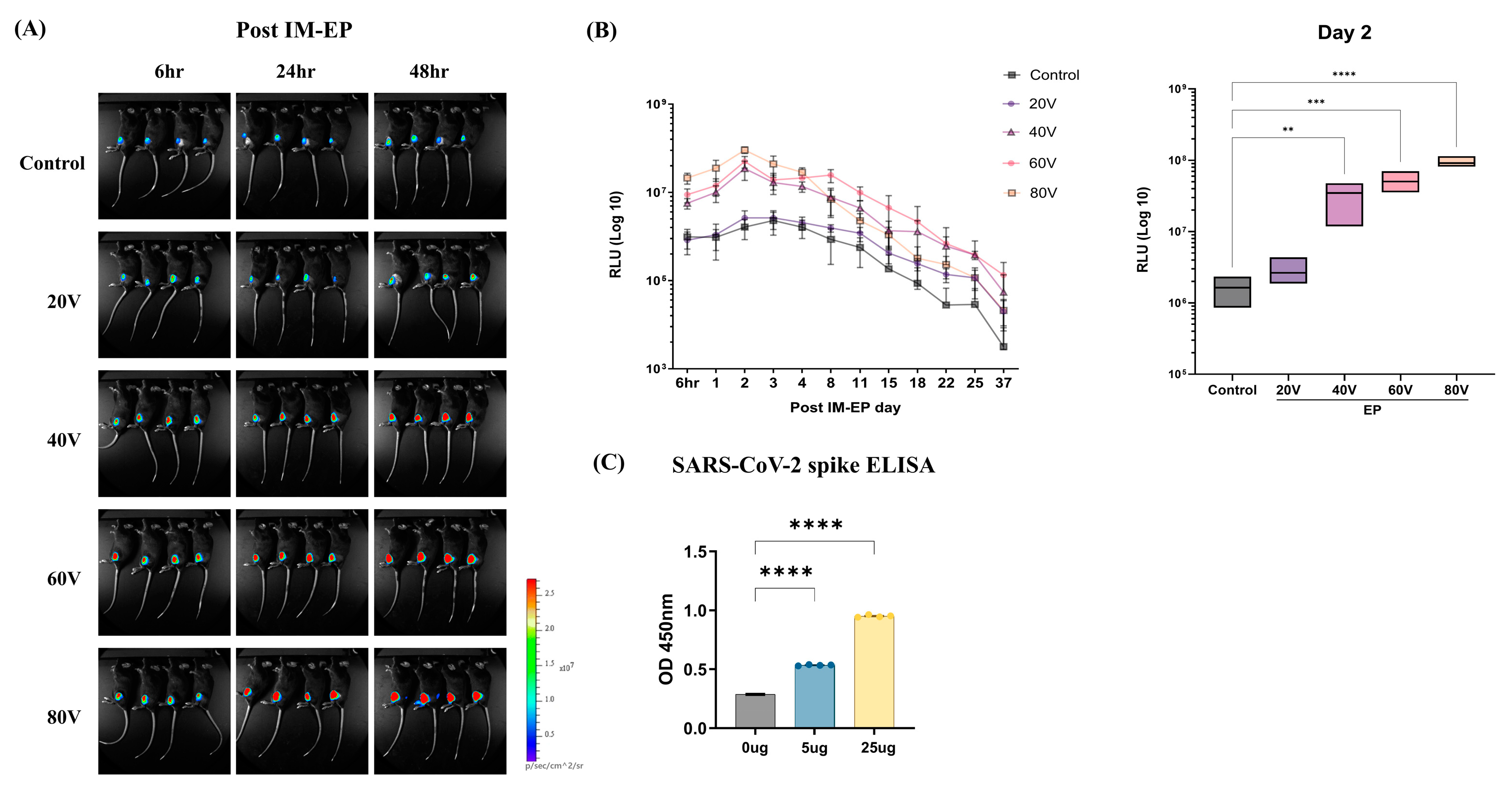
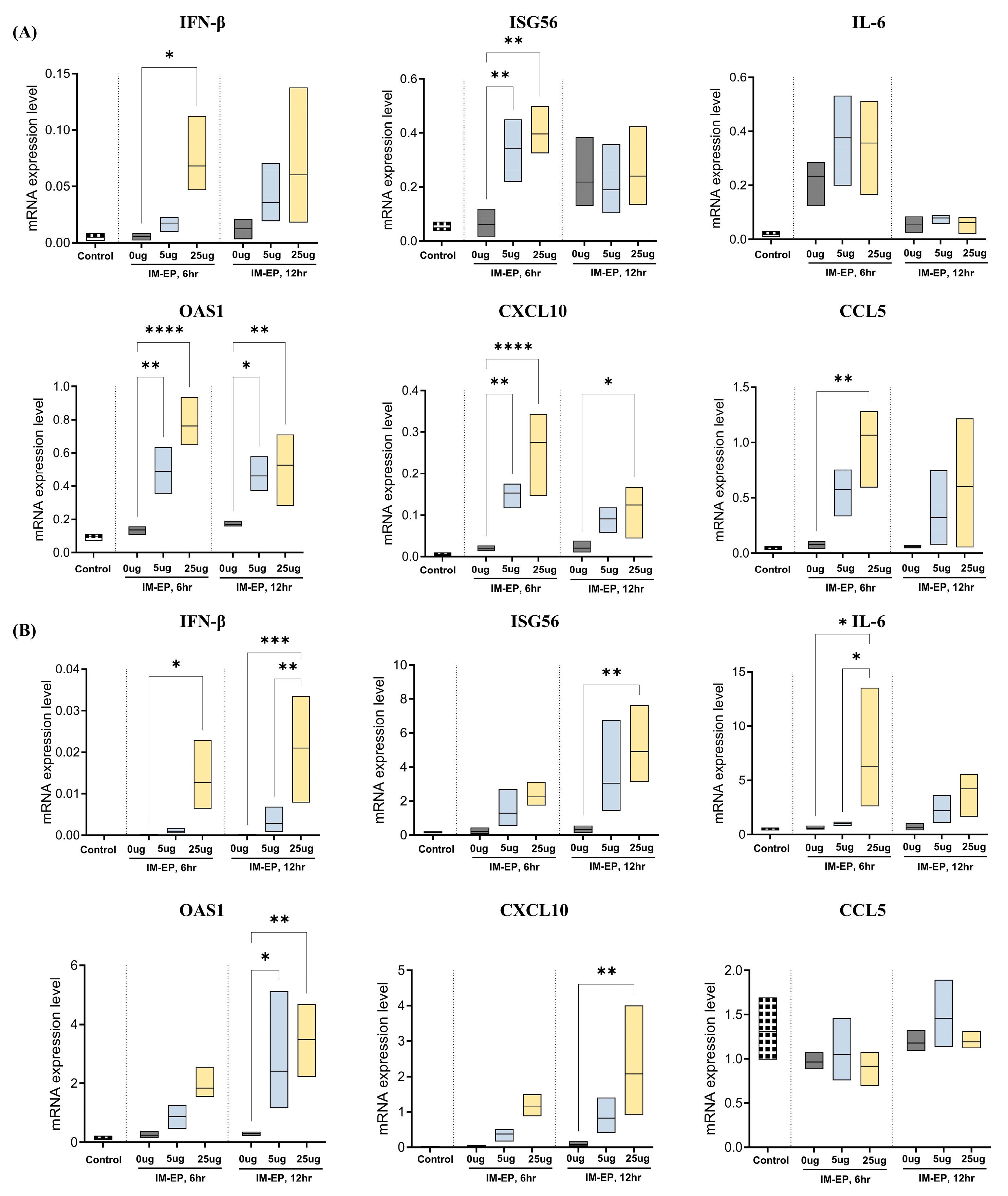
3.1. Optimization of mRNA Vaccine Delivery by Electroporation

To optimize EP conditions for mRNA vaccine delivery, we injected 10 μg of firefly luciferase (Fluc) mRNA into the biceps femoris muscle of the mice and then performed IM-EP at varying voltages (20, 40, 60, and 80 V), while keeping the duration (10 ms) and pulse number (12) constant, repeated twice (Figure 1A). Bioluminescence imaging revealed that EP significantly increased Fluc expression in a voltage- and time-dependent manner. As shown in Figure 1B (left panel), we noted the highest expression at 80 V, peaking at 48 h post-EP. Fluc activity was predominant on day 2 and persisted until day 37 at all voltages. The analysis performed on day 2 after IM-EP revealed a voltage-dependent enhancement of Fluc mRNA expression. Compared with the controls, we noted significantly higher expression levels at 40, 60, and 80 V (Figure 1B, right panel). However, as the expression levels did not differ significantly between 60 and 80 V, we considered 60 V as the optimal voltage, balancing efficacy and safety concerns. Therefore, in all subsequent experiments, we performed IM-EP at 60 V.
We next evaluated whether the SARS-CoV-2 mRNA vaccine could be efficiently delivered to the muscle and translated into protein via IM-EP (60 V). After injecting the mRNA vaccine at 5 and 25 μg doses, we performed EP to enhance delivery efficiency. As shown in Figure 1C, ELISA of muscle tissue extracts revealed a dose-dependent increase in spike protein expression, with the 25 μg group showing significantly higher expression than the 5 μg group. These results suggest that IM-EP effectively promotes the expression of mRNA vaccines, further supporting its potential as an efficient delivery method for mRNA-based immunization.

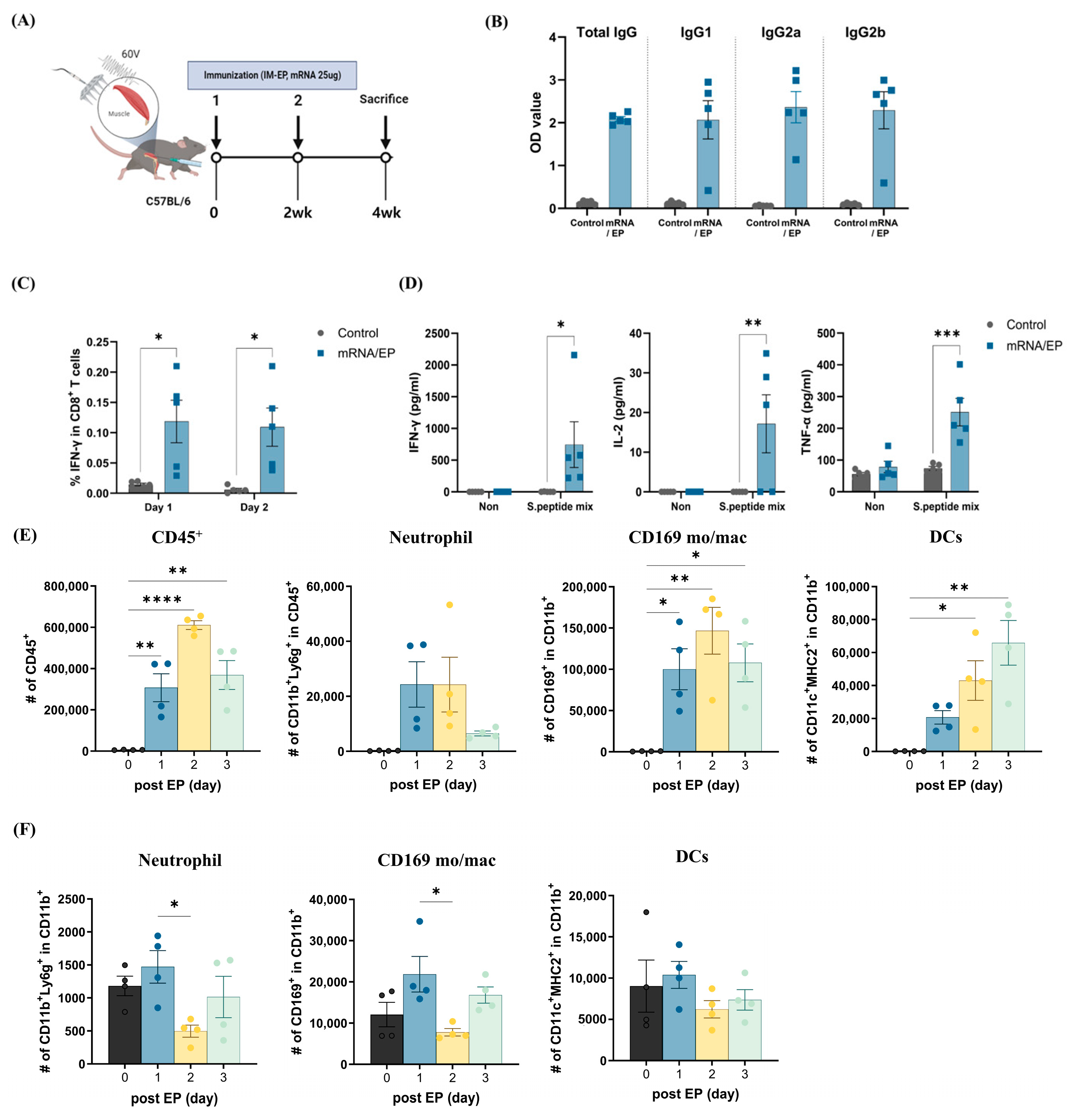
3.2. Naked mRNA Vaccination via IM-EP Effectively Induces Antigen-Specific Immune Responses in Mice

To evaluate the immune response induced by IM-EP-mediated mRNA vaccine delivery, we first examined cytokine expression in muscle tissue and draining lymph nodes following the administration of SARS-CoV-2 mRNA vaccine. EP-mediated delivery led to a dose-dependent increase in cytokine mRNA expression levels, with IFN-β, ISG56, IL-6, CXCL10, and CCL5 levels significantly elevated in muscle tissue, indicating strong local innate immune activation (Figure 2A). Similarly, dLNs from vaccinated mice exhibited a dose-dependent increase in cytokine expression levels, suggesting the induction of innate immune responses (Figure 2B).
We next assessed systemic humoral and cellular immune responses (Figure 3A). Serum analysis conducted 2 weeks after the second immunization revealed substantial elevations in RBD-specific IgG, IgG1, IgG2a, and IgG2b levels, indicating a strong antigen-specific humoral response (Figure 3B). In addition to humoral immunity, IM-EP induced a potent cellular immune response. Immunized mice exhibited enhanced IFN-γ production by CD8+ T cells, indicating robust cytotoxic T lymphocyte (CTL) activation (Figure 3C). Further analysis of splenocytes stimulated with spike protein peptides confirmed a significant increase in IFN-γ, IL-2, and tumor necrosis factor-alpha (TNF-α) secretion (Figure 3D), reinforcing the ability of IM-EP to induce a significant cell-mediated immune response.
Interestingly, we also found that IM-EP alone, without mRNA (PBS control), triggered mild immune activation. To further assess the potential immunomodulatory effects of EP, we analyzed immune cell infiltration in muscle tissue by flow cytometry at different time points following IM-EP without mRNA administration. We observed an initial increase in total immune cell infiltration on day 1 and a further increase on day 2, followed by a decline on day 3. Neutrophils exhibited a similar trend; however, the differences were not statistically significant because of high individual variability. In contrast, monocytes/macrophages increased on day 1, peaked on day 2, and remained elevated on day 3, suggesting their role in prolonged local inflammation and immune modulation. Notably, dendritic cells (DCs), key professional antigen-presenting cells (APCs), continuously increased up to day 3, indicating an extended antigen-presenting capacity that could contribute to enhanced adaptive immune responses upon mRNA vaccine delivery (Figure 3E,F), with detailed gating strategies and representative plots provided in Supplementary Figure S1.
Collectively, these findings highlight IM-EP as an effective strategy for mRNA vaccine delivery that can induce potent antigen-specific immune responses and facilitate immune cell recruitment to the injection site.

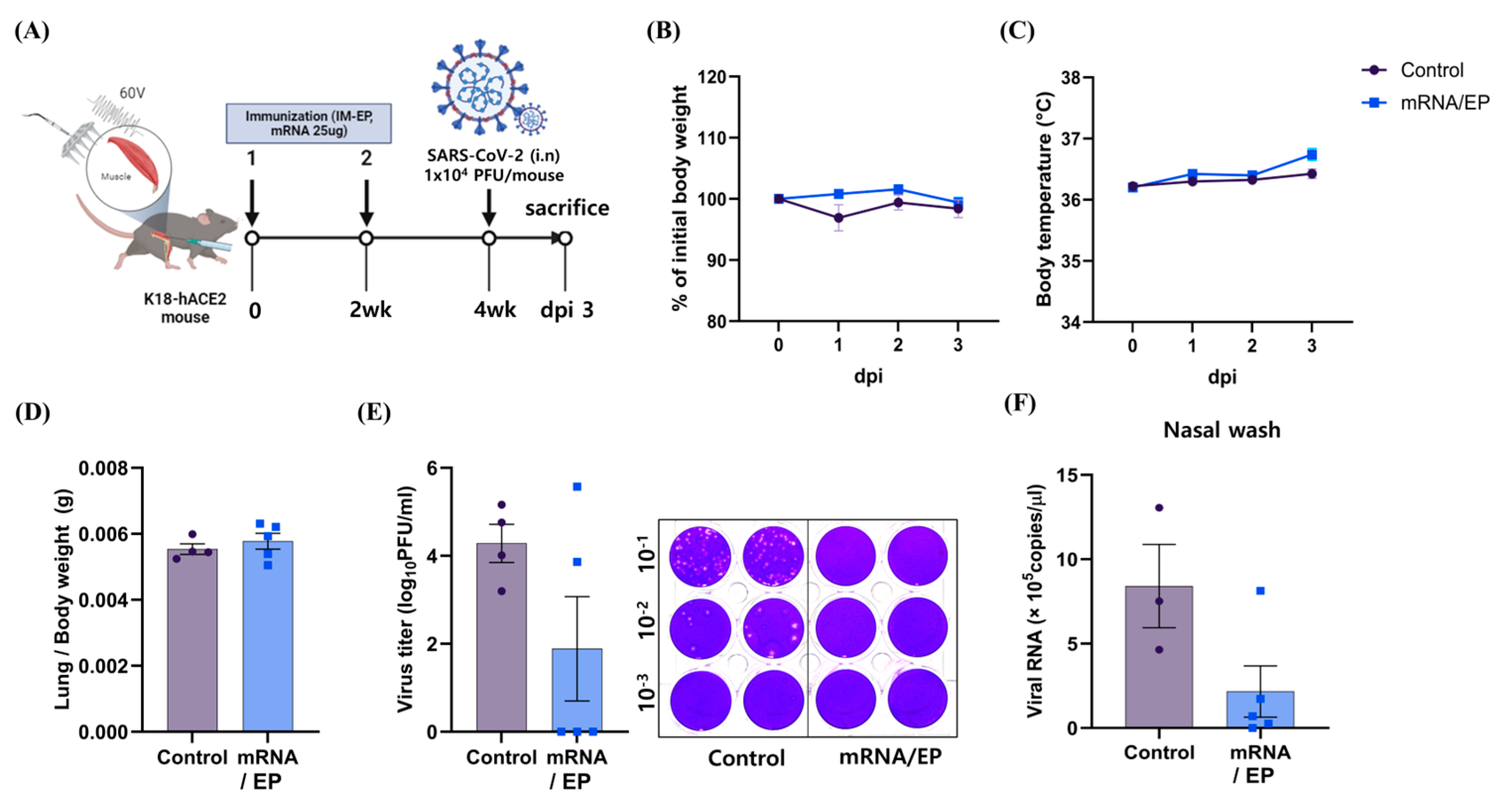
3.3. IM-EP-Mediated Naked mRNA Vaccination Reduces SARS-CoV-2 Load in K18-hACE2 Tg Mice

To evaluate the immunogenicity of naked mRNA vaccines delivered via IM-EP, we immunized K18-hACE2 Tg mice thrice with 5 or 25 µg of SARS-CoV-2 spike mRNA at 2-week intervals. We noted significant RBD-specific antibody production and IFN-γ responses only in the 25 µg group. Hence, we performed subsequent experiments using the 25 µg mRNA vaccine dose (Supplementary Figure S2). Two weeks after the final immunization, we intranasally challenged the mice with a lethal dose of SARS-CoV-2 (1 × 10⁴ PFU). We then euthanized the mice at 3 dpi (Figure 4A). While body weight and body temperature remained comparable between the vaccinated and unvaccinated groups (Figure 4B,C), the lung-to-body weight ratio showed a slightly higher trend in the vaccinated group than in the unvaccinated group; the difference was not statistically significant (Figure 4D). However, viral titers in lung tissues measured by the plaque assay exhibited a significant reduction in the vaccinated mice. All unvaccinated control mice showed a detectable viral load in lung tissues. In contrast, 60% of vaccinated mice showed complete viral clearance, with no detectable infectious virus (Figure 4E). Furthermore, analysis of nasal washes showed a marked reduction in viral RNA in the vaccinated group, indicating that IM-EP-mediated mRNA vaccination effectively limited viral replication at the primary site of infection (Figure 4F).
These findings indicate that two doses of IM-EP-mediated mRNA vaccination confer strong immune protection against SARS-CoV-2 infection, significantly reducing viral burden in lung tissues and nasal washes.

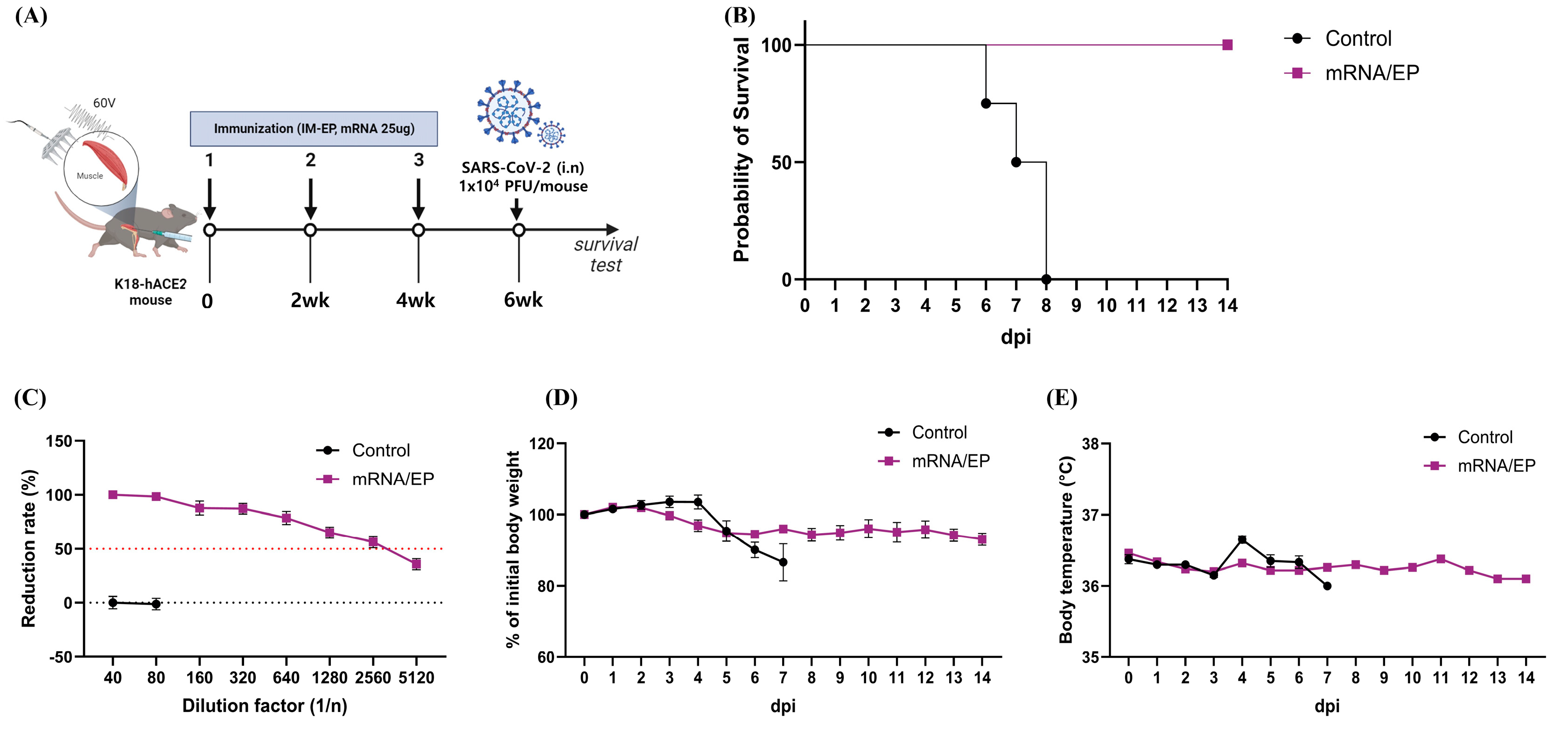
3.4. Naked mRNA Vaccine Protects K18-hACE2 Mice from Lethal Infection of SARS-CoV-2

We next assessed whether an additional booster dose could provide complete protection against lethal SARS-CoV-2 infection. We immunized K18-hACE2 Tg mice thrice with 25 µg of the mRNA vaccine at 2-week intervals via IM-EP (60 V). To assess vaccine-induced protection, we challenged the mice with a lethal dose of SARS-CoV-2 via the intranasal route (Figure 5A). Two weeks after the final immunization, we measured neutralizing antibody titers by performing a PRNT (Figure 5C). The vaccinated group exhibited a high PRNT50 titer of ≥2560, indicating a strong and sustained neutralizing immune response. In the unvaccinated control group, body weight loss began at 4 dpi (Figure 5D) and mortality began at 6 dpi, with all mice succumbing to the infection by 8 dpi. In contrast, all vaccinated mice survived the lethal challenge, demonstrating robust protection against severe disease (Figure 5B). Throughout the monitoring period, the body weight (Figure 5D) and body temperature (Figure 5E) of the vaccinated mice remained stable, indicating that IM-EP-mediated vaccination effectively prevented disease symptoms.
These results demonstrate that three doses of IM-EP-mediated SARS-CoV-2 mRNA vaccination can induce a highly protective immune response, preventing weight loss, reducing disease severity, and achieving 100% survival in K18-hACE2 mice following a lethal viral challenge. These findings further support the potential of IM-EP as a powerful strategy for enhancing the protective efficacy of naked mRNA vaccines against respiratory pathogens.

4. Discussion

Extensive research has been conducted on mRNA delivery systems, with LNPs emerging as the primary platform for COVID-19 mRNA vaccines used worldwide [26,27]. While LNP-based delivery has enabled the rapid development and deployment of mRNA vaccines, challenges related to stability [28], biodistribution [29], and potential inflammatory responses have driven continued efforts to explore alternative delivery methods. In this study, we investigated IM-EP as a non-carrier-based approach for mRNA vaccine delivery, aiming to enhance immune responses without the need for LNP encapsulation. Our results demonstrate that IM-EP successfully delivers naked SARS-CoV-2 spike mRNA into cells, leading to strong humoral and cellular immune responses and providing robust protection against lethal viral challenge in mice. By facilitating direct cytoplasmic entry of mRNA, IM-EP circumvents the limitations associated with LNPs, offering a safer and more accessible alternative for mRNA vaccine administration. These findings highlight the potential of IM-EP as an effective delivery platform for mRNA vaccines, not only against SARS-CoV-2 but also for other infectious diseases and emerging pathogens requiring rapid vaccine deployment.
EP-mediated nucleic acid delivery has been widely explored for both DNA and RNA vaccines. Intramuscular DNA immunization with in vivo EP has been shown to significantly enhance both cellular and humoral immune responses, further highlighting the immune-boosting potential of EP-based vaccine delivery [30]. EP-based DNA vaccines have induced enhanced antigen expression and immune responses in various infectious disease models, including HIV, Zika virus, and influenza, exhibiting promising results in preclinical and clinical trials [31]. However, unlike DNA vaccines, which require nuclear localization for transcription, mRNA vaccines are directly translated in the cytoplasm, allowing for faster antigen expression and immune activation [32]. Furthermore, although the clinical application of RNA vaccines remains limited, electroporation has been proposed as a promising strategy to enhance their delivery. Broderick and Humeau emphasized that EP significantly improves the intracellular uptake and expression of DNA vaccines, resulting in stronger immune responses in preclinical models [9]. Self-amplifying mRNA vaccines, such as those targeting influenza have demonstrated enhanced immunogenicity [33], while studies using EP-based delivery further support EP as an effective platform for self-amplifying RNA-based vaccine administration [19]. These findings validate the efficacy of IM-EP in delivering naked mRNA vaccines and inducing protective immune responses
Given the success of EP in vaccine delivery, we sought to evaluate its potential for mRNA vaccine administration as a viable alternative to LNP formulations. Recent studies have reported various adverse effects associated with LNP-mRNA vaccines, including vaccine-induced immune thrombotic thrombocytopenia [34], myocarditis [35], IgA vasculitis [36], and autoimmune disorders [37,38]. Although the exact mechanisms underlying these adverse effects remain unclear, LNP components, particularly cationic lipids, have been implicated in triggering innate immune activation and systemic inflammation [39]. In contrast, EP allows for the direct cytoplasmic delivery of mRNA without requiring lipid encapsulation, reducing concerns related to carrier-induced immune activation. Given these limitations, developing an EP-based delivery system for mRNA vaccines represents a promising approach to achieving efficient antigen expression while overcoming LNP-associated limitations and improving vaccine safety.
Our findings indicate that EP triggers immune activation, as indicated by the significant upregulation of cytokines such as IFN-β, IL-6, and CXCL10 in the muscle tissue and draining lymph nodes following IM-EP. In addition, we found that IM-EP enhances immune responses by recruiting innate immune cells to the injection site, which makes it an effective strategy for naked mRNA vaccine delivery. In K18-hACE2 mice, IM-EP-mediated SARS-CoV-2 mRNA vaccination reduced viral replication in respiratory tissues while maintaining stable body weight and body temperature. These results highlight the potential of IM-EP to control SARS-CoV-2 replication and prevent severe disease progression. This is consistent with previous studies showing that EP-based DNA vaccines for SARS-CoV-2 elicit strong neutralizing antibody responses and induce T-cell activation, further supporting the effectiveness of EP in enhancing immune response [39,40]. IM-EP enhances immune responses by promoting immune cell recruitment and activation, reinforcing its effectiveness as a strategy for naked mRNA vaccine delivery. Similar studies support these findings, including research on non-invasive delivery methods, such as jet injectors for naked mRNA, which also induce strong immune responses without systemic reactogenicity [41]. Interestingly, wet cupping has been demonstrated to reduce pro-inflammatory cytokines such as IL-6 and TNF-α, thereby enhancing immune responses [42]. This cytokine modulation suggests that local immune activation, similar to the effects observed with EP, could play a significant role in improving vaccine efficacy by enhancing antigen presentation and immune cell recruitment. These results reinforce the growing potential of EP and similar delivery methods as viable alternatives to LNP-based systems.

5. Conclusions

Our findings show that IM-EP-mediated delivery of naked mRNA encoding the SARS-CoV-2 spike protein induces high levels of neutralizing antibodies and strong cellular immunity, effectively preventing lethal viral infections in mice. These results demonstrate that delivering mRNA directly into the cytoplasm via IM-EP, without the need for lipid carriers, not only simplifies the processes of vaccine production and distribution but also reduces adverse effects associated with LNPs. Given the rising interest in optimizing mRNA vaccine platforms, our study supports IM-EP as a promising alternative for enhancing vaccine efficacy while improving safety and accessibility. Furthermore, given the ongoing advancements in mRNA-based cancer vaccines, EP-mediated delivery could also be considered as a potential platform for tumor mRNA vaccine delivery. Further studies are necessary to optimize and expand the application of this approach in both infectious disease and cancer vaccine development.

Supplementary Materials

The following supporting information can be downloaded at: https://www.mdpi.com/article/10.3390/app15084428/s1, Figure S1. Gating strategy for flow cytometry analysis of muscle and lymph node samples. A: Lymphocytes were initially gated based on forward scatter area (FSC-A) vs. side scatter area (SSC-A) characteristics. Live cells were then selected using a Live/Dead fixable violet dead cell stain. Single cells were further gated based on forward scatter area (FSC-A) vs. forward scatter height (FSC-H) to exclude doublets, leaving only single cells. CD45+ immune cells were then gated for further analysis. B–E: Representative flow cytometry plots from muscle samples are shown for CD45+ immune cell subsets, including neutrophils (CD11b+Ly6G+), dendritic cells (DCs, CD11c+MHCII+CD11b+), and monocytes/macrophages (CD169+CD115+CD11b+). Figure S2: Immune response induced by IM-EP immunization. A: Immunization schedule for C57BL/6 mice receiving three doses of the SARS-CoV-2 mRNA vaccine via IM-EP. B: RBD-specific total IgG serum measured by RBD-specific ELISA after the third dose and one week post-immunization serum. C: IFN-γ concentration in splenocytes stimulated with a SARS-CoV-2 spike peptide library for 48 h, measured by IFN-gamma ELISA. (* p < 0.05 Control vs. EP group).

Author Contributions

Conceptualization, Y.J.L. and S.-M.L.; methodology, S.-H.P., Y.K., M.K. and Y.S.; investigation, S.-H.P., Y.K., M.K. and Y.S.; data curation, S.-H.P., Y.K., M.K. and Y.S.; writing—original draft preparation, S.-H.P., M.K., Y.S. and H.J.; writing—review and editing, S.-M.L. and H.J.; project administration, Y.J.L. and S.-M.L. All authors have read and agreed to the published version of the manuscript. GenAI tools were utilized to assist with English editing and proofreading. The authors take full responsibility for the final content of the manuscript.

Funding

This research was supported by a grant of the Korea Health Technology R&D Project through the Korea Health Industry Development Institute (KHIDI), funded by the Ministry of Health and Welfare, Republic of Korea (Grant No: RS-2022-KH128441). It was also supported by the Technology Innovation Program (20019452, Development of LNP-free mRNA vaccine using new concept vaccination equipment based on Korean technology), funded by the Ministry of Trade, Industry and Energy (MOTIE, Korea). Additional support was provided by the Ministry of Food and Drug Safety through a grant from the Korean government (Grant No: 22213MFDS421).

Institutional Review Board Statement

The animal studies were performed with the approval of the Institutional Animal Care Committee (IACUC) of CBNUA-2046-22-01 (23 December 2022).

Informed Consent Statement

Not applicable.

Data Availability Statement

The original contributions presented in this study are included in the article/Supplementary Material. Further inquiries can be directed to the corresponding authors.

Conflicts of Interest

Authors Mina Kim, Yong Jin Lee and Yeji Seo were employed by Department of R&D, Hulux. The remaining authors declare that the research was conducted in the absence of any commercial or financial relationships that could be construed as a potential conflict of interest.

Abbreviations

The following abbreviations are used in this manuscript:
APCsAntigen-presenting cells
BSL3Biosafety level 3
CBACytometric bead array
CTLCytotoxic T lymphocytes
COVID-19Coronavirus disease 2019
DCsDendritic cells
DMEMDulbecco’s modified eagle medium
DPIDays post infection
FBSFetal bovine serum
FlucFirefly luciferase
IM-EPIntramuscular electroporation
ID-EPIntradermal electroporation
ISG56IFN-stimulated gene 56
LNPLipid nanoparticle
mRNAMessenger RNA
PFUPlaque-forming units
PRNTPlaque reduction neutralization test
RBDReceptor-binding domain
ROIRegion of interest
Th1Type 1 helper T cells
TNF-αTumor necrosis factor alpha

References

  1. Semple, S.C.; Leone, R.; Barbosa, C.J.; Tam, Y.K.; Lin, P.J.C. Lipid Nanoparticle Delivery Systems to Enable mRNA-Based Therapeutics. Pharmaceutics 2022, 14, 398. [Google Scholar] [CrossRef] [PubMed]
  2. Zhang, G.; Tang, T.; Chen, Y.; Huang, X.; Liang, T. mRNA vaccines in disease prevention and treatment. Signal Transduct. Target. Ther. 2023, 8, 365. [Google Scholar] [CrossRef] [PubMed]
  3. Kiaie, S.H.; Majidi Zolbanin, N.; Ahmadi, A.; Bagherifar, R.; Valizadeh, H.; Kashanchi, F.; Jafari, R. Recent advances in mRNA-LNP therapeutics: Immunological and pharmacological aspects. J. Nanobiotechnol. 2022, 20, 276. [Google Scholar] [CrossRef] [PubMed]
  4. Zhang, B.; Sim, W.K.; Shen, T.L.; Lim, S.K. Engineered EVs with pathogen proteins: Promising vaccine alternatives to LNP-mRNA vaccines. J. Biomed. Sci. 2024, 31, 9. [Google Scholar] [CrossRef]
  5. Heidecker, B.; Dagan, N.; Balicer, R.; Eriksson, U.; Rosano, G.; Coats, A.; Tschöpe, C.; Kelle, S.; Poland, G.A.; Frustaci, A.; et al. Myocarditis following COVID-19 vaccine: Incidence, presentation, diagnosis, pathophysiology, therapy, and outcomes put into perspective. A clinical consensus document supported by the Heart Failure Association of the European Society of Cardiology (ESC) and the ESC Working Group on Myocardial and Pericardial Diseases. Eur. J. Heart Fail. 2022, 24, 2000–2018. [Google Scholar] [CrossRef]
  6. Kapoor, M.; Spillane, J.; Englezou, C.; Sarri-Gonzalez, S.; Bell, R.; Rossor, A.; Manji, H.; Reilly, M.M.; Lunn, M.P.; Carr, A. Thromboembolic risk with IVIg: Incidence and risk factors in patients with inflammatory neuropathy. Neurology 2020, 94, e635–e638. [Google Scholar] [CrossRef]
  7. Muir, K.-L.; Kallam, A.; Koepsell, S.A.; Gundabolu, K. Thrombotic thrombocytopenia after Ad26.COV2.S vaccination. N. Engl. J. Med. 2021, 384, 1964–1965. [Google Scholar] [CrossRef]
  8. Fahrni, M.L.; Ismail, I.A.; Refi, D.M.; Almeman, A.; Yaakob, N.C.; Saman, K.M.; Mansor, N.F.; Noordin, N.; Babar, Z.U. Management of COVID-19 vaccines cold chain logistics: A scoping review. J. Pharm. Policy Pract. 2022, 15, 16. [Google Scholar] [CrossRef]
  9. Broderick, K.E.; Humeau, L.M. Electroporation-enhanced delivery of nucleic acid vaccines. Expert Rev. Vaccines 2015, 14, 195–204. [Google Scholar] [CrossRef]
  10. Gehl, J. Electroporation: Theory and methods, perspectives for drug delivery, gene therapy and research. Acta Physiol. Scand. 2003, 177, 437–447. [Google Scholar] [CrossRef]
  11. Laddy, D.J.; Yan, J.; Kutzler, M.; Kobasa, D.; Kobinger, G.P.; Khan, A.S.; Greenhouse, J.; Sardesai, N.Y.; Draghia-Akli, R.; Weiner, D.B. Heterosubtypic protection against pathogenic human and avian influenza viruses via in vivo electroporation of synthetic consensus DNA antigens. PLoS ONE 2008, 3, e2517. [Google Scholar] [CrossRef] [PubMed]
  12. Todorova, B.; Adam, L.; Culina, S.; Boisgard, R.; Martinon, F.; Cosma, A.; Ustav, M.; Kortulewski, T.; Le Grand, R.; Chapon, C. Electroporation as a vaccine delivery system and a natural adjuvant to intradermal administration of plasmid DNA in macaques. Sci. Rep. 2017, 7, 4122. [Google Scholar] [CrossRef]
  13. Lin, F.; Shen, X.; McCoy, J.R.; Mendoza, J.M.; Yan, J.; Kemmerrer, S.V.; Khan, A.S.; Weiner, D.B.; Broderick, K.E.; Sardesai, N.Y. A novel prototype device for electroporation-enhanced DNA vaccine delivery simultaneously to both skin and muscle. Vaccine 2011, 29, 6771–6780. [Google Scholar] [CrossRef] [PubMed]
  14. Han, K.T.; Sin, J.I. DNA vaccines targeting human papillomavirus-associated diseases: Progresses in animal and clinical studies. Clin. Exp. Vaccine Res. 2013, 2, 106–114. [Google Scholar] [CrossRef] [PubMed]
  15. Hirao, L.A.; Draghia-Akli, R.; Prigge, J.T.; Yang, M.; Satishchandran, A.; Wu, L.; Hammarlund, E.; Khan, A.S.; Babas, T.; Rhodes, L.; et al. Multivalent smallpox DNA vaccine delivered by intradermal electroporation drives protective immunity in nonhuman primates against lethal monkeypox challenge. J. Infect. Dis. 2011, 203, 95–102. [Google Scholar] [CrossRef]
  16. Golzio, M.; Rols, M.P.; Teissie, J. In vitro and in vivo electric field-mediated permeabilization, gene transfer, and expression. Methods 2004, 33, 126–135. [Google Scholar] [CrossRef]
  17. Golzio, M.; Mazzolini, L.; Moller, P.; Rols, M.P.; Teissie, J. Inhibition of gene expression in mice muscle by in vivo electrically mediated siRNA delivery. Gene Ther. 2005, 12, 246–251. [Google Scholar] [CrossRef]
  18. Van Nuffel, A.M.; Corthals, J.; Neyns, B.; Heirman, C.; Thielemans, K.; Bonehill, A. Immunotherapy of cancer with dendritic cells loaded with tumor antigens and activated through mRNA electroporation. Methods Mol. Biol. 2010, 629, 405–452. [Google Scholar] [CrossRef]
  19. Cu, Y.; Broderick, K.E.; Banerjee, K.; Hickman, J.; Otten, G.; Barnett, S.; Kichaev, G.; Sardesai, N.Y.; Ulmer, J.B.; Geall, A. Enhanced Delivery and Potency of Self-Amplifying mRNA Vaccines by Electroporation In Situ. Vaccines 2013, 1, 367–383. [Google Scholar] [CrossRef]
  20. Paz, S.; Vilasco, M.; Werden, S.J.; Arguello, M.; Joseph-Pillai, D.; Zhao, T.; Nguyen, T.L.; Sun, Q.; Meurs, E.F.; Lin, R.; et al. A functional C-terminal TRAF3-binding site in MAVS participates in positive and negative regulation of the IFN antiviral response. Cell Res. 2011, 21, 895–910. [Google Scholar] [CrossRef]
  21. Unterholzner, L.; Keating, S.E.; Baran, M.; Horan, K.A.; Jensen, S.B.; Sharma, S.; Sirois, C.M.; Jin, T.; Latz, E.; Xiao, T.S.; et al. IFI16 is an innate immune sensor for intracellular DNA. Nat. Immunol. 2010, 11, 997–1004. [Google Scholar] [CrossRef] [PubMed]
  22. Seth, S.; Matsui, Y.; Fosnaugh, K.; Liu, Y.; Vaish, N.; Adami, R.; Harvie, P.; Johns, R.; Severson, G.; Brown, T.; et al. RNAi-based therapeutics targeting survivin and PLK1 for treatment of bladder cancer. Mol. Ther. 2011, 19, 928–935. [Google Scholar] [CrossRef] [PubMed]
  23. Gebremeskel, S.; Nelson, A.; Walker, B.; Oliphant, T.; Lobert, L.; Mahoney, D.; Johnston, B. Natural killer T cell immunotherapy combined with oncolytic vesicular stomatitis virus or reovirus treatments differentially increases survival in mouse models of ovarian and breast cancer metastasis. J. Immunother. Cancer 2021, 9, e002096. [Google Scholar] [CrossRef]
  24. Yang, M.S.; Park, M.J.; Lee, J.; Oh, B.; Kang, K.W.; Kim, Y.; Lee, S.M.; Lim, J.O.; Jung, T.Y.; Park, J.H.; et al. Non-invasive administration of AAV to target lung parenchymal cells and develop SARS-CoV-2-susceptible mice. Mol. Ther. 2022, 30, 1994–2004. [Google Scholar] [CrossRef]
  25. Hong, S.H.; Oh, H.; Park, Y.W.; Kwak, H.W.; Oh, E.Y.; Park, H.J.; Kang, K.W.; Kim, G.; Koo, B.S.; Hwang, E.H.; et al. Immunization with RBD-P2 and N protects against SARS-CoV-2 in nonhuman primates. Sci. Adv. 2021, 7, eabg7156. [Google Scholar] [CrossRef] [PubMed]
  26. Polack, F.P.; Thomas, S.J.; Kitchin, N.; Absalon, J.; Gurtman, A.; Lockhart, S.; Perez, J.L.; Perez Marc, G.; Moreira, E.D.; Zerbini, C.; et al. Safety and Efficacy of the BNT162b2 mRNA COVID-19 Vaccine. N. Engl. J. Med. 2020, 383, 2603–2615. [Google Scholar] [CrossRef]
  27. Baden, L.R.; El Sahly, H.M.; Essink, B.; Kotloff, K.; Frey, S.; Novak, R.; Diemert, D.; Spector, S.A.; Rouphael, N.; Creech, C.B.; et al. Efficacy and Safety of the mRNA-1273 SARS-CoV-2 Vaccine. N. Engl. J. Med. 2021, 384, 403–416. [Google Scholar] [CrossRef]
  28. Schoenmaker, L.; Witzigmann, D.; Kulkarni, J.A.; Verbeke, R.; Kersten, G.; Jiskoot, W.; Crommelin, D.J.A. mRNA-lipid nanoparticle COVID-19 vaccines: Structure and stability. Int. J. Pharm. 2021, 601, 120586. [Google Scholar] [CrossRef]
  29. Hassett, K.J.; Benenato, K.E.; Jacquinet, E.; Lee, A.; Woods, A.; Yuzhakov, O.; Himansu, S.; Deterling, J.; Geilich, B.M.; Ketova, T.; et al. Optimization of Lipid Nanoparticles for Intramuscular Administration of mRNA Vaccines. Mol. Ther. Nucleic Acids 2019, 15, 1–11. [Google Scholar] [CrossRef]
  30. Shoji, M.; Katayama, K.; Tachibana, M.; Tomita, K.; Sakurai, F.; Kawabata, K.; Mizuguchi, H. Intramuscular DNA immunization with in vivo electroporation induces antigen-specific cellular and humoral immune responses in both systemic and gut-mucosal compartments. Vaccine 2012, 30, 7278–7285. [Google Scholar] [CrossRef]
  31. Gary, E.N.; Weiner, D.B. DNA vaccines: Prime time is now. Curr. Opin. Immunol. 2020, 65, 21–27. [Google Scholar] [CrossRef]
  32. Pardi, N.; Hogan, M.J.; Porter, F.W.; Weissman, D. mRNA vaccines—A new era in vaccinology. Nat. Rev. Drug Discov. 2018, 17, 261–279. [Google Scholar] [CrossRef] [PubMed]
  33. Vogel, A.B.; Lambert, L.; Kinnear, E.; Busse, D.; Erbar, S.; Reuter, K.C.; Wicke, L.; Perkovic, M.; Beissert, T.; Haas, H.; et al. Self-Amplifying RNA Vaccines Give Equivalent Protection against Influenza to mRNA Vaccines but at Much Lower Doses. Mol. Ther. 2018, 26, 446–455. [Google Scholar] [CrossRef] [PubMed]
  34. Parums, D.V. Editorial: SARS-CoV-2 mRNA vaccines and the possible mechanism of vaccine-induced immune thrombotic thrombocytopenia (VITT). Med. Sci. Monit. Int. Med. J. Exp. Clin. Res. 2021, 27, e932899. [Google Scholar] [CrossRef]
  35. Verma, A.K.; Lavine, K.J.; Lin, C.-Y. Myocarditis after COVID-19 mRNA vaccination. N. Engl. J. Med. 2021, 385, 1332–1334. [Google Scholar] [CrossRef] [PubMed]
  36. Nishimura, N.; Shiomichi, Y.; Takeuchi, S.; Akamine, S.; Yoneda, R.; Yoshizawa, S. IgA vasculitis following COVID-19 vaccination. Mod. Rheumatol. Case Rep. 2023, 7, 122–126. [Google Scholar] [CrossRef]
  37. Flemming, A. mRNA vaccine shows promise in autoimmunity. Nat. Rev. Immunol. 2021, 21, 72. [Google Scholar] [CrossRef]
  38. Chen, Y.; Xu, Z.; Wang, P.; Li, X.M.; Shuai, Z.W.; Ye, D.Q.; Pan, H.F. New-onset autoimmune phenomena post-COVID-19 vaccination. Immunology 2022, 165, 386–401. [Google Scholar] [CrossRef]
  39. Ndeupen, S.; Qin, Z.; Jacobsen, S.; Bouteau, A.; Estanbouli, H.; Igyártó, B.Z. The mRNA-LNP platform’s lipid nanoparticle component used in preclinical vaccine studies is highly inflammatory. iScience 2021, 24, 103479. [Google Scholar] [CrossRef]
  40. Khobragade, A.; Bhate, S.; Ramaiah, V.; Deshpande, S.; Giri, K.; Phophle, H.; Supe, P.; Godara, I.; Revanna, R.; Nagarkar, R.; et al. Efficacy, safety, and immunogenicity of the DNA SARS-CoV-2 vaccine (ZyCoV-D): The interim efficacy results of a phase 3, randomised, double-blind, placebo-controlled study in India. Lancet 2022, 399, 1313–1321. [Google Scholar] [CrossRef]
  41. Abbasi, S.; Matsui-Masai, M.; Yasui, F.; Hayashi, A.; Tockary, T.A.; Mochida, Y.; Akinaga, S.; Kohara, M.; Kataoka, K.; Uchida, S. Carrier-free mRNA vaccine induces robust immunity against SARS-CoV-2 in mice and non-human primates without systemic reactogenicity. Mol. Ther. 2024, 32, 1266–1283. [Google Scholar] [CrossRef] [PubMed]
  42. Ekrami, N.; Ahmadian, M.; Nourshahi, M.; Shakouri, G.H. Wet-cupping induces anti-inflammatory action in response to vigorous exercise among martial arts athletes: A pilot study. Complement. Ther. Med. 2021, 56, 102611. [Google Scholar] [CrossRef] [PubMed]
Figure 1. Optimization and evaluation of mRNA delivery via IM-EP in mice. (A):Administration of 10 µg firefly luciferase (Fluc) mRNA into the biceps femoris muscle, followed by IM-EP. Control: without IM-EP. Bioluminescence imaging at 6, 24, and 48 h (each group, n = 8). (B) (Left) Quantification of bioluminescence signals up to 37 days post-IM-EP. (Right) Analysis on day 2 post-IM-EP under different voltages (control, 20 V, 40 V, 60 V, and 80 V; duration: 10 ms; pulse: 12, two repetitions) (** p < 0.01, *** p < 0.001, and **** p < 0.0001; control vs. EP). (C) Mice were intramuscularly injected with 5 or 25 µg of the SARS-CoV-2 mRNA vaccine, followed by IM-EP. Levels of SARS-CoV-2 spike protein were evaluated by performing ELISA using muscle tissue lysates (**** p < 0.001; 0 μg vs. 5 or 25 μg mRNA vaccine group).
Applsci 15 04428 g001
Figure 2. Expression of innate immune response-related genes in muscle and draining lymph nodes. (A) Muscle tissues were harvested from mice to measure expression levels of IFN-β, ISG56, IL-6, OAS1, CXCL10, and CCL5 genes using real-time PCR (* p < 0.05, ** p < 0.01, and **** p < 0.0001; compared to the 0 µg IM-EP group at each time point). (B) Draining lymph nodes were harvested from mice to measure expression levels of IFN-β, ISG56, IL-6, OAS1, CXCL10, and CCL5 genes using real-time PCR (* p < 0.05, ** p < 0.01, and *** p < 0.001; compared to the 0 µg IM-EP group at each time point).
Applsci 15 04428 g002
Figure 3. Immunization with SARS-CoV-2 mRNA vaccine via EP efficiently induces humoral and cellular immunity. (A) C57BL/6 mice received 25 µg of the SARS-CoV-2 mRNA vaccine via IM-EP. Serum and spleen samples were collected 2 weeks after the second immunization (each group, n = 8) (B) RBD-specific total IgG and IgG isotypes (IgG1, IgG2a, and IgG2b) in the serum. (C) IFN-γ+CD8+ cytotoxic T lymphocytes in the spleen analyzed by intracellular cytokine staining (* p < 0.05; control vs. EP). (D) Secretion of cytokines (IFN-γ, IL-2, and TNF-α) from stimulated splenocytes measured after 48 h (* p < 0.05, ** p < 0.01, and *** p < 0.001; Non vs. peptide mix). (E,F) Immune cell populations in muscle and lymph nodes analyzed post-EP. Gating: CD45+ (immune cells), CD11b+Ly6G+ (neutrophils), CD169+CD11b+ (monocytes/macrophages), and CD11c+MHC II+CD11b+ (DCs) (* p < 0.05, ** p < 0.01, and **** p < 0.0001; between indicated groups).
Applsci 15 04428 g003
Figure 4. IM-EP-mediated SARS-CoV-2 mRNA vaccination reduces viral replication in lethal infection mouse model. (A) Immunization schedule for K18-hACE2 mice; the mice received two EP-mediated mRNA vaccine doses, followed by a SARS-CoV-2 challenge. (B) Body weight changes post-challenge (n = 4–5 per group). (C) Body temperature changes post-challenge. (D) Lung-to-body weight ratio at 3 dpi. (E) SARS-CoV-2 titers in lung tissues measured by a plaque assay. (F) Viral RNA in nasal washes quantified by real-time PCR using SARS-CoV-2 RNA standards.
Applsci 15 04428 g004
Figure 5. SARS-CoV-2 mRNA vaccine delivered via IM-EP protects mice from lethal SARS-CoV-2 infection. (A) Schematic of the immunization and challenge protocols in K18-hACE2 mice, showing three immunizations with EP and subsequent SARS-CoV-2 challenge. (B) Post-challenge monitoring of K18-hACE2 female mice (n = 4–5/group) for survival. (C) Neutralizing antibody levels measured by performing a PRNT (red line indicates the PRNT50 threshold). (D,E) Post-challenge monitoring of K18-hACE2 female mice for body weight (D) and body temperature (E).
Applsci 15 04428 g005
Table 1. RT-PCR primer sequences.
Title 1Forward (5′-3′)Reverse (5′-3′)
IFNβATGGTGGTCCGAGCAGAGATCCACCACTCATTCTGAGGCA
ISG56 [20]CTCTGAAAGTGGAGCCAGAAAACAAATCTTGGCGATAGGCTACGA
IL-6 [21]AGAATTGCCATTGCACACTCCCAACAGACCTGTCTATA
OAS1 [22]CTTTGATGTCCTGGGTCATGTGCTCCGTGAAGCAGGTAGAG
CXCL10GCAACTGCATCCATATCGATGACGGATTCCGGATTCAGACATCTCTGC
CCL5 [23]CTCACCATATGGCTCGGACAACAAACACGACTGCAAGATTGG
ActinTCCAGCCTTCCTTCTTGGGTGCACTGTGTTGGCATAGAGGT
Disclaimer/Publisher’s Note: The statements, opinions and data contained in all publications are solely those of the individual author(s) and contributor(s) and not of MDPI and/or the editor(s). MDPI and/or the editor(s) disclaim responsibility for any injury to people or property resulting from any ideas, methods, instructions or products referred to in the content.

