Article

Interaction Between Human Microbiota, Immune System, and Hepatitis C Virus Infection: A Narrative Review

1
Department of Pharmacy, University of Genoa, Viale Benedetto XV 7, 16132 Genoa, Italy
2
The Directorate of Research, Development and Innovation Management (DMCDI), The Technical University of Cluj-Napoca, Constantin Daicoviciu St., no. 15, 400020 Cluj-Napoca, Cluj County, Romania
*
Authors to whom correspondence should be addressed.
Appl. Sci. 2025, 15(6), 3157; https://doi.org/10.3390/app15063157
Submission received: 29 January 2025 / Revised: 8 March 2025 / Accepted: 10 March 2025 / Published: 14 March 2025

Abstract

:
Hepatitis C virus (HCV) accounts for approximately 17.5% of acute hepatitis cases, with around 65% of individuals progressing to chronic infection after the acute phase. The role of intestinal microbiota in the pathogenesis of chronic liver diseases associated with HCV is an emerging area of scientific interest. However, the precise mechanisms by which microbiota influence chronic HCV infection remain inadequately understood, warranting further investigation. While comprehensive data on human microbiota–HCV interactions are limited, existing studies provide valuable insights that can inform future research and theoretical exploration. These studies lay the foundation for hypothesizing potential mechanisms linking microbiota and HCV within the gut–liver axis. The existing knowledge regarding the interactions between microbiota and hepatitis C virus (HCV) remains insufficient; however, recent findings highlight the significant influence of the gut–liver axis on the progression of HCV infection and its associated complications. The possible therapeutic advantages of strategies that modulate the microbiome, such as fecal microbiota transplantation (FMT), merit further investigation in relation to HCV. Future studies should focus on elucidating the reciprocal relationship between gut microbiota and HCV pathogenesis, identifying microbial markers linked to disease advancement, and assessing the effectiveness of microbiota-targeted therapies in enhancing clinical outcomes for individuals infected with HCV. This review aims to summarize the current understanding of microbiota–HCV interactions and propose a novel perspective based on literature findings, thereby paving the way for new research initiatives on HCV infections. We surveyed the existing literature and described it by conducting both a thematic and conceptual analysis.

1. Introduction

The human body hosts a vast and complex array of microbial communities, collectively termed the microbiota, which colonize various anatomical regions, including the skin, oral cavity, gastrointestinal tract, and other mucosal surfaces. These microbial consortia predominantly consist of bacteria, alongside fungi, archaea, viruses, and other microorganisms, forming intricate ecological networks that interact with host physiological processes. The estimated number of microbial cells within the human body has been suggested to either approximate or exceed the total number of human cells, highlighting the profound extent of microbial colonization [1,2,3].
The microbiota plays a fundamental role in maintaining homeostasis by contributing to metabolic functions, synthesizing essential nutrients, and engaging in immunomodulatory activities that shape host defense mechanisms. The intestinal microbiota, in particular, is indispensable for the proper development of the immune system, influencing immune tolerance and enhancing the host’s ability to recognize and eliminate pathogenic invaders. The relationship between host immunity and gut microbiota is increasingly recognized as a critical determinant of health and disease, with growing evidence implicating microbial dysbiosis—an imbalance in microbial composition—in the progression of various chronic conditions, including hepatic disorders [4,5].
One area of emerging scientific interest is the potential involvement of intestinal microbiota in the pathogenesis of chronic liver diseases associated with hepatitis C virus (HCV) infection. HCV remains a major global health burden, often leading to progressive hepatic fibrosis, cirrhosis, and hepatocellular carcinoma. Despite advances in antiviral therapies, the underlying mechanisms through which the gut microbiota influences the persistence and progression of chronic HCV infection remain incompletely understood, warranting further investigation [6]. The dynamic interplay between microbiota and viral pathogens extends beyond simple coexistence; microbiota can actively modulate viral pathogenesis through multiple mechanisms, including direct interactions with viral particles, competition for nutrients and ecological niches, and the regulation of host immune responses.
The influence of microbiota on viral infections is multifaceted and remains a subject of ongoing debate. Certain microbial species may exert antiviral effects by producing metabolites that inhibit viral replication or enhance antiviral immune responses. Conversely, other microbial components may facilitate viral persistence by suppressing immune activation or by modulating the integrity of epithelial barriers, thereby influencing viral dissemination within the host. It is also conceivable that microbiota may exert a neutral effect, neither enhancing nor inhibiting viral infections, depending on the specific host and environmental context [7,8,9,10].
In the case of HCV infection, microbiota-derived metabolites and bacterial components, such as lipopolysaccharides, have been implicated in shaping hepatic immune responses and inflammatory pathways. Additionally, the gut–liver axis—a bidirectional communication network between the intestinal microbiota and the liver—plays a central role in liver disease progression. Disruptions in this axis, particularly in the context of gut dysbiosis and increased intestinal permeability, have been associated with heightened systemic inflammation and hepatic fibrosis, further underscoring the relevance of microbiota in chronic HCV pathogenesis [11,12,13,14].
Beyond viral infections, intestinal microbiota has been widely acknowledged as a key player in the development and progression of autoimmune diseases, including type 1 diabetes (T1D), wherein microbial alterations contribute to immune dysregulation and the breakdown of self-tolerance [15,16,17]. Given the parallels between immune dysfunction in autoimmune disorders and chronic viral infections, it is plausible that microbiota-mediated immune modulation similarly influences the trajectory of HCV-related liver diseases.
The objective of this review is to synthesize current knowledge regarding microbiota–HCV interactions and to critically assess the potential mechanistic pathways through which microbiota may influence HCV infection and its associated pathologies. By integrating insights from existing studies, this review aims to offer a novel perspective that highlights the complexities of the gut–liver axis in the context of chronic HCV infection, thereby paving the way for future research initiatives aimed at elucidating microbiota-driven therapeutic strategies.

2. Methodology

This overview was conducted by systematically searching relevant literature to explore the relationship between HCV infection, its extrahepatic complications, the molecular evolution of the virus, and its interplay with the human microbiota. A comprehensive analysis of studies was undertaken to provide an in-depth understanding of how HCV affects multiple organ systems, with particular attention to its impact on metabolic, immunological, and renal functions, as well as its associated microbiota alterations. The methodology for compiling and analyzing the data for this review involved the following steps: literature search and selection, data extraction and synthesis, microbiota–HCV relationship, assessment of animal models, and review and synthesis of findings.

2.1. Research Strategy

We performed a comprehensive search in databases such as PubMed, Scopus, Web of Science, and Google Scholar, using search terms related to the interaction between the human microbiota, immune system, and HCV infection (i.e., HCV and microbiota, microbiota immune system Hepatitis C, gut microbiota immune modulation HCV, immune response to HCV, HCV and microbial dysbiosis)
We included studies exploring both human and animal models to gather insights into the interaction mechanisms. Qualitative studies, experimental research, clinical trials, case studies, and review articles were included to provide a complete perspective, prioritizing studies about the relationship between gut microbiota and immune system modulation in the context of HCV infection.

2.2. Eligibility Criteria

Human studies (clinical trials, observational studies, cohort studies) focused on HCV infection were considered eligible, as were animal studies or microbiome-related studies that provide insight into the human immune response during HCV infection and studies investigating the role of the gut microbiota in modulating immunity and alterations (innate and adaptive immunity) during HCV infection. Research that explores the modulation of HCV progression by microbial dysbiosis was also considered eligible.
Studies that do not specifically address interactions between the microbiota, immune system, and HCV were excluded.

2.3. Data Extraction

Data were extracted by considering the type of analyzed microbiota (i.e., gut microbiota, oral microbiota, skin microbiota, etc.), as well as specific microbial species or communities, not to mention the methods used to analyze it (e.g., 16S rRNA sequencing, shotgun metagenomics).
Immune system data were reviewed by considering markers or immune cells analyzed (e.g., T cells, macrophages, cytokines) and immune modulation in HCV infection.
Effects of microbiota on immune modulation (e.g., changes in cytokine production, immune cell activation). The impact of microbiota and immune system interactions on HCV progression or clearance was also used as a parameter to extract information.

3. HCV Infection

HCV is a major global health concern, contributing significantly to the burden of viral-hepatitis-related morbidity and mortality. Epidemiological data indicate that HCV accounts for approximately 17.5% of acute hepatitis cases, with studies suggesting that around 65% of individuals who contract an acute HCV infection fail to achieve spontaneous viral clearance and subsequently progress to chronic infection [18]. Chronic hepatitis C (CHC) is a persistent, long-term condition that can lead to progressive liver damage, ultimately culminating in severe hepatic complications if left untreated.
According to the World Health Organization (WHO), an estimated 170 million individuals worldwide are currently living with HCV infection, underscoring its substantial prevalence and public health significance. The natural course of CHC varies among individuals; however, a considerable proportion of those infected remain asymptomatic for years, during which ongoing hepatic inflammation contributes to progressive fibrosis and liver dysfunction. Over time, persistent HCV infection poses a significant risk for the development of cirrhosis, hepatic decompensation, and hepatocellular carcinoma (HCC), which together constitute the most severe complications associated with chronic HCV infection [19,20,21]. Cirrhosis, characterized by extensive hepatic fibrosis and architectural distortion, is a critical turning point in disease progression, as it significantly increases the likelihood of liver failure and the need for liver transplantation. Additionally, hepatocellular carcinoma, the most common primary malignancy of the liver, represents a major cause of cancer-related mortality in individuals with chronic HCV infection, particularly in cases of advanced fibrosis or cirrhosis.
Beyond its direct hepatic manifestations, HCV infection is also associated with a range of extrahepatic complications, which further contribute to its complex clinical presentation. A growing body of research has established a link between chronic HCV infection and several metabolic, immunological, and renal disorders. Notably, HCV has been implicated in the pathogenesis of type 2 diabetes mellitus and insulin resistance, conditions that are believed to arise due to viral-induced disruptions in glucose metabolism and inflammatory signaling pathways. The association between HCV and metabolic dysfunction suggests a broader impact of the virus beyond the liver, reinforcing its role as a systemic disease rather than a strictly hepatotropic infection [22,23,24,25].
Virus molecular evolution is a critical factor influencing HCV transmission, the progression of the disease, and the outcomes of therapeutic interventions. The significant genetic diversity inherent to HCV serves as a fundamental aspect that enables the rapid adaptation of the viral population within a host to various selective pressures, such as immune responses and antiviral treatments. The molecular evolution of HCV is influenced by several mechanisms, including a high mutation rate, genetic bottlenecks, genetic drift, recombination, temporal fluctuations, and compartmentalization. These evolutionary dynamics continuously modify the composition of the HCV population within the host in a staged manner. Recent advancements in understanding the molecular mechanisms governing HCV replication have led to the development of numerous direct-acting antiviral agents, resulting in enhanced sustained viral response rates [26].
Additionally, emerging evidence suggests a correlation between chronic HCV infection and deteriorating oral health, with studies reporting an increased prevalence of periodontal disease, dental caries, and salivary gland dysfunction among affected individuals. The underlying mechanisms of these associations remain an area of active investigation, though they are hypothesized to involve chronic inflammation, immune dysregulation, and alterations in oral microbiota composition. Furthermore, HCV has been identified as a contributing factor in various glomerular diseases, including membranoproliferative glomerulonephritis (MPGN) and cryoglobulinemic vasculitis, both of which are characterized by immune complex deposition and renal impairment. These renal manifestations highlight the virus’s ability to provoke systemic immunological responses, leading to multi-organ involvement and complications that extend beyond hepatic pathology. Given the extensive and multifaceted impact of HCV on human health, a comprehensive understanding of both hepatic and extrahepatic disease mechanisms is crucial for developing effective therapeutic and management strategies.
The experimental infection of chimpanzees, alongside human studies, has been crucial in the identification of hepatitis C virus (HCV) and has significantly contributed to our understanding of host–virus interactions, particularly regarding cellular immunity and the preclinical evaluation of antiviral therapies. Research has shown that genomes with cell-culture-adaptive mutations exhibit a marked attenuation in chimpanzees or revert to their wild-type sequences when introduced into mice with humanized livers, highlighting the limitations of in vitro systems and the differences between these experimental models [27]. Notably, subtle distinctions in HCV behavior between chimpanzees and humans have emerged; while a small percentage of humans can spontaneously clear the infection, only a few chimpanzees progress to chronic infection, with minimal cases of fibrosis and a single instance of hepatocellular carcinoma reported thus far. In light of increasing ethical concerns, the limited availability of chimpanzees, and the high costs associated with such studies, researchers have explored the potential of other animal models for HCV infection. Although HCV has been shown to infect induced pluripotent stem cell (iPSC)-derived hepatocyte-like cells from pigtail macaques, no viremia has been documented in macaques or other primates aside from chimpanzees. Various other species have been assessed for their susceptibility to HCV, with most demonstrating resistance to infection. An exception to this trend is the tree shrew (Tupaia belangeri), a non-rodent mammal resembling a squirrel, which has been found to support persistent low-level HCV viremia and associated liver disorders in certain individuals. The restricted host range of HCV has posed significant challenges in developing effective small animal models, such as laboratory mice and rats, for HCV research. Given that rodents are inherently resistant to HCV infection, several strategies have been employed to facilitate the study of the virus in murine models [27].

4. Microbiota and Viral Infections

Microbial communities, collectively known as the microbiota, colonize various anatomical sites that serve as primary entry points for viral pathogens. The presence of these microbial populations can influence the outcomes of viral infections, either by conferring resistance against viral invasion or by facilitating viral replication and dissemination within the host. The intricate interplay between host-associated microbiota and viral pathogens has become an area of increasing scientific interest, as it holds significant implications for understanding host immunity, viral pathogenesis, and potential therapeutic interventions.
One notable example of microbiota-mediated antiviral effects is observed in Aedes aegypti, the primary vector responsible for transmitting Dengue virus (DENV). Studies have demonstrated that the natural commensal microbiota of Aedes aegypti indirectly contributes to reducing DENV transmission by modulating the mosquito’s immune response. Specifically, mosquitoes subjected to antibiotic treatment exhibit a marked depletion of their commensal microbial populations, which correlates with significantly elevated viral titers compared to untreated controls. This suggests that microbiota depletion compromises the mosquito’s innate antiviral defenses, thereby facilitating enhanced viral replication and transmission efficiency. Conversely, mosquitoes harboring a robust and diverse microbial community display an upregulation of key immune-related genes, including those responsible for the synthesis of antimicrobial peptides, which are predominantly regulated via the Toll-like receptor (TLR) signaling pathway [28]. These findings underscore the role of microbiota in enhancing the host’s immune readiness, thereby mitigating viral propagation within the insect vector.
However, while the microbiota can confer protective effects against viral infections, it can also play a paradoxical role by facilitating viral pathogenesis through both direct and indirect mechanisms. Several studies have highlighted how microbiota may contribute to viral replication by promoting the proliferation and activation of target cells, creating a more permissive environment for viral invasion and replication. This phenomenon is particularly relevant in the context of retroviral infections, where viral replication is inherently linked to the proliferative state of host cells [29].
For instance, murine models of leukemia induced by murine leukemia virus (MuLV) provide compelling evidence of microbiota-driven modulation of viral pathogenesis. Germ-free mice infected with MuLV exhibit a degree of resistance to virus-induced leukemia when compared to conventionally raised mice, suggesting that microbiota-derived factors contribute to disease progression [30,31]. Furthermore, immunization of MuLV-infected mice with sheep red blood cells leads to a significant increase in leukemia incidence, mirroring the levels observed in infected specific pathogen-free (SPF) mice [31]. These findings indicate that microbiota-driven immune activation may inadvertently promote viral oncogenesis by stimulating the proliferation of infected target cells, thereby accelerating disease progression [11,13].
The role of microbiota in shaping viral infections extends beyond retroviruses and has been extensively studied in enteric viral infections, such as those caused by poliovirus. Research conducted by Kuss et al. [11] revealed that antibiotic-treated mice exhibited lower mortality rates following poliovirus infection compared to untreated controls. This observation suggests that the microbiota plays a crucial role in enhancing poliovirus virulence and pathogenesis. Further analysis demonstrated that the replication and pathogenicity of poliovirus within the gastrointestinal tract are significantly influenced by the presence of commensal bacteria. Notably, antibiotic-treated and germ-free mice produced viral strains with reduced virulence, implying that the microbiota not only facilitates viral replication but may also drive the evolution of more pathogenic viral variants.
Mechanistically, both Gram-positive and Gram-negative bacteria have been shown to enhance poliovirus infectivity in vitro. Interestingly, this enhancement does not require the presence of live bacteria; instead, bacterial surface polysaccharides, including peptidoglycan (PG) and lipopolysaccharides (LPSs), are sufficient to modulate viral infectivity. These bacterial components interact directly with viral particles, potentially stabilizing them or enhancing their ability to bind and enter host cells. Importantly, this microbiota-mediated enhancement of viral infection is not exclusive to poliovirus, as similar findings have been reported for reovirus, where gut microbial populations were associated with more severe infections and increased viral dissemination [11,13]. Collectively, these studies highlight the dual nature of microbiota in viral pathogenesis, demonstrating that microbial communities can serve as both barriers and facilitators of infection, depending on the host, pathogen, and environmental context.

5. HCV and Microbiota

Despite growing interest in this field, the precise nature of the interactions between HCV and the microbiota is still inadequately explored. A deeper understanding of these interactions is essential, as emerging evidence suggests that the gut microbiota may play a critical role in modulating both viral pathogenesis and the progression of HCV-associated liver disease.
A recent clinical study involving 95 patients diagnosed with chronic HCV infection, with and without cirrhosis, provided significant insights into the alterations of intestinal microbiota associated with disease progression. The findings revealed substantial differences in the composition of gut microbiota between individuals with cirrhosis and those without. Notably, the study highlighted a marked reduction in alpha diversity—a measure of microbial richness and evenness—in patients with more advanced liver disease. This decline in microbial diversity is hypothesized to be driven by either a direct interaction between HCV and host-associated bacterial communities or an indirect consequence mediated by immune system dysfunction [32]. These observations underscore the potential role of microbiota in influencing both viral persistence and the progression of hepatic fibrosis. Furthermore, the inverse correlation between microbial diversity and the severity of cirrhosis complications suggests that microbiota composition may serve as a biomarker for disease progression.
Alterations in gut microbiota have been reported across various stages of HCV-related liver disease, including chronic hepatitis C (CHC), cirrhosis, and HCC. Studies have demonstrated that patients with CHC exhibit significant dysbiosis, characterized by an increase in the relative abundance of Proteobacteria and a concomitant decrease in the phyla Bacteroidetes and Firmicutes. This microbial imbalance is even more pronounced in individuals with cirrhosis and HCC, suggesting a progressive deterioration of microbial homeostasis as liver disease advances [33]. The expansion of Proteobacteria, a phylum that includes numerous pathogenic bacteria, has been implicated in inflammatory processes and gut permeability dysfunction, potentially exacerbating hepatic damage through systemic endotoxemia and immune activation. Consequently, these findings suggest that the gut microbiota may not only reflect the severity of HCV-induced liver disease but also contribute to its pathophysiology.
In this context, the disruption of gut microbial communities in HCV-infected individuals highlights the potential for microbiota-targeted therapeutic strategies to mitigate disease progression. Several studies have proposed that interventions aimed at restoring microbial balance—such as the administration of prebiotics, probiotics, or fecal microbiota transplantation (FMT)—could offer therapeutic benefits by modulating gut–liver axis interactions. FMT is widely regarded as a safe and well-tolerated procedure, even among high-risk patient populations. The majority of short-term risks are mild and are linked to the methods of administration. Currently, long-term adverse effects have not been definitively identified, and no evidence of harm has been observed thus far. Nevertheless, it is essential to establish causality for various diseases associated with the microbiome [34]. Similarly, probiotic bacteria, upon colonizing the small intestine, have the capacity to deconjugate and dehydroxylate bile salts, which may lead to diarrhea and intestinal damage. Probiotic strains are known to produce bile salt hydrolase (BSH), resulting in the accumulation of deconjugated bile salts. Subsequently, these bile salts can be transformed into potentially harmful secondary bile acids by the intestinal microbiota. The presence of these cytotoxic substances in the enterohepatic circulation may elevate the risk of cholestasis and colorectal cancer [35]. Septic complications are common contributors to morbidity associated with liver diseases and subsequent hepatic surgeries. The majority of these infections arise from the individual’s own intestinal microflora. The composition of the intestinal microflora plays a significant role in both physiological and pathophysiological processes within the human gastrointestinal tract; however, the extent of their impact on the liver under various conditions remains ambiguous [36,37,38]. While further clinical research is required to validate these approaches, maintaining microbial diversity through dietary or probiotic interventions appears to be a promising avenue for preventing or managing liver disorders linked to HCV.
To further elucidate the microbiota–HCV relationship, a comparative analysis of gut microbiomes in HCV-infected patients and healthy individuals was conducted. Stool sample sequencing from six HCV-positive individuals and eight healthy controls revealed marked differences in microbial composition. Notably, alpha diversity was significantly lower in HCV-infected individuals, suggesting a depletion of microbial richness. While Bacteroidetes were found to be more prevalent in HCV patients, healthy individuals exhibited higher relative abundances of Firmicutes, Proteobacteria, and Actinobacteria. Additionally, the probiotic genus Bifidobacterium was exclusively detected in samples obtained from healthy participants, suggesting that beneficial microbial taxa may be diminished in HCV-infected individuals [39].
Despite these insights, the impact of HCV eradication on microbiota composition remains an area of ongoing investigation. A study involving 105 patients with HCV-related cirrhosis sought to determine whether achieving a sustained virological response (SVR) following antiviral therapy influences gut microbiota diversity. Surprisingly, the analysis revealed no statistically significant differences in overall microbiota diversity between individuals who achieved SVR and those who did not (UNIFRAC p = 0.3) [40]. These findings suggest that while the resolution of HCV infection eliminates viral replication, it does not necessarily restore gut microbiome composition to a pre-infection state. The persistence of microbial dysbiosis post-SVR may be attributed to longstanding hepatic damage, immune alterations, or residual inflammatory processes that persist even after viral clearance.
Despite these advancements, research on the relationship between microbiota and HCV remains in its infancy, with a paucity of large-scale studies examining the gut microbiome in a substantial cohort of HCV-infected individuals. Future research should focus on conducting longitudinal analyses to determine how microbiota composition evolves throughout different stages of HCV infection, as well as assessing the potential for microbiota-based therapies to complement existing antiviral treatments.

6. Microbiota Alterations in HCV Patients

HCV infection has been extensively associated with significant alterations in gut microbiota composition, a phenomenon referred to as dysbiosis. This disruption in microbial equilibrium has been implicated in various pathological processes, including immune dysregulation, systemic inflammation, and the progression of liver disease. The gut–liver axis, a bidirectional communication system between the gut microbiota and the liver, plays a critical role in maintaining homeostasis. However, HCV-induced dysbiosis can compromise this balance, leading to adverse health outcomes.
A study conducted by Aly et al. (2016) [39] provided compelling evidence of a marked decrease in microbial diversity among cirrhotic patients infected with HCV compared to healthy individuals. This decline in microbial richness is particularly concerning, as microbial diversity is a key determinant of gut ecosystem stability. Reduced diversity has been linked to compromised gut barrier integrity, increased intestinal permeability, and translocation of bacterial endotoxins into the systemic circulation. Such events exacerbate liver inflammation, promote hepatic fibrogenesis, and contribute to the progression of chronic liver disease [39]. These findings underscore the notion that gut dysbiosis is not merely a secondary consequence of liver dysfunction but may actively drive disease pathogenesis.
Further supporting this concept, conducted a large-scale study involving 212 patients diagnosed with HCV at varying stages of disease progression. Their findings revealed that gut microbiota alterations were evident even in asymptomatic patients, suggesting that dysbiosis may develop at an early stage of HCV infection. Moreover, the severity of microbial dysbiosis exhibited a direct correlation with disease progression, reinforcing the potential utility of microbiota composition as a biomarker for monitoring liver disease severity and predicting clinical outcomes [41].
The persistence of gut dysbiosis following antiviral therapy has also been explored. Sultan et al. (2021) [42] investigated longitudinal changes in the gut microbiome of HCV-infected individuals before and after undergoing direct-acting antiviral (DAA) therapy. Their analysis identified significant alterations in specific bacterial taxa, highlighting the impact of both HCV infection and antiviral treatment on the gut microbiota. However, despite achieving sustained virological response (SVR), the overall microbial composition did not revert to a pre-infection state. This finding suggests that while viral clearance mitigates some aspects of dysbiosis, it does not fully restore microbial homeostasis, possibly due to long-term liver damage, immune alterations, or residual inflammatory processes [43].
The intricate interplay between the gut microbiota and the immune system plays a pivotal role in shaping the host’s response to HCV infection. The immune system exerts selective pressure on microbial populations, influencing their composition and function. Conversely, microbial metabolites and structural components, such as lipopolysaccharides (LPSs) and short-chain fatty acids (SCFAs), modulate immune responses, thereby impacting viral pathogenesis. In individuals with HCV who have achieved sustained virologic response (SVR), a correlation was observed between the extent of liver fibrosis and the dysbiosis of mucosa-associated SCFA-producing bacterial genera, which may influence the integrity of the intestinal barrier and the synthesis of bile acids in the ascending colon. In the context of HCV infection, dysbiosis has been associated with an impaired antiviral immune response, which may facilitate viral persistence and increase the likelihood of chronic infection. Furthermore, specific microbial metabolites can modulate the expression of key immunoregulatory molecules, including interferons and pro-inflammatory cytokines, further influencing the host’s ability to mount an effective response against HCV [44].
Given the emerging evidence linking gut dysbiosis to HCV pathogenesis and liver disease progression, microbiota-targeted therapeutic strategies are increasingly being explored as adjunctive approaches in HCV management. Probiotics and prebiotics, which selectively modulate beneficial microbial populations, have been proposed as potential interventions to restore gut homeostasis, enhance intestinal barrier integrity, and mitigate inflammation. Additionally, dietary modifications aimed at promoting microbiota diversity may offer protective effects against liver disease progression. Fecal microbiota transplantation (FMT), an innovative therapeutic strategy designed to reconstitute gut microbial communities, has shown promise in correcting dysbiosis in various gastrointestinal and metabolic disorders. However, further research is required to establish its efficacy, safety, and potential application in the context of HCV infection [44].
Variability in individual responses to medications can be attributed to several factors, one of which is the metabolism of these drugs by the human gut microbiota. This suggests that the gut microbiota may significantly influence the metabolism of antiviral agents used to treat HCV infection. Although there exists a body of research detailing the metabolic processes of various drugs mediated by gut microbiota, there remains a notable lack of studies specifically addressing the metabolism of antiviral drugs in this context. A set of microbiome-derived esterase enzymes, including acetyl esterase, lysophospholipase L1, and sialate O-acetylesterase, that participate in the metabolism of antiviral compounds were identified. However, further investigation is necessary to elucidate the complete array of microbiota genes involved in antiviral drug metabolism, potentially utilizing meta-proteomic and meta-transcriptomic approaches. Evidence that deleobuvir is converted into the metabolite CD 6168 primarily through the metabolic activity of gut microbiota was provided. This conclusion was supported by experiments involving the incubation of deleobuvir with fecal homogenates from humans and rats, as well as in vivo studies conducted in a pseudo-germ-free rat model, where the gut microbiota was largely eradicated through antimicrobial treatment, compared to control rats. Overall, these findings underscore the importance of evaluating the metabolism of antiviral drugs by gut microbiota and the implications of their metabolites in the context of drug development [42].
Research investigating the impact of hepatitis C virus (HCV) on microbiota dysbiosis has demonstrated that patients infected with HCV exhibit reduced bacterial diversity compared to healthy individuals. This alteration in diversity correlates directly with the severity and progression of the disease. Such findings may be linked to the production of immunoglobulin A (IgA) by gastric B lymphocytes infected with HCV, which subsequently induces modifications in the composition of the gut microbiota. Notably, advanced stages of chronic liver disease are associated with more significant changes in gut microbiota compared to patients with less severe disease. Furthermore, the reduction in gut microbiota diversity associated with HCV infection was found to be alleviated following treatment with antiviral medications. Conversely, a recent investigation indicated that healthy adults exhibited lower microbial diversity compared to treatment-naïve patients newly diagnosed with HCV, highlighting the potential confounding influence of treatment in microbiome research. Additionally, variations in HCV-related diversity may stem from differences in ethnic backgrounds, therapeutic interventions, and stages of the disease among cohorts. During HCV infection, a significant depletion of the Clostridiales order has been observed, while the genera Lactobacillus and Streptococcus show notable increases. Furthermore, it is well established that chronic HCV patients experience an increase in the phylum Bacteroidetes, the family Enterobacteriaceae, and viridans streptococci, alongside a decrease in the phylum Firmicutes. It is also important to note that HCV infection is linked to elevated serum levels of lipopolysaccharides (LPSs), which suggest a compromised intestinal barrier, microbial translocation, and inflammation as the disease progresses [42].
Interestingly, since the intestinal microbiota was shown to be a powerful tool in balancing the immune system homeostasis, it is reasonable to consider it as a promising means to help the body counteract viral infections, such as HCV. Considering this, probiotic administration to HCV patients could enhance antiviral therapy efficacy by sustaining the immune system [45].
On the other hand, dysbiosis impacts the integrity of the gut barrier, resulting in increased bacterial translocation. This process can lead to infections, systemic inflammation, and vasodilation, all of which play a significant role in acute decompensation and subsequent fibrosis, cirrhosis, and organ failure [46].
Furthermore, the onset and advancement of viral hepatitis are associated with a notable reduction in the diversity of gut microbiota. This decline in gut microbial alpha diversity, along with an elevated presence of genera such as Butyricimonas, Escherichia-Shigella, Veillonella, and Lactobacillus, holds significant promise as a potential biomarker for assessing the risk of viral hepatitis [47]. Such findings could be the foundation for elaborating a well-founded microbiota-based predictive HCV diagnostic system. Since the diversity and composition of gut microbiota are significantly influenced by both macronutrients, including fiber, protein, and fatty acids, and micronutrients, such as vitamins and minerals [48], and knowing Butyricimonas, Escherichia-Shigella, Veillonella, and Lactobacillus are potential HCV biomarkers, it could be reasonable to think of diet as an HCV infection risk factor.
From a risk point of view, commensal bacteria, primarily anaerobic in nature, play a crucial role in preserving microbial equilibrium and resistance to colonization by inhibiting the proliferation of pathogenic bacteria. This is achieved through complex interactions within the microbial community and between the microbiota and the host. In individuals experiencing immune suppression as a result of high-dose chemotherapy or hematopoietic stem cell transplantation, the use of antibiotics can disrupt the microbiota. This disruption, combined with compromised host immunity, creates conditions that favor the dominance of pathogenic species in the intestines, resulting in heightened bacterial translocation and an increased risk of systemic infections. After these considerations, it is safe to infer that microbiota-targeted interventions could be harmful in immunocompromised HCV patients [49].
Since variations in gut microbiota attributable to geographic location and seasonal fluctuations are closely associated with alterations in dietary patterns, and knowing that even minor adjustments to one’s diet can have a profound effect on the composition of the gut microbiota, we could consider such parameters as related to microbiota–HCV interactions [50].
To further elucidate the evolving landscape of research on gut microbiota alterations in HCV-infected patients, Table 1 provides an overview of the most up-to-date clinical trials investigating the relationship between HCV infection and gut microbiota dysbiosis. These studies aim to enhance our understanding of microbiota-mediated mechanisms in HCV pathogenesis and explore novel microbiota-based therapeutic interventions.

7. HCV and Immune System

The cellular innate immune response plays a pivotal role in the host defense against HCV infection, with natural killer (NK) cells being among the most crucial effectors of early antiviral immunity. NK cells constitute approximately 30–50% of the total lymphocyte population in the liver, underscoring their significant role in immunosurveillance and antiviral defense within this organ [51]. These cells are primarily classified based on the expression patterns of the surface markers CD16 and CD56, which influence their functional properties. NK cells expressing CD16 + CD56dim are predominantly cytotoxic, exhibiting strong effector functions, including direct target cell lysis, whereas CD16 − CD56bright NK cells exhibit a primarily immunoregulatory and cytokine-producing phenotype. Numerous studies have documented viral infections’ influence on NK activity, which was also found to be affected in HCV patients. Specifically, infections caused by cytomegalovirus, Epstein–Barr virus, and herpes virus have been associated with a reduction in spontaneous NK cell cytotoxic activity. This observed decline in cytotoxic response may arise from either the direct infection of NK cells by the viruses or the release of soluble factors from infected cells that modulate the spontaneous cytotoxicity of NK cells [51].
One of the principal mechanisms through which NK cells contribute to the control of HCV infection is the secretion of pro-inflammatory cytokines such as tumor necrosis factor-alpha (TNF-α) and interferon-gamma (IFN-γ). These cytokines exert antiviral effects by suppressing HCV replication and modulating the broader immune response. Additionally, NK cells employ direct cytolytic mechanisms, including the release of perforin and granzyme, to induce apoptosis in infected hepatocytes. However, this cytotoxic response, while crucial for viral clearance, may also contribute to unintended liver injury by promoting tissue damage and inflammation [52].
During HCV infection, an upregulation of killer immunoglobulin-like receptors (KIRs) on NK cells has been observed, reflecting the critical role of these immune cells in mediating target cell lysis. KIRs are essential in regulating NK cell activity, as their interaction with major histocompatibility complex (MHC) class I molecules determines whether NK cells execute cytotoxic responses or remain inhibited [52]. The engagement of NK cells in antiviral defense is particularly important in the initial phase of HCV infection, during which they contribute to an early reduction in systemic viral load through multiple mechanisms, including direct cytolysis of infected cells, secretion of antiviral cytokines, and the activation of other immune effectors.
Following this initial innate immune response, the adaptive arm of the immune system is activated. Antigen-presenting cells (APCs), particularly dendritic cells (DCs), play a central role in bridging innate and adaptive immunity by processing and presenting viral antigens to naïve T cells. DCs activate virus-specific CD4+ helper T cells, CD8+ cytotoxic T cells, and B cells, facilitating a coordinated immune response against HCV [53,54,55]. The interaction between dendritic cells and NK cells is mediated through the NKp30 receptor, which triggers the release of interleukin-12 (IL-12) and interleukin-15 (IL-15). These cytokines serve as potent activators of NK cell function, further enhancing their ability to produce IFN-γ and TNF-α. In turn, these cytokines contribute to the maturation and antigen-presenting capabilities of dendritic cells, reinforcing a positive feedback loop that amplifies antiviral immunity [56].
Beyond conventional NK cells, natural killer T (NKT) cells represent another subset of innate lymphoid cells involved in the immune response to HCV infection. These cells comprise approximately 26% of intrahepatic lymphocytes [57,58] and are capable of secreting a range of cytokines, including IFN-γ, TNF-α, and interleukin-2 (IL-2) [58]. The precise role of NKT cells in chronic HCV infection remains incompletely understood. However, existing evidence suggests that they may be involved in modulating the balance between T-helper 1 (Th1) and T-helper 2 (Th2) immune responses, which are crucial in determining the outcome of viral infections [59].
Studies examining the frequency of NKT cells in individuals with chronic HCV infection have reported conflicting findings. One study indicated an increased presence of NKT cells in the liver of chronically infected individuals [60], whereas another study suggested a significant reduction in their numbers [61]. Despite these discrepancies, functional analyses of NKT cells derived from HCV-infected patients have revealed alterations in their cytokine production profiles, specifically with regard to interleukin-13 (IL-13) secretion [62]. IL-13 is a cytokine predominantly associated with the Th2 immune response and exhibits considerable functional overlap with IL-4. It plays a critical role in regulating cell-mediated immunity and has also been implicated in the pathogenesis of allergic asthma [63]. Given its immunomodulatory properties, IL-13 may influence the progression of chronic HCV infection by shaping the inflammatory milieu within the liver. Taken together, these findings highlight the complexity of the innate immune response to HCV, particularly the roles of NK and NKT cells in viral control and immune modulation. While NK cells contribute to the early containment of HCV through cytolytic and cytokine-mediated mechanisms, NKT cells may be involved in orchestrating broader immunological shifts that impact disease progression. A summarization of the above-described HCV-immune system interplay can be found in Table 2.

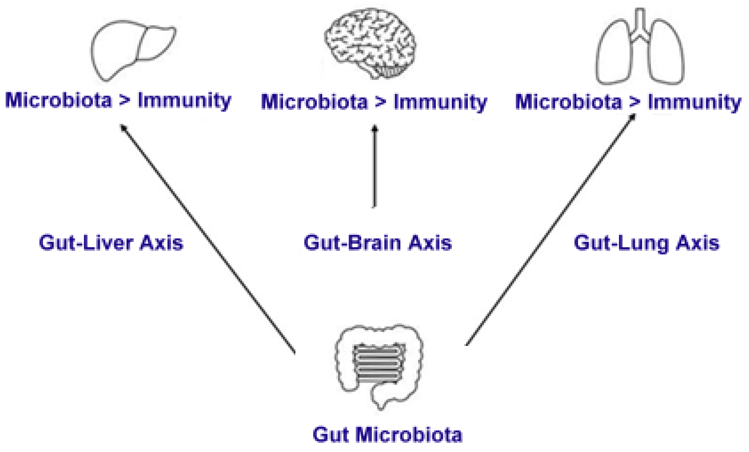
8. The Immune Response and Its Interaction with the Gut Microbiota

The immune response is a fundamental determinant of an individual’s susceptibility to infections, as well as the duration and resolution of such infections. The immune system is broadly classified into two primary components: the innate immune system and the adaptive immune system [64]. The innate immune system provides a nonspecific yet immediate line of defense through various protective mechanisms. These mechanisms include physical barriers, such as the skin and mucosal membranes, which serve as the first layer of defense against pathogenic invasion. Additionally, chemical barriers, such as antimicrobial proteins and enzymes, further contribute to host protection. Moreover, innate immune cells—including granulocytes, macrophages, and natural killer (NK) cells—play a crucial role in pathogen recognition and elimination [65].
In contrast, the adaptive immune system is highly specific and is primarily mediated by T and B lymphocytes. T cells are responsible for identifying and responding to pathogens that have invaded host cells, thereby constituting the cellular arm of adaptive immunity. Moreover, T cells exert regulatory control over B cells, which are responsible for antibody production. These antibodies bind to specific antigens and facilitate pathogen neutralization, a process that occurs within the bodily fluids, thus forming the basis of humoral immunity [66].

8.1. The Gut Microbiota and Immune System Development

The maturation and functional efficacy of the immune system are intrinsically linked to the composition and development of the gut microbiota. This intricate relationship has been substantiated by comparative studies involving age- and sex-matched germ-free mice, which lack commensal microflora, and conventionally raised counterparts of the same strain. Furthermore, the use of gnotobiotic mice, which are germ-free but reconstituted with a specific microbiota, has significantly advanced our understanding of how individual bacterial strains, combinations of strains, microbial gene expression, and microbe-derived metabolites influence both intestinal homeostasis and systemic immune responses [67].
Innate immunity plays a pivotal role in the initial detection of and response to microbial-derived products. Within the gastrointestinal tract, innate immune activation is primarily mediated by the intestinal epithelial cells (IECs), which form a single-layered barrier in direct contact with luminal antigens and microbial metabolites. The delicate balance between host immunity and the gut microbiota is maintained through the recognition of microbial components by pattern-recognition receptors (PRRs), which are expressed both extracellularly and intracellularly. PRRs are responsible for detecting microbe-associated molecular patterns (MAMPs), allowing for immune surveillance of the intestinal lumen. Key PRR families include TLRs, C-type lectin receptors (CLRs), nucleotide-binding oligomerization domain (NOD)-like receptors (NLRs), and cytosolic nucleic acid sensors, each of which contributes to host defense by detecting specific microbial signatures [68].
The engagement of PRRs initiates signaling cascades that result in the production of chemokines and cytokines, which coordinate innate immune responses. Myeloid differentiation primary response gene 88 (MyD88) is a critical adaptor molecule in PRR signaling, linking PRR activation to the nuclear factor kappa-light-chain-enhancer of activated B cells (NF-κB) pathway, a key regulator of inflammatory gene transcription. Consequently, deficiencies in MyD88 result in impaired immune responses, increasing host susceptibility to infections [69,70,71]. However, dysregulated activation of PRRs can provoke excessive immune responses, leading to chronic inflammation and autoimmune pathologies. To prevent such aberrant responses, PRR-mediated signaling is tightly regulated through positive and negative feedback mechanisms, as well as cross-regulatory interactions, which have been extensively characterized in the previous literature [72].
IECs also produce antimicrobial peptides (AMPs), which serve as effector molecules of the innate immune system, exhibiting bactericidal, anti-inflammatory, and endotoxin-neutralizing properties. AMPs contribute to host defense by restricting pathogen interactions with epithelial surfaces. Certain microbial pathogens have evolved mechanisms to downregulate AMP expression, while beneficial commensal bacteria can enhance their production. Thus, the composition of the gut microbiota is instrumental in shaping the innate immune response [73].

8.2. Microbial Metabolites and Immune Modulation

The gut microbiota influences immune function through the production of bioactive metabolites derived from dietary components, host-secreted factors, or microbial metabolic interactions. These metabolites exert immunoregulatory effects and contribute to the protective functions of commensal bacteria. Among these, SCFAs, tryptophan-derived metabolites, and bile acid derivatives have been extensively studied for their immunomodulatory properties [74].
SCFAs—primarily acetate, propionate, and butyrate—are produced through bacterial fermentation of dietary fiber. These metabolites reinforce intestinal barrier integrity by stimulating the production of mucus and antimicrobial peptides by IECs. Additionally, SCFAs promote the maturation and expansion of regulatory T (Treg) cells in the colon, thereby mitigating excessive inflammatory responses to microbial antigens [75]. SCFAs also support intestinal homeostasis by facilitating epithelial repair through the proliferation and differentiation of intestinal epithelial cells. Furthermore, they contribute to the expansion of innate lymphoid cells (ILC3), which secrete interleukin-22 (IL-22), a cytokine that enhances epithelial cell production of antimicrobial molecules [76].
Tryptophan metabolites, particularly indoles, are derived from the microbial metabolism of dietary tryptophan and serve as ligands for the aryl hydrocarbon receptor (AhR), a key regulator of mucosal homeostasis. Deficiencies in these metabolites have been associated with the pathogenesis of inflammatory bowel disease (IBD) [77,78]. Similarly, bile acid derivatives influence host immunity by modulating signaling pathways involving the farnesoid X receptor (FXR) and the G-protein-coupled bile acid receptor (TGR5). The enzymatic activity of bacterial bile salt hydrolases (BSHs) contributes to the metabolic transformation of bile acids. Notably, a reduction in BSH gene prevalence has been linked to an increased risk of IBD [79,80].
Mucosal homeostasis within the gastrointestinal tract is characterized by a dynamic interplay among gut microbiota, microbial metabolites, and host immune components. This precisely regulated state of low-grade inflammation is essential for maintaining immune tolerance and determining host susceptibility to infections [81,82].

8.3. Gut Microbiota and Systemic Immune Regulation

The influence of the gut microbiome extends beyond local mucosal immunity, affecting systemic immune responses mediated by both innate and adaptive immune cells. One mechanism involves the secretion of microbial-derived soluble factors into the circulation, which subsequently influences immune cell activation in peripheral tissues [83]. Additionally, resident immune cells in extraintestinal organs possess the ability to recognize and respond to microbial metabolites. The absence of these microbiota-derived signaling molecules leads to alterations in systemic immune function, increasing the host’s vulnerability to infections [83].
One of the most well-characterized mechanisms through which the gut microbiota modulates systemic immunity is its impact on the differentiation of adaptive immune cells, particularly T lymphocytes. Studies have demonstrated that the gut microbiota influences the balance among T-helper (Th) subsets, including Th1, Th2, and Th17 cells, as well as the induction of regulatory T cells [84,85,86]. SCFAs, particularly butyrate, facilitate the differentiation of peripherally induced Treg cells, thereby suppressing systemic inflammatory responses [87]. Additionally, pentanoate, another microbial metabolite, has been shown to regulate immune cell metabolism by promoting regulatory B cell differentiation while inhibiting Th17 cell expansion. This mechanism is particularly relevant in the pathogenesis of autoimmune and inflammatory disorders [88].
Other microbial-derived metabolites further shape adaptive immune responses. For instance, ATP produced by the gut microbiota stimulates Th17 cell proliferation, whereas tryptophan metabolites enhance intraepithelial CD4+CD8αα+ T cell populations. Additionally, bacterial polysaccharides have been identified as key activators of regulatory T cells, thereby dampening excessive inflammatory responses [89,90].
The ability of the gut microbiome to regulate both local and systemic immune responses has profound implications for host defense, immune homeostasis, and disease susceptibility. As research continues to uncover the intricate interactions between the microbiota and the immune system, a more comprehensive understanding of these mechanisms will inform therapeutic strategies aimed at modulating the gut microbiome to enhance immune resilience and mitigate inflammatory diseases. Interestingly, microbiota have the potential to bolster the immune response in patients with chronic HCV by stimulating an increase in the counts of CD56+ natural killer (NK) cells and CD3+ T cells. The augmentation of the cytotoxic function of NK cells may play a crucial role in suppressing HCV replication [91].
Figure 1 and Figure 2 provide an overview of the primary microbiota–immune system interactions.

9. Conclusions

Research exploring the complex interactions between human microbiota and hepatitis C virus (HCV) infections remains relatively limited. However, the existing body of literature offers valuable insights that could inform future investigations and contribute to the development of theoretical frameworks in this field. HCV infection, along with its progression to chronic liver diseases such as fibrosis, cirrhosis, and HCC, has been increasingly linked to alterations in gut microbiota composition. These alterations are often characterized by dysbiosis, a condition marked by disruptions in microbial diversity and imbalances in bacterial phyla.
A growing body of evidence suggests that gut microbial dysbiosis plays a critical role in the pathogenesis of HCV-related liver complications. The gut–liver axis—a bidirectional communication system between the gastrointestinal tract and the liver—facilitates the exchange of microbial metabolites, endotoxins, and inflammatory mediators. This axis is particularly relevant in HCV infection, as bacterial translocation and the leakage of microbial products from the intestine into the portal circulation have been implicated in exacerbating liver inflammation, promoting fibrosis, and contributing to the progression of liver disease. Notably, studies have identified reductions in beneficial bacterial taxa, such as those within the Firmicutes and Bacteroidetes phyla, alongside an increase in potentially pathogenic bacteria, including members of the Proteobacteria phylum, in patients with chronic HCV infection. These compositional shifts may influence systemic immune responses, modulate hepatic inflammation, and disrupt metabolic homeostasis, further complicating the course of HCV-related diseases.
Given the established role of the gut microbiota in maintaining immune homeostasis and liver function, therapeutic strategies aimed at restoring microbial equilibrium have garnered significant interest. One such intervention is fecal microbiota transplantation (FMT), which involves the transfer of a healthy donor’s gut microbiota to a recipient exhibiting dysbiosis. FMT has demonstrated efficacy in modulating gut microbial diversity, reducing inflammation, and improving metabolic parameters in various liver diseases, including non-alcoholic fatty liver disease (NAFLD) and cirrhosis. Therefore, it may hold therapeutic potential for HCV-infected patients experiencing gut dysbiosis, particularly those with advanced liver disease. However, while FMT represents a promising approach, its application in the context of HCV remains largely unexplored. Further research is required to determine its efficacy, optimal protocols, and long-term outcomes in this patient population.
A fundamental question that remains unresolved is whether dysbiosis precedes and exacerbates HCV infection or if chronic HCV infection itself drives microbial alterations. It is possible that pre-existing gut microbial imbalances create a microenvironment conducive to viral persistence and immune dysregulation, thereby predisposing individuals to more severe HCV-related liver damage. Conversely, chronic HCV infection, through mechanisms such as immune-mediated inflammation, hepatic metabolic alterations, and bile acid dysregulation, may contribute to progressive changes in gut microbiota composition. Longitudinal studies and mechanistic investigations are essential to elucidate the causal relationships between HCV infection and microbiota alterations, which could pave the way for microbiome-targeted interventions in the management of HCV-associated liver diseases.
In summary, while the current understanding of microbiota–HCV interactions is limited, emerging evidence underscores the critical role of the gut–liver axis in shaping the course of HCV infection and its complications. The potential therapeutic benefits of microbiome-modulating strategies, including FMT, warrant further exploration in the context of HCV. Future research should aim to clarify the bidirectional relationship between gut microbiota and HCV pathogenesis, identify microbial signatures associated with disease progression, and evaluate the efficacy of targeted microbiota-based therapies in improving clinical outcomes for HCV-infected individuals.

Author Contributions

Conceptualization, methodology, data curation, formal analysis, investigation, validation, supervision, writing original draft preparation, D.F.; resources, software, funding acquisition, project administration, writing—review and editing, Ș.Ț. All authors have read and agreed to the published version of the manuscript.

Funding

This research received no external funding.

Institutional Review Board Statement

Not applicable.

Informed Consent Statement

Not applicable.

Data Availability Statement

The data used to support the findings of this study are available from the corresponding authors upon request.

Conflicts of Interest

The authors declare no conflicts of interest.

Abbreviations

The following abbreviations are used in this manuscript:
APCsAntigen-Presenting Cells
CDCluster of Differentiation
CLRsC-type Lectin Receptors
DCsDendritic Cells
HCCHepatocellular Carcinoma
HCVHepatitis C Virus
IECsIntestinal Epithelial Cells
ILInterleukin
KIRKiller Immunoglobulin-like Receptors
NKNatural Killer
NKTNatural Killer T
NLRsNucleotide-binding Oligomerization Domain-like Receptors
PRRsPattern-Recognition Receptors
SCFAsShort-Chain Fatty Acids
TLRsToll-like Receptors
TregsRegulatory T Cells
UNIFRACUniFrac distance

References

  1. Bianconi, E.; Piovesan, A.; Facchin, F.; Beraudi, A.; Casadei, R.; Frabetti, F.; Vitale, L.; Pelleri, M.C.; Tassani, S.; Piva, F.; et al. An estimation of the number of cells in the human body. Ann. Hum. Biol. 2013, 40, 463–471. [Google Scholar] [CrossRef] [PubMed]
  2. Hao, W.L.; Lee, Y.K. Microflora of the gastrointestinal tract: A review. Methods Mol. Biol. 2004, 268, 491–502. [Google Scholar] [CrossRef] [PubMed]
  3. Whitman, W.B.; Coleman, D.C.; Wiebe, W.J. Prokaryotes: The unseen majority. Proc. Natl. Acad. Sci. USA 1998, 95, 6578–6583. [Google Scholar] [CrossRef]
  4. Grice, E.A.; Kong, H.H.; Conlan, S.; Deming, C.B.; Davis, J.; Young, A.C.; NISC Comparative Sequencing Program; Bouffard, G.G.; Blakesley, R.W.; Murray, P.R.; et al. Topographical and temporal diversity of the human skin microbiome. Science 2009, 324, 1190–1192. [Google Scholar] [CrossRef]
  5. Keijser, B.J.; Zaura, E.; Huse, S.M.; van der Vossen, J.M.; Schuren, F.H.; Montijn, R.C.; ten Cate, J.M.; Crielaard, W. Pyrosequencing analysis of the oral microflora of healthy adults. J. Dent. Res. 2008, 87, 1016–1020. [Google Scholar] [CrossRef]
  6. Preveden, T.; Scarpellini, E.; Milić, N.; Luzza, F.; Abenavoli, L. Gut microbiota changes and chronic hepatitis C virus infection. Expert Rev. Gastroenterol. Hepatol. 2017, 11, 813–819. [Google Scholar] [CrossRef] [PubMed]
  7. Buffie, C.G.; Pamer, E.G. Microbiota-mediated colonization resistance against intestinal pathogens. Nat. Rev. Immunol. 2013, 13, 790–801. [Google Scholar] [CrossRef]
  8. Maynard, C.L.; Elson, C.O.; Hatton, R.D.; Weaver, C.T. Reciprocal interactions of the intestinal microbiota and immune system. Nature 2012, 489, 231–241. [Google Scholar] [CrossRef]
  9. Hill, D.A.; Hoffmann, C.; Abt, M.C.; Du, Y.; Kobuley, D.; Kirn, T.J.; Bushman, F.D.; Artis, D. Metagenomic analyses reveal antibiotic-induced temporal and spatial changes in intestinal microbiota with associated alterations in immune cell homeostasis. Mucosal Immunol. 2010, 3, 148–158. [Google Scholar] [CrossRef]
  10. Mazmanian, S.K.; Liu, C.H.; Tzianabos, A.O.; Kasper, D.L. An immunomodulatory molecule of symbiotic bacteria directs maturation of the host immune system. Cell 2005, 122, 107–118. [Google Scholar] [CrossRef]
  11. Kuss, S.K.; Best, G.T.; Etheredge, C.A.; Pruijssers, A.J.; Frierson, J.M.; Hooper, L.V.; Dermody, T.S.; Pfeiffer, J.K. Intestinal microbiota promote enteric virus replication and systemic pathogenesis. Science 2011, 334, 249–252. [Google Scholar] [CrossRef]
  12. Robinson, C.M.; Jesudhasan, P.R.; Pfeiffer, J.K. Bacterial lipopolysaccharide binding enhances virion stability and promotes environmental fitness of an enteric virus. Cell Host Microbe 2014, 15, 36–46. [Google Scholar] [CrossRef]
  13. Kane, M.; Case, L.K.; Kopaskie, K.; Kozlova, A.; MacDearmid, C.; Chervonsky, A.V.; Golovkina, T.V. Successful transmission of a retrovirus depends on the commensal microbiota. Science 2011, 334, 245–249. [Google Scholar] [CrossRef] [PubMed]
  14. Miura, T.; Sano, D.; Suenaga, A.; Yoshimura, T.; Fuzawa, M.; Nakagomi, T.; Nakagomi, O.; Okabe, S. Histo-blood group antigen-like substances of human enteric bacteria as specific adsorbents for human noroviruses. J. Virol. 2013, 87, 9441–9451. [Google Scholar] [CrossRef] [PubMed]
  15. Vaarala, O. Is the origin of type 1 diabetes in the gut? Immunol. Cell Biol. 2012, 90, 271–276. [Google Scholar] [CrossRef] [PubMed]
  16. Vaarala, O. Human intestinal microbiota and type 1 diabetes. Curr. Diab. Rep. 2013, 13, 601–607. [Google Scholar] [CrossRef]
  17. Zipris, D. The interplay between the gut microbiota and the immune system in the mechanism of type 1 diabetes. Curr. Opin. Endocrinol. Diabetes Obes. 2013, 20, 265–270. [Google Scholar] [CrossRef]
  18. Li, H.C.; Lo, S.Y. Hepatitis C virus: Virology, diagnosis and treatment. World J. Hepatol. 2015, 7, 1377–1389. [Google Scholar] [CrossRef]
  19. Alberti, A.; Chemello, L.; Benvegnù, L. Natural history of hepatitis C. J. Hepatol. 1999, 31 (Suppl. S1), 17–24. [Google Scholar] [CrossRef]
  20. Hoofnagle, J.H. Course and outcome of hepatitis C. Hepatology 2002, 36 (Suppl. S1), S21–S29. [Google Scholar] [CrossRef]
  21. Chen, S.L.; Morgan, T.R. The natural history of hepatitis C virus (HCV) infection. Int. J. Med. Sci. 2006, 3, 47–52. [Google Scholar] [CrossRef] [PubMed]
  22. Carrozzo, M.; Scally, K. Oral manifestations of hepatitis C virus infection. World J. Gastroenterol. 2014, 20, 7534–7543. [Google Scholar] [CrossRef]
  23. Ozkok, A.; Yildiz, A. Hepatitis C virus associated glomerulopathies. World J. Gastroenterol. 2014, 20, 7544–7554. [Google Scholar] [CrossRef] [PubMed]
  24. Grimbert, S.; Valensi, P.; Lévy-Marchal, C.; Perret, G.; Richardet, J.P.; Raffoux, C.; Trinchet, J.C.; Beaugrand, M. High prevalence of diabetes mellitus in patients with chronic hepatitis C: A case-control study. Gastroenterol. Clin. Biol. 1996, 20, 544–548. [Google Scholar] [PubMed]
  25. Montenegro, L.; De Michina, A.; Misciagna, G.; Guerra, V.; Di Leo, A. Virus C hepatitis and type 2 diabetes: A cohort study in southern Italy. Am. J. Gastroenterol. 2013, 108, 1108–1111. [Google Scholar] [CrossRef]
  26. Preciado, M.V.; Valva, P.; Escobar-Gutierrez, A.; Rahal, P.; Ruiz-Tovar, K.; Yamasaki, L.; Vazquez-Chacon, C.; Martinez-Guarneros, A.; Carpio-Pedroza, J.C.; Fonseca-Coronado, S.; et al. Hepatitis C virus molecular evolution: Transmission, disease progression and antiviral therapy. World J. Gastroenterol. 2014, 20, 15992–16013. [Google Scholar] [CrossRef]
  27. Vercauteren, K.; de Jong, Y.P.; Meuleman, P. Animal models for the study of HCV. Curr. Opin. Virol. 2015, 13, 67–74. [Google Scholar] [CrossRef]
  28. Xi, Z.; Ramirez, J.L.; Dimopoulos, G. The Aedes aegypti toll pathway controls dengue virus infection. PLoS Pathog. 2008, 4, e1000098. [Google Scholar] [CrossRef]
  29. Wilks, J.; Golovkina, T. Influence of microbiota on viral infections. PLoS Pathog. 2012, 8, e1002681. [Google Scholar] [CrossRef]
  30. Isaak, D.D.; Bartizal, K.F.; Caulfield, M.J. Decreased pathogenicity of murine leukemia virus-Moloney in gnotobiotic mice. Leukemia 1988, 2, 540–544. [Google Scholar] [PubMed]
  31. Kouttab, N.M.; Jutila, J.W. Friend leukemia virus infection in germfree mice following antigen stimulation. J. Immunol. 1972, 108, 591–595. [Google Scholar] [CrossRef] [PubMed]
  32. Heidrich, B.; Vital, M.; Plumeier, I.; Döscher, N.; Kahl, S.; Kirschner, J.; Ziegert, S.; Solbach, P.; Lenzen, H.; Potthoff, A.; et al. Intestinal microbiota in patients with chronic hepatitis C with and without cirrhosis compared with healthy controls. Liver Int. 2018, 38, 50–58. [Google Scholar] [CrossRef] [PubMed]
  33. Trivedi, Y.; Bolgarina, Z.; Desai, H.N.; Senaratne, M.; Swami, S.S.; Aye, S.L.; Mohammed, L. The role of gut microbiome in hepatocellular carcinoma: A systematic review. Cureus 2023, 15, e43862. [Google Scholar] [CrossRef] [PubMed]
  34. Park, S.Y.; Seo, G.S. Fecal Microbiota Transplantation: Is It Safe? Clin. Endosc. 2021, 54, 157–160. [Google Scholar] [CrossRef] [PubMed]
  35. Zawistowska-Rojek, A.; Tyski, S. Are Probiotic Really Safe for Humans? Pol. J. Microbiol. 2018, 67, 251–258. [Google Scholar] [CrossRef]
  36. Adawi, D.; Ahrné, S.; Molin, G. Effects of different probiotic strains of Lactobacillus and Bifidobacterium on bacterial translocation and liver injury in an acute liver injury model. Int. J. Food Microbiol. 2001, 70, 213–220. [Google Scholar] [CrossRef]
  37. Bajaj, J.S.; Heuman, D.M.; Hylemon, P.B.; Sanyal, A.J.; Puri, P.; Sterling, R.K.; Luketic, V.; Stravitz, R.T.; Siddiqui, M.S.; Fuchs, M.; et al. Randomised clinical trial: Lactobacillus GG modulates gut microbiome, metabolome and endotoxemia in patients with cirrhosis. Aliment. Pharmacol. Ther. 2014, 39, 1113–1125. [Google Scholar] [CrossRef]
  38. Chiva, M.; Soriano, G.; Rochat, I.; Peralta, C.; Rochat, F.; Llovet, T.; Mirelis, B.; Schiffrin, E.J.; Guarner, C.; Balanzó, J. Effect of Lactobacillus johnsonii La1 and antioxidants on intestinal flora and bacterial translocation in rats with experimental cirrhosis. J. Hepatol. 2002, 37, 456–462. [Google Scholar] [CrossRef]
  39. Aly, A.M.; Adel, A.; El-Gendy, A.O.; Essam, T.M.; Aziz, R.K. Gut microbiome alterations in patients with stage 4 hepatitis C. Gut Pathog. 2016, 8, 42. [Google Scholar] [CrossRef]
  40. Bajaj, J.S.; Sterling, R.K.; Betrapally, N.S.; Nixon, D.E.; Fuchs, M.; Daita, K.; Heuman, D.M.; Sikaroodi, M.; Hylemon, P.B.; White, M.B.; et al. HCV eradication does not impact gut dysbiosis or systemic inflammation in cirrhotic patients. Aliment. Pharmacol. Ther. 2016, 44, 638–643. [Google Scholar] [CrossRef]
  41. Inoue, T.; Nakayama, J.; Moriya, K.; Kawaratani, H.; Momoda, R.; Ito, K.; Iio, E.; Nojiri, S.; Fujiwara, K.; Yoneda, M.; et al. Gut dysbiosis associated with hepatitis C virus infection. Clin. Infect. Dis. 2018, 67, 869–877. [Google Scholar] [CrossRef] [PubMed]
  42. El-Mowafy, M.; Elgaml, A.; El-Mesery, M.; Sultan, S.; Ahmed, T.A.E.; Gomaa, A.I.; Aly, M.; Mottawea, W. Changes of Gut-Microbiota-Liver Axis in Hepatitis C Virus Infection. Biology 2021, 10, 55. [Google Scholar] [CrossRef]
  43. Hsu, Y.C.; Chen, C.C.; Lee, W.H.; Chang, C.Y.; Lee, F.J.; Tseng, C.H.; Chen, T.H.; Ho, H.J.; Lin, J.T.; Wu, C.Y. Compositions of gut microbiota before and shortly after hepatitis C viral eradication by direct antiviral agents. Sci. Rep. 2022, 12, 5481. [Google Scholar] [CrossRef] [PubMed]
  44. Midori, Y.; Nosaka, T.; Hiramatsu, K.; Akazawa, Y.; Tanaka, T.; Takahashi, K.; Naito, T.; Matsuda, H.; Ohtani, M.; Nakamoto, Y. Isolation of mucosa-associated microbiota dysbiosis in the ascending colon in hepatitis C virus post-sustained virologic response cirrhotic patients. Front. Cell. Infect. Microbiol. 2024, 14, 1371429. [Google Scholar] [CrossRef] [PubMed]
  45. Yang, M.; Yang, Y.; He, Q.; Zhu, P.; Liu, M.; Xu, J.; Zhao, M. Intestinal Microbiota—A Promising Target for Antiviral Therapy? Front. Immunol. 2021, 12, 676232. [Google Scholar] [CrossRef]
  46. Trebicka, J.; Macnaughtan, J.; Schnabl, B.; Shawcross, D.L.; Bajaj, J.S. The microbiota in cirrhosis and its role in hepatic decompensation. J. Hepatol. 2021, 75 (Suppl. S1), S67–S81. [Google Scholar] [CrossRef]
  47. Yang, X.; Mai, H.; Zhou, J.; Li, Z.; Wang, Q.; Lan, L.; Lu, F.; Yang, X.; Guo, B.; Ye, L.; et al. Alterations of the gut microbiota associated with the occurrence and progression of viral hepatitis. Front. Cell. Infect. Microbiol. 2023, 13, 1119875. [Google Scholar] [CrossRef]
  48. Soldán, M.; Argalášová, Ľ.; Hadvinová, L.; Galileo, B.; Babjaková, J. The Effect of Dietary Types on Gut Microbiota Composition and Development of Non-Communicable Diseases: A Narrative Review. Nutrients 2024, 16, 3134. [Google Scholar] [CrossRef]
  49. Taur, Y.; Pamer, E.G. The intestinal microbiota and susceptibility to infection in immunocompromised patients. Curr. Opin. Infect Dis. 2013, 26, 332–337. [Google Scholar] [CrossRef]
  50. Adhikary, S.; Esmeeta, A.; Dey, A.; Banerjee, A.; Saha, B.; Gopan, P.; Duttaroy, A.K.; Pathak, S. Impacts of gut microbiota alteration on age-related chronic liver diseases. Dig. Liver Dis. 2024, 56, 112–122. [Google Scholar] [CrossRef]
  51. Corado, J.; Toro, F.; Rivera, H.; Bianco, N.E.; Deibis, L.; De Sanctis, J.B. Impairment of natural killer (NK) cytotoxic activity in hepatitis C virus (HCV) infection. Clin. Exp. Immunol. 1997, 109, 451–457. [Google Scholar] [CrossRef] [PubMed]
  52. Khakoo, S.I. HLA and NK cell inhibitory receptor genes in resolving hepatitis C virus infection. Science 2004, 305, 872–874. [Google Scholar] [CrossRef]
  53. Brown, M.G. Vital involvement of a natural killer cell activation receptor in resistance to viral infection. Science 2001, 292, 934–937. [Google Scholar] [CrossRef]
  54. Golden-Mason, L.; Rosen, H.R.; Golden-Mason, L. Natural killer cells: Primary target for hepatitis C virus immune evasion strategies? Liver Transplant. 2006, 12, 363–372. [Google Scholar] [CrossRef] [PubMed]
  55. Guidotti, L.G.; Chisari, F.V. Noncytolytic control of viral infections by the innate and adaptive immune response. Annu. Rev. Immunol. 2001, 19, 65–91. [Google Scholar] [CrossRef]
  56. Lunemann, S.; Schlaphoff, V.; Cornberg, M.; Wedemeyer, H. NK cells in hepatitis C: Role in disease susceptibility and therapy. Dig. Dis. 2012, 30, 48–54. [Google Scholar] [CrossRef]
  57. Doherty, D.G.; O’Farrelly, C. Innate and adaptive lymphoid cells in the human liver. Immunol. Rev. 2000, 174, 5–20. [Google Scholar] [CrossRef]
  58. Ahmad, A.; Alvarez, F. Role of NK and NKT cells in the immunopathogenesis of HCV-induced hepatitis. J. Leukoc. Biol. 2004, 76, 743–759. [Google Scholar] [CrossRef] [PubMed]
  59. Swain, M.G. Hepatic NKT cells: Friend or foe? Clin. Sci. 2008, 114, 457–466. [Google Scholar] [CrossRef]
  60. Lucas, M.; Gadola, S.; Meier, U.; Young, N.T.; Harcourt, G.; Karadimitris, A.; Coumi, N.; Brown, D.; Dusheiko, G.; Cerundolo, V.; et al. Frequency and phenotype of circulating Valpha24/Vbeta11 double-positive natural killer T cells during hepatitis C virus infection. J. Virol. 2003, 77, 2251–2257. [Google Scholar] [CrossRef]
  61. Deignan, T.; Curry, M.P.; Doherty, D.G.; Golden-Mason, L.; Volkov, Y.; Norris, S.; Nolan, N.; Traynor, O.; McEntee, G.; Hegarty, J.E.; et al. Decrease in hepatic CD56(+) T cells and V alpha 24(+) natural killer T cells in chronic hepatitis C viral infection. J. Hepatol. 2002, 37, 101–108. [Google Scholar] [CrossRef] [PubMed]
  62. Inoue, M.; Kanto, T.; Miyatake, H.; Itose, I.; Miyazaki, M.; Yakushijin, T.; Sakakibara, M.; Kuzushita, N.; Hiramatsu, N.; Takehara, T.; et al. Enhanced ability of peripheral invariant natural killer T cells to produce IL-13 in chronic hepatitis C virus infection. J. Hepatol. 2006, 45, 190–196. [Google Scholar] [CrossRef]
  63. Wynn, T.A. IL-13 effector functions. Annu. Rev. Immunol. 2003, 21, 425–456. [Google Scholar] [CrossRef]
  64. Parkin, J.; Cohen, B. An overview of the immune system. Lancet 2001, 357, 1777–1789. [Google Scholar] [CrossRef] [PubMed]
  65. Hillion, S.; Arleevskaya, M.I.; Blanco, P.; Bordron, A.; Brooks, W.H.; Cesbron, J.Y.; Kaveri, S.; Vivier, E.; Renaudineau, Y. The Innate Part of the Adaptive Immune System. Clin. Rev. Allergy Immunol. 2020, 58, 151–154. [Google Scholar] [CrossRef]
  66. Bonilla, F.A.; Oettgen, H.C. Adaptive immunity. J. Allergy Clin. Immunol. 2010, 125, S33–S40. [Google Scholar] [CrossRef]
  67. Fiebiger, U.; Bereswill, S.; Heimesaat, M.M. Dissecting the interplay between intestinal microbiota and host immunity in health and disease: Lessons learned from germfree and gnotobiotic animal models. Eur. J. Microbiol. Immunol. 2016, 6, 253–271. [Google Scholar] [CrossRef] [PubMed]
  68. Fukata, M.; Arditi, M. The role of pattern recognition receptors in intestinal inflammation. Mucosal Immunol. 2013, 6, 451–463. [Google Scholar] [CrossRef]
  69. Frantz, A.L.; Rogier, E.W.; Weber, C.R.; Shen, L.; Cohen, D.A.; Fenton, L.A.; Bruno, M.E.C.; Kaetzel, C.S. Targeted deletion of MyD88 in intestinal epithelial cells results in compromised antibacterial immunity associated with downregulation of polymeric immunoglobulin receptor, mucin-2, and antibacterial peptides. Mucosal Immunol. 2012, 5, 501–512. [Google Scholar] [CrossRef]
  70. Araki, A.; Kanai, T.; Ishikura, T.; Makita, S.; Uraushihara, K.; Iiyama, R.; Totsuka, T.; Takeda, K.; Akira, S.; Watanabe, M. MyD88-deficient mice develop severe intestinal inflammation in dextran sodium sulfate colitis. J. Gastroenterol. 2005, 40, 16–23. [Google Scholar] [CrossRef]
  71. Kubinak, J.L.; Petersen, C.; Stephens, W.Z.; Soto, R.; Bake, E.; O’Connell, R.M.; Round, J.L. MyD88 signaling in T cells directs IgA-mediated control of the microbiota to promote health. Cell Host Microbe 2015, 17, 153–163. [Google Scholar] [CrossRef] [PubMed]
  72. Cao, X. Self-regulation and cross-regulation of pattern-recognition receptor signalling in health and disease. Nat. Rev. Immunol. 2016, 16, 35–50. [Google Scholar] [CrossRef]
  73. Vandamme, D.; Landuyt, B.; Luyten, W.; Schoofs, L. A comprehensive summary of LL-37, the factotum human cathelicidin peptide. Cell. Immunol. 2012, 280, 22–35. [Google Scholar] [CrossRef]
  74. Chen, F.; Stappenbeck, T.S. Microbiome control of innate reactivity. Curr. Opin. Immunol. 2019, 56, 107–113. [Google Scholar] [CrossRef] [PubMed]
  75. Schnupf, P.; Gaboriau-Routhiau, V.; Cerf-Bensussan, N. Modulation of the gut microbiota to improve innate resistance. Curr. Opin. Immunol. 2018, 54, 137–144. [Google Scholar] [CrossRef]
  76. Chun, E.; Lavoie, S.; Fonseca-Pereira, D.; Bae, S.; Michaud, M.; Hoveyda, H.R.; Fraser, G.L.; Comeau, C.A.G.; Glickman, J.N.; Fuller, M.H.; et al. Metabolite-sensing receptor Ffar2 regulates colonic group 3 innate lymphoid cells and gut immunity. Immunity 2019, 51, 871–884. [Google Scholar] [CrossRef] [PubMed]
  77. Rannug, A. How the AHR became important in intestinal homeostasis-a diurnal FICZ/AHR/CYP1A1 feedback controls both immunity and immunopathology. Int. J. Mol. Sci. 2020, 21, 5681. [Google Scholar] [CrossRef]
  78. Lloyd-Price, J.; IBDMDB Investigators; Arze, C.; Ananthakrishnan, A.N.; Schirmer, M.; Avila-Pacheco, J.; Poon, T.W.; Andrews, E.; Ajami, N.J.; Bonham, K.S.; et al. Multi-omics of the gut microbial ecosystem in inflammatory bowel diseases. Nature 2019, 569, 655–662. [Google Scholar] [CrossRef]
  79. Baars, A.; Oosting, A.; Knol, J.; Garssen, J.; Van Bergenhenegouwen, J. The gut microbiota as a therapeutic target in IBD and metabolic disease: A role for the bile acid receptors FXR and TGR5. Microorganisms 2015, 3, 641–666. [Google Scholar] [CrossRef]
  80. Labbé, A.; Ganopolsky, J.G.; Martoni, C.J.; Prakash, S.; Jones, M.L. Bacterial bile metabolising gene abundance in Crohn’s, ulcerative colitis and type 2 diabetes metagenomes. PLoS ONE 2014, 9, e115175. [Google Scholar] [CrossRef]
  81. Biagi, E.; Candela, M.; Turroni, S.; Garagnani, P.; Franceschi, C.; Brigidi, P. Ageing and gut microbes: Perspectives for health maintenance and longevity. Pharmacol. Res. 2013, 69, 11–20. [Google Scholar] [CrossRef]
  82. Negi, S.; Das, D.K.; Pahari, S.; Nadeem, S.; Agrewala, J.N. Potential role of gut microbiota in induction and regulation of innate immune memory. Front. Immunol. 2019, 10, 2441. [Google Scholar] [CrossRef] [PubMed]
  83. Blander, J.M.; Longman, R.S.; Iliev, I.D.; Sonnenberg, G.F.; Artis, D. Regulation of inflammation by microbiota interactions with the host. Nat. Immunol. 2017, 18, 851–860. [Google Scholar] [CrossRef] [PubMed]
  84. Honda, K.; Littman, D.R. The microbiota in adaptive immune homeostasis and disease. Nature 2016, 535, 75–84. [Google Scholar] [CrossRef]
  85. Francino, M.P. Early development of the gut microbiota and immune health. Pathogens 2014, 3, 769–790. [Google Scholar] [CrossRef] [PubMed]
  86. Owaga, E.; Hsieh, R.-H.; Mugendi, B.; Masuku, S.; Shih, C.-K.; Chang, J.-S. Th17 cells as potential probiotic therapeutic targets in inflammatory bowel diseases. Int. J. Mol. Sci. 2015, 16, 20841–20858. [Google Scholar] [CrossRef]
  87. Arpaia, N.; Campbell, C.; Fan, X.; Dikiy, S.; Van Der Veeken, J.; DeRoos, P.; Liu, H.; Cross, J.R.; Pfeffer, K.; Coffer, P.J.; et al. Metabolites produced by commensal bacteria promote peripheral regulatory T-cell generation. Nature 2013, 504, 451–455. [Google Scholar] [CrossRef]
  88. Luu, M.; Pautz, S.; Kohl, V.; Singh, R.; Romero, R.; Lucas, S.; Hofmann, J.; Raifer, H.; Vachharajani, N.; Carrascosa, L.C.; et al. The short-chain fatty acid pentanoate suppresses autoimmunity by modulating the metabolic-epigenetic crosstalk in lymphocytes. Nat. Commun. 2019, 10, 760. [Google Scholar] [CrossRef]
  89. Ahern, P.P.; Maloy, K.J. Understanding immune-microbiota interactions in the intestine. Immunology 2020, 159, 4–14. [Google Scholar] [CrossRef]
  90. Grainger, J.; Daw, R.; Wemyss, K. Systemic instruction of cell-mediated immunity by the intestinal microbiome. F1000Research 2018, 7, 1910. [Google Scholar] [CrossRef]
  91. Doskali, M.; Tanaka, Y.; Ohira, M.; Ishiyama, K.; Tashiro, H.; Chayama, K.; Ohdan, H. Possibility of adoptive immunotherapy with peripheral blood-derived CD3CD56+ and CD3+CD56+ cells for inducing antihepatocellular carcinoma and antihepatitis C virus activity. J. Immunother. 2011, 34, 129–138. [Google Scholar] [CrossRef] [PubMed]
Figure 1. Depiction of microbiota axes.
Applsci 15 03157 g001
Figure 2. Depiction of digestive interactions with the immune system.
Applsci 15 03157 g002
Table 1. Summary of studies on HCV infection and gut microbiota alterations.
Study/ReferencePopulationKey Findings
Aly et al., 2016 [39]6 HCV-infected cirrhotic patients vs. 8 healthy controlsSignificant reduction in gut microbiome diversity in HCV-infected patients. The potential alteration of the microbiome in chronic hepatitis C may be influenced by bacterial translocation, alongside the liver’s compromised functions in digestion and protein synthesis.
Inoue et al., 2018 [41]166 HCV-infected patients vs. 23 healthy controlsGut dysbiosis observed even in asymptomatic patients; progression of clinical stage associated with further dysbiosis. In individuals infected with HCV, there was a notable reduction in bacterial diversity when contrasted with healthy subjects. This decline was particularly evident in the Clostridiales order, while an increase was observed in the populations of Streptococcus and Lactobacillus.
Hsu et al., 2022 [43]42 HCV-infected patients vs. 84 healthy controlsAltered gut microbiome composition observed; specific bacterial taxa changes noted. The gut microbiota in patients infected with HCV exhibits alterations when compared to uninfected controls; however, the overall composition of the microbiome does not show significant changes in the short term following the eradication of HCV.
Table 2. HCV and immune system—key aspects.
AspectDetails
Innate Immune Response in HCVCellular innate immune response is crucial in host defense against HCV infection
Role of NK CellsNK cells are essential for early antiviral immunity, comprising 30–50% of liver lymphocytes
NK Cell ClassificationCD16 + CD56dim NK cells: cytotoxic, strong effector functions (target cell lysis)
CD16 − CD56bright NK cells: immunoregulatory, cytokine-producing
Impact of Other Infections on NK CellsInfections like cytomegalovirus, Epstein-Barr, herpes virus reduce NK cell cytotoxic activity
NK Cell Functions in HCV InfectionSecrete pro-inflammatory cytokines (TNF-α, IFN-γ) to suppress HCV replication.
NK-Cell-Induced Liver InjuryCytotoxic response can lead to unintended liver injury and inflammation
Killer Immunoglobulin-like Receptors (KIRs)KIRs interact with MHC class I molecules, regulating NK cell activity
NK Cells in Early HCV InfectionContribute to early viral load reduction through cytolysis, cytokine secretion, and activation of other immune cells
Adaptive Immune Response ActivationDendritic cells (APCs) process and present viral antigens to activate CD4+ helper T cells, CD8+ cytotoxic T cells, and B cells
Interaction between Dendritic Cells and NK Cells NKp30 receptor triggers IL-12 and IL-15 release from dendritic cells, enhancing NK cell function and cytokine production (IFN-γ, TNF-α)
Natural Killer T (NKT) CellsNKT cells comprise 26% of intrahepatic lymphocytes, secreting IFN-γ, TNF-α, IL-2
NKT Cells in Chronic HCVConflicting findings on NKT cell frequency: some studies show increased, others show decreased NKT cells in the liver of chronic HCV patients
NKT Cells Cytokine Production in HCVAltered cytokine profiles, particularly IL-13, which regulates cell-mediated immunity and may influence chronic HCV progression
Disclaimer/Publisher’s Note: The statements, opinions and data contained in all publications are solely those of the individual author(s) and contributor(s) and not of MDPI and/or the editor(s). MDPI and/or the editor(s) disclaim responsibility for any injury to people or property resulting from any ideas, methods, instructions or products referred to in the content.

