Article

Comparative Effects of Rubber Dam and Traditional Isolation Techniques on Orthodontic Bracket Positioning: A 3D Digital Model Evaluation

by
Türkan Sezen Erhamza
*,
Kadir Can Küçük
and
İsmayil Malikov
Department of Orthodontics, Faculty of Dentistry, Kırıkkale University, Kırıkkale 71450, Turkey
*
Author to whom correspondence should be addressed.
Appl. Sci. 2025, 15(5), 2552; https://doi.org/10.3390/app15052552
Submission received: 22 January 2025 / Revised: 25 February 2025 / Accepted: 26 February 2025 / Published: 27 February 2025

Abstract

:
Dental professionals face an increased risk of exposure to biological fluids, aerosols, and droplets due to close patient contact, which heightens the risk of infectious diseases. Rubber dam, commonly used in dentistry, not only isolates treatment areas but also reduces aerosol and droplet dispersion. Accurate orthodontic bracket positioning is crucial for optimal treatment, and isolation techniques like rubber dam and traditional methods are essential for ensuring precise bracket placement and bonding. This study aims to compare the effects of rubber dam and traditional isolation techniques on orthodontic bracket positioning using 3D digital models, while also evaluating the impact of these methods on the patient’s chair time during the procedure. The study group (RDI—Rubber Dam Isolation) included individuals isolated with a rubber dam, while the control group (TI—Traditional Isolation) consisted of those isolated using retractors and cotton rolls. Digital models were taken from these groups before bracketing (BB) and after bracketing (AB). BB models were transferred to the OrthoanalyzerTM program for virtual bracketing and a virtual bonding model (VB) was created. AB and VB models were superimposed in the GOM InspectTM program in order to determine the accuracy of the bracket positions. Linear measurements were taken along the X, Y, and Z axes, while angular measurements were recorded on the XY, XZ, and YZ planes. There was no significant difference in deviation values along the X-axis between the RDI and TI groups. In both groups, the lowest deviation values in linear measurements were found in the Z-axis, while the highest deviation values were found in the Y-axis. In the Y-axis, it was found that the deviation values were higher in the RDI group for tooth numbers 32 and 33, and in the Z-axis, the deviation values were higher in the RDI group for tooth numbers 34 and 44. In angular measurements, it was observed that in the XY plane, the deviation values in tooth number 35 were higher in the TI group. RDI proves to be an effective method for ensuring accurate bracket positioning in orthodontic procedures when compared to traditional isolation techniques. Especially considering infectious diseases, the use of RDI is considered appropriate.

1. Introduction

Infectious and contagious diseases such as Hepatitis C virus, Hepatitis A virus, Hepatitis B virus, Human Immunodeficiency Virus, Severe Acute Respiratory Syndrome, Coronavirus Disease 2019, measles, and tuberculosis pose a significant risk to healthcare workers [1,2,3]. Dental professionals are at high risk for nosocomial infection, particularly in the context of the widespread transmission of SARS-CoV-2. This risk stems from the unique nature of dental interventions, which often involve aerosol generation, handling sharps, and the provider being in close proximity to the patient’s oropharyngeal region [4]. The most significant factors contributing to the heightened risk of infection for dental practitioners are their frequent exposure to saliva, blood, and the creation of aerosols or droplets during various dental procedures. In these procedures, the transmission of SARS-CoV-2 can occur through several routes, including the inhalation of aerosols or droplets from infected patients, direct contact with mucous membranes, oral fluids, and contaminated instruments or surfaces [5].
To reduce the risk of exposure to harmful microorganisms during dental procedures, the Center for Disease Control and Prevention (CDC) recommends that dental healthcare providers (DHCPs) implement protective barriers on clinical surfaces and wear personal protective equipment (PPE) such as gloves, masks, goggles, and gowns. These precautions are intended to safeguard the skin and mucous membranes of the eyes, nose, and mouth from potential exposure to infectious agents. Furthermore, the CDC advises using high-velocity suction and rubber dams to limit the spread of aerosols generated during rotary dental procedures [6].
A rubber dam, a thin, disposable rubber sheet, is applied around the tooth or teeth being treated, creating a barrier that isolates the treatment area from saliva. It is a standard tool in restorative and endodontic procedures and has been linked to higher success rates in these treatments [6]. In one particular study, Cochran et al. found that the combination of high-speed rotary instruments and air–water syringes, either with or without a rubber dam, demonstrated that the rubber dam alone prevented up to 90–98% of bio-aerosols from spreading. Similarly, Samaranayake and colleagues reported a 70% decrease in airborne particles within a 1 m radius of the treatment site, with no notable effect on surfaces located 3 m away [7].
In addition to the advantages of rubber dam usage, there are also certain disadvantages associated with its application. These include the time required for placement, the difficulty of use, and the material cost [3]. However, the time considered as a loss during placement is more than compensated for by the time saved through the retraction of the tongue and lips, the reduction in contamination risk, and the elimination of the constant need for replacing cotton rolls [8,9]. In a study using three different rubber dam systems, the average time for rubber dam application was found to be 51 s (ranging from 38 to 79 s). The application times for OptiDam™ was 42 s, for the traditional method it was 51 s, and for OptraDam™ Plus it was 53 s. The removal times for the rubber dam were 12 s for the traditional method, 14 s for OptraDam™ Plus, and 10 s for OptiDam™ [10]. When comparing isolation techniques used in pediatric dentistry, including the Isolite system, rubber dam, and cotton roll isolation methods, the average times spent at the patient’s chair were approximately 248, 256, and 243 s, respectively. No significant differences were observed in procedure times among the isolation methods, and it was found that the rubber dam placement time decreased as children grew older [11].
Fixed appliances in orthodontics began to be used in 1887 by Edward Angle. This system, defined as the ‘Edgewise system’, forms the basis of current orthodontic approaches. Due to its disadvantages for both orthodontists and patients, this technique has been largely abandoned in favor of the ‘Straightwire technique’ defined by Andrews. In this technique, the brackets contain values such as tip, torque, and in/out that are crucial for transferring forces to the teeth. The most important aspect is determining the correct placement of these self-contained value brackets [12].
Andrews defined the reference point for bracket placement as the Facial Axis of Clinical Crown (FACC) of the tooth. The midpoint of this axis (FA) is where the bracket should be positioned. The facial axis of the bracket must be parallel to the FACC, and the center of the bracket must be located at the FA point. The base of the bracket is sloped to achieve the appropriate torque value, and the bracket slot itself does not contain torque values. The bracket base has both vertical and horizontal contours, which ensure a reliable placement of the bracket [12].
The distance from the slot base to the bracket base varies for each tooth, reflecting the in/out values. The in/out, tip, and torque values contained in the bracket minimize the need for bending, thereby reducing treatment time and the duration of patient visits. Brackets incorporating anti-tip and anti-rotation features facilitate translational movement in extraction treatments [12].
The straight wire technique is dependent on the precise positioning of brackets, followed by the application of the archwire to align the teeth in all three planes [12,13]. Therefore, in orthodontic treatment, the position, height, torque, and angulation of the brackets are crucial components that have an impact on the treatment outcome [14].
This study aims to compare the effects of rubber dam and traditional isolation techniques on orthodontic bracket positioning using 3D digital models, while also evaluating the impact of these methods on the patient’s chair time during the procedure.

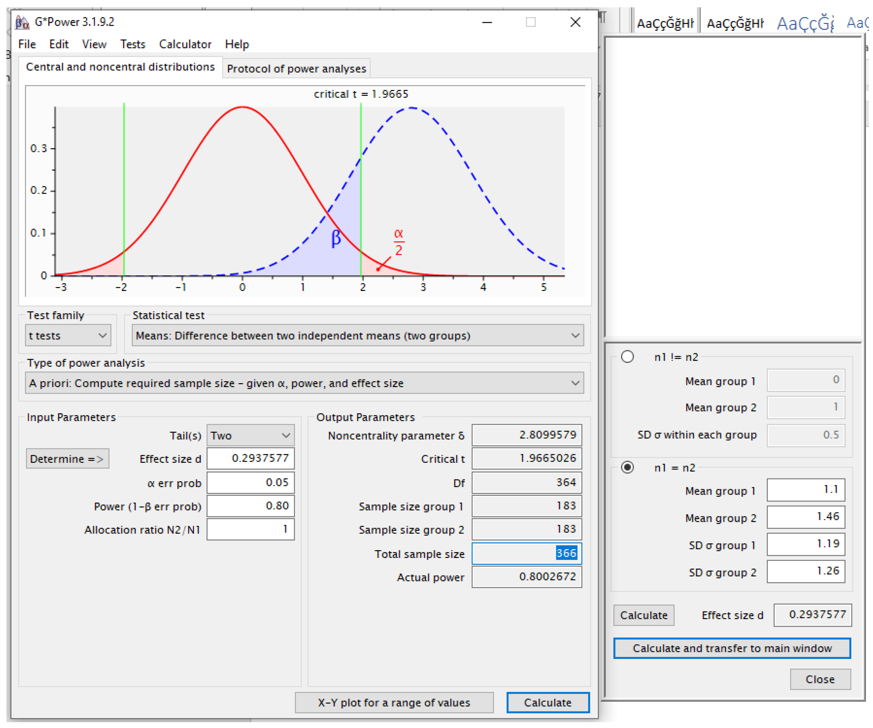
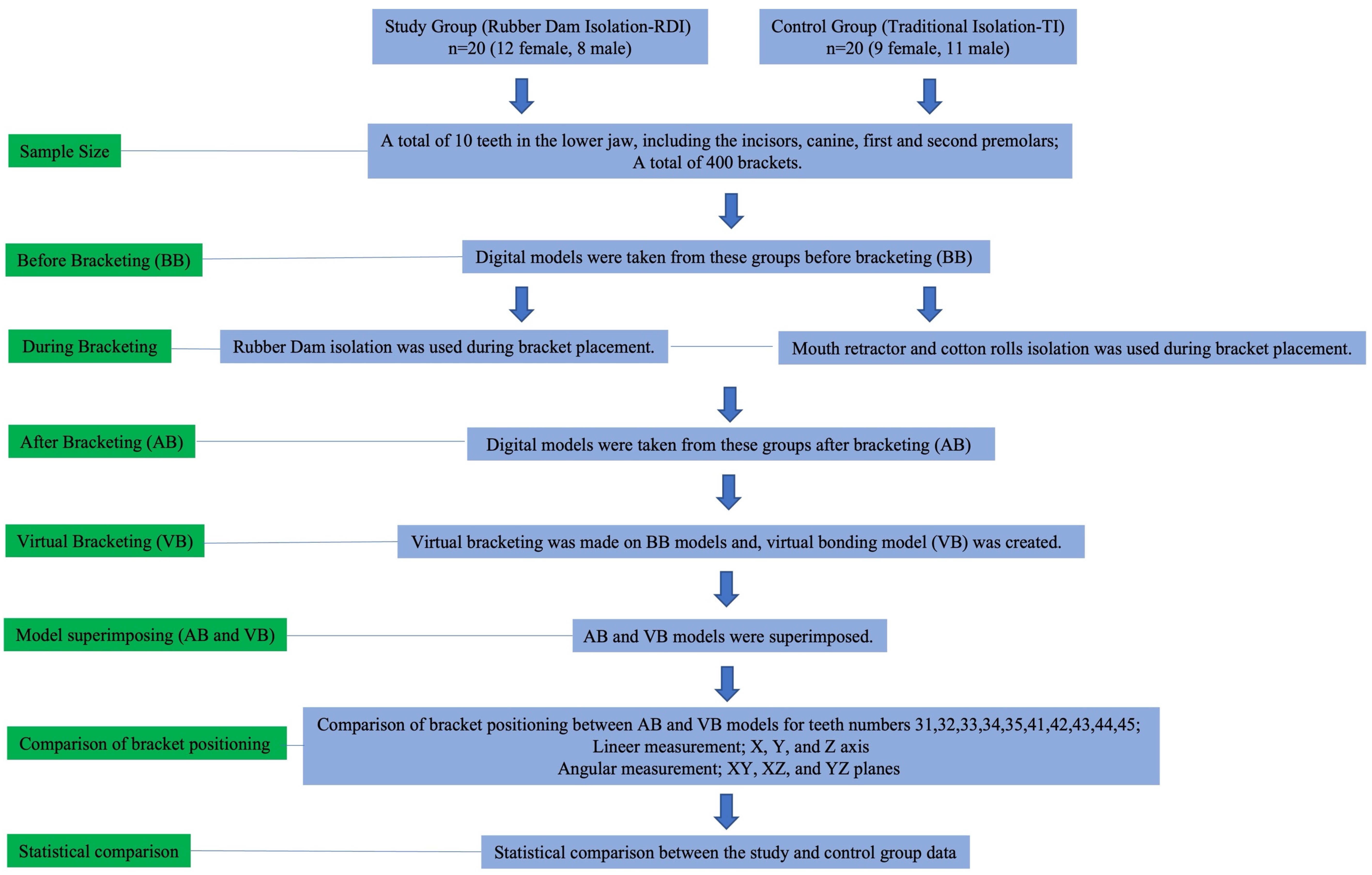
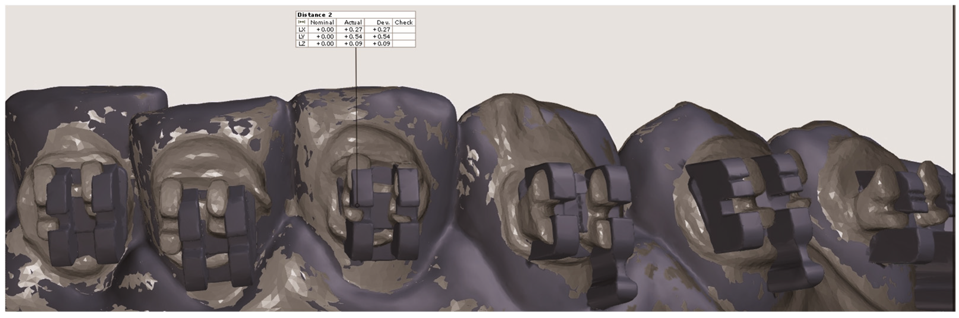
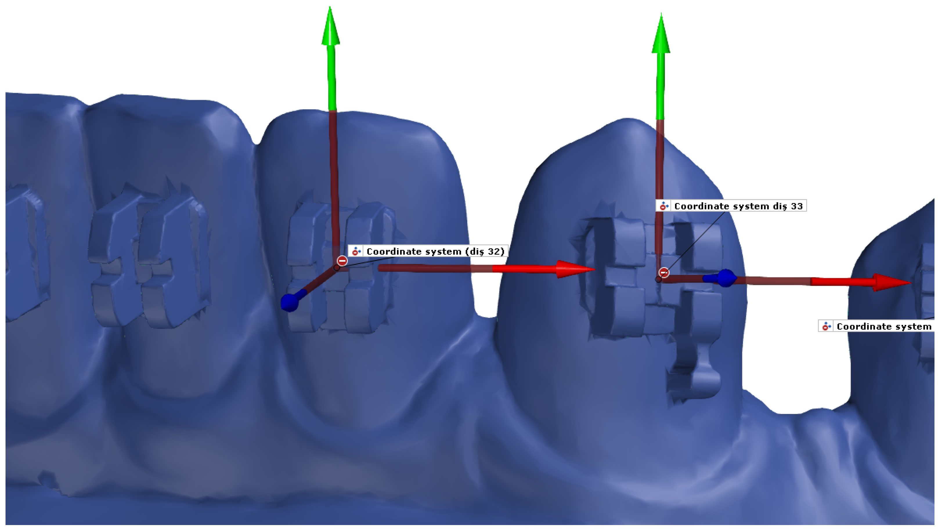
2. Materials and Methods

This study, designed as a retrospective analysis, obtained ethical approval from the Kırıkkale University Non-Interventional Research Ethics Committee (Decision number: 2022.03.30) and has been supported by project number 2022/115 from the Kırıkkale University Scientific Research Projects Coordination Unit. Informed consent to participate was obtained from all the participants in this study.
The inclusion and exclusion criteria for participants were defined to ensure the ideal positioning of brackets. These criteria included [15,16,17] the following:
-
No missing teeth in the mandible. To maintain statistical consistency when comparing the teeth only patients with a complete dentition in the arch were included in this study.
-
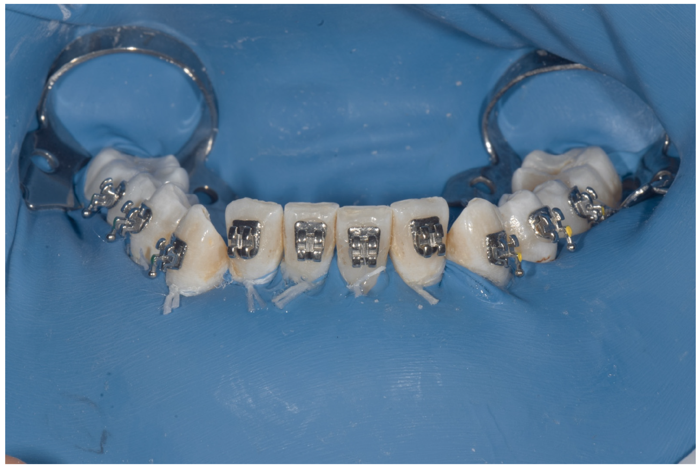
Complete permanent dentition with all teeth fully erupted. Attention was given to ensuring the presence of fully erupted teeth for the correct placement of the brackets.
-
The mandible was used. Based on previous studies [18,19], the mandible was preferred due to the minimal crowding when comparing this study and control group patients.
-
Crowding of less than 4 mm.
-
No active dental caries, fractures, dimensional anomalies, or dental prostheses.
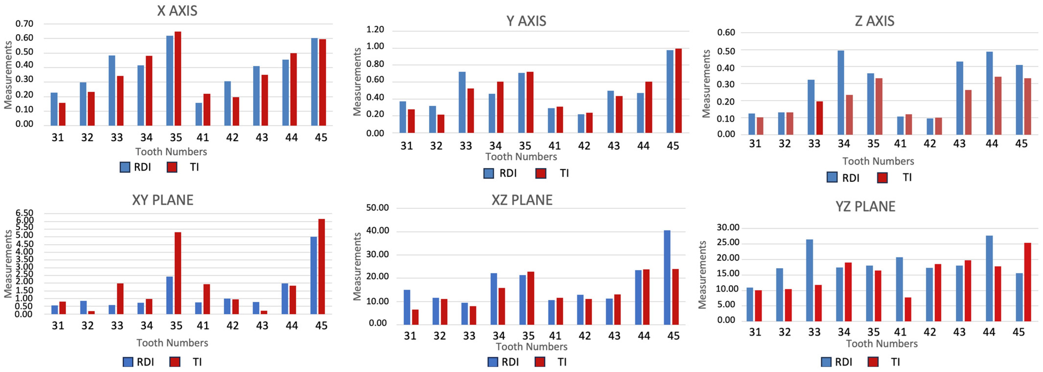
These criteria were selected to ensure that all participants had a stable and well-aligned dental structure, allowing for optimal bracket placement and minimizing potential confounding factors that could affect the outcomes of this study. The study group was selected from patients who were bracketed using rubber dam isolation during the pandemic, while the control group consisted of patients bracketed with mouth retractors and cotton rolls isolation.
In the study by Kim et al. [20], the mean values of bracket superimposition for the study group were found to be 1.10 ± 1.19, while the mean values for the control group were 1.46 ± 1.26. In our study, it was assumed that the 0.35-degree difference in superimposition between the two groups would be considered significant. The effect size (d) was calculated as 0.29, with 80% power and a 0.05 significance level. Based on these parameters, a minimum of 183 brackets per group, totaling 366 brackets, was required for this study. However, in our study, a total of 400 brackets were included. The calculations were performed using the ’GPower 3.1.9.2’ software (Figure 1). To increase the power of this study, 400 brackets have been included. For each individual, a total of 10 teeth in the lower jaw, including the incisors, canine, first and second premolars, were examined, resulting in the assessment of a total of 400 brackets.
A total of 40 individuals (21 females and 19 males, aged 15 to 25) were divided into two groups: the study group (12 females and 8 males, with a mean age of 21.2 years) and the control group (9 females and 11 males, with a mean age of 21.3 years). In this study, the group that received bracketing with rubber dam isolation (RDI) was designated as the study group, while the control group (TI—traditional isolation) included patients who underwent bracketing using a mouth retractor and cotton rolls. The flowchart of this study is presented in Figure 2.
OptiDam™ (Kerr Hawe SA, Bioggio, Switzerland) rubber dam was used in the RDI group. The protrusions found on the rubber dam were punctured using a dental bur. The rubber dam was placed onto the teeth and secured with a clamp. To prevent the rubber dam from slipping coronally, a ligation process was performed using dental floss (Figure 3).
In both groups, 0.022-inch MBT Prescription Gemini Twin brackets (3M Unitek, Monrovia, CA, USA) were used. The brackets were positioned according to MBT (McLaughlin, Bennett, Trevisi) guidelines [13], with measurements taken from the cusp or incisal edge using a gauge. Bonding was performed by the same operator with 4 years of experience (KCK).
To determine the chair time with each patient, a stopwatch was managed by the dentist’s assistant for all individuals. The time measured included, for the RDI group, the patient’s positioning in the dental chair, application of the rubber dam, bonding, and removal of the rubber dam; and for the TI group, the patient’s positioning in the chair, placement of the mouth retractor, bonding, and removal of the retractor.
For the individuals included in this study, before-bracketing scan models (BB) and after-bracketing intraoral scan models (AB) were obtained using the 3 Shape intraoral scanner device (Trios 3, Copenhagen, Denmark). The scan data in STL format were transferred to the Orthoanalyzer software (Orthoanalyzer 1.11.1.1, Copenhagen, Denmark). On the BB models, 0.022-inch MBT Prescription Gemini Twin brackets were placed according to MBT criteria by an orthodontic expert and checked by three experts (KCK, TSE, IM) [14]. The digital bracket placement led to the creation of virtual bonding models (VB) (Figure 4) and these models are considered the gold standard.
In both groups (TI-RDI), superimposition was performed using the GOM Inspect software (GOM, V8 SR1, Braunschweig, Germany) to enable comparison of the AB models with the VB models (Figure 5).
A local coordinate system was defined on each tooth in order to ensure the standardization of the measurements made. Planes were defined by selecting three points on the bracket surfaces to create local coordinate systems (Figure 6). For each local coordinate system, the X-axis was designated to represent the mesiodistal direction, the Y-axis the occlusogingival direction, and the Z-axis the buccolingual direction.
For each tooth, the angle between the planes in the superimposed model was created. The measurements of the resulting angle were taken along the XY, XZ, and YZ axes of the tooth’s local coordinate system, and the angular deviations for each bracket were calculated in degrees.
The data were analyzed using IBM SPSS Statistics for Windows, version 29.0.1.0 (IBM Corp., Armonk, NY, USA). Descriptive statistics for continuous data included mean, standard deviation, median, minimum, and maximum values, while for discrete data, frequency and percentage values were provided. The normality of continuous data was assessed using the Shapiro–Wilk test. The analysis revealed that more than half of the variables did not follow a normal distribution. In the variables that were normally distributed, it was observed that the variances were not equal, leading to the decision to use non-parametric methods. Among the non-parametric test methods, the Mann–Whitney U test was chosen, considering the need to examine two independent variables, the equality of sample sizes in the test groups, and the continuous and quantitative nature of the data obtained in this study. After superimposing the models, the Mann–Whitney U test was used to compare the linear and angular values between the RDI and TI groups. A p-value of less than 0.05 indicates a significant difference within the limits of 0.5 mm for linear measurements and 2° for angular measurements, while a p-value greater than 0.05 indicates no significant difference. These limits were selected based on the professional standards of the Objective Grading System of the American Board of Orthodontics.
The chair time measurements with each patient were compared using the parametric t-test for independent groups.

3. Results

There was no statistically significant difference (p > 0.05) between the 2 groups for the X-axis. For the Y-axis, a statistically significant difference (p < 0.05) was found between the two groups for teeth numbers 32 and 33, with the deviation being higher in the RDI group. For the Z-axis, a statistically significant difference (p < 0.05) was found between the two groups for teeth numbers 34 and 44. In the RDI group, the deviation values for teeth numbers 34 and 44 were measured to be higher (Table 1, Figure 7).
For the XY plane, a statistically significant difference (p < 0.05) was found between the two groups for tooth number 35, with the deviation being higher in the TI group. For the XZ plane, a statistically significant difference (p < 0.05) was found between the two groups for tooth number 45, with the deviation values being higher in the RDI group. For the YZ plane, a statistically significant difference (p < 0.05) was found between the two groups for teeth numbers 33 and 41, with the deviation being higher in the RDI group (Table 2, Figure 7).
Upon examining the application times, a statistically significant difference (p < 0.05) was found between the two groups, with the application time being higher in the RDI group (Table 3).

4. Discussion

Healthcare workers are 10 times more likely to contract infectious diseases than other workers. Infectious diseases such as HCV, HAV, HBV, and HIV pose a great risk for healthcare professionals [1]. During the COVID-19 pandemic, healthcare workers constituted 21% of all cases and 9.6% of all fatalities [21]. Since infections are usually transmitted through droplets, dentists use all protective barrier equipment, including protective goggles, masks, gloves, bonnets, face shields, protective outerwear [22]. Particularly during periods like the COVID-19 pandemic, additional protective measures are considered essential. Rubber dams, combined with powerful suction, minimize the likelihood of aerosol or spatter contamination from saliva and blood. Studies indicate that employing a rubber dam can lead to a 70% decrease in airborne particles within a radius of approximately three feet from the treatment area [7]. Upon reviewing the literature, it was observed that bracketing using rubber dam isolation has not been practiced.
Aerosols ranging in size from 1 to 5 μm can remain suspended in the air and spread up to 1–3 m, potentially contaminating surfaces and individuals outside the immediate work area [23,24]. Previous studies have demonstrated that bacterial and viral particles can be detected in air samples taken 30 min post-procedure, with levels returning to baseline after 2 h [25,26]. In one case series, aerosols generated by ultrasonic handpieces during procedures on a patient with an active infection led to contamination of the dentist and assistant within the clinic. These findings underscore the heightened vulnerability of dental professionals and their staff to infectious diseases [27]. The use of methods that minimize microbial contamination, such as rubber dam isolation, can significantly reduce the risk of such exposures, ensuring a safer environment for both dental professionals and patients. This highlights the critical importance of employing effective isolation techniques to prevent the transmission of infectious diseases in dental settings [28].
In the traditionally preferred direct bonding method, the placement of brackets can be challenging, especially in the posterior region, due to the presence of cheeks, saliva, and the use of cotton and suction for isolation, which can limit visibility. Due to these obstructions, determining the long axis and marginal edge fit of the bracket can be challenging. In orthodontic measurements made by the orthodontist using a gauge, the margin of error may increase, and verifying and visualizing the bracket’s position with a mirror can become more difficult [28].
In our study, deviations exceeding 0.5 mm and clinically significant were found in teeth numbers 35 and 45 for the X-axis and Y-axis in both RDI and TI groups. In the RDI group, deviations exceeding 0.5 mm and clinically significant were also observed for the Y-axis in teeth numbers 34 and 44. In their study, Panayi et al. [29], found more errors in bracket positions for posterior teeth than anterior teeth and explained the reason as visual difficulty in the posterior region.
McMullan and Richardson saw [30] more rotation in second premolar teeth than first premolar teeth. In teeth with rotation, the bracket can be slightly shifted in the direction of rotation for excessive correction [31]. This situation may have appeared as a mesiodistal positional error in our study. Additionally, the presence [32,33] of different crown variations in premolar teeth may have increased the deviation values.
When placing brackets on canine and premolar teeth, the brackets should be kept parallel to the occlusal plane using a gauge [31]. During this procedure, a small mistake or difficulty in determining the occlusal plane can lead to incorrect gauge usage.
McLaughlin and Bennet [34] reported that factors such as gingival hyperplasia, gingival thickness, the presence of rotation, and the tooth’s location in the vestibular or palatal area can affect the bracket’s position. Errors may have occurred depending on the presence of one or several of these factors.
In a study conducted by Stasinopoulos et al. [35], a high rate of bracket breakage in premolar brackets was found, and they attributed this to moisture contamination. Experienced clinicians, being aware of the risk of moisture contamination, may experience stress during the bracket placement process. This can lead to a situation where they are unable to allocate sufficient time for bracket positioning.
In our study, deviations exceeding 2 degrees were observed in teeth numbers 35 and 45 for the XY plane and in all teeth for the XZ and YZ planes. These deviations were clinically significant. The deviations exceeding 2 degrees observed in the XY plane are consistent with findings in other studies, indicating a parallel trend [29,36].
When reviewing the literature, it can be observed that there is limited information available regarding the comparison of angular values in the XZ and YZ planes [37,38]. Xue et al. [38] intersected XZ and YZ angular evaluations, grouping them as canine, premolar, and molar. All the results obtained are below 2 degrees. They performed bracketing on the guide obtained with a 3D scanner. The margin of error is at a minimum level. The methodological differences in this study may affect the reliability of comparisons.
In another study [37] that conducted angular evaluations in the XZ plane, it is observed that the reference teeth used were maxillary anterior teeth, and bracketing was performed on phantom teeth. The study’s differences, such as conducting it on maxillary anterior and phantom teeth, ensuring the desired viewing angle, and eliminating patient-dependent negative factors, contribute to its uniqueness.
Studies have highlighted that the additional time required for rubber dam placement is one of the barriers to its use [3]. For this reason, our study decided to calculate the time required during bracket placement using the RDI and TI methods. Studies indicate that the average time for direct bonding procedures using the TI method ranges from 22 to 42 min [39,40,41,42]. In our study, the average procedure time was found to be 20 min and 38 s for the RDI group and 16 min and 47 s for the TI group. These findings support the idea that the time considered as lost during rubber dam placement is compensated due to the improved working conditions provided by its use, such as the retraction of the tongue and lips, the elimination of saliva contamination, the need for fewer changes in cotton rolls, and the reduction in the patient’s need for rinsing minutes [8,9].
Image losses may have occurred due to reasons such as the inability of the sprays used to reduce the reflections caused by the light of the scanning device on the brackets, and the inability to achieve complete elimination of reflections due to the uneven distribution of sprays. This factor can be considered as a limitation of this study. Future research could be conducted with advanced scanners that provide high image quality across different types and models. If matte-surfaced brackets are provided by the manufacturer, the glare and scattering occurring on the brackets would be minimized, potentially leading to higher image and scanning results. Additionally, in future studies, the use of indirect bonding techniques instead of direct bonding, as well as the implementation of different scanners, may result in variations in the outcomes.

5. Conclusions

This study evaluated the use of rubber dam isolation during orthodontic bonding and its potential impact on bracket positioning. No significant difference was found between the two groups along the X-axis. However, deviations of 0.1 mm and 0.2 mm were observed on teeth 32 and 33 along the Y-axis, and 0.2 mm and 0.1 mm on teeth 34 and 44 along the Z-axis. Considering that linear deviations greater than 0.5 mm are clinically significant, these findings suggest that the use of the rubber dam does not significantly affect the accuracy of bracket placement and can be used without compromising bracket alignment. The findings suggest that the use of rubber dam does not significantly affect the accuracy of bracket placement, indicating that it can be used without compromising bracket alignment. While the rubber dam procedure led to a slight increase in chair time, with a difference of approximately four minutes between the groups, this time extension may be considered minimal, particularly for orthodontists seeking to reduce the risk of infectious disease transmission. Overall, the use of rubber dam, despite the small increase in time, appears to be a viable option for maintaining accurate bracket placement while providing additional protection against potential contamination.

Author Contributions

K.C.K.: Conceptualization, methodology, software, validation, formal analysis, investigation, data curation, Writing—original draft; T.S.E., Validation, software, investigation, resources, writing, review; İ.M., Investigation, Resources, Data Curation, Review. All authors have read and agreed to the published version of the manuscript.

Funding

This study was supported by the Kırıkkale University Scientific Research Projects Coordination Unit with the project number 2022/115.

Institutional Review Board Statement

The Non-Invasive Research Ethics Committee approved this study of Kırıkkale University (Decision No: 2022.03.30).

Informed Consent Statement

Informed consent to participate was obtained from all the participants in this study.

Data Availability Statement

The datasets analyzed during the current study are available from the corresponding author upon reasonable request.

Conflicts of Interest

The authors declare no competing interests.

References

  1. Sepkowitz, K.A.; Eisenberg, L. Occupational Deaths among Healthcare Workers. Emerg. Infect. Dis. 2005, 11, 1003–1008. [Google Scholar] [CrossRef]
  2. Izzetti, R.; Nisi, M.; Gabriele, M.; Graziani, F. COVID-19 Transmission in Dental Practice: Brief Review of Preventive Measures in Italy. J. Dent. Res. 2020, 99, 1030–1038. [Google Scholar] [CrossRef]
  3. Ahmad, I.A. Rubber Dam Usage for Endodontic Treatment: A Review. Int. Endod. J. 2009, 42, 963–972. [Google Scholar] [CrossRef]
  4. Ostrovskyi, P.; Smirnova, V.; Boitsaniuk, S.; Kochan, O.; Levkiv, M. COVID-19 transmission in dental practice: Risks and prevention–narrative review. EUREKA 2023, 31, 67–72. [Google Scholar] [CrossRef]
  5. Singh, R.; Goel, A.; Pani, P.; Negi, B.S.; Rajguru, J.P.; Somayaji, N.S. Coronavirus: An emerging and future challenge for dental clinicians. Nat. J. Maxillofac. Surg. 2022, 13, 20–26. [Google Scholar] [CrossRef]
  6. Al-Amad, S.H.; Awad, M.A.; Edher, F.M.; Khalil, S.; Omran, T.A. The effect of rubber dam on atmospheric bacterial aerosols during restorative dentistry. J. Infect. Public Health 2017, 10, 195–200. [Google Scholar] [CrossRef]
  7. Samaranayake, L.P.; Fakhruddin, K.S.; Buranawat, B.; Panduwawala, C. The efficacy of bio-aerosol reducing procedures used in dentistry: A systematic review. Acta Odontol. Scand. 2021, 79, 69–80. [Google Scholar] [CrossRef]
  8. Reuter, J.E. The isolation of teeth and the protection of the patient during endodontic treatment. Int. Endod. J. 1983, 16, 173–181. [Google Scholar] [CrossRef]
  9. Filipović, J.; Jukić, S.; Miletić, I.; Pavelić, B.; Malčić, A.; Anić, I. Patient’s attitude to rubber dam use. Acta Stomatolog. Croat. 2004, 38, 313–317. Available online: https://hrcak.srce.hr/file/3699 (accessed on 22 January 2025).
  10. Kapitan, M.; Sustova, Z.; Ivancakova, R.; Suchanek, J. A comparison of different rubber dam systems on a dental simulator. Acta Med. 2014, 57, 15–20. [Google Scholar] [CrossRef]
  11. Mattar, R.; Sulimany, A.M.; Binsaleh, S.S.; Al-Majed, I.M. Comparison of Fissure Sealant Chair Time and Patients’ Preference Using Three Different Isolation Techniques. Children 2021, 8, 444. [Google Scholar] [CrossRef]
  12. Andrews, L.F. The Straight-Wire Appliance. Br. J. Orthod. 1979, 6, 125–143. [Google Scholar] [CrossRef]
  13. McLaughlin, R.P.; Bennett, J.C. Finishing and Detailing with a Preadjusted Appliance System. J. Clin. Orthod. 1991, 25, 251–264. [Google Scholar] [PubMed]
  14. Shin, S.H.; Yu, H.S.; Cha, J.Y.; Kwon, J.S.; Hwang, C.J. Scanning Accuracy of Bracket Features and Slot Base Angle in Different Bracket Materials by Four Intraoral Scanners: An in Vitro Study. Materials 2021, 14, 365. [Google Scholar] [CrossRef]
  15. Aboujaoude, R.; Kmeid, R.; Gebrael, C.; Amm, E. Comparison of the Accuracy of Bracket Positioning between Direct and Digital Indirect Bonding Techniques in the Maxillary Arch: A Three-Dimensional Study. Prog. Orthod. 2022, 23, 31. [Google Scholar] [CrossRef] [PubMed]
  16. Hodge, T.M.; Dhopatkar, A.; Rock, P.A. Randomized clinical trial comparing the accuracy of direct versus indirect bracket placement. J. Orthod. 2004, 31, 132–137. [Google Scholar] [CrossRef] [PubMed]
  17. Kang, S.; Kee, Y.; Lee, K.C. Effect of the presence of orthodontic brackets on intraoral scans. Angle Orthod. 2021, 1, 98–104. [Google Scholar] [CrossRef]
  18. Gelgör, I.E.; Karaman, A.I.; Ercan, E. Prevalence of malocclusion among adolescents in central anatolia. Eur. J. Dent. 2007, 1, 125–131. [Google Scholar] [CrossRef] [PubMed]
  19. Celikoglu, M.; Akpinar, S.; Yavuz, I. The pattern of malocclusion in a sample of orthodontic patients from Turkey. Med. Oral. Patolog. Oral. Cir. Bucal 2010, 15, 791–796. [Google Scholar] [CrossRef] [PubMed]
  20. Kim, J.; Youn-Sic, C.; Minji, K. Accuracy of bracket positions with a CAD/CAM indirect bonding system in posterior teeth with different cusp heights. Am. J. Orthod. Dentofac. Orthop. 2018, 153, 298–307. [Google Scholar] [CrossRef]
  21. World Health Organization. Keep Health Workers Safe to Keep Patients Safe: WHO. 2020. Available online: https://www.who.int/news/item/17-09-2020-keep-health-workers-safe-to-keep-patients-safe-who (accessed on 22 January 2025).
  22. Peng, X.; Xu, X.; Li, Y.; Cheng, L.; Zhou, X.; Ren, B. Transmission Routes of 2019-NCoV and Controls in Dental Practice. Int. J. Oral. Sci. 2020, 12, 9. [Google Scholar] [CrossRef] [PubMed]
  23. Volgenant, C.M.C.; de Soet, J.J. Cross-transmission in the Dental Office: Does This Make You Ill? Cur. Oral. Health Rep. 2018, 5, 221–228. [Google Scholar] [CrossRef] [PubMed]
  24. Marui, V.C.; Souto, M.L.S.; Rovai, E.S.; Romito, G.A.; Chambrone, L.; Pannuti, C.M. Efficacy of preprocedural mouthrinses in the reduction of microorganisms in aerosol: A systematic review. J. Am. Dent. Assoc. 2019, 150, 1015–1026. [Google Scholar] [CrossRef] [PubMed]
  25. Dutil, S.; Meriaux, A.; de Latremoille, M.-C.; Lazure, L.; Barbeau, J.; Duchaine, C. Measurement of airborne bacteria and endotoxin generated during dental cleaning. J. Occup. Environ. Hyg. 2009, 6, 121–130. [Google Scholar] [CrossRef] [PubMed]
  26. Nikitin, N.; Petrova, E.; Trifonova, E.; Karpova, O. Influenza virus aerosols in the air and their infectiousness. Adv. Virol. 2014, 2014, 859090. [Google Scholar] [CrossRef]
  27. Browning, W.D.; McCarthy, J.P. A case series: Herpes simplex virus as an occupational hazard. J. Esthet. Restor. Dent. 2012, 24, 61–66. [Google Scholar] [CrossRef] [PubMed]
  28. Phillips, H.W. Dr. Homer W. Phillips on Bonding (Part 2). Interview by EL Gottlieb. J. Clin. Orthod. 1980, 14, 462–480. [Google Scholar] [PubMed]
  29. Panayi, N.C.; Tsolakis, A.I.; Athanasiou, A.E. Digital Assessment of Direct and Virtual Indirect Bonding of Orthodontic Brackets: A Clinical Prospective Cross-Sectional Comparative Investigation. Int. Orthod. 2020, 18, 714–721. [Google Scholar] [CrossRef]
  30. McMullan, R.E.; Richardson, A. Spontaneous Changes in the Rotation of Premolar Teeth from Eruption until the Established Dentition. Eur. J. Orthod. 1991, 13, 392–396. [Google Scholar] [CrossRef] [PubMed]
  31. Bennett, J.C.; McLaughlin, R.P. Orthodontic Management of the Dentition with the Preadjusted Appliance; ISIS Medical Media: Mosby, MD, USA, 1997. [Google Scholar]
  32. Ludwig, F.J. The Mandibular Second Premolars: Morphologic Variation and Inheritance. J. Dent. Res. 1957, 36, 263–273. [Google Scholar] [CrossRef]
  33. Carlsen, O.; Alexandersen, V. Mandibular Premolar Differentiation. Scand. J. Dent. Res. 1994, 102, 81–87. [Google Scholar] [CrossRef]
  34. McLaughlin, R.P. Bracket Placement with the Preadjusted Appliance. J. Clin. Orthod. 1995, 29, 302–311. [Google Scholar] [PubMed]
  35. Stasinopoulos, D.; Papageorgiou, S.N.; Kirsch, F.; Daratsianos, N.; Jäger, A.; Bourauel, C. Failure Patterns of Different Bracket Systems and Their Influence on Treatment Duration: A Retrospective Cohort Study. Angle Orthod. 2018, 88, 338–347. [Google Scholar] [CrossRef] [PubMed]
  36. Fowler, P.V. Variations in the Perception of Ideal Bracket Location and Its Implications for the Pre-Adjusted Edgewise Appliance. Br. J. Orthod. 1990, 17, 305–310. [Google Scholar] [CrossRef] [PubMed]
  37. Taylor, N.G.; Cook, P.A. The Reliability of Positioning Pre-Adjusted Brackets: An in Vitro Study. Br. J. Orthod. 1992, 19, 25–34. [Google Scholar] [CrossRef] [PubMed]
  38. Xue, C.; Xu, H.; Guo, Y.; Xu, L.; Dhami, Y.; Wang, H.; Liu, Z.; Ma, J.; Bai, D. Accurate Bracket Placement Using a Computer-Aided Design and Computer-Aided Manufacturing–Guided Bonding Device: An in Vivo Study. Am. J. Orthod. Dentofac. Orthop. 2020, 157, 269–277. [Google Scholar] [CrossRef]
  39. Armstrong, D.; Shen, G.; Petocz, P.; Darendeliler, M.A. A Comparison of Accuracy in Bracket Positioning between Two Techniques—Localizing the Centre of the Clinical Crown and Measuring the Distance from the Incisal Edge. Eur. J. Orthod. 2007, 29, 430–436. [Google Scholar] [CrossRef] [PubMed]
  40. Deahl, S.T.; Salome, N.; Hatch, J.P.; Rugh, J.D. Practice-Based Comparison of Direct and Indirect Bonding. Am. J. Orthod. Dentofac. Orthop. Off. Publ. Am. Assoc. Orthod. Its Const. Soc. Am. Board. Orthod. 2007, 132, 738–742. [Google Scholar] [CrossRef] [PubMed]
  41. Dindaroğlu, F.; Doğan, E.; Doğan, S. The Evaluation Of Clinical Chair Time And Bracket Failure Amount Between Direct And Indirect Bonding Methods. J. EGE Univ. Sch. Dent. 2016, 37, 120–125. [Google Scholar] [CrossRef]
  42. Aguirre, M.J.; King, G.J.; Waldron, J.M. Assessment of Bracket Placement and Bond Strength When Comparing Direct Bonding to Indirect Bonding Techniques. Am. J. Orthod. 1982, 82, 269–276. [Google Scholar] [CrossRef] [PubMed]
Figure 1. Sample size calculation.
Applsci 15 02552 g001
Figure 2. The flowchart of the study.
Applsci 15 02552 g002
Figure 3. The application of rubber dam.
Applsci 15 02552 g003
Figure 4. (A,B) The placement of brackets on BB models and the creation of VB models.
Applsci 15 02552 g004
Figure 5. The visual representation of superimposed models in the GOM program.
Applsci 15 02552 g005
Figure 6. Selection of three points and the creation of planes.
Applsci 15 02552 g006
Figure 7. The comparison of linear and angular measurements between the AB and VB models in the RDI and TI groups.
Applsci 15 02552 g007
Table 1. The comparison of linear measurements (millimeter) between the AB and VB models in the RDI and TI groups.
X Y Z
nRDITI RDITI RDITI
Mean ± SDMean ± SDpMean ± SDMean ± SDpMean ± SDMean ± SDp
310.23 ± 0.150.16 ± 0.110.1800.37 ± 0.230.28 ± 0.200.1920.12 ± 0.110.10 ± 0.060.862
320.30 ± 0.230.23 ± 0.190.1960.32 ± 0.170.21 ± 0.160.0430.13 ± 0.110.13 ± 0.090.799
330.49 ± 0.360.34 ± 0.220.1730.72 ± 0.310.52 ± 0.310.040.32 ± 0.220.20 ± 0.160.056
340.42 ± 0.360.48 ± 0.280.9260.46 ± 0.350.60 ± 0.400.2010.49 ± 0.260.23 ± 0.180.002
350.62 ± 0.380.65 ± 0.380.4970.71 ± 0.580.72 ± 0.470.7180.36 ± 0.190.33 ± 0.330.327
410.16 ± 0.120.22 ± 0.170.8910.29 ± 0.200.31 ± 0.230.8410.11 ± 0.100.12 ± 0.080.429
420.31 ± 0.210.20 ± 0.120.6430.22 ± 0.210.24 ± 0.210.820.10 ± 0.050.10 ± 0.050.355
430.41 ± 0.250.35 ± 0.230.2540.50 ± 0.250.44 ± 0.260.3980.43 ± 0.290.26 ± 0.210.091
440.46 ± 0.410.50 ± 0.450.8620.47 ± 0.380.61 ± 0.470.4290.49 ± 0.220.34 ± 0.230.035
450.60 ± 0.480.60 ± 0.350.9580.97 ± 0.740.99 ± 0.630.7180.41 ± 0.230.33 ± 0.190.231
n, Tooth number; RDI, rubber dam isolation group; TI, traditional isolation group.
Table 2. The comparison of angular measurements (degrees) between the AB and VB models in the RDI and TI groups.
XY XZ YZ
nRDITI RDITI RDITI
Mean ± SDMean ± SDpMean ± SDMean ± SDpMean ± SDMean ± SDp
310.55 ± 1.20.81 ± 2.60.44515.1 ± 18.66.6 ± 5.60.0610.9 ± 6.810.1 ± 8.80.398
320.85 ± 2.10.18 ± 0.50.49511.5 ± 9.511.1 ± 17.20.47817.2 ± 14.110.4 ± 12.20.096
330.58 ± 1.81.99 ± 5.20.2659.49 ± 5.128 ± 6.40.20126.5 ± 25.111.8 ± 8.50.021
340.73 ± 2.10.98 ± 2.70.86222.2 ± 21.215.8 ± 9.60.98917.5 ± 20.419 ± 17.50.64
352.4 ± 8.45.31 ± 100.02821.4 ± 19.322.9 ± 21.70.8218.1 ± 15.916.5 ± 11.10.883
410.76 ± 2.91.94 ± 3.60.41410.6 ± 11.211.5 ± 15.40.65920.8 ± 34.57.8 ± 7.20.021
420.99 ± 2.90.94 ± 3.70.77912.9 ± 13.211.1 ± 16.40.56517.3 ± 14.818.5 ± 26.50.265
430.78 ± 2.70.23 ± 0.80.10211.2 ± 14.113.1 ± 18.70.77918 ± 20.119.8 ± 30.10.327
441.98 ± 4.71.85 ± 4.70.13423.5 ± 1623.7 ± 15.80.8227.7 ± 24.917.8 ± 16.40.114
455.01 ± 9.36.17 ± 13.50.67840.6 ± 29.423.9 ± 18.30.04315.6 ± 9.825.4 ± 330.925
n, Tooth number; RDI, rubber dam isolation group; TI, traditional isolation group.
Table 3. Comparison of the chair time between the RDI and TI groups.
RDITI
Chair TimeMeanMedianSDMeanMedianSDt Valuep
Time (min)20.3820.380.2216.4716.590.6325.344<0.05
RDI, rubber dam isolation group; TI, traditional isolation group.
Disclaimer/Publisher’s Note: The statements, opinions and data contained in all publications are solely those of the individual author(s) and contributor(s) and not of MDPI and/or the editor(s). MDPI and/or the editor(s) disclaim responsibility for any injury to people or property resulting from any ideas, methods, instructions or products referred to in the content.

