Article

Preliminary Investigation of a Cd0.9Zn0.1Te Detector for Small-Field Dosimetry Applications Using Therapeutic MV Beams

1
Global Health Technology Research Center, Korea University, Seoul 02841, Republic of Korea
2
Department of Radiation Oncology, Chonnam National University Medical School, Gwangju 61469, Republic of Korea
*
Authors to whom correspondence should be addressed.
These authors contributed equally to this work.
These corresponding authors contributed equally to this work.
Appl. Sci. 2025, 15(4), 1693; https://doi.org/10.3390/app15041693
Submission received: 14 January 2025 / Revised: 4 February 2025 / Accepted: 5 February 2025 / Published: 7 February 2025

Abstract

:
Stereotactic radiosurgery (SRS) and stereotactic body radiation therapy (SBRT) require precise small-field dosimetry, verified through patient-specific quality assurance (PSQA). This study evaluated the feasibility of using a single-crystal cadmium–zinc–telluride (Cd0.9Zn0.1Te, CZT) detector for PSQA in SRS and SBRT. We fabricated a CZT detector with Au electrodes and examined its fundamental characteristics, including dose linearity, dose rate dependence, energy dependence, angular dependence, source-to-surface distance (SSD) dependence, field size dependence, depth dependence, and reproducibility, under 6 and 10 MV LINAC beam irradiation and compared the results with those from a standard ionization chamber. The results revealed that the CZT detector demonstrated excellent linearity across 0–1000 cGy with minimal deviation in the low-dose region, negligible dose rate dependence, and minimal energy dependence, exhibiting a 2.2% drop at 15 MV relative to 6 MV. Its angular and SSD dependencies deviated slightly from the ionization chamber, consistent with the expected physical behaviors and correctable in clinical practice. The detector also revealed consistent performance over time with excellent reproducibility, and its depth dependence results were consistent with those of the ionization chamber. Thus, the CZT detector provides consistent performance in small-field measurements under varying conditions, satisfying the requirements for SRS and SBRT.

1. Introduction

Stereotactic radiosurgery (SRS) and stereotactic body radiation therapy (SBRT) have become prevalent because of their effectiveness in treating various types of tumors with the simultaneous minimization of exposure to surrounding healthy tissues [1,2]. SRS and SBRT achieve high tumor control rates and short treatment times by delivering highly focused dose distributions to tumors while ensuring rapid dose fall-off to protect the surrounding organs at risk [3,4,5,6]. To achieve these outcomes, SRS and SBRT rely on the precise control of small fields, multibeam irradiation, and positional accuracy and consistency, which are verified through patient-specific quality assurance (PSQA) prior to treatment [7]. Guidelines for PSQA in SRS and SBRT are actively being developed, emphasizing the necessity for strict tolerance in dose delivery and efficient measurement tools, particularly for small-field dosimetry [7,8,9].
Various types of detectors, including film detectors, ionization chambers, diode detector arrays, and amorphous silicon (a-Si) electronic portal imaging devices (EPIDs), have been tested for PSQA [7,10,11,12,13]. Film detectors achieve the highest spatial resolution and do not exhibit limitations associated with the dose rate, temperature, angle, and energy dependence in the MV beam condition [14]. However, they require long measurement times and are prone to errors due to processing steps such as scaling and scanning, necessitating high technical expertise for accurate measurements [15,16,17,18,19,20]. Although ionization chambers exhibit excellent detection performance, their spatial resolution is limited due to size and leakage current, and their outputs exhibit dose rate, pulse, and angle dependence [21,22]. Similarly, although diode detector arrays contain a low number of embedded detectors, their outputs are still influenced by the dose rate, temperature, and photon incident angles [23,24,25]. a-Si EPIDs have emerged as devices for in vivo and high-resolution measurements in PSQA applications, although saturation effects can occur during the measurement of unfiltered high dose rate beams and the over-response to low-energy photons [26,27,28]. Additionally, errors exceeding 20% have been reported for the output factors of small field sizes [29].
In this study, we investigated the feasibility of using a new detector material, cadmium–zinc–telluride (Cd0·9Zn0·1Te, CZT), for PSQA in SRS and SBRT. Single-crystal CZT detectors are widely used in various applications, such as national security, medical imaging, industrial nondestructive testing, and astronomical observation, because of their high resistivity, wide bandgap, room-temperature operation, and high resolution with short response times [30,31,32,33]. Recently reported CZT detectors demonstrate extremely low leakage currents, high resolution, and stable operation in the γ-ray energy region [34]. Moreover, CZT can boost the miniaturization of the sensor size, which is a critical requirement for detectors in small-field dosimetry. The array-type detector approach, which configures the surface of the CZT detector as a series of small, pixelated detectors, reveals excellent performance, and the fabrication processes are sufficiently mature. The advantage of this array-type detector method is that as the pixel size decreases, the electric field formed near the pixels increases, enhancing spatial resolution. This effect, known as the “small pixel effect”, is one of the factors that can make the CZT detector particularly suitable for small-field dosimetry [35]. The primary objectives of this study were to apply CZT detectors in PSQA, collect fundamental data, and provide reference information for potential clinical applications. Fundamental data were obtained by evaluating the output characteristics of CZT, including dose linearity, dose rate dependence, energy dependence, angular dependence, depth dependence, and reproducibility.

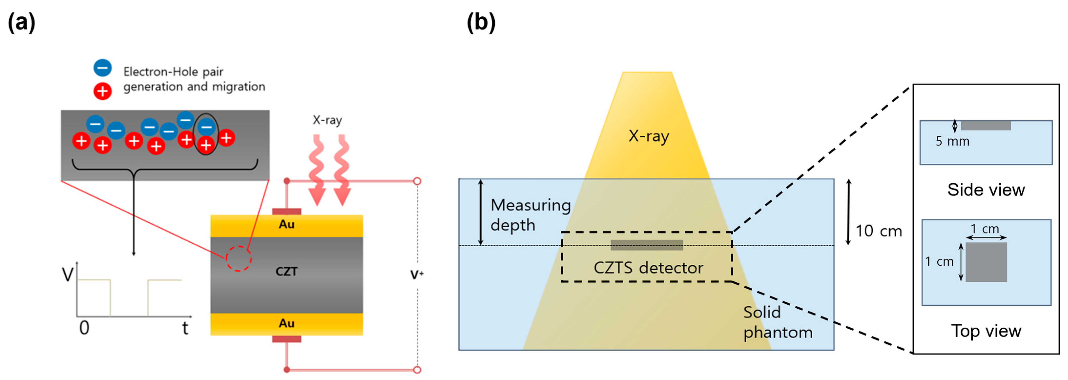
2. Materials and Methods

2.1. Growth of CZT

First, a single-crystal CZT detector was fabricated using the traveling heater method. Cd and Te of 7N purity were purchased from Nippon Mining & Metals Co., Ltd. (Tokyo, Japan), and Zn of 5N purity was used. To enhance the performance of the single crystal, 6 ppm of In, purchased from the same company, was doped into it. InTe was synthesized prior to the CZT synthesis to finely adjust the doping concentration. After crystal growth, the ingots were cooled and cut using a diamond saw. The cut CZT wafer was then mechanically polished and chemically etched. Next, 20–30 nm thick Au electrodes were formed on both ends of the dosimeter using a Au2Cl3 solution.

2.2. Measurement Configuration
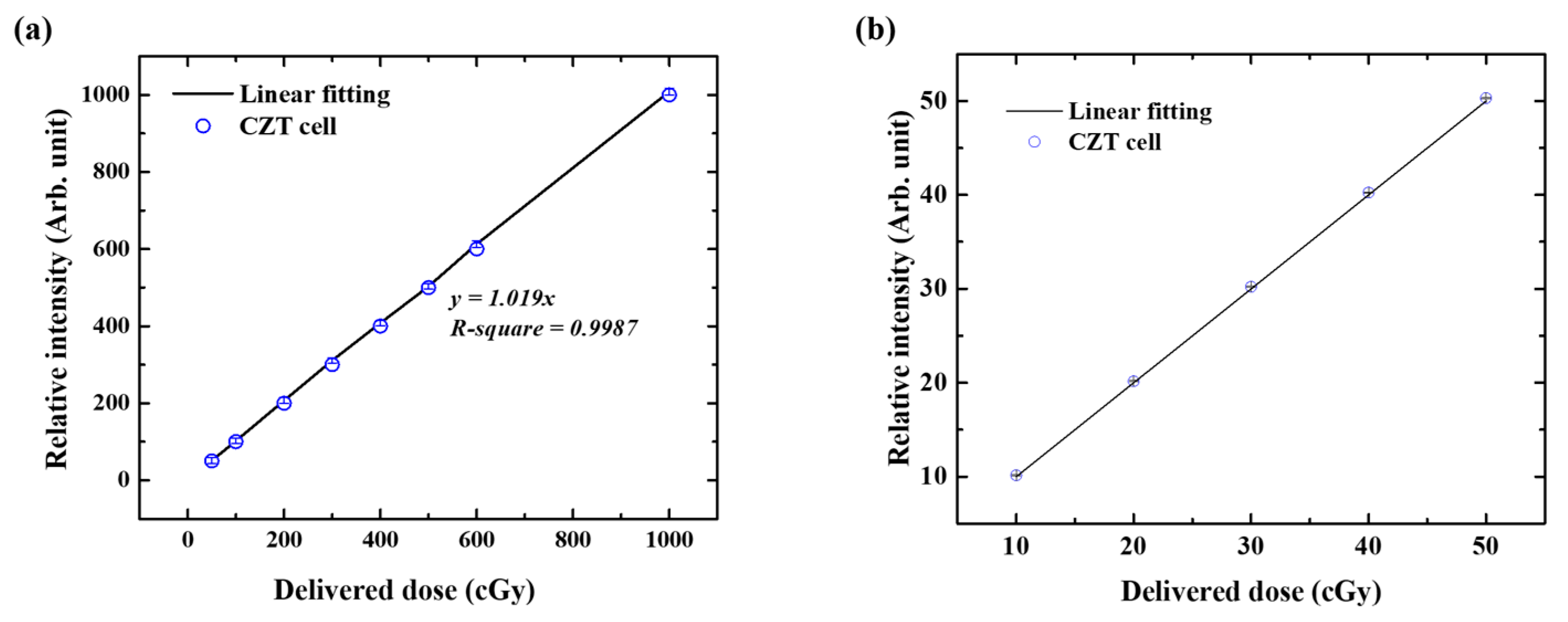
In this study, we fabricated a solid CZT detector, with a metal–semiconductor–metal planar structure (Figure 1a) and electrodes attached to both sides of the CZT crystal. When high-energy radiation with a wavelength that exceeds the bandgap of the CZT layer is incident, electrons in the valence band are excited to the conduction band because of the generation of electron–hole pairs. These electrons and holes are collected by the electrodes upon the application of a bias, which can lead to the generation of a signal. In this study, an external bias of 100 V/mm was applied to the electrodes by using a Keithley 236 high-voltage source measurement unit to facilitate the collection of the generated electrons and holes. The current generated by the collected electrons and holes was measured using a B2911A Precision Source/Measure unit at a sampling rate of 1000 points/s. Figure 1b illustrates the measurement setup used to extract the reference dose data. The sample was positioned at a source-to-surface distance (SSD) of 100 cm and depth of 10 cm in a solid water phantom and irradiated with therapeutic 6 and 10 MV beams using a LINAC device (Trilogy Clinac Ix, Varian, Palo Alto, CA, USA). The final results were obtained by averaging those of five measurements. The characteristics of the CZT detector, including dose linearity, dose rate dependence, energy dependence, angular dependence, SSD dependence, field size dependence, reproducibility, and percentage depth dose (PDD), were evaluated under defined conditions.

2.3. Dose Linearity

A 6 MV X-ray beam at a dose rate of 400 MU/min was irradiated to deliver doses of 50, 100, 200, 300, 400, 500, 600, and 1000 cGy at a gantry angle of 0°. The SSD and field size were 100 cm and 10 cm × 10 cm, respectively. To verify linearity in the low-dose region, measurements were performed at doses of 10, 20, 30, 40, and 50 cGy as well. A calibrated ionization chamber was used to adjust 1 MU to deliver a dose of 1 cGy at an SSD of 100 cm.

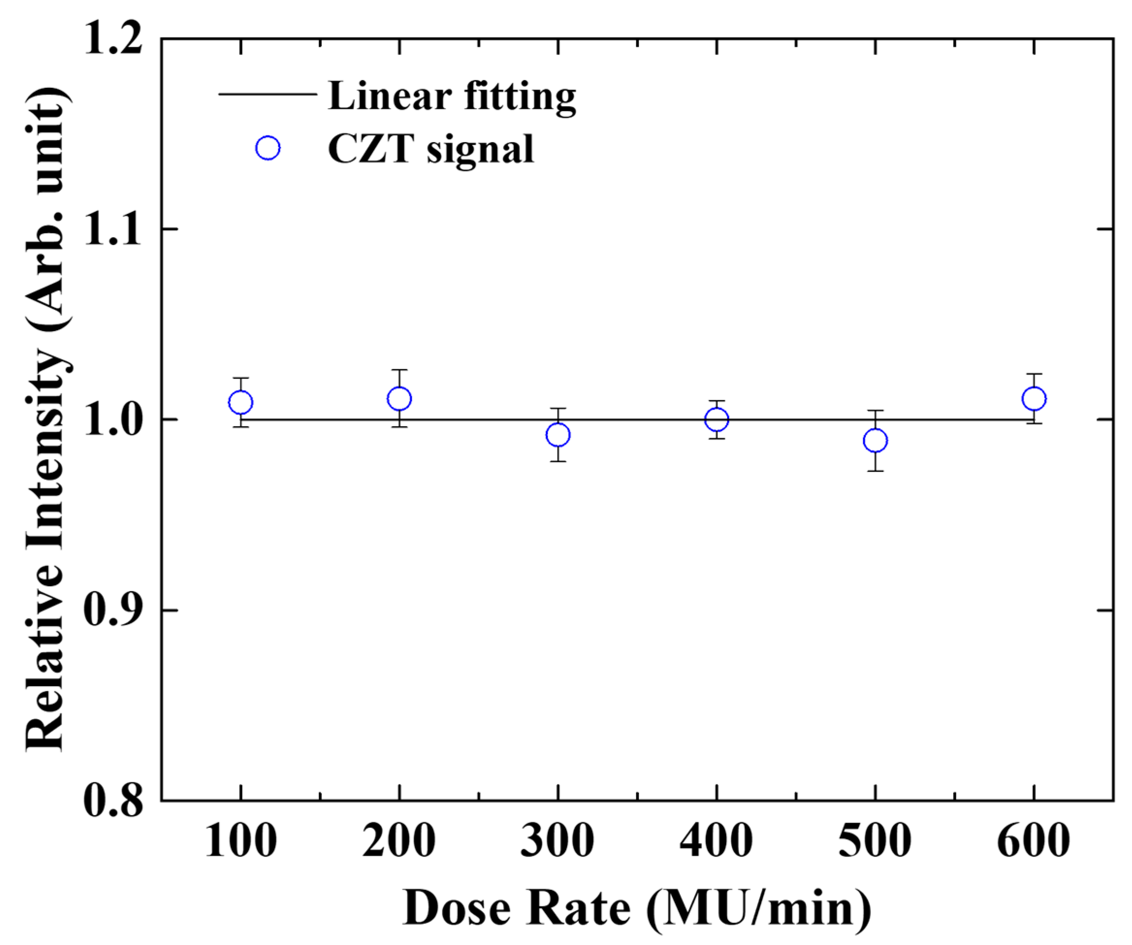
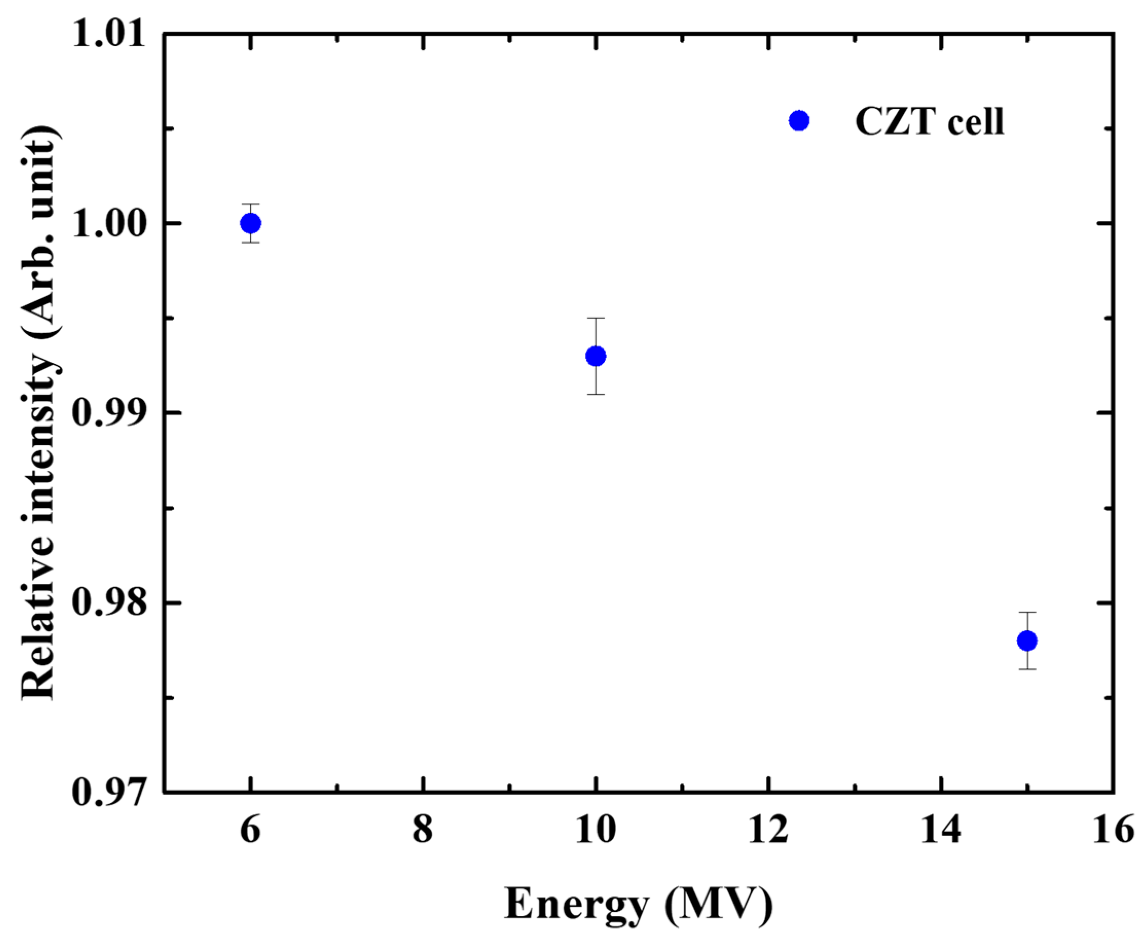
2.4. Dose Rate and Energy Dependence

To verify the dose rate dependence of the CZT detector’s output signal, 200 cGy was delivered to the detector, while varying the dose rate from 100 to 600 MU/min, using a 6 MV beam. To measure the energy dependence, 6, 10, and 15 MV beams were irradiated on the sample and delivered a dose of 200 cGy at a dose rate of 400 MU/min.

2.5. Angular and SSD Dependence

The angular dependence of the detector’s output was determined by rotating the gantry angle from 0° to 180° at 30° intervals while delivering a beam to the detector. Furthermore, the SSD dependence was tested by varying the SSD from 80 to 120 cm while delivering a dose of 200 cGy. In both the experiments, the same measurements were performed using an ionization chamber for comparison.

2.6. Field Size Dependence and Reproducibility

The field size dependence of the detector’s response was measured by varying the field size from 5 cm × 5 cm to 25 cm × 25 cm while delivering 200 cGy to the CZT detector at a dose rate of 400 MU/min and an SSD of 100 cm. The same measurements were performed using an ionization chamber as well for comparison. The reproducibility of the measured values was confirmed by conducting weekly measurements at a dose of 200 cGy over a 1-month period and comparing the results.

2.7. Percentage Depth Dose

The percentage depth dose (PDD) of the detector’s response was measured at a dose of 500 cGy by varying the depth of the CZT detector from 0 to 25 cm under 6 and 10 MV beam irradiation. The corresponding results were compared with those obtained using an ionization chamber. Table 1 presents the measurement conditions used to verify the characteristics of the CZT detector.

3. Results

All measurements with error bars were obtained from five repeated measurements.

3.1. Dose Linearity

Figure 2a shows that the intensity of the signal generated by the CZT detector increases linearly when the delivered dose is increased from 50 to 1000 cGy, indicating a linear correlation between the signal intensity and the delivered dose. The slope of the fitted curve is 1.019, with an R2 value of 0.9987, demonstrating an excellent linearity of the detector within the measured dose range. Furthermore, measurements in the low-dose region (Figure 2b) were performed to assess the precision of the measured values. The same linear fit was used for both high-dose and low-dose ranges. Data measured from 10 to 50 cGy also revealed excellent linearity, with a standard deviation close to zero.

3.2. Dose Rate Dependence

Figure 3 depicts the dose rate dependence of the detector’s output measured from 100 to 600 MU/min by using a 6 MV beam. The relative dose is normalized to the dose rate measured at 400 MU/min. The measured relative dose rates are similar within an error of 1%, and those of repeated measurements at the same dose are within an error of 2%. Furthermore, the dose rate does not reveal an increasing or decreasing trend.

3.3. Energy Dependence

Figure 4 reveals the response signal intensities, measured under 6, 10, and 15 MV beam irradiation, normalized to that recorded under 6 MV beam irradiation. The response signal’s intensity decreases as the beam energy increases, with the normalized response-signal intensity at 15 MV being 2.2% lower than that recorded at 6 MV. The error bars in the repeatedly measured datapoints are less than 0.5% for all the beam energies, demonstrating the excellent energy characteristics of the fabricated detector.

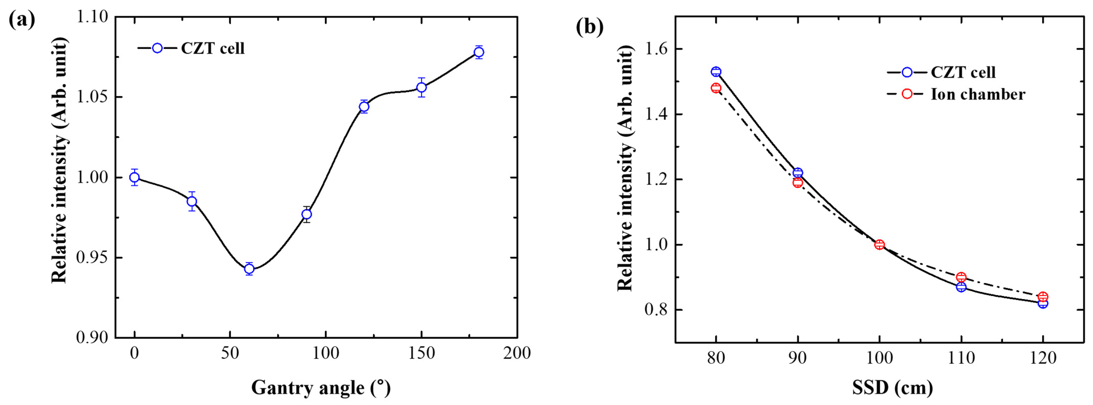
3.4. Angular and SSD Dependence

The angular dependence of the CZT detector was determined by normalizing the measured signal intensities to the signal recorded at a gantry angle of 0°. Figure 5a depicts the results obtained at 6 MV. Because the solid water phantom has a hexahedral shape, the signal intensities measured by the CZT detector at each gantry angle were divided by those measured by the ionization chamber to correct for depth dependence occurring during the angular dependence measurements. Figure 5b represents the changes in the signal intensities obtained from the CZT detector when varying the SSD from 80 to 120 cm. At an SSD of 100 cm, the normalized signal intensity obtained from the CZT detector is higher than that output from the ionization chamber, which reveals decreasing signal intensities with increasing SSD.

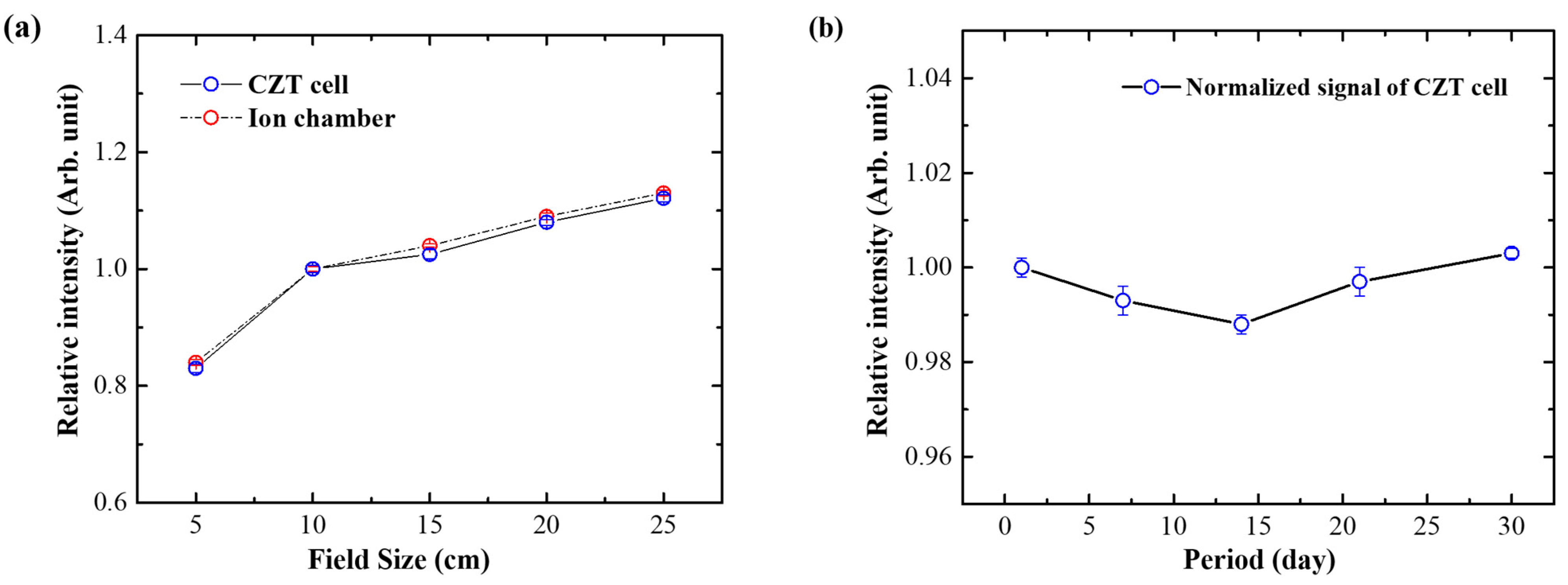
3.5. Field Size Dependence and Reproducibility

The field size dependence of the detector’s response was measured by varying the field size from 5 cm × 5 cm to 25 cm × 25 cm while delivering 200 cGy to the detector, and the results were compared to those obtained from the ionization chamber (Figure 6a). On normalizing the intensities recorded at 10 cm × 10 cm, the results obtained from the CZT detector are consistent with those obtained from the ionization chamber within an error of 1% (Figure 6b). Furthermore, the signal intensities recorded by the CZT detector under a delivered dose of 400 cGy remain consistent within 1% over a 1-month period, without changing with time.

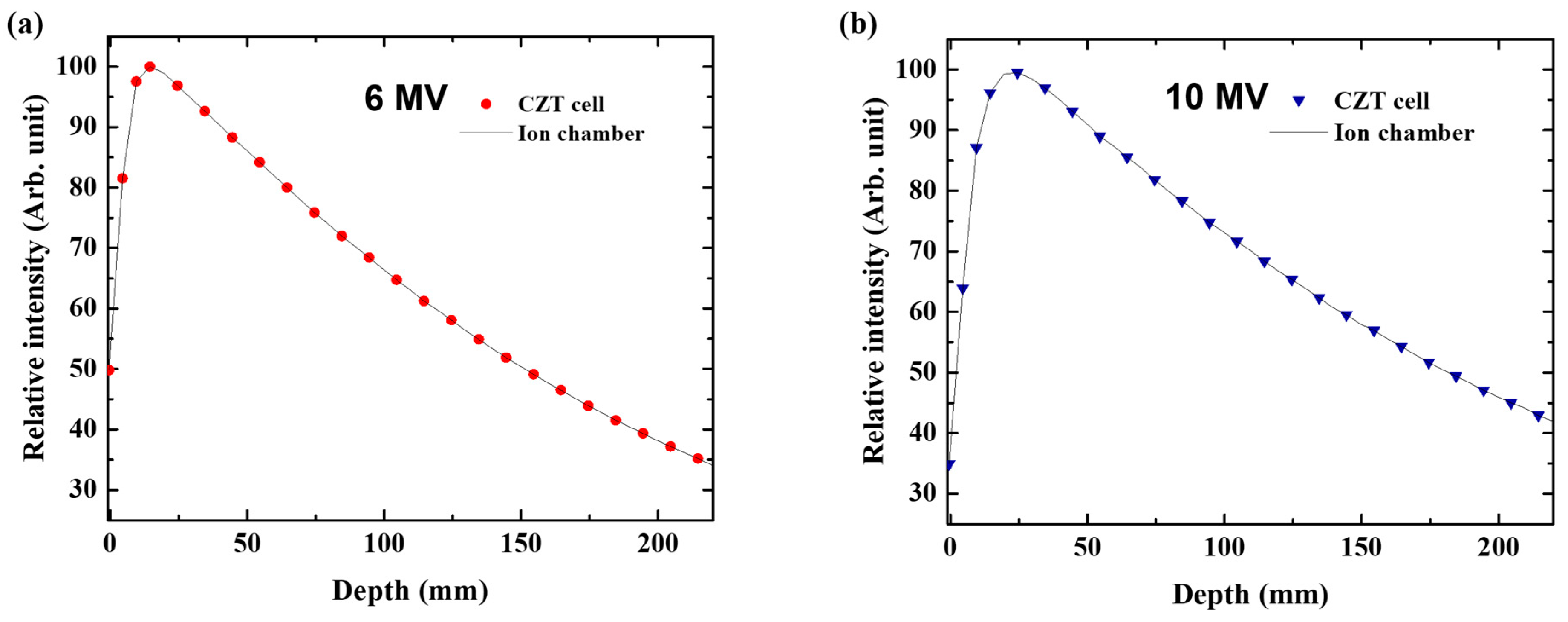
3.6. Percentage Depth Dose

Figure 7 reveals that the signal intensities recorded by the CZT detector as functions of depth are almost identical to those obtained from the ionization chamber. Under 6 and 10 MV beam irradiation, the signal intensities recorded by the CZT detector at depths ranging from 0 to 25 cm are consistent with those obtained from the ionization chamber.

4. Discussion

In this study, we evaluated the fundamental characteristics of a CZT detector and conducted validation tests to assess its potential for SRS and SBRT applications. The results indicate that the CZT detector satisfies the various requirements for utilization with therapeutic X-ray beams. The elements constituting the CZT detector exhibit high atomic numbers (Z), providing excellent radiation absorption characteristics. Additionally, the detector’s large carrier mobility–lifetime product is conducive to high-energy radiation measurements.
In the dose linearity test, the CZT detector exhibited nearly perfect linearity across a dose range from 0 to 1000 cGy, including the low-dose region. The single-crystal CZT exhibited excellent linearity, as shown in Figure 2, with very small error bars. This characteristic suggests that the CZT detector can provide reliable values under various treatment conditions, especially in SRS and SBRT, where the dose distribution is concentrated in specific regions and rapid dose fall-off is required outside the target area.
In the dose rate dependence experiment, the signal generated by the CZT detector exhibited minimal variations with changes in the dose rate. Dose rate dependence issues—particularly over-response with increasing dose rate caused by electron–hole recombination in semiconductor junction structures—are typically observed in diode-type detectors. In such cases, dose rate correction factors are required to monitor and adjust for dose rate variations continuously. However, this dose rate-dependent over-response is not exhibited by the fabricated CZT detector, which generates signals by collecting electron–hole pairs under an applied bias. Thus, this detector does not require extensive dose rate correction processes.
The energy dependence analysis reveals that the intensity of the signal obtained from the CZT detector decreases slightly with the increase in beam energy. The gradual decrease in signal with increasing energy indicates a reduction in absorption efficiency at higher energy regions, despite the high-Z characteristics of CZT. This dependency is a common issue in solid-state detectors, requiring calibration based on the energy range used. However, within the primary energy range (6 and 10 MV) used in this study, the intensity difference is only about 1%. Although the detector is composed of high-Z materials, the observed energy dependence could be attributed to the Z-effect, where interactions with higher-energy photons are less probable in high-Z materials.
The angular dependence deviation, particularly near 60° and 120°, could be attributed to the detector’s asymmetric structure in the upper and lower regions. This phenomenon is typically observed in fixed detector structures and can be corrected in clinical practice by compensating the results or by positioning the detector perpendicular to the beam path. Although the SSD dependence results of the CZT detector differ from those of the ionization chamber, they follow the inverse square law and can be corrected in practice.
Reproducibility and depth dependence experiments yielded favorable results. The CZT detector maintained consistent performance with minimal changes when subjected to beam exposure over a period of more than 1 month. The depth dependence results were consistent with those of the ionization chamber at both 6 and 10 MV. This consistent performance could be attributed to the characteristics of the detector, which uses a single material rather than a junction or hybrid structure.
The field size dependence results were consistent with those obtained using the ionization chamber, which is crucial for small-field measurements. Guidelines for SRS and SBRT measurements discuss the challenges of small-field dosimetry and the necessity for high detector densities and large sample treatment volumes [36]. Studies on CZT detectors have investigated various electrode configurations, and a pixelated detector structure can enhance the applicability of CZT detectors in SRS and SBRT [35,37,38,39]. Detectors intended for use in SRS and SBRT need to observe dose variations within small areas, indicating the necessity of pixel formation in the detector. In the case of CZT, the smaller the pixels formed on the surface, the more the detection performance is enhanced due to the localized strengthening effect of the electric field. This suggests the potential for significantly improved performance compared to the currently used single-electrode CZT. Adapting the pixelated structures proposed in the previous studies could enhance the sensitivity and signal-collection efficiency of CZT detectors in small-field measurements. Such a pixelated CZT detector could surpass ionization chambers in terms of performance.

5. Conclusions

In this study, we evaluated the characteristics of a single-crystal CZT detector to validate its applicability in SRS and SBRT. The fabricated CZT detector demonstrated excellent performance in terms of dose linearity, dose rate dependence, energy dependence, field size dependence, reproducibility, and depth dependence. Notably, the stable linearity observed in both the high- and low-dose regions is conducive for both SRS and SBRT, in which abrupt dose variations are common. However, further investigations are required with the established techniques for developing submicron pixel structures and to assess the potential for performance enhancement. Detectors with a pixelated CZT structure are expected to have significantly higher performance and an optimized design for SRS and SBRT compared to the single-pixel detector studied in this research. The CZT detector is a highly promising device for application in SRS and SBRT and can be used in various radiation therapy environments.

Author Contributions

Conceptualization, S.C. and S.K.; methodology, J.-Y.S. and J.-U.J.; software, M.S.Y.; validation, S.C., S.K. and J.-U.J.; formal analysis, J.-Y.S. and Y.-H.K.; investigation, T.-K.N. and Y.-H.K.; resources, J.-U.J.; data curation, T.-K.N.; writing—original draft preparation, S.K.; writing—review and editing, M.S.Y. and S.-J.A.; visualization, M.S.Y.; supervision, S.C.; project administration, S.C.; funding acquisition, S.C. All authors have read and agreed to the published version of the manuscript.

Funding

This research was funded by a grant (BCRI24053) of Chonnam National University Hospital Biomedical Research Institute.

Institutional Review Board Statement

Not applicable.

Informed Consent Statement

Not applicable.

Data Availability Statement

Data underlying the results presented in this paper are not publicly available at this time but may be obtained from the authors upon reasonable request.

Acknowledgments

This study was supported by a grant (BCRI24053) from the Chonnam National University Hospital Biomedical Research Institute.

Conflicts of Interest

The authors declare no conflicts of interest. The funders had no role in the design of the study; in the collection, analyses, or interpretation of data; in the writing of the manuscript, or in the decision to publish the results.

References

  1. Lo, S.S.; Fakiris, A.J.; Chang, E.L.; Mayr, N.A.; Wang, J.Z.; Papiez, L.; Teh, B.S.; McGarry, R.C.; Cardenes, H.R.; Timmerman, R.D. Stereotactic body radiation therapy: A novel treatment modality. Nat. Rev. Clin. Oncol. 2010, 7, 44–54. [Google Scholar] [CrossRef]
  2. Kann, B.H.; Park, H.S.; Johnson, S.B.; Chiang, V.L.; Yu, J.B. Radiosurgery for brain metastases: Changing practice patterns and disparities in the United States. J. Natl. Compr. Canc. Netw. 2017, 15, 1494–1502. [Google Scholar] [CrossRef] [PubMed]
  3. Levy, A.; Guckenberger, M.; Hurkmans, C.; Nestle, U.; Belderbos, J.; De Ruysscher, D.; Faivre-Finn, C.; Le Péchoux, C. SBRT dose and survival in non-small cell lung cancer: In regard to Koshy et al. Int. J. Radiat. Oncol. Biol. Phys. 2015, 92, 945–946. [Google Scholar] [CrossRef] [PubMed]
  4. Hoffman, D.; Dragojević, I.; Hoisak, J.; Hoopes, D.; Manger, R. Lung stereotactic body radiation therapy (SBRT) dose gradient and PTV volume: A retrospective multicenter analysis. Radiat. Oncol. 2019, 14, 162. [Google Scholar] [CrossRef] [PubMed]
  5. Narayanasamy, G.; Desai, D.; Maraboyina, S.; Penagaricano, J.; Zwicker, R.; Johnson, E.L. A dose falloff gradient study in RapidArc planning of lung stereotactic body radiation therapy. J. Med. Phys. 2018, 43, 147–154. [Google Scholar] [CrossRef]
  6. Shaw, M.; Lye, J.; Alves, A.; Keehan, S.; Lehmann, J.; Hanlon, M.; Kenny, J.; Baines, J.; Porumb, C.; Geso, M.; et al. Characterisation of a synthetic diamond detector for end-to-end dosimetry in stereotactic body radiotherapy and radiosurgery. Phys. Imaging Radiat. Oncol. 2021, 20, 40–45. [Google Scholar] [CrossRef] [PubMed]
  7. Halvorsen, P.H.; Cirino, E.; Das, I.J.; Garrett, J.A.; Yang, J.; Yin, F.-F.; Fairobent, L.A. AAPM-RSS medical physics practice Guideline 9.a. for SRS-SBRT. J. Appl. Clin. Med. Phys. 2017, 18, 10–21. [Google Scholar] [CrossRef] [PubMed]
  8. Benedict, S.H.; Yenice, K.M.; Followill, D.; Galvin, J.M.; Hinson, W.; Kavanagh, B.; Keall, P.; Lovelock, M.; Meeks, S.; Papiez, L.; et al. Stereotactic body radiation therapy: The report of AAPM Task Group 101. Med. Phys. 2010, 37, 4078–4101. [Google Scholar] [CrossRef]
  9. Agency, I.A.E. Dosimetry of Small Static Fields Used in External Beam Radiotherapy: An International Code of Practice for Reference and Relative Dose Determination; Technical Reports Series; International Atomic Energy Agency (IAEA): Vienna, Austria, 2017; Volume 483. [Google Scholar]
  10. Foote, M.; Bailey, M.; Smith, L.; Siva, S.; Hegi-Johnson, F.; Seeley, A.; Barry, T.; Booth, J.; Ball, D.; Thwaites, D. Guidelines for safe practice of stereotactic body (ablative) radiation therapy. J. Med. Imaging Radiat. Oncol. 2015, 59, 646–653. [Google Scholar] [CrossRef]
  11. Sahgal, A.; Roberge, D.; Schellenberg, D.; Purdie, T.G.; Swaminath, A.; Pantarotto, J.; Filion, E.; Gabos, Z.; Butler, J.; Letourneau, D.; et al. The Canadian association of radiation oncology scope of practice guidelines for lung, liver and spine stereotactic body radiotherapy. Clin. Oncol. (R Coll Radiol) 2012, 24, 629–639. [Google Scholar] [CrossRef]
  12. Matuszak, M.M.; Hadley, S.W.; Feng, M.; Hayman, J.A.; Brock, K.K.; Burger, P.; Owen, D.; Suresh, K.; Schipper, M.; Lawrence, T.S.; et al. Enhancing safety and quality through preplanning peer review for patients undergoing stereotactic body radiation therapy. Pract. Radiat. Oncol. 2016, 6, e39–e46. [Google Scholar] [CrossRef] [PubMed]
  13. Van Elmpt, W.; McDermott, L.; Nijsten, S.; Wendling, M.; Lambin, P.; Mijnheer, B. A literature review of electronic portal imaging for radiotherapy dosimetry. Radiother. Oncol. 2008, 88, 289–309. [Google Scholar] [CrossRef] [PubMed]
  14. Bjärngard, B.E.; Tsai, J.S.; Rice, R.K. Doses on the central axes of narrow 6-MV x-ray beams. Med. Phys. 1990, 17, 794–799. [Google Scholar] [CrossRef]
  15. Niroomand-Rad, A.; Blackwell, C.R.; Coursey, B.M.; Gall, K.P.; Galvin, J.M.; McLaughlin, W.L.; Meigooni, A.S.; Nath, R.; Rodgers, J.E.; Soares, C.G. Radiochromic film dosimetry: Recommendations of AAPM radiation therapy committee task group 55. American Association of Physicists in Medicine. Med. Phys. 1998, 25, 2093–2115. [Google Scholar] [CrossRef]
  16. Butson, M.J. Scanning orientation effects on Gagchromic EBT film dosimetry. Australas. Phys. Eng. Sci. Med. 2006, 293, 281–284. [Google Scholar] [CrossRef] [PubMed]
  17. Paelinck, L.; Neve, W.D.; Wagter, C.D. Precautions and strategies in using a commercial flatbed scanner for radiochromic film dosimetry. Phys. Med. Biol. 2007, 521, 231–242. [Google Scholar] [CrossRef]
  18. Tyler, M.; Liu, P.Z.Y.; Chan, K.W.; Ralston, A.; McKenzie, D.R.; Downes, S.; Suchowerska, N. Characterization of small-field stereotactic radiosurgery beams with modern detectors. Phys. Med. Biol. 2013, 58, 7595–7608. [Google Scholar] [CrossRef] [PubMed]
  19. Schoenfeld, A.A.A.; Wieker, S.; Harder, D.; Poppe, B. The origin of the flatbed scanner artifacts in radiochromic film dosimetry—Key experiments and theoretical descriptions. Phys. Med. Biol. 2016, 61, 7704–7724. [Google Scholar] [CrossRef]
  20. Darafsheh, A.; Zhao, T.; Khan, R. Spectroscopic analysis of irradiated radiochromic EBT-XD films in proton and photon beams. Phys. Med. Biol. 2020, 65, 205002. [Google Scholar] [CrossRef] [PubMed]
  21. Chung, E.; Davis, S.; Seuntjens, J. Experimental analysis of general ion recombination in a liquid-filled ionization chamber in high energy photon beams. Med. Phys. 2013, 40, 062104. [Google Scholar] [CrossRef]
  22. Markovic, M.; Stathakis, S.; Mavroidis, P.; Jurkovic, I.A.; Papanikolaou, N. Characterization of a two-dimensional liquid-filled ion chamber detector array used for verification of the treatments in radiotherapy. Med. Phys. 2014, 41, 051704. [Google Scholar] [CrossRef]
  23. Lechner, W.; Palmans, H.; Sölkner, L.; Grochowska, P.; Georg, D. Detector comparison for small field output factor measurements in flattening filter free photon beams. Radiother. Oncol. 2013, 109, 356–360. [Google Scholar] [CrossRef] [PubMed]
  24. Yarahmadi, M.; Wegener, S.; Sauer, O.A. Energy and field size dependence of a silicon diode designed for small-field dosimetry. Med. Phys. 2017, 44, 1958–1964. [Google Scholar] [CrossRef] [PubMed]
  25. Lim, S.B.; Kuo, L.; Li, T.; Li, X.; Ballangrud, A.M.; Lovelock, M.; Chan, M.F. Comparative study of SRS end-to-end QA processes of a diode array device and an anthropomorphic phantom loaded with GafChromic XD film. J. Appl. Clin. Med. Phys. 2022, 23, e13747. [Google Scholar] [CrossRef] [PubMed]
  26. Camilleri, J.; Mazurier, J.; Franck, D.; Dudouet, P.; Latorzeff, I.; Franceries, X. 2D EPID dose calibration for pretreatment quality control of conformal and IMRT fields: A simple and fast convolution approach. Phys. Med. 2016, 32, 133–140. [Google Scholar] [CrossRef] [PubMed]
  27. Pardo, E.; Novais, J.C.; Molina López, M.Y.; Ruiz Maqueda, S. On flattening filter-free portal dosimetry. J. Appl. Clin. Med. Phys. 2016, 17, 132–145. [Google Scholar] [CrossRef]
  28. Podesta, M.; Nijsten, S.M.J.J.G.; Persoon, L.C.G.G.; Scheib, S.G.; Baltes, C.; Verhaegen, F. Time dependent pre-treatment EPID dosimetry for standard and FFF VMAT. Phys. Med. Biol. 2014, 59, 4749–4768. [Google Scholar] [CrossRef]
  29. Agarwal, A.; Rastogi, N.; Maria Das, K.J.; Yoganathan, S.A.; Udayakumar, D.; Kumar, S. Investigating the electronic portal imaging device for small radiation field measurements. J. Med. Phys. 2017, 42, 59–64. [Google Scholar] [CrossRef]
  30. Arlt, R.; Brutscher, J.; Gunnink, R.; Ivanov, V.; Parnham, K.; Soldner, S.; Stein, J. Use of CdZnTe detectors in hand-held and portable isotope identifiers to detect illicit trafficking of nuclear material and radioactive sources. In Proceedings of the 2000 IEEE Nuclear Science Symposium Conference Record (cat. No. 00CH37149), Lyon, France, 15–20 October 2000; Volume 11, pp. 4–14. [Google Scholar] [CrossRef]
  31. Sordo, S.D.; Abbene, L.; Caroli, E.; Mancini, A.M.; Zappettini, A.; Ubertini, P. Progress in the development of CdTe and CdZnTe semiconductor radiation detectors for astrophysical and medical applications. Sensors 2009, 9, 3491–3526. [Google Scholar] [CrossRef] [PubMed]
  32. Li, X.; Chu, J.H.; Li, L.X.; Dai, N.; Zhang, F.J. Investigation of room temperature nuclear radiation CdZnTe pixel array detector. J. Optoelectron. Laser 2008, 19, 751–753. [Google Scholar]
  33. Haijun, X. Data Acquisition System of CZT Nuclear Detector Based on FPGA. Master’s Thesis, Chongqing University, Chongqing, China, 2013. [Google Scholar]
  34. Roy, U.N.; Bolotnikov, A.E.; Camarda, G.S.; Cui, Y.; Hossain, A.; Lee, K.; Marshall, M.; Yang, G.; James, R.B. Growth of CdTexSe1-x from a Te-rich solution for applications in radiation detection. J. Cryst. Growth 2014, 386, 43–46. [Google Scholar] [CrossRef]
  35. Veale, M.C.; Bell, S.J.; Jones, L.L.; Seller, P.; Wilson, M.D.; Allwork, C.; Kitou, D.; Sellin, P.J.; Veeramani, P.; Cernik, R.C. An ASIC for the study of charge sharing effects in small pixel CdZnTe X-ray detectors. IEEE Trans. Nucl. Sci. 2011, 58, 2357–2362. [Google Scholar] [CrossRef]
  36. Alashrah, S.; El-Taher, A. Intensity modulated radiation therapy plans verification using a Gaussian convolution kernel to correct the single chamber response Function of the I’mRT MatriXX Array. J. Appl. Sci. 2015, 15, 483–491. [Google Scholar] [CrossRef]
  37. Zhu, W.; Wu, J.; Tang, X.; Zhao, R.; Lu, P.; Qv, B.; Li, M.; Qin, F. Simulation of energy spectrum characteristics of planar CdZnTe detector. Nucl. Technol. 2023, 46, 62–68. [Google Scholar]
  38. Luke, P.N.; Amman, M.; Lee, J.S.; Ludewigt, B.A.; Yaver, H. A CdZnTe coplanar-grid detector array for environmental remediation. Nucl. Instrum. Methods Phys. Res. Sect. A 2001, 458, 319–324. [Google Scholar] [CrossRef]
  39. Bennett, P.R.; Shah, K.S.; Klugerman, M.; Squillante, M.R. High efficiency pixellated CdTe detector. Nucl. Instrum. Methods Phys. Res. 1997, 392, 260–263. [Google Scholar] [CrossRef]
Figure 1. Schematics of (a) an X-ray beam converted to an electrical signal and (b) reference experiment setup of the CZT cell.
Applsci 15 01693 g001
Figure 2. Linear relation between the intensity of the CZT signal with the delivered dose (a) in the range of 50–1000 cGy and (b) in the low-dose region (≤50 cGy) under 6 MV beam irradiation.
Applsci 15 01693 g002
Figure 3. Dose rate dependence of the signal intensities obtained from the CZT cell under 6 MV beam irradiation. Data are normalized to the intensity of the signal generated at a dose rate of 400 MU/min.
Applsci 15 01693 g003
Figure 4. Energy dependence of the signal intensities obtained from the CZT cell at various irradiated photon beam energies.
Applsci 15 01693 g004
Figure 5. (a) Angular and (b) SSD dependence of the signal intensities output obtained from the CZT cell and ionization chamber under 6 MV beam irradiation. Data are normalized to the signal intensity recorded at a gantry angle of 0° and an SSD of 100 cm.
Applsci 15 01693 g005
Figure 6. (a) Field size dependence of the outputs obtained from the CZT cell and ionization chamber. The values are normalized to 1 for a field size of 10 cm × 10 cm. (b) Reproducibility of the results obtained from the CZT cell under 6 MV beam irradiation over a period of 1 month. The data from day 0 are represented as the reference value of 1.
Applsci 15 01693 g006
Figure 7. Relative signal intensities measured by the CZT cell and ionization chamber at various depths under (a) 6 MV and (b) 10 MV photon beam irradiation.
Applsci 15 01693 g007
Table 1. Measurement conditions used in this study.
Energy (MV)Field Size (cm2)SSD (cm)Dose (cGy)Dose Rate (MU/min)Measuring Depth (cm)
Dose linearity610 × 101000–100040010
Dose rate dependence610 × 10100200100–60010
Energy dependence6, 10, and 1510 × 1010020040010
Angular dependence610 × 10100200400-
SSD dependence610 × 1080–12020040010
Field size dependence65 × 5 to
25 × 25
10020040010
Reproducibility610 × 1010020040010
PDD6, 1010 × 101005004000–25
Disclaimer/Publisher’s Note: The statements, opinions and data contained in all publications are solely those of the individual author(s) and contributor(s) and not of MDPI and/or the editor(s). MDPI and/or the editor(s) disclaim responsibility for any injury to people or property resulting from any ideas, methods, instructions or products referred to in the content.

