Article

Differences in EEG Functional Connectivity in the Dorsal and Ventral Attentional and Salience Networks Across Multiple Subtypes of Depression

1
Brain-Behaviour Research Group, School of Science & Technology, University of New England, Armidale, NSW 2351, Australia
2
Department of Neurosurgery, The Alfred Hospital, Melbourne, VIC 3004, Australia
3
Griffith Health Group, Griffith University, Southport, QLD 4222, Australia
*
Author to whom correspondence should be addressed.
Appl. Sci. 2025, 15(3), 1459; https://doi.org/10.3390/app15031459
Submission received: 28 November 2024 / Revised: 13 January 2025 / Accepted: 29 January 2025 / Published: 31 January 2025
(This article belongs to the Special Issue Brain Functional Connectivity: Prediction, Dynamics, and Modeling)

Abstract

:
Depression remains one of the most widespread and costly mental disorders, with the current first-line treatment efficacy of about a third, possibly due to its heterogeneous nature. Consequently, there is a need to identify reliable biomarkers for specific subtypes of depression, particularly neurological signatures that may help with targeted treatments. This study aimed to explore the connectivity between two important networks in the brain: the dorsal and ventral attention networks and the salience network, to determine their potential as biomarkers of depression subtypes. From resting electroencephalogram (EEG) data collected on 54 males and 46 females aged between 18 and 75 yr (M = 33 yr), functional network connectivity data were examined for their relationships with four depression subtypes. Beta and gamma wave connectivity was significantly associated with Anhedonia and Cognitive depression subtypes across and within all three networks while no significant results were found for alpha wave activity connectivity, and only one result was found for either the Mood or Somatic depression subtypes. In conclusion, these results provide further support for the concept of depression as heterogeneous rather than homogeneous and identify the novel neurophysiological signatures of two depression subtypes.

1. Introduction

Despite carrying a major disease burden [1] and high risk for all-cause mortality [2], and bringing severe personal distress [3], depression has also been defined as an adaptive response to uncontrollable stress [4,5]. The ‘adaptive’ nature of depression is defined by the shaping effects of negative reinforcers (e.g., social withdrawal, which helps the individual avoid unpleasant events and/or escape from threat) and positive reinforcers (e.g., receiving sympathy or relaxing medication) that follow depressive behaviours [6,7,8,9]. The longer-term adaptive outcome of these consequences of depressive behaviour was noted some time ago by Charles Darwin, who commented that “Pain or suffering of any kind, if long continued, causes depression and lessens the power of action; yet, it is well adapted to make a creature guard itself against any great or sudden evil” [10], p. 51.
As such, it has been hypothesised that depression (or depressive behaviour) may be profitably analysed for its functionality, or how the depressive behaviour helps to protect the depressed individual from further discomfort [11]. Arising from this model of depression, one therapeutic approach applies behavioural activation strategies to address the depressed individual’s disinclination to confront their behavioural withdrawal from the uncontrollable stressors that have led them to feel depressed. This approach to treatment has met with some success, as demonstrated in a meta-analysis of 28 research studies including 1853 participants [12], and providing support for the adaptive model for depressive behaviour.
However, although most studies of depression utilise a unitary model and metric of depression, such as Major Depressive Disorder (MDD), this disorder may occur in almost 1500 different combinations of the established diagnostic criteria and associated features for MDD [13], indicating that it is a heterogeneous entity. Consequently, some attention has been given to the identification of particular depression symptoms and how these may group together, often referred to as ‘depression subtypes’. Several examples of these subtypes have been described, including melancholia, psychotic depression, atypical depression, and anxious depression [14]. Another model that has received some research attention focuses on subtypes of depression known as depressed Mood, Anhedonic depression, Cognitive depression, and Somatic depression [15,16]. Both of these models were developed using procedures that grouped MDD symptoms according to their clinical coherence, and then tested for significant associations between the defined subtype and other variables.
One particularly relevant set of variables that are valuable to investigate for their association with MDD subtypes are those generated by electroencephalography (EEG). EEG enables the identification of the relative electrical activity in specific areas of the brain, as well as the connections between those brain areas. The associations between this kind of information and MDD symptoms can make an obvious contribution to understanding the particular ways in which brain activity is related to specific MDD symptoms or specific depression subtypes, and thence to the eventual development of individualised treatment of depression based upon that brain activation–MDD symptomatology relationship [17,18,19]. Understanding depression from a neurological perspective has been a major and developing theme in the research literature [20,21,22], but mostly via the studies of depression as a unitary construct such as MDD. Although relatively easy to measure, depression as a unitary phenomenon may ignore variations in the way that EEG data relate to groups (i.e., subtypes) of depression symptoms.
There are many sites on the skull that can be used to measure an electrical signal representing brain activity, but it is worthwhile focusing on those which have a theoretical association with aspects of MDD. In particular, the attentional (DAN/VAN) and salience (SN) networks represent brain activities that are of relevance to depression when it is conceived as behavioural withdrawal. The dorsal and ventral attention networks (DAN/VAN) have been described as having multiple nodes located throughout the grey matter, each of which is related to specific aspects of attention [23]. These are (1) alerting (increases and maintains response readiness in response to a specific stimulus; located in the frontal and parietal regions, mostly in the right hemisphere); (2) orienting (selecting pertinent information; located in the pulvinar, superior colliculus, superior parietal lobe, temporoparietal junction, superior temporal lobe, and frontal eye fields); and (3) executive attention (the monitoring and resolution of conflict between information from different neural regions; the dorsal anterior cingulate cortex (ACC) in cognitive conflict tasks, and activation in the rostral ACC after the commission of errors. Whether the ACC monitors or resolves conflict is not clear, but the ACC function seems to preferentially relate to conflict at the response level [24]).
The salience network (SN; [24,25]) is mostly located in the cortical and subcortical prominent nodes of the anterior cingulate cortex, as well as the rostral prefrontal cortex and the supramarginal gyrus. The SN has a major role in emotional control [26]. Altered SN function has been associated with reduced motivation to engage with the external environments of depressed patients [27]. It has been suggested that the primary function of the SN is to identify the most relevant stimuli that reach the brain, including both the cognitive and affective aspects of the stimulus [28]. If these were associated with the emotional and cognitive aspects of MDD, then the SN may well be conceptualised as helping the depressed patient identify which stimuli held the most emotional and/or cognitive valence vis à vis the MDD symptomatology and the individual’s response to those stimuli. With relevance to the attentional networks, the SN may assist in deciding if the information identified by the DAN or VAN requires a behavioural response from the individual. That response may differ across depressed and non-depressed individuals and reflect the underlying propensity towards behavioural withdrawal from stressful stimuli that is a hallmark of MDD. Connectivity between the DAN, VAN, and SN might provide a confirmation of the behavioural withdrawal model of MDD, and assist in the development of effective treatment models for depression or its subtypes.
Therefore, the aim of the study was to examine the DAN, VAN, and SN network activity (and their connectivity with each other), and their association with MDD as well as the four MDD subtypes (Anhedonic, Cognitive, Somatic, and Mood) described by Sharpley and Bitsika [15]. Although the lack of previous conclusive research findings on these issues prevented the raising of specific directional hypotheses, it was expected that high depression scores (characterised by behavioural withdrawal in the form of MDD symptoms) would be associated with lowered activity in frequency bands associated with cognitive activation such as beta and gamma. That is, the depressed individual would exert neurological energy on the environmental cues that contributed to their depression but fail to find them sufficiently relevant to initiate behavioural activity.

2. Materials and Methods

2.1. Participants

A sample of 54 males and 46 females (M age = 32.53 yr, SD = 14.13 yr, and range = 18–75 yr) was drawn from a larger study undertaken in the New England region of New South Wales, Australia [29]. The participants were selected on the basis of not having a medical history of severe physical brain injury, brain surgery, or history of epilepsy or seizure disorder.

2.2. Depression Scales

The Zung Self-Rating Depression Scale (SDS) [30] comprises ten positively worded and ten negatively worded statements based on the Diagnostic Criteria and Associated Features of MDD [3]. Participants are required to state their frequency of these 20 items during the last two weeks on a four-point scale (None or a little of the time = 1, Some of the time = 2, Good part of the time = 3, and Most or all of the time = 4) so that the total raw scores range from 20 to 80 [30,31]. SDS raw scores of 40 or above indicate “clinically significant depression” [31], p. 335, a threshold that was met by 34 members of the overall sample. Split-half reliability for the SDS has been reported as 0.81, [30], 0.79 [32], and 0.94 [33], and internal consistency (Cronbach alpha) of 0.88 for depressed patients and 0.93 for non-depressed patients [34]. The four depression subtypes described by these data were derived from the 20 SDS items; for more information on each depression subtype see [15]. Rather than divide the sample into depressed and non-depressed groups, the depression score for the overall MDD and each subtype was calculated for each individual to be used for the subsequent correlation analyses.

2.3. EEG Data Processing

A 40-channel Neuroscan QuikCap EEG machine (Compumedics USA 144 Ltd., El Paso, TX, USA) with sintered Ag/AgCl electrodes was used to record EEG signals with electrode arrangement in consonance with the international 10/20 system and aligned with the anatomical inion and nasion points. A NuAmps digital amplifier (Compumedics USA Ltd., El Paso, TX, USA) was used for signal acquisition and digitalization at a sampling rate of 1000 Hz and passed through a bandpass filter of DC to 250 Hz. The amplifier was connected to the Curry 7 Acquisition software (Compumedics USA Ltd., El Paso, TX, USA) running on a Dell Optiflex 9020 desktop PC.
EEG recordings were initially referenced to the average of the A1-A2 earlobe electrodes, and subsequently converted to a common average reference offline. By using the four electrodes, EOG data were collected. Specifically, two electrodes were placed above and below the left eye to measure vertical eye movement, and the other two electrodes were located on the outside left and right canthus to measure horizontal eye movement. Prior to the commencement of EEG recording, the electrode impedance values were adjusted to <5 kΩ at all the electrodes to ensure the quality of signal acquisition. The EEG signals were processed using a 1–45 Hz 2nd order Butterworth bandpass filter, then re-referenced to a common average. Data tapering was performed using a Hann window with a 10% width to prevent data loss. This was then followed by a visual examination of the EEG signals to identify artefacts (eye movements, muscle movements, spontaneous discharges or electrode pops etc.), which were then removed from the recorded data. Bad block and eye blink detection (using the magnitude of eye blink deflections as a set threshold criterion to detect artefacts) was undertaken by three automated methods (subtraction, covariance and principal component analysis) to produce clean EEG data.
From the cleaned EEG data generated, 2 secs back-to-back epochs were then created and re-examined such that epochs with bad blocks were removed from averaged data; in this study, all the participants had a minimum of 87% usable epochs in the Eyes Open condition. By using the Key Institute eLORETA (exact low-resolution brain electromagnetic tomography) [35] software, the functional lagged linear connectivity (also known as coherence) estimates of four EEG frequency band activity were obtained, i.e., theta (4.05–8 Hz), alpha (8.05–12 Hz), beta (12.05–30 Hz), and gamma (30.05–45 Hz). This technique provides a single weighted minimum norm solution to the inverse problem and has been demonstrated to provide zero error (despite low spatial resolution) in localising cortical grey matter test sources [36,37]. The weights utilised by eLORETA (based on the EEG montage used in the recording) are used to calculate the current source density throughout the grey matter of a standardised realistic head model [38] based on the MNI template [39]. The resulting current density distribution is used to calculate measures of linear dependence between ‘virtual’ electrodes placed at regions of interest (ROIs) within the grey matter; for a comprehensive description of the mathematics underlying the eLORETA methodology and how the weighted norms are calculated, see [35,40]. ROIs were selected using commonly identified grey matter nodes in the DAN, VAN, and SN based on MNI coordinates identified in previous studies [41,42], with all the grey matter tissue within 10 mm of the identified source included as part of that node. This resulted in 15 ROIs being selected (see Table 1). In Table 1, ‘Location’ refers to the generally identified brain region. However, MNI coordinates locate each region more specifically. MNI X describes the brain region according to its location from left (negative values) to right (positive values); MNI Y coordinates refer to the front (positive values) to rear (negative values); and MNI Z coordinates position a brain site on the transverse plane of the brain, from top (positive values) to the bottom (negative values). Some regions listed in Table 1 are widespread, and thus occur in more than one network. In those cases, the regions may be more precisely identified by their MNI coordinates.

2.4. Procedure

The participants signed a consent form, filled out a background questionnaire (age and sex) and the SDS, and had their scalps prepared and the electrode cap fitted. Headphones for the participants were used to minimise the effect of external stimuli. Following 15 min of sitting still (adaptation), the audio-recorded experimental protocol (not used in this study) was presented via headphones to ensure consistency across the participants. Ethics approval was received from the Human Research Ethics Committee of the University of New England, Australia (Approval No. HE14-051).

3. Results

3.1. Age, Sex, and SDS Scores

SDS internal consistency (Cronbach’s alpha) was 0.905. The normal Q-Q plots for the SDS revealed an almost completely straight line, indicative of normality. The mean SDS score was 36.70 (SD = 11.25), minimum = 21, and maximum = 66. Using Zung’s [31] cutoff score of 40, there were 33 participants who had clinically significant depression (mean SDS score = 50.39, SD = 7.43) and 67 participants whose SDS scores did not meet this category (mean SDS score = 29.95, SD = 4.83; F (1,99) = 273.729, p < 0.001, ηp2 = 0.736). There were no significant correlations between age or sex and SDS total score, and any of the four SDS subtype scores.

3.2. MDD Subtypes

Mean scores for the four MDD subtypes were depressed Mood = 1.736 (SD = 0.653; range = 1.00 to 3.67), Anhedonia = 1.850 (SD = 0.657; range = 1.00 to 3.75), Cognitive depression = 2.183 (SD = 0.810; range = 1.00 to 4.00), and Somatic depression = 1.618 (SD = 0.511; range = 1.00 to 3.50) using the 4-point scale described above. These means were used as cutoffs to identify the most depressed from the least depressed participants in each SDS subtype. All four SDS subtype scores were significantly correlated with each other (all ρ ≥ 0.646, p < 0.001).

3.2.1. Theta Band

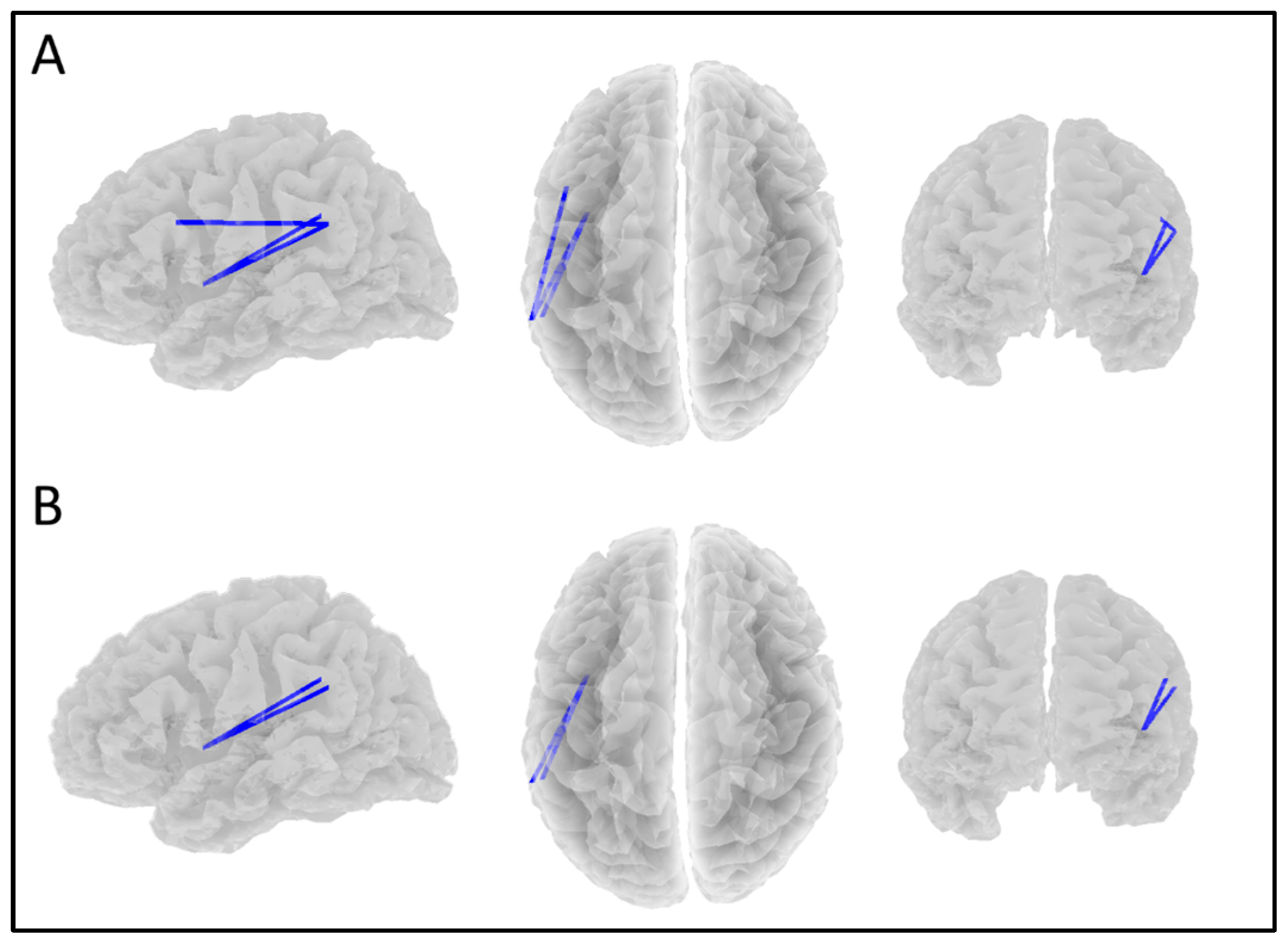
Correlation analyses were conducted in eLORETA using each depression score (MDD and the four subtypes) and functional connectivity between all the ROIs. Significant positive correlations were found between theta coherence and the Anhedonia scores. Two distinct neural systems were observed: firstly, connectivity between the right middle frontal gyrus (VAN) and both the left and right posterior IPS (DAN). Secondly, a four-node ring-like system including the left FEF (DAN), the left middle frontal gyrus (VAN), the dorsal ACC (SN), and the left insula (SN); significant r-values from 0.311 to 0.342 and p-values from 0.037 to 0.082 (see Figure 1A and Table 2).
Significant positive correlations were also found between theta coherence and the Cognitive scores, with a five-node system including the right FEF (DAN), both the left and right posterior IPS (DAN), the right middle frontal gyrus (VAN), and the right insula (SN); significant r-values from 0.289 to 0.358 and p-values from 0.009 to 0.089 (see Figure 1B and Table 3).
Significant positive correlations were found between theta band coherence and the SDS scores in a small system consisting of the right insula (SN), the left posterior IPS (DAN), and the right middle frontal gyrus (VAN); significant r-values from 0.294 to 0.300 and p-values from 0.082 to 0.096 (see Figure 1C and Table 4).

3.2.2. Alpha Band

No significant results were found in the alpha band across all the subtypes and the overall SDS scores.

3.2.3. Beta Band

Significant negative correlations were found between beta band coherence and the Cognitive scores. This connectivity system consisted of a four-node chain involving the left middle frontal gyrus (VAN), the left lateral parietal lobule (SN), the left insula (SN), and the left supramarginal gyrus (VAN); significant r-values from −0.253 to −0.279 and p-values from 0.037 to 0.092 (see Figure 2A and Table 5).
Significant negative correlations were also found between beta band coherence and the SDS scores between the left supramarginal gyrus (VAN), the left insula (SN), and the left lateral parietal lobule (SN); r = −0.239, p-values from 0.093 to 0.097 (see Figure 2B and Table 6). Negative correlations between beta band coherence and the Anhedonic scores were also found in the same connectivity pattern; significant r-values from −0.266 to −0.271 and p-values from 0.032 to 0.040 (see Table 6).

3.2.4. Gamma Band

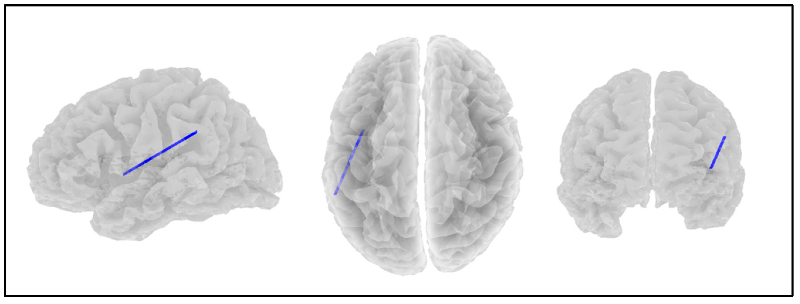
Significant negative correlations were also found between gamma band coherence and the SDS scores in connectivity between the left supramarginal gyrus (VAN) and the left insula (SN); r = −0.275, p = 0.024 (see Figure 3). Negative correlations between gamma band coherence and the Anhedonia (r = −0.261, p = 0.049), Cognitive (r = −0.301, p = 0.010), and Mood subtype scores (r = −0.247, p = 0.065) were also found in the same connectivity pattern.

4. Discussion

The primary aim of this study was to investigate and describe any differences in the attentional and salience network connectivity in the four MDD subtypes as defined by the scores on groups of items from the SDS obtained by the participants regardless of whether or not they met the criteria for clinical depression.
Theta coherence increased along with the overall SDS scores in a short three-node system typically involved in orienting visual attention across all the networks used in this study [43,44]. As increases in theta activity are typically associated with drowsiness or similar low-energy states, this indicates that people with MDD are increasingly likely to have less interest in monitoring their surroundings, leading to a sense of withdrawal from the environment given that it is less likely that attentional networks will identify anything considered worth engaging with. This finding is congruent with the ‘depression as behavioural withdrawal’ hypothesis mentioned in the Introduction.
This situation is increasingly the case with the Cognitive and particularly the Anhedonic depression subtypes. The theta band coherence system associated with the Cognitive depression subtype includes all the nodes found in those listed in the broader SDS depression and two more nodes from the DAN (the right frontal eye field and right posterior IPS). Given that activation in the DAN is indicative of increased top-down processing effects, this indicates that Cognitive depression may also be affected by the top-down control of attention [45,46]. The theta results relating to Anhedonic depression are even broader in scope but mostly involve the DAN and VAN, indicating that Anhedonic depression may be the result of a more chronic cause. This view is backed by the inclusion of the dorsal ACC (as part of the SN) in the theta results, given the broad range of mental processes that the dACC is associated with [47,48].
As distinct from theta coherence, beta coherence decreased as the overall SDS scores increased in a short three-node system primarily in the SN. Similar results were also found with the Cognitive subtypes with the addition of the middle frontal gyrus node from the VAN. Each node here plays an important role in identifying relevant stimuli based on episodic memory, particularly the supramarginal gyrus and the lateral parietal lobule [49,50]. This function would still be active during resting states such as those used in this study because monitoring the environment is an ongoing process rather than a task-positive one. Since beta and gamma activation is typically associated with active cognitive processing, this would indicate that greater degrees of MDD are associated with a fall in cognitive resources available for engaging with the environment (perhaps contributing to the behavioural withdrawal noted above). Similar results were found in the gamma band as those in the beta band where gamma coherence between the left supramarginal gyrus and left insula decreased as Anhedonia, Cognitive, Mood, and overall SDS scores increased. The overlap between the beta and gamma bands is likely the result of the same processes discussed above in the beta band results.
The Mood and Somatic depression subtypes are conspicuous in their absence from these results; apart from a single connectivity result in the gamma band, there was no relationship between either depression subtype and any measure of connectivity in the attentional networks. This can be explained by, or may be a product of, the more inward-focused nature of these depression subtypes; Mood-based depression is associated with negative emotional states (sadness; crying; and feeling hopeless, useless, or irritable) while Somatic-based depression is associated with negative bodily states (sleeping difficulties, weight loss, high pulse, and feeling tired and restless [15]). The absence of any relationship between the Mood or Somatic depression subtypes and attentional network activation indicates that these forms of depression are primarily driven by internal psychological and/or physical factors rather than a response to external stimuli.

4.1. Clinical Implications

This study was based on the assumption of MDD being heterogeneous, and this was confirmed by the different connectivity results across the four MDD subtypes used here. As such, the use of a homogeneous construct of MDD in clinical settings is challenged by these data. Instead, depressed patients might benefit from the consideration of their MDD subtype symptom profiles and a consequent therapeutic focus upon specific treatments for the subtypes they present with most severely. Those treatment choices could also be subsequently driven by the inclusion of the underlying cognitive processes inherent in the attentional and salience networks. This kind of diagnostic and therapeutic approach could move depression diagnosis and treatment from a ‘one-size fits all’ model, in which all depressions are treated similarly, towards a more personalised medicine approach [51,52]. The first step in that process is to recognise that, at a neurophysiological level (i.e., brain network connectivity), different groupings of MDD symptoms are more likely to be of benefit than a single score on an inventory or clinical interview that assumes a dichotomous classification is the best diagnosis available. The current study purposely examined only three major brain networks, and there are others that could also repay investigation such as task-positive networks.

4.2. Limitations

The Zung SDS is a creditable and well-justified method of measuring multiple MDD symptoms; however, it is not the only such instrument, and the use of alternative self-report instruments could add some greater validity to the overall results. Additionally, the DSM-5-TR Diagnostic Criteria are complemented by Diagnostic Features, but no current instrument exists that includes all of these. In fact, the tendency has been towards the development and use of briefer lists of MDD symptoms to diagnose MDD, such as the Personal Health Questionnaire-9 [53], but there are reasons from this study for moving towards more inclusive diagnostic instruments that measure all the aspects of depressive behaviour that contribute to the final diagnosis. This study used a ‘snap-shot’ research design, with data being collected at a single time point, whereas individuals’ mood states vary across days or weeks, and so repeated sampling of mood states would enhance the generalizability of this research. The sample was composed of volunteers from a single cultural and geographical region; comparative data from other settings could help extend the current findings. EEG does produce precise measures of site-specific electrical activity in the brain, but might be profitably matched with fMRI (for example) to provide a more detailed account of neurocognitive activity. The method for identifying MDD subtypes used in this study might be described as ‘a priori’ because the subtypes were grouped according to the commonalities between specific MDD symptoms, performed by a panel of clinicians [15]. Another method of identifying subtypes is ‘a posteriori’ by applying cluster analysis to EEG data and then regressing those data upon MDD symptoms from a scale like the SDS. Neither method has yet shown superiority, and both represent valuable models for defining MDD subtypes. Although some of these limitations do limit generalizability, they do not invalidate these results. Finally, research is always strengthened by testing hypotheses based on previous findings, but that was not possible here due to the lack of studies in this field.

5. Conclusions

Although each of the four MDD subtypes did not show a specific set of inter-network connections, the fact that two of them did so (Anhedonia and Cognitive depression) provides strong support for the notion of MDD as heterogeneous, and also provides some basis for the subtype model tested here. It may be that Somatic depression and depressed Mood are also able to be identified by reference to alternative neurocognitive variables, and that work is being undertaken by the authors.

Author Contributions

I.D.E.: conceptualisation, methodology, software, formal analysis, writing (original draft), and visualisation. C.F.S.: conceptualisation, methodology, resources, and writing (review and editing). V.B.: conceptualisation and writing (review and editing). K.A.V.: writing (review and editing). R.J.W.: writing (review and editing). E.J.: investigation and data curation. L.L.A.: resources. All authors have read and agreed to the published version of the manuscript.

Funding

This research did not receive any specific grant from funding agencies in the public, commercial, or not-for-profit sectors.

Institutional Review Board Statement

Ethics approval for this study was provided by the Human Research Ethics Committee of the University of New England, Australia (Approval No. HE14-051), consistent with the Code of Ethics of the World Medical Association (Declaration of Helsinki).

Informed Consent Statement

Written and informed consent was obtained from all the subjects involved in the study.

Data Availability Statement

EEG and source localization data will be made available upon reasonable request.

Conflicts of Interest

The authors declare no conflicts of interest.

References

  1. Liu, Q.; He, H.; Yang, J.; Feng, X.; Zhao, F.; Lyu, J. Changes in the global burden of depression from 1990 to 2017: Findings from the Global Burden of Disease study. J. Psychiatr. Res. 2020, 126, 134–140. [Google Scholar] [CrossRef] [PubMed]
  2. Li, W.; Chen, D.; Ruan, W.; Peng, Y.; Lu, Z.; Wang, D. Associations of depression, sleep disorder with total and cause-specific mortality: A prospective cohort study. J. Affect. Disord. 2022, 298, 134–141. [Google Scholar] [CrossRef] [PubMed]
  3. American Psychiatric Association. Diagnostic and Statistical Manual of Mental Disorders, 5th ed.; American Psychiatric Association: Washington DC, USA, 2022. [Google Scholar]
  4. LeMoult, J. From stress to depression: Bringing together cognitive and biological science. Curr. Dir. Psychol. Sci. 2020, 29, 592–598. [Google Scholar] [CrossRef] [PubMed]
  5. Ferster, C.B. A Functional Analysis of Depression. Am. Psychol. 1973, 28, 857–870. [Google Scholar] [CrossRef]
  6. Dougher, M.J.; Hackbert, L. A Behavior-Analytic Account of Depression and a Case Report Using Acceptance-Based Procedures. Behav. Anal. 1994, 17, 321–334. [Google Scholar] [CrossRef]
  7. Bolling, M.; Kohlenberg, R.; Parker, C. Behavior Analysis and Depression. In Clinical Behavior Analysis; Dougher, M.J., Ed.; Context Press: Reno, NV, USA, 1999; pp. 127–153. [Google Scholar]
  8. Kanter, J.; Cautilli, J.; Busch, A.; Baruch, D. Towards a comprehensive functional analysis of depressive behavior: Five environmental factors and a possible sixth and seventh factor. Int. J. Behav. Consult. Ther. 2011, 7, 5–14. [Google Scholar] [CrossRef]
  9. Santos, M.M.; Nagy, G.A.; Kanter, J.W.; López, S.R. Applying a Process-Oriented Model of Cultural Competence to Behavioral Activation for Depression. Cogn. Behav. Pract. 2021, 28, 127–146. [Google Scholar] [CrossRef]
  10. Darwin, C. Recollections of the development of my mind and character. In Autobiographies; Penguin: London, UK, 2002. [Google Scholar]
  11. Sturmey, P. Functional Analysis in Clinical Psychology; John Wiley & Sons: New York, NY, USA, 1996. [Google Scholar]
  12. Stein, A.T.; Carl, E.; Cuijpers, P.; Karyotaki, E.; Smits, J.A. Looking beyond depression: A meta-analysis of the effect of behavioral activation on depression, anxiety, and activation. Psychol. Med. 2021, 51, 1491–1504. [Google Scholar] [CrossRef]
  13. Ostergaard, S.; Jensen, S.; Bech, P. The heterogeneity of the depressive syndrome: When numbers get serious. Acta Psychiatr. Scand. 2011, 124, 495–496. [Google Scholar] [CrossRef]
  14. Baumeister, H.; Parker, G. Meta-review of depressive subtyping models. J. Affect. Disord. 2012, 139, 126–140. [Google Scholar]
  15. Sharpley, C.F.; Bitsika, V. Differences in neurobiological pathways of four “clinical content” subtypes of depression. Behav. Brain Res. 2013, 256, 368–376. [Google Scholar] [CrossRef] [PubMed]
  16. Sharpley, C.F.; Bitsika, V. Validity, reliability and prevalence of four ‘Clinical Content’ subtypes of depression. Behav. Brain Res. 2014, 259, 9–15. [Google Scholar] [CrossRef] [PubMed]
  17. Milaneschi, Y.; Lamers, F.; Berk, M.; Penninx, B. Depression Heterogeneity and Its Biological Underpinnings: Toward Immunometabolic Depression. Biol. Psychiatry 2020, 88, 369–380. [Google Scholar] [CrossRef] [PubMed]
  18. Beijers, L.; Wardenaar, K.; van Loo, H.; Schoevers, R. Data-driven biological subtypes of depression: Systematic review of biological approaches to depression subtyping. Mol. Psychiatry 2019, 24, 888–900. [Google Scholar] [CrossRef]
  19. Buch, A.; Liston, C. Dissecting diagnostic heterogeneity in depression by integrating neuroimaging and genetics. Neuropsychopharmacology 2021, 46, 156–175. [Google Scholar] [CrossRef]
  20. Pandya, M.; Altinay, M.; Malone, D.A.; Anand, A. Where in the brain is depression? Curr. Psychiatry Rep. 2012, 14, 634–642. [Google Scholar] [CrossRef]
  21. Drevets, W.C.; Price, J.L.; Furey, M.L. Brain structural and functional abnormalities in mood disorders: Implications for neurocircuitry models of depression. Brain Struct. Funct. 2008, 213, 93–118. [Google Scholar] [CrossRef]
  22. Zhang, F.F.; Peng, W.; Sweeney, J.A.; Jia, Z.Y.; Gong, Q.Y. Brain structure alterations in depression: Psychoradiological evidence. CNS Neurosci. Ther. 2018, 24, 994–1003. [Google Scholar] [CrossRef]
  23. Raz, A.; Buhle, J. Typologies of attentional networks. Nat. Rev. Neurosci. 2006, 7, 367–379. [Google Scholar] [CrossRef]
  24. Huang, H.; Chen, C.; Rong, B.; Wan, Q.; Chen, J.; Liu, Z.; Zhou, Y.; Wang, G.; Wang, H. Resting-state functional connectivity of salience network in schizophrenia and depression. Sci. Rep. 2022, 12, 11204. [Google Scholar] [CrossRef]
  25. Han, S.; Cui, Q.; Wang, X.; Li, L.; Li, D.; He, Z.; Guo, X.; Fan, Y.; Guo, J.; Sheng, W.; et al. Resting state functional network switching rate is differently altered in bipolar disorder and major depressive disorder. Hum. Brain Mapp. 2020, 41, 3295–3304. [Google Scholar] [CrossRef] [PubMed]
  26. Goulden, N.; Khusnulina, A.; Davis, N.J.; Bracewell, R.M.; Bokde, A.L.; McNulty, J.P.; Mullins, P.G. The salience network is responsible for switching between the default mode network and the central executive network: Replication from DCM. Neuroimage 2014, 99, 180–190. [Google Scholar] [CrossRef] [PubMed]
  27. Nolen-Hoeksema, S.; Wisco, B.E.; Lyubomirsky, S. Rethinking rumination. Perspect. Psychol. Sci. 2008, 3, 400–424. [Google Scholar] [CrossRef] [PubMed]
  28. Menon, V.; Uddin, L.Q. Saliency, switching, attention and control: A network model of insula function. Brain Struct. Funct. 2010, 214, 655–667. [Google Scholar] [CrossRef]
  29. Sharpley, C.F.; Agnew, L.L. The New England Mental Health Study, 1st ed.; University of New England: Amidale, NSW, Australia, 2016. [Google Scholar]
  30. Zung, W. A self-rating depression scale. Arch. Gen. Psychiatry 1965, 12, 63–70. [Google Scholar] [CrossRef]
  31. Zung, W. From art to science: The diagnosis and treatment of depression. Arch. Gen. Psychiatry 1973, 29, 328–337. [Google Scholar] [CrossRef]
  32. DeJonge, J.; Baneke, J. The Zung Self-rating Depression Scale: A replication study on reliability, validity and prediction. Psychol. Rep. 1989, 64, 833–834. [Google Scholar] [CrossRef]
  33. Gabrys, J.; Peters, K. Reliability, discriminant and predictive validity of the Zung Self-Rating Depression Scale. Psychol. Rep. 1985, 57, 1091–1096. [Google Scholar] [CrossRef]
  34. Schaefer, A.; Brown, J.; Watson, C.; Plenel, D.; DeMotts, J.; Howard, M.; Norman, P.; Balleweg, B.J.; Douglas, A. Comparison of the validities of the Beck, Zung and MMPI depression scales. J. Consult. Clin. Psychol. 1985, 53, 415–418. [Google Scholar] [CrossRef]
  35. Pascual-Marqui, R.D.; Lehmann, D.; Koukkou, M.; Kochi, K.; Anderer, P.; Saletu, B.; Tanaka, H.; Hirata, K.; John, E.R.; Prichep, L.; et al. Assessing interactions in the brain with exact low-resolution electromagnetic tomography. Philos. Trans. R. Soc. A Math. Phys. Eng. Sci. 2011, 369, 3768–3784. [Google Scholar] [CrossRef]
  36. Marco-Pallarés, J.; Grau, C.; Ruffini, G. Combined ICA-LORETA analysis of mismatch negativity. Neuroimage 2005, 25, 471–477. [Google Scholar] [CrossRef] [PubMed]
  37. Pascual-Marqui, R.D.; Esslen, M.; Kochi, K.; Lehmann, D. Functional imaging with low-resolution brain electromagnetic tomography (LORETA): A review. Methods Find. Exp. Clin. Pharmacol. 2002, 24 (Suppl. C), 91–95. [Google Scholar] [PubMed]
  38. Fuchs, M.; Kastner, J.; Wagner, M.; Hawes, S.; Ebersole, J.S. A standardized boundary element method volume conductor model. Clin. Neurophysiol. 2002, 113, 702–712. [Google Scholar] [CrossRef] [PubMed]
  39. Mazziotta, J.; Toga, A.; Evans, A.; Fox, P.; Lancaster, J.; Zilles, K.; Woods, R.; Paus, T.; Simpson, G.; Pike, B.; et al. A probabilistic atlas and reference system for the human brain: International Consortium for Brain Mapping (ICBM). Philos. Trans. R. Soc. Lond. Ser. B Biol. Sci. 2001, 356, 1293–1322. [Google Scholar] [CrossRef]
  40. Pascual-Marqui, R.D. Discrete, 3D distributed, linear imaging methods of electric neuronal activity. Part 1: Exact, zero error localization. arXiv 2007, arXiv:07103341. [Google Scholar]
  41. Fox, M.D.; Corbetta, M.; Snyder, A.Z.; Vincent, J.L.; Raichle, M.E. Spontaneous neuronal activity distinguishes human dorsal and ventral attention systems. Proc. Natl. Acad. Sci. USA 2006, 103, 10046–10051. [Google Scholar] [CrossRef]
  42. Raichle, M.E. The restless brain. Brain Connect. 2011, 1, 3–12. [Google Scholar] [CrossRef]
  43. Japee, S.; Holiday, K.; Satyshur, M.D.; Mukai, I.; Ungerleider, L.G. A role of right middle frontal gyrus in reorienting of attention: A case study. Front. Syst. Neurosci. 2015, 9, 23. [Google Scholar] [CrossRef]
  44. Suo, X.; Ding, H.; Li, X.; Zhang, Y.; Liang, M.; Zhang, Y.; Yu, C.; Qin, W. Anatomical and functional coupling between the dorsal and ventral attention networks. Neuroimage 2021, 232, 117868. [Google Scholar] [CrossRef]
  45. Astafiev, S.V.; Shulman, G.L.; Corbetta, M. Visuospatial reorienting signals in the human temporo-parietal junction are independent of response selection. Eur. J. Neurosci. 2006, 23, 591–596. [Google Scholar] [CrossRef]
  46. Corbetta, M.; Shulman, G.L. Control of goal-directed and stimulus-driven attention in the brain. Nat. Rev. Neurosci. 2002, 3, 201–215. [Google Scholar] [CrossRef] [PubMed]
  47. Margulies, D.S.; Kelly, A.C.; Uddin, L.Q.; Biswal, B.B.; Castellanos, F.X.; Milham, M.P. Mapping the functional connectivity of anterior cingulate cortex. Neuroimage 2007, 37, 579–588. [Google Scholar] [CrossRef] [PubMed]
  48. Posner, M.I.; Rothbart, M.K.; Sheese, B.E.; Tang, Y. The anterior cingulate gyrus and the mechanism of self-regulation. Cogn. Affect. Behav. Neurosci. 2007, 7, 391–395. [Google Scholar] [CrossRef] [PubMed]
  49. Menon, V.; D’Esposito, M. The role of PFC networks in cognitive control and executive function. Neuropsychopharmacology 2022, 47, 90–103. [Google Scholar] [CrossRef] [PubMed]
  50. Rubinstein, D.Y.; Camarillo-Rodriguez, L.; Serruya, M.D.; Herweg, N.A.; Waldman, Z.J.; Wanda, P.A.; Sharan, A.D.; Weiss, S.A.; Sperling, M.R. Contribution of left supramarginal and angular gyri to episodic memory encoding: An intracranial EEG study. Neuroimage 2021, 225, 117514. [Google Scholar] [CrossRef]
  51. Kambeitz-Ilankovic, L.; Koutsouleris, N.; Upthegrove, R. The potential of precision psychiatry: What is in reach? Br. J. Psychiatry 2022, 220, 175–178. [Google Scholar] [CrossRef]
  52. Tozzi, L.; Zhang, X.; Pines, A.; Olmsted, A.M.; Zhai, E.S.; Anene, E.T.; Chesnut, M.; Holt-Gosselin, B.; Chang, S.; Stetz, P.C.; et al. Personalized brain circuit scores identify clinically distinct biotypes in depression and anxiety. Nat. Med. 2024, 30, 2076–2087. [Google Scholar] [CrossRef]
  53. Kroenke, K.; Spitzer, R.; Williams, J. The PHQ-9: Validity of a brief depression severity measure. J. Gen. Intern. Med. 2001, 16, 606–613. [Google Scholar] [CrossRef]
Figure 1. Significant positive correlations (p < 0.1) between theta coherence and the (A) Anhedonic depression scores, (B) Cognitive depression scores, and (C) overall MDD depression scores. Views are from the left, top, and front, respectively.
Applsci 15 01459 g001
Figure 2. Significant negative correlations (p < 0.1) between beta coherence and (A) the Cognitive depression scores and (B) both the overall MDD depression scores and Anhedonic depression scores. Views are from the left, top, and front, respectively.
Applsci 15 01459 g002
Figure 3. Significant negative correlations (p < 0.1) between gamma coherence and the Anhedonic, Cognitive, Mood, and overall MDD depression scores. Views are from the left, top, and front, respectively.
Applsci 15 01459 g003
Table 1. Neural networks, brain sites, and MNI coordinates of centroid for each ROI.
NetworkLocationMNI (X, Y, Z)
Dorsal Attention Networkleft frontal eye field−25, 12, 55
right frontal eye field28, −10, 53
left posterior intraparietal sulcus−22, −68, 46
right posterior intraparietal sulcus20, −67, 51
Ventral Attention Networkleft middle frontal gyrus−47, 14, 32
right middle frontal gyrus47, 14, 32
left supramarginal gyrus−57, −43, 34
right supramarginal gyrus57, −43, 34
Salience Networkdorsal anterior cingulate0, −21, 36
left anterior prefrontal cortex−35, 45, 30
right anterior prefrontal cortex32, 45, 30
left insula−41, 3, 6
right insula41, 3, 6
left lateral parietal lobule−62, −45, 30
right lateral parietal lobule62, −45, 30
Table 2. Pearson’s r- and p-values for each significant connection in the theta band Anhedonic depression condition.
ROINetworkrp
left frontal eye fieldDAN0.3110.082
right middle frontal gyrusVAN
right middle frontal gyrusVAN0.3420.037
dorsal anterior cingulateSN
dorsal anterior cingulateSN0.3230.064
left insulaSN
left insulaSN0.3250.060
left frontal eye fieldDAN
left posterior IPSDAN0.3230.063
left middle frontal gyrusVAN
left middle frontal gyrusVAN0.3260.059
right posterior IPSDAN
Table 3. Pearson’s r- and p-values for each significant connection in the theta band Cognitive depression condition.
ROINetworkrp
right insulaSN0.3090.049
left posterior IPSDAN
left posterior IPSDAN0.3580.009
left middle frontal gyrusVAN
left middle frontal gyrusVAN0.3300.022
right posterior IPSDAN
right posterior IPSDAN0.3100.047
right insulaSN
right frontal eye fieldDAN0.2890.089
left posterior IPSDAN
Table 4. Pearson’s r- and p-values for each significant connection in the theta band overall SDS depression condition.
ROINetworkrp
right insulaSN0.2940.096
left posterior IPSDAN
left posterior IPSDAN0.3000.082
left middle frontal gyrusVAN
Table 5. Pearson’s r- and p-values for each significant connection in the beta band Cognitive depression condition.
ROINetworkrp
right supramarginal gyrusVAN−0.2730.046
left insulaSN
left insulaSN−0.2790.037
left lateral parietal lobuleSN
left lateral parietal lobuleSN−0.2530.092
right middle frontal gyrusVAN
Table 6. Pearson’s r- and p-values for each significant connection in the beta band Anhedonic and SDS depression conditions.
AnhedonicSDS
ROINetworkrprp
right supramarginal gyrusVAN−0.2660.040−0.2390.097
left insulaSN
left insulaSN−0.2710.032−0.2390.093
left lateral parietal lobuleSN
Disclaimer/Publisher’s Note: The statements, opinions and data contained in all publications are solely those of the individual author(s) and contributor(s) and not of MDPI and/or the editor(s). MDPI and/or the editor(s) disclaim responsibility for any injury to people or property resulting from any ideas, methods, instructions or products referred to in the content.