Article

Fate of the Mandible in Class III Patients Subjected to Bimaxillary Surgery with a New 3D Planning Reference

by
Federico Hernández-Alfaro
1,2,
Carlos de la Fuente-Vázquez
1,
Adaia Valls-Ontañón
1,2,*,
Orion-Luiz Haas-Junior
3,
Maria Giralt-Hernando
1,2 and
Jorge Masià-Gridilla
1,2
1
Institute of Maxillofacial Surgery, Teknon Medical Center, 08022 Barcelona, Spain
2
Department of Oral and Maxillofacial Surgery, Universitat Internacional de Catalunya, Sant Cugat del Vallès, 08194 Barcelona, Spain
3
Department of Oral and Maxillofacial Surgery, Pontifical Catholic University of Rio Grande do Sul, Porto Alegre 6681, Brazil
*
Author to whom correspondence should be addressed.
Appl. Sci. 2025, 15(3), 1069; https://doi.org/10.3390/app15031069
Submission received: 8 October 2024 / Revised: 24 December 2024 / Accepted: 8 January 2025 / Published: 22 January 2025
(This article belongs to the Section Applied Dentistry and Oral Sciences)

Abstract

:
Class III patients have classically been managed through monomaxillary techniques, mainly involving mandibular setback movements. More recently, according to the upper incisor to soft tissue plane (UI-STP) or Barcelona Line (BL) planning protocol, bimaxillary procedures for upper maxilla advancement and reduced mandibular setback have been recommended in order to secure better aesthetic outcomes and avoid upper airway constriction. The present study describes the jaw movements in the sagittal plane performed in class III patients subjected to bimaxillary surgery following the BL protocol. A retrospective evaluation was performed on 124 class III patients subjected to bimaxillary surgery. All subjects underwent upper maxilla advancement. A total of 112 patients received mandible advancement movement (90.3%), nine received mandibular setback (7.25%), and the mandible underwent no movement along the sagittal dimension in the three remaining patients (2.4%). Mandibular advancement was significantly the most frequent treatment option. The presented results suggest that when the BL planning protocol is used as an aesthetic and functional reference, class III occlusion appears mostly related to maxillary sagittal hypoplasia instead of mandible hyperplasia, so bimaxillary advancement surgery should be considered as one of the first-choice procedures for the treatment of these patients.

1. Introduction

The class III growth pattern is characterized by disproportionate forward mandibular growth and/or deficient maxillary growth [1]. The treatment of class III patients can start as soon as the deciduous dentition appears. In growing patients, it is attempted to treat class III malocclusions using orthopedic techniques such as chin cups or protraction facemasks. Little long-term success has been documented with the use of these devices [2]. However, once the patient has stopped growing, the treatment options for skeletal class III range from orthodontic camouflage in non-severe cases [3] to orthognathic surgery in combination with orthodontic treatment in more severe cases [4,5]. Nevertheless, non-surgical treatment generally falls short of expectations and rarely produces acceptable facial esthetics. Accordingly, orthognathic surgery is considered to be the first treatment choice in the majority of cases.
Over the last few years, the authors have been trying to establish a reference that, moving beyond classical cephalometric analyses, and independently of the occlusal situation of the patient, could serve as an esthetic guide for the sagittal repositioning of the maxillomandibular complex [6]. Previous surgical strategies have been aimed at achieving class I occlusion by combining different levels of maxillary advancement and mandibular retrusion. However, the authors believed that most treated class III patients had deficient jawline and neck definition, and they also found that a significant proportion of them presented with posterior airway involvement, resulting in variable degrees of obstructive sleep apnea hypopnea syndrome (OSAHS) [7,8].
In 2010, after studying a series of visually attractive individuals, the senior author proposed a new sagittal anterior repositioning reference. The latter related a plane tangential to the soft tissue nasion and perpendicular to the floor with the head in the natural head position (NHP) to the upper incisor. In 101 out of 112 cases (92%), the upper incisor was at this vertical plane or in front of it [6]. In 2022, the upper incisor to soft tissue plane (UI-STP), or the so-called “Barcelona Line” (BL), was first validated as a guide for the sagittal repositioning of the maxillomandibular complex [7,9], where, first, the mandible is brought to occlusion, and then the maxillomandibular complex is moved to the BL.
After more than a decade using this vertical reference for both facial diagnostic analysis and surgical planning, the authors realized that most class III patients required large maxillary advancement and even the mandible was usually moved forward. Therefore, the previously mentioned deficient jawline and neck definition that characterizes the majority of these class III patients was improved. Thus, the authors aimed to demonstrate the fate of the mandible in the sagittal plane in a series of class III patients subjected to orthognathic surgery using the previously described UI-STP [6,7].

2. Material and Methods

2.1. Sample Selection

A retrospective analysis was performed on consecutive class III patients subjected to bimaxillary orthognathic surgery at the Maxillofacial Institute—Teknon Medical Center (Barcelona, Spain). Data were collected from the patients’ medical records between January 2016 and December 2018.
The following inclusion criteria were applied: age ≥ 18 years, Caucasian patients, skeletal class III malocclusion in need of surgical correction, and skeletal growth completed at the time of the operation. Patients were excluded as study subjects if they received a single jaw surgery, had a history of condylar hyperplasia of the mandible or any craniofacial syndrome, or there was a lack of preoperative cone-beam computed tomography (CBCT) scanning and facial pictures.
The Ethics Committee of the Teknon Medical Center approved the study (number ISF). The guidelines of the Declaration of Helsinki on medical protocols and ethics were followed at all treatment stages. Written informed consent was obtained from all subjects.

2.2. Virtual Planning

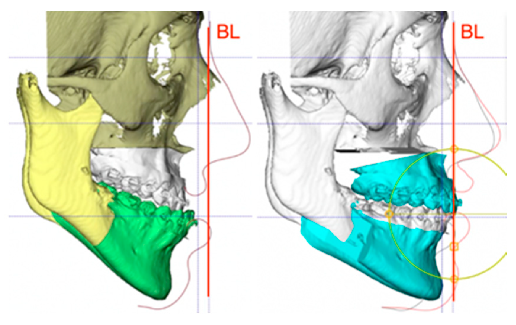
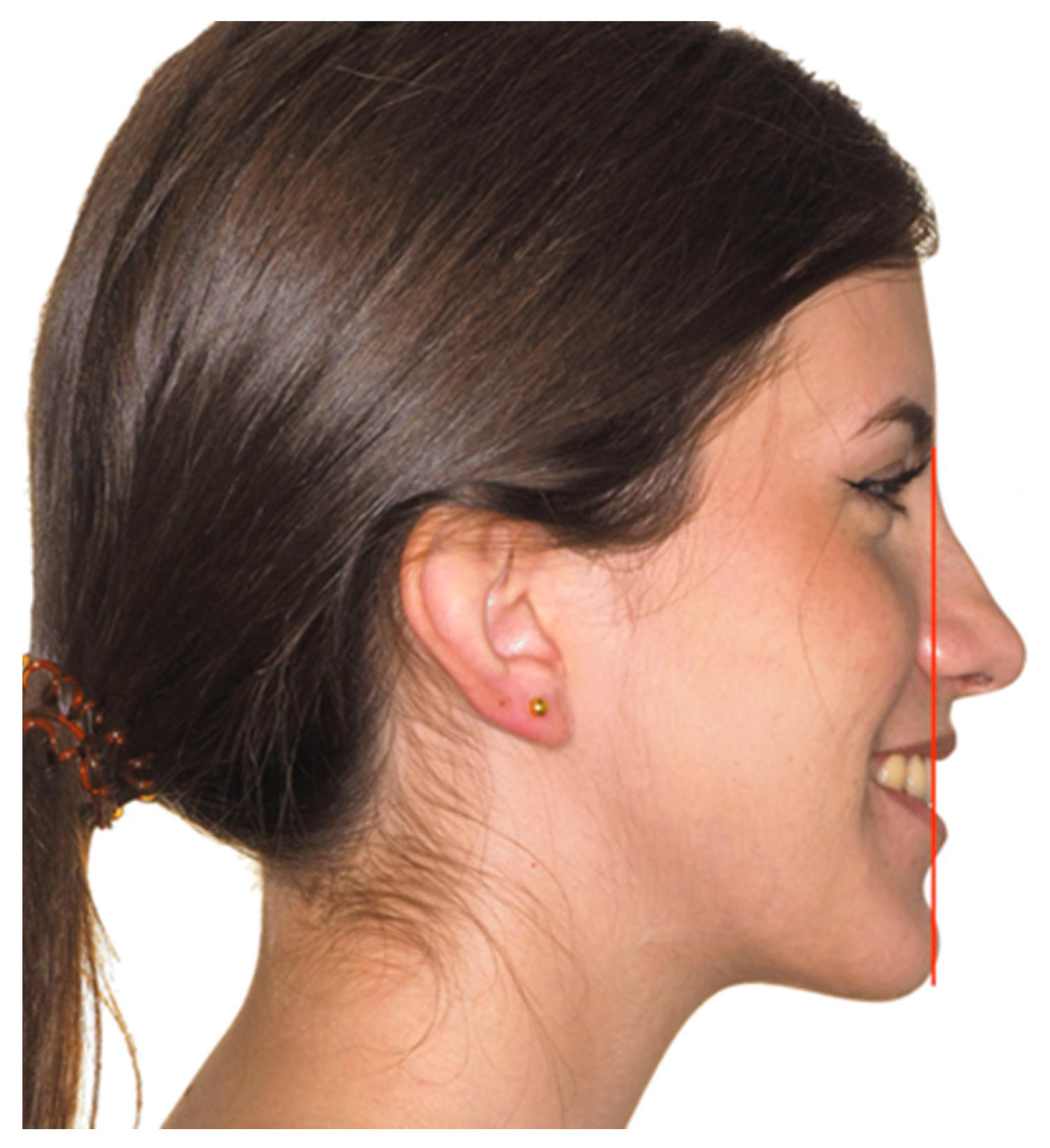
In all cases, the routine protocol for diagnostic work-up and three-dimensional (3D) surgical planning of the study center was followed. This protocol has been validated and described in detail elsewhere [6]. Patients’ records were obtained preoperatively, which included occlusal and facial pictures (sitting upright in the natural head orientation (NHO)) and a cone-beam computed tomography (CBCT) scan (iCAT, Imaging Sciences International, Hatfield, PA, USA). The DICOM datasets were exported to the Dolphin Imaging 3D version 11.8 software (Dolphin Imaging & Management Solutions, Chatsworth, CA, USA), where the virtual head was oriented according to the NHO, using the photographs as a reference. Then, virtual simulations of orthognathic surgery and computer-aided design and computer-aided manufacturing (CAD/CAM) production of the intermediate and final splints were performed in all cases. All surgeries were virtually planned by the same surgeon (FHA), and the treatment objectives were established according to the UI-STP as the reference line [4], which places the upper incisor (provided that it has adequate torque) at a vertical plane through the soft tissue nasion perpendicular to the floor, with the patient in the natural head position (NHP) (Figure 1).
The virtual planning steps included (a) the definition of the desired final occlusion in the physical dental models; (b) the digital scanning of the final occlusion and introduction into the 3D software; (c) the use of this desired final occlusion, positioning the maxilla and mandible together in class I; (d) moving the maxillomandibular complex with its upper incisor to the abovementioned plane; (e) applying the appropriate vertical modifications; and (f) the repositioning of the maxillomandibular complex in all spatial planes (pitch, roll, and yaw) in order to set the virtual treatment objectives. The clockwise (CW) or counter-clockwise (CCW) rotation of the maxillomandibular complex was performed to achieve the adequate projection of the chin (Figure 2).

2.3. Surgery

All patients were operated upon under general anesthesia by the same surgeon (FHA). Mandibular osteotomies were performed first according to the standardized bilateral sagittal split osteotomy (BSSO) technique [10]. Then, Le Fort I osteotomies were performed in a minimally invasive manner with the previously published “twist technique” [11]. Finally, genioplasty was carried out when necessary. All patients were extubated in the operating room and discharged 24 h after the procedure, and dynamic intermaxillary fixation was maintained with guiding elastics.

2.4. Evaluation

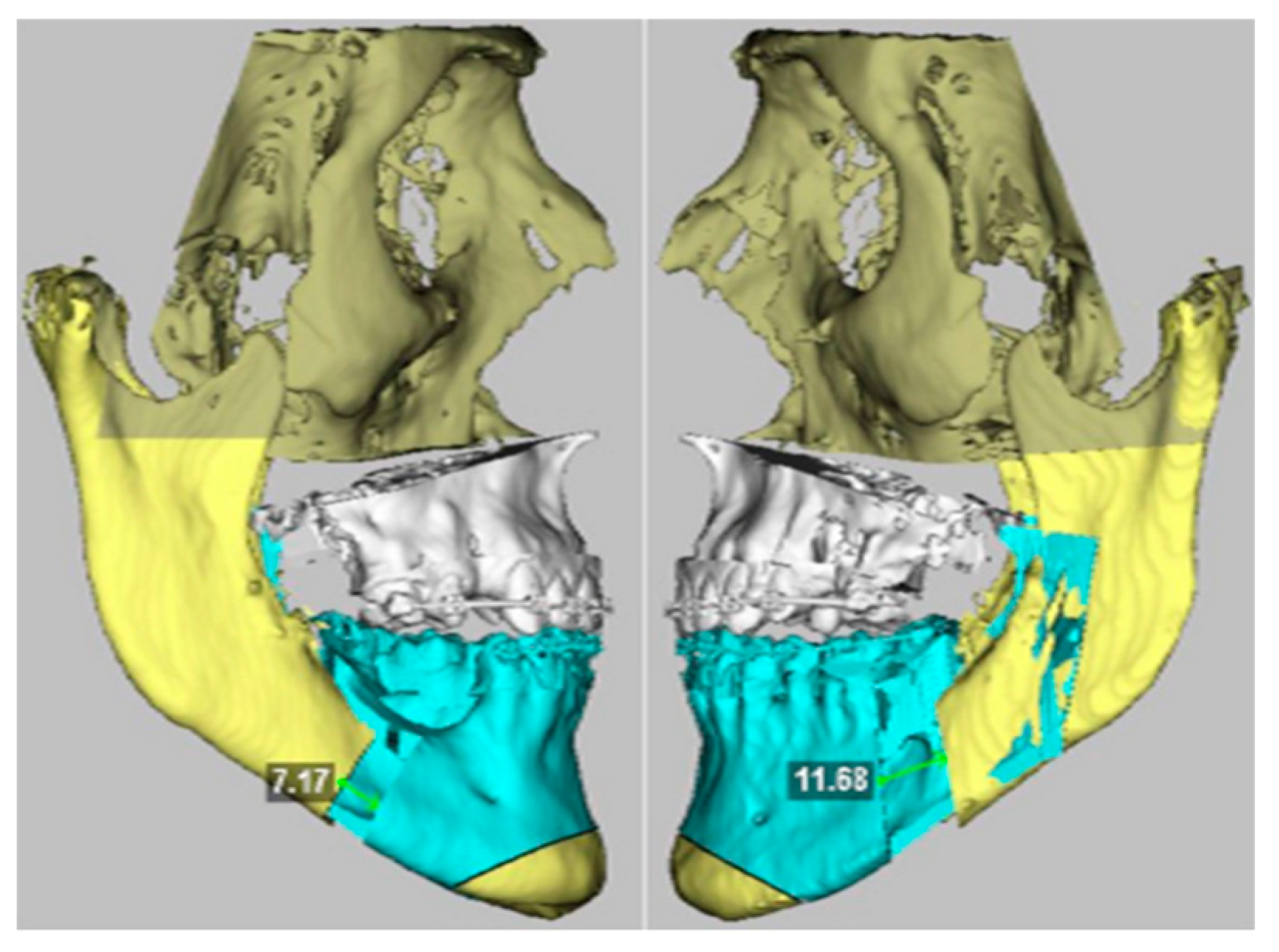
The virtual surgical planning with the Dolphin Imaging version 11.8 software was used to evaluate the direction and amount of sagittal movement of the mandible and the maxilla in the class III patients, always positioning the virtual head of the patient in the NHO. The following measurements were carried out: (a) sagittal movement of the osseous B point (pre- and postoperatively according to the surgical planning) (Figure 2); (b) linear measurement of both osteotomy gaps (a perpendicular line was traced between both vertical osteotomy lines on each side) (Figure 3); maxillary sagittal movement was evaluated measuring pre- and post-treatment values at the osseous A point. Moreover, the type and magnitude of occlusal plane rotation (clockwise versus counter-clockwise) and the absolute mandible centering value were recorded.

2.5. Statistical Analysis

Statistical analysis was carried out using SPSS for Windows (version 15.0.1, SPSS Inc., Chicago, IL, USA). The descriptive analysis was based on the mean, standard deviation (SD), and median (for continuous variables) and absolute and relative frequencies (for categorical variables).
Regarding the inferential analysis, the chi-squared test was applied to compare the proportions between mandibular advancement, mandibular setback, and cases without mandibular sagittal movement. The Mann–Whitney test was used to assess whether the above result depended on specific centering and/or rotation movements. The significance level used in all analyses was 5% (α = 0.05).

3. Results

During the two-year study period, a total of 124 patients met all the inclusion criteria and were therefore included in the study. The mean patient age at the time of virtual surgery planning was 26.3 years (range 18–49). Fifty patients were male (40.3%) and 74 were female (59.7%).
All patients underwent upper maxilla advancement (mean 6.79 ± 3.53 mm), as is typical in class III patients. However, regarding mandibular sagittal movement at the B point, 112 patients (90.3%) received mandibular advancement (mean 5.22 ± 4.82 mm), only nine (7.3%) received mandibular setback (mean −2.55 ± 1.16 mm), and no changes along the sagittal dimension occurred in three patients (2.4%). Therefore, in this series of class III patients, the chi-squared test confirmed mandibular advancement as being a significantly more frequent option than mandibular setback or mandibular sagittal non-movement (p = 2.7 × 10⁻¹² (p < 0.001)).
The mean distance at the bilateral sagittal split osteotomy gaps was 5.86 ± 4.64 mm on the right side and 5.87 ± 4.60 mm on the left side, which is in concordance with the mandibular advancement amount at the B point.
Regarding the occlusal plane changes, the CCW rotation of the occlusal plane was performed in 65 patients (52.4%, mean 3.6 ± 1.76°), while CW rotation was not carried out in any patient. A centering movement was performed in 84 of the patients (67.7%, mean 0.8 mm), and 39 (31.5%) received both centering and CCW rotation of the mandible (Table 1). Out of the 112 patients subjected to mandibular advancement, only six received a pure sagittal movement (5.4%), while, in the remaining 106 cases, mandibular advancement was accompanied by centering, CCW rotation, or both (94.6%). Thus, mandibular CCW rotation always involved its advancement (p < 0.001, MW), as well as the combination of mandibular CCW rotation and centering (p = 0.012, MW). On the other hand, mandibular setback never needed centering (p = 0.001, MW).
There were no significant differences in mandibular sagittal movement according to sex (p = 0.564, MW) or whether maxillary segmentation was performed (p = 0.582, MW).

4. Discussion

In coincidence with previous studies [12], the authors firmly believe that classical cephalometric analyses cannot be used as a roadmap in facial planning: analyses based on intracranial horizontal lines, such as the Sella-Nasion [13] or Frankfort plane [14], add errors due to positional variations and skull base disparities, while extracranial references such as the NHO are preferred for surgical planning. In accordance with this, some esthetic reference lines for antero-posterior maxillomandibular guide positioning have been created. The most recent and widely accepted approach was defined by Arnett [15] and uses a vertical line running through the subnasale point, but it will be altered in hypoplastic maxillaries and modified by surgery. On the other hand, the UI-STP or BL reference [6,7], used in this study, constitutes a stable reference that is not modified with surgery and can be analyzed clinically in a lateral smiling picture in the NHP, without the need for any radiological examination [16].
During the past few decades, and basing the treatment plan on “classical” cephalometric analyses, the most common movement of the jaw when performing orthognathic surgery in class III patients used to be isolated mandibular setback, since such patients were most likely to be diagnosed with mandibular excess [3]. Nowadays, maxillary advancement or two-jaw surgery combining these procedures has become the most popular treatment option [17].
According to the authors’ protocol, during 3D planning, the upper and lower jaws are placed in class I and moved forward until the upper incisor reaches the BL. Then, vertical adjustments are made. The chin is positioned appropriately according to the occlusal plane and the lower incisor position (around 90°). Finally, the required occlusal plane rotational movements are performed in order to obtain a true linear distance between the A and B points around 3 mm [18]. This planning is irrespective of the occlusal situation of the patient or the amount of maxilla–mandibular discrepancy.
The results of this study show that, when using this reference line as an antero-posterior repositioning reference in Caucasian patients, most cases diagnosed with and treated for class III dentofacial deformities underwent forward movements of both the upper maxilla and the mandible (although the latter to a lesser extent), in order to maximize the facial esthetics, improve soft tissue support, and avoid a reduction in the upper airway volume (90.3% of all patients, p = 2.7 × 10⁻¹² (p < 0.001)) (Figure 4).
With regard to mandibular setback, it only appears to be necessary in a minority of cases (7.3% of our patients), which are mostly coincident with hemimandibular elongation or hyperplasia and underlying acromegaly (three cases in our sample).
In comparing the functional advantages of mandibular advancement or setback, there are several points favorable for the forward movement of the jaw. In relation to stability, it has been widely demonstrated that isolated mandibular setback, especially when large movements are performed, results in poorer outcomes than when performing two-jaw surgery with maxilla advancement and less mandibular setback [19,20].
On the other hand, several investigators have reported the induction of sleep-related breathing disorders after mandibular setback procedures [21,22,23]. Mandibular setback results in the reorganization of the pharyngeal wall, and research has evidenced the postoperative narrowing of the retrolingual and hypopharyngeal airway and posteroinferior displacement of the hyoid bone and tongue. This new relationship in the upper airway may compromise the air flow and predispose the patient to OSAHS [24,25,26].
Lastly, setback can also entail esthetically detrimental secondary effects, since an undesired jowl may appear [1]. Thus, as mandible setback surgery involves both functional and esthetic drawbacks, CCW rotation can be performed in some cases with a relatively prominent mandible (52.4% of our cases). Mandibular CCW rotation can be also combined with setback in order to minimize the side effects of the latter.
The present study has some limitations, such as its retrospective cohort and single-center design, with the inherent biases involved. Thus, although a multicenter study would be desirable in order to draw firm conclusions, and the present study is focused on Caucasian patients, we have reported that mandibular advancement is significantly the most frequent treatment option in skeletal class III patients.
The authors understand that this new planning paradigm can be disruptive when confronted with the classical concepts. However, if an “orthofacial” instead of an “orthognathic” approach is embraced, this redefinition of surgical planning could improve the outcomes in terms of facial esthetics, soft tissue support, and the airway volume.

5. Conclusions

Therefore, in contrast to the classical class III dentofacial deformity approach, the results of this study suggest that, when using the BL as a sagittal maxilla–mandibular repositioning reference, most cases diagnosed with and treated for class III dentofacial deformities require the forward movement of both the upper maxilla and the mandible in order to maximize the facial esthetics, improve soft tissue support, and avoid the constriction of the upper airway.

Author Contributions

F.H.-A. designed the work; C.d.l.F.-V. contributed to the data acquisition; M.G.-H., O.-L.H.-J. and A.V.-O. substantively revised the work; and J.M.-G. drafted and revised the work. All authors have read and agreed to the published version of the manuscript.

Funding

This research did not receive any specific grant from funding agencies in the public, commercial, or not-for-profit sectors.

Institutional Review Board Statement

The Ethics Committee of the Teknon Medical Center approved the study under number 2019/89-CMF-TEK (21/11/2019). The guidelines of the Declaration of Helsinki on medical protocols and ethics were followed at all treatment stages.

Informed Consent Statement

Written informed consent was obtained from all subjects and from all patients with submitted images.

Data Availability Statement

The datasets used and/or analyzed during the current study are available from the corresponding author on reasonable request.

Acknowledgments

The authors would like to thank all staff members at the Institute of Maxillofacial Surgery, Teknon Medical Center (Barcelona, Spain), for their administrative and clinical support.

Conflicts of Interest

All authors declare that they have no competing interests.

References

  1. Zere, E.; Chaudhari, P.K.; Sharan, J.; Dhingra, K.; Tiwari, N. Developing Class III malocclusions: Challenges and solutions. Clin. Cosmet. Investig. Dent. 2018, 10, 99–116. [Google Scholar] [CrossRef] [PubMed]
  2. Mousoulea, S.; Tsolakis, I.; Ferdianakis, E.; Tsolakis, A.I. The effect of chin-cup therapy in Class III malocclusion: A systematic review. Open Dent. J. 2016, 10, 664–679. [Google Scholar] [CrossRef]
  3. Ngan, P.; Moon, W. Evolution of Class III treatment in orthodontics. Am. J. Orthod. Dentofac. Orthop. 2015, 148, 22–36. [Google Scholar] [CrossRef]
  4. Profit, W.; Fields, H., Jr. Contemporary Orthodontics, 3rd ed.; Mosby: St. Louis, MI, USA, 2007. [Google Scholar]
  5. Alhammadi, M.S.; Almashraqi, A.A.; Khadhi, A.H.; Arishi, K.A.; Alamir, A.A.; Beleges, E.M.; Halboub, E. Orthodontic camouflage versus orthodontic-orthognathic surgical treatment in borderline class III malocclusion: A systematic review. Clin. Oral Investig. 2022, 26, 6443–6455. [Google Scholar] [CrossRef] [PubMed]
  6. Hernández-Alfaro, F. Upper incisor to Soft Tissue Plane (UI-STP): A new reference for diagnosis and planning in dentofacial deformities. Med. Oral Patol. Oral Cir. Bucal 2010, 15, e779–e781. [Google Scholar] [CrossRef] [PubMed]
  7. Hernández-Alfaro, F.; Vivas-Castillo, J.; Belle de Oliveira, R.; Hass-Junior, O.; Giralt-Hernando, M.; Valls-Ontañón, A. Barcelona line. A multicentre validation study of a facial projection reference in orthognathic surgery. Br. J. Oral Maxillofac. Surg. 2023, 61, 3–11. [Google Scholar] [CrossRef] [PubMed]
  8. Moscarino, S.; Kötter, F.; Brandt, M.; Modabber, A.; Kniha, K.; Hölzle, F.; Wolf, M.; Möhlhenrich, S.C. Influence of different surgical concepts for moderate skeletal class II and III treatment on the nasopharyngeal airway space. J. Craniomaxillofac. Surg. 2019, 47, 1489–1497. [Google Scholar] [CrossRef]
  9. Chen, M.W.J.; Lum, J.L.; Yong, C.W.; Lai, W.M.C.; Wong, R.C.W. Placing the maxilla in the most aesthetic sagittal position: Validation of several reference lines in relation to the forehead shape. Int. J. Oral Maxillofac. Surg. 2024, 53, 405–412. [Google Scholar] [CrossRef]
  10. Hunsuck, E.E. A modified intraoral sagittal splitting technique for correction of mandibular prognathism. J. Oral Surg. 1968, 26, 250–253. [Google Scholar]
  11. Hernández Alfaro, F.; Guijarro-Martínez, R. “Twist Technique” for pterygomaxillary dysjunction in minimally invasive Le Fort I Osteotomy. J. Oral Maxillofac. Surg. 2013, 71, 389–392. [Google Scholar] [CrossRef] [PubMed]
  12. Wylie, G.A.; Fish, L.C.; Epker, B.N. Cephalometrics: A comparison of five analyses currently used in the diagnosis of dentofacial deformities. Int. J. Adult Orthodon Orthognath. Surg. 1987, 2, 15–36. [Google Scholar]
  13. Steiner, C.C. Cephalometrics for you and me. Am. J. Orthod. 1953, 39, 729–755. [Google Scholar] [CrossRef]
  14. Ricketts, R. Cephalometric analysis and synthesis. Angle Orthod. 2009, 31, 141–156. [Google Scholar]
  15. Arnett, G.W.; Bergman, R.T. Facial keys to orthodontic diagnosis and treatment planning-Part II. Am. J. Orthod. Dentofac. Orthop. 1993, 103, 395–411. [Google Scholar] [CrossRef] [PubMed]
  16. Maspero, C.; Cenzato, N.; Inchingolo, F.; Cagetti, M.G.; Isola, G.; Sozzi, D.; Del Fabbro, M.; Tartaglia, G.M. The Maxilla-Mandibular Discrepancies through Soft-Tissue References: Reliability and Validation of the Anteroposterior Measurement. Children 2023, 10, 459. [Google Scholar] [CrossRef]
  17. Bailey, L.J.; White, R.P.; Proffit, W.R.; Turvey, T.A. Segmental Le Fort I osteotomy for management of transverse maxillary de deficiency. J. Oral Maxillofac. Surg. 1997, 55, 728. [Google Scholar] [CrossRef]
  18. Ertty, E.; Méndez-Manjón, I.; Haas, O.L., Jr.; Hernández-Alfaro, F.; Meloti, F. Definition of New Three-Dimensional Cephalometric Analysis of Maxillomandibular Sagittal Relationship for Orthodontics and Orthognathic Surgery: Normative Data Based on 700 CBCT Scans. J. Craniofac Surg. 2023, 34, 1291–1295. [Google Scholar] [CrossRef] [PubMed]
  19. Proffit, W.R.; Phillips, C.; Turvey, T.A. Stability after mandibular setback: Mandible-only versus 2-jaw surgery. J. Oral Maxillofac. Surg. 2012, 70, e408–e414. [Google Scholar] [CrossRef]
  20. Jakobsone, G.; Stenvik, A.; Sandvik, L.; Espeland, L. Three-year follow-up of bimaxillary surgery to correct skeletal Class III malocclusion: Stability and risk factors for relapse. Am. J. Orthod. Dentofac. Orthop. 2011, 139, 80–89. [Google Scholar] [CrossRef]
  21. Giralt-Hernando, M.; Valls-Ontañón, A.; Junior, O.L.H.; Masià-Gridilla, J.; Hernández-Alfaro, F. What are the Surgical Movements in Orthognathic Surgery That Most Affect the Upper Airways? A Three-Dimensional Analysis. J. Oral Maxillofac. Surg. 2021, 79, 450–462. [Google Scholar] [CrossRef]
  22. Hochban, W.; Schurmann, R.; Brandenburg, U. Mandibular setback for surgical correction of mandibular hyperplasia— Does it provoke sleep-related breathing disorders? Int. J. Oral Maxillofac. Surg. 1996, 25, 333. [Google Scholar] [CrossRef] [PubMed]
  23. Liukkonen, M.; Vähätalo, K.; Peltomäki, T.; Tiekso, J.; Happonen, R.-P. Effect of mandibular setback surgery on the posterior airway size. Int. J. Adult Orthod. Orthognath. Surg. 2002, 17, 41–46. [Google Scholar]
  24. Riley, R.W.; Powell, N.B.; Guilleminault, C.; Ware, W. Obstructive sleep apnea syndrome following surgery for mandibular prognathism. J. Oral Maxillofac. Surg. 1987, 45, 450. [Google Scholar] [CrossRef] [PubMed]
  25. Demetriades, N.; Chang, D.J.; Laskarides, C.; Papageorge, M. Effects of mandibular retropositioning, with or without maxillary advancement, on the oro-naso-pharyngeal airway and development of sleep-related breathing disorders. J. Oral Maxillofac. Surg. 2010, 68, 2431–2436. [Google Scholar] [CrossRef] [PubMed]
  26. Foltán, R.; Hoffmannová, J.; Pavlíková, G.; Hanzelka, T.; Klíma, K.; Horká, E.; Adámek, S.; Šedý, J. The influence of orthognathic surgery on ventilation during sleep. Int. J. Oral Maxillofac. Surg. 2011, 40, 146–149. [Google Scholar] [CrossRef]
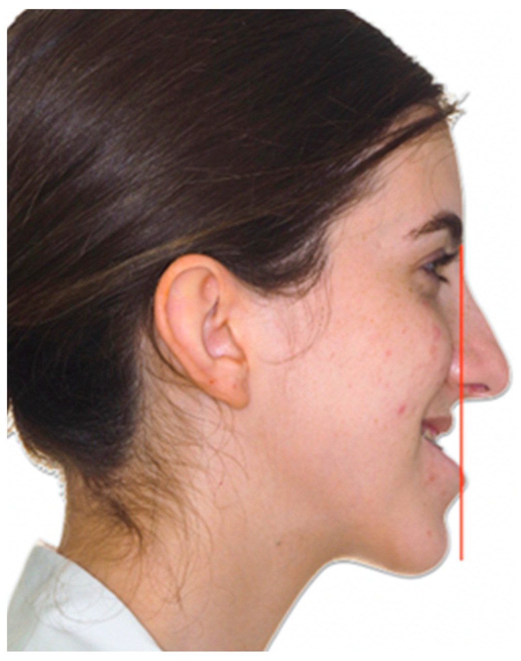
Figure 1. Preoperative picture of a class III patient where the Barcelona Line is drawn.
Applsci 15 01069 g001
Figure 2. Initial and final situations in the virtual planning of the same class III patient using the UI-STP or so-called BL reference.
Applsci 15 01069 g002
Figure 3. Measures taken during three-dimensional (3D) virtual planning of a class III patient.
Applsci 15 01069 g003
Figure 4. Same patient showing class III correction after bimaxillary advancement.
Applsci 15 01069 g004
Table 1. Percentage of patients treated by mandibular advancement, mandibular centering, mandibular counter-clockwise rotation, or combined centering and counter-clockwise rotation.
N%
Mandibular sagittal movementTotal124100
Setback97.3
Advancement11290.3
No sagittal movement32.4
Mandibular centering movementTotal124100
Yes8467.7
No4032.3
Mandibular counter-clockwise rotationTotal124100
Yes6552.4
No5947.6
Mandibular centering and counter-clockwise rotationTotal124100
Yes8568.5
No3931.5
Disclaimer/Publisher’s Note: The statements, opinions and data contained in all publications are solely those of the individual author(s) and contributor(s) and not of MDPI and/or the editor(s). MDPI and/or the editor(s) disclaim responsibility for any injury to people or property resulting from any ideas, methods, instructions or products referred to in the content.