Article

Resonant Soft X-Ray Scattering Reveals Chromophore Domains in Polymer Doped with Disperse Orange 11 Dye

1
Department of Physics and Astronomy, Washington State University, Pullman, WA 99164, USA
2
Department of Physics, University of Arizona, Tucson, AZ 85721, USA
3
Department of Astronomy and Astrophysics, University of California, San Diego, La Jolla, CA 92093, USA
*
Author to whom correspondence should be addressed.
Appl. Sci. 2025, 15(22), 12006; https://doi.org/10.3390/app152212006
Submission received: 16 September 2025 / Revised: 30 October 2025 / Accepted: 4 November 2025 / Published: 12 November 2025
(This article belongs to the Special Issue Self-Healing Materials and Applications)

Featured Application

Any application that uses high light intensities that damage the device, such as lasers and all-optical switches, would benefit from self-healing materials. These include but are not limited to high-power solid- state dye-doped polymer lasers, single-mode polymer fiber lasers, all-optical waveguide switches based on the Kerr Effect of a guest-host polymer, Nonlinear optical devices for signal processing, and Organic light-emitting diodes (OLEDs) and displays.

Abstract

Chromophore domains were proposed in a previous work as the mediators of self-healing of optical properties in dye-doped polymers. A statistical mechanical model based on domains matches all observed self-healing dynamics as a function of dye concentration, temperature and light intensity. This suggests that domains are responsible. However, there is no direct observation of domains, nor has their physical morphology been determined. This work reports the first observation of domains in a self-healing polymer using Resonant Soft X-ray Scattering (RSoXS), which gives a domain size in the range of 39.3 Å to 62.8 Å. This range includes the domain model’s prediction of an average domain size of roughly 30 molecules, which is about 56 Å, if the molecules form a loosely packed ball. X-ray scattering of samples of concentration spanning from neat polymer to the saturation limit of Disperse Orange 11 (DO11) dye in poly (methyl methacrylate) (PMMA) polymer shows domains in the expected size scales, with the mode of the effective scattering width varying little with concentration. However, for constant domain shape, the mode peak would decrease in q with increasing concentration, according to the domain model. This work suggests that the domain shape might change with concentration, which warrants further investigations of domain topology and geometry. The important evidence presented in this work is the direct experimental observation of domains, which is central to self-healing models.

1. Introduction

1.1. Background

Organic dyes incorporated in a solid polymer solution, called a dye-doped polymer, are versatile nonlinear optical [1] and lasing materials [2,3,4,5,6,7] with significant tunability due to the broad range of dopant dyes available [8]. Dye-doped polymers are also easy to process into thin films [9] and polymer optical fibers [10], making them well suited for fabrication into solid-state components that allow for a wide range of applications, from fiber optics to optical technology and data storage. They can also be formed into self-healing complex lasing structures [11], elastomeric switches [12], and shape-memory mechanosalient molecular crystals [13]. Notably, self-healing can be triggered with light [14]. There is no unified theory of self-healing, but rather the material systems chosen are based on intuition and trial end error. The work here is the first step into producing such a model. Our model applies to a broad range of materials, so it has promise in unifying these apparently disparate material types [15,16,17] and configurations. This study also complements work performed by chemists who have characterized dye-doped polymer photochemistry [18].
Lasing and nonlinear optical applications require high light levels that are often near the polymer damage threshold. As a result, most polymeric materials photodegrade over time, which is typically an irreversible process that renders the material unusable [19,20,21,22,23,24,25,26,27,28,29,30,31,32,33,34,35,36,37].
Previous work focused on mitigating photodegradation at high power outputs. For instance, materials can be prepared with higher damage thresholds, or dopant molecules can be redesigned for increased nonlinear/lasing efficiency, thus decreasing the required input light intensity. However, it is often found that increased optical nonlinearity/lasing efficiency originates in the unimpeded movement of electrons over long distances within the dopant molecules, which also decreases the molecules’ damage threshold [21,22,23,24,25,26,27,28,29,30,31,32,33,34,35,38,39]. The most promising approach to increase the damage threshold is through materials processing to remove singlet and triplet oxygen [40] to reduce impurities that convert absorbed light to heat, which damages the material. Unfortunately, this painstaking approach does not lead to significant enough improvements for high-intensity applications.
Self-healing is another way to improve resistance to photodegradation. This phenomenon was serendipitously discovered when a sample’s amplified stimulated emission (ASE) was re-measured after photo-induced damage in a prior experiment, where it was observed the ASE had improved dramatically, indicating self-healing [41]. The overall goal of this series of studies has been to improve our understanding of this self-healing process.
Simple population dynamic models of photodegradation and healing [42,43,44] predict decay and recovery dynamics in a broad range of optical phenomena including in ASE [44], fluorescence [3,4], imaging [19,45,46], white light interferometry [47], and applied electric fields [42]. In addition, decay and recovery dynamics are functions of dye concentration, temperature, and time.
All models that predict the measured population dynamics posit that molecules within domains of healthy dyes heal faster and photodegrade at a lower rate in inverse proportion to the number of molecules in a domain. These domain models also explain why the self-healing rate decreases at higher temperatures, since higher temperatures result in the thermal breakup of domains [43,44]. While these models are useful, they describe domains whose morphology is unknown and of which no direct observations have been made. The purpose of this paper is to use RSoXS experiments to test the hypothesis that there are physical domains of DO11 molecules in PMMA polymer as predicted by domain models.

1.2. Approach

For the RSoXS experiment, an X-ray wavelength that is resonant with the dopant DO11 molecules was chosen. Therefore, observed scattering features indicate the presence of the dopant and not the presence of the polymer host, which is demonstrated by contrast functions described later. This measurement showed that the scattering intensity increased with increased dopant concentration, which confirms the signal was indeed from the dopant molecules. Furthermore, subtracting the neat PMMA scattering signal from the dye-doped sample scattering signal nulls other possible sources of scattering from the polymer. These techniques together establish that the observed scattering originates from DO11 molecules. In addition, a peak in the scattering data gives a measure of the size scale of the molecular aggregates, which we will see is corroborated by the domain model predictions of self-healing given some simple constraints.

1.3. Motivation

The self-healing process described here is an uncommon phenomenon. The vast majority of the existing research using this keyword is studying one of many entirely different processes [48,49,50,51]. This leads to confusion on the nature and audience of this work, necessitating the following explanations.
The following sections will discuss important points to appreciate about the motivation of the work and the significance of the results. Section 1.3.1 briefly describes the common mechanisms and explanations of self-healing that were eliminated. Section 1.3.2 summarizes the observed behavior for which the most consistently suitable explanation is domains. Section 1.3.3 describes the nature and purpose of the physical experiment that is performed in this study to broadly characterize the domains that are predicted by the domain model.

1.3.1. Ruling out Mechanisms

There are multiple hypotheses that might explain the observation of a decrease in a material’s functionality, such as light-emitting efficiency under high-intensity illumination, that would also be reversible. Most hypotheses have been ruled out: the diffusion of molecules away from the hot spot induced by the pump light [52], which is not observed by direct imaging [45] or second harmonic imaging [53]; the reorientation of molecules under polarized light, which is not observed with dichroism [54]; and proton transfer or twisted intramolecular charge transfer [18,55,56]. For proton transfer, tautomers may play a role in the self-healing process, but the process is more complex than a simple conversion between two tautomers.

1.3.2. Observed Behavior

Observations imply the photodegradation and recovery pathways are not simple single-mechanism processes. Characterization studies that led to this conclusion consider thermal activation, concentration dependence, and the role of the polymer host in self-healing. These considerations, including general optical properties and empirical healing data, among others, are the motivating factors for the hypothesis that domains are responsible for healing, laser hardening during cycling, and for the mitigation of photodegradation.
The rates of most reactions increase with temperature, as should self-healing if it is a similar process [57]. If the damaged species is of higher energy than the undamaged one, then its lifetime will be determined by either the thermal or quantum tunneling rate through a barrier. Increased temperature increases the dopant molecule’s energy, making it more likely that it will overcome the barrier energy, thus increasing its recovery rate. However, DO11-doped PMMA shows a decreased rate of recovery at increased temperature [44].
This observation suggests that associations of dopant molecules might be responsible for self-healing, since if domains are broken apart by thermal energy greater than the binding energy, the recovery rate will decrease due to the loss of the domain’s protective effect.
The recovery rate is observed to be higher, and the decay rate lower, with higher concentration of DO11 molecules [43,44]. This behavior is consistent with the thermodynamics of domain formation in a condensation-type process, where larger domains form with a higher concentration. Furthermore, the healing process is most pronounced when the dyes are at the saturation limit of solubility in PMMA polymer, and samples made by polymerizing a solution of DO11 dye in MMA—the monomer that makes PMMA polymer when linked—heal faster and are more resistant to photodegradation compared to those made by spin-coating a solution of PMMA and DO11 in solvent. This suggests that the polymerization process may enhance DO11 domain formation by concentrating molecules in the ever-decreasing portions of MMA.
PMMA doped with DO11 self-heals, but DO11 in aqueous solution with MMA does not heal [2,41,58]. The PMMA polymer host thus appears to be an important part of the self-healing process, which is consistent with the polymer immobilizing the molecules in a domain, while in liquid form, the domains are smaller or do not form. Additionally, the change in the absorption spectrum in the decay process is different in MMA versus PMMA, further buttressing the hypothesis that the decay processes are different.
These observations and more than two decades of research result in a general model that fits all data [43,44]. Central to these models is the hypothesis that dyes embedded within a domain are partially protected from damage and heal at a higher rate. The original model [43] posits that the recovery rate depends on the number of undamaged molecules in a domain, while in the newer Correlated Chromophore Model (CCM), which fits the data more precisely, the recovery rate depends instead on the domain size [44]. This is consistent with observations that total normalized recovery of the ASE efficiency can increase under the right conditions, as damaging the molecules may not remove them from the domains. This can be cycled to refine the sample further [20]. The effect of the addition of nanoparticles on this process has also been studied by Anderson et al. [20,59,60]. Copolymers of PMMA and ester-containing polymers also show this enhancement [61].
The CCM posits dyes are found within a distribution of tightly packed clusters of molecules, ranging in size/number from one (uncorrelated molecules) to the total available number of dye molecules, where the number of dye molecules in a domain is distributed according to a statistical mechanical cluster condensation model. The observations of healing and recovery thus depend on the thermal size distribution of these domains, where the larger domains are highly protective, while the small domains are marginally more protective than free molecules. Newer forms of the model also include an irreversibly damaged species and the effects of applied electric fields [42]. Additionally, the role of the polymer matrix appears to provide scaffolding that supports domains, which enable the damaged pieces to reunite by virtue of their decreased mobility within a domain. This hypothesis is based on the central role played by the PMMA polymer in the healing process, as demonstrated by the fact that neither DO11 alone nor DO11 in the unpolymerized MMA is observed to self-heal.
These observations give great insight into the nature of the domains postulated in the domain model but do not provide a direct representation of the domains in physical space. This paper will bridge this gap using resonant scattering, which characterizes the distribution of dye molecules in reciprocal space [62].

1.3.3. Scattering

We use X-ray experiments to characterize the sizes and distances between physical entities that scatter the light. When the light is tuned specifically to the DO11 molecules, they can be characterized separately from the surrounding material. The observed size distribution can then be correlated with the predicted molecular domains.
X-ray scattering experiments use collimated beams in which the scattered X-ray intensity from the molecules in a sample is recorded with a planar detector that is perpendicular to the incident beam and downstream from the sample. X-ray characterization has various applications and sub-methods that make it extremely versatile [63,64,65,66]. Scattered X-ray intensity is measured as a function of momentum transfer q, defined by q = 2 k sin ( θ / 2 ) —where k is the wavevector magnitude, and θ is the angle of deflection from the incident trajectory [67]. k is unchanged in the collision [68]. RSoXS is an anomalous scattering method where the incident light energy is selected based on its proximity to the resonant energy of target bonds in the molecular structure of interest [69], in this case, the SP2 carbon bond in DO11 dye, which enhances the scattering cross section of DO11 by orders of magnitude [70]. This technique combines insights from both scattering and spectroscopy to increase sensitivity to specific low-Z organic materials [71]. This experiment involves sample preparation for the synchrotron, 2D scattering data collection, 1D reduction in the data, and graphical analysis.

2. Materials and Methods

2.1. Sample Preparation

For the RSoXS experiment, light is ar normal incidence to the sample, requiring free-standing thin film samples with thicknesses on the order of 100–300 nm for adequate transmission and to avoid thermal effects [9]. Samples prepared by spin-coating from dissolved PMMA/DO11 have also been observed to self-heal, but not as well as those prepared with in-situ polymerization.
To prepare these samples, first, 75,000 g/mol PMMA powder from Polysciences, Inc. at Warrington, PA, USA and DO11 crystalline powder from Aldrich Chemical Company at Milwaukee, WI, USA are co-dissolved into solution using solvents propylene glycol methyl ether acetate (PGMEA) from Aldrich Chemical Company at Milwaukee, WI, USA and Oxolan-2-one ( γ -but) from Sigma-Aldrich at St. Louis, MO, USA. The formulation uses a constant mass ratio of solids to solvents. The solvents as a volume are 7.3 mL of PGMEA and 3.1 mL of γ -but, and the solids are a total mass of approximately 1.875 g. This mass is distributed between the two solids based on the concentration measure of weight-percent (wt%) DO11 in proportion to the total solid mass. The four components are added in the order of PGMEA, DO11, γ -but, and PMMA, and left to mix for a minimum of 12 h. Finally, a glass syringe and 25 mm 0.2 µm GHP filters from Pall Corporation, Ann Arbor, MI, USA were used to strain the solution into fresh vials for storage and use.
Silicon substrates were cleaved to an area of ∼1 cm2 and cleaned sequentially in deionized water (twice), acetone, and isopropyl alcohol (IPA), both from University Stores in Pullman, WA and HPLC-certified, for 20 min each. IPA was removed from the substrates with nitrogen gas before placing them in a UV–ozone environment for 10 min. A water-soluble sacrificial layer [72] made from a 1:4 dilution of Poly(Sodium 4-Styrenesulfate) (NaPSS) from Sigma-Aldrich at St. Louis, MO, USA in deionized water was spin-coated onto the substrates with rotation reaching 1000 rpm at an acceleration of 1000 rpm/s to form films with a thickness of ∼100 nm. The NaPSS layer was dried at 150 °C for 5–10 min. The sample solution was spin-coated onto the sacrificial layer with maximum rotation of 4000 rpm at an acceleration of 6000 rpm/s to form films with a thickness of 200–300 nm. The thicknesses of both layers were measured for multiple test samples with only either the sacrificial layer or the sample layer using profilometry or ellipsometry. The samples prepared for this experiment are listed in terms of weight percent of DO11 as a fraction of the film mass, which serves as the only variable intentionally varied between samples. To prepare samples for RSoXS measurements, the substrates were then submerged in deionized water to dissolve the NaPSS and lift the sample films from the substrates. Samples were then placed onto silicon nitride membrane windows from Norcada in Edmonton, AB, Canada and dried under a heat lamp.

2.2. Resonant Soft X-Ray Scattering

RSoXS was carried out at the 7-ID-1 (SST-1) beamline of the NSLS-II synchrotron [73]. It is a method often used to quantify solid-state nanostructure to determine average phase-separated domain size and concentration. The linearly polarized, undulator-produced beam is transmitted through the sample with the scattering pattern centered on and measured by a CCD detector. Before the full experiment is conducted, non-resonant scattering data is collected at about 50 different spots on the sample to check for parasitic scattering due to sample nonuniformity, and 3–5 spots with little or no parasitic scattering are selected to measure with the complete experiment. The resulting 2D scattering images were azimuthally averaged around the direct beam to create a 1D scattering intensity versus q profile using the Nika processing software version 1.84 as an extension to Igor Pro version 8.04 [74]. The q range was determined by the maximum measurable angle, which is in turn determined by the distance between the detector and sample, the surface area of the detector, and the wavelength of the light, as shown by k = 2 π / λ . Unique to resonant scattering, this method used energies corresponding to molecular bond energies with significant contrast from the surrounding matrix [75]. These energies are found with Near-Edge X-ray Absorption Fine Structure (NEXAFS) across the carbon K-edge (photoionization at 290 eV), as shown in Figure 1 [76]. Scattering intensity from roughness, density, or porosity fluctuations in the mainly PMMA sample will vary with the green trace as a function of photon energy, while scattering intensity from fluctuations in DO11 concentration (molecular domains) will vary with the blue trace as a function of energy. In this case, the bond probed was an SP2 bond of N with C at 284.5 eV, and 270 eV was used as a baseline non-resonant energy for a point of comparison to eliminate the contribution due to roughness, porosity, or other density fluctuations not related to chemical domains. This resonant contrast gives much more pronounced features in scattering profiles related to the morphology of the resonant bonds in reciprocal q space and can be analyzed to indicate real-space size-scales of features [69,76,77]. This technique allowed us to observe primarily that such a feature exists, and secondarily that one of the model’s basic predictions, the mode domain size, is within the range of the features observed.
The component materials are well-studied, particularly PMMA, and optical constants have been measured for both DO11 and PMMA. The contrast between the two, | Δ n | 2 , with complex index of refraction n, is derived from their individual indices. The maximum contrast shows the greatest enhancement of scattering intensity due to molecules of DO11, while the minimum functions as the background without this enhancement. Maximizing this difference reveals the features of interest most clearly.

3. Results

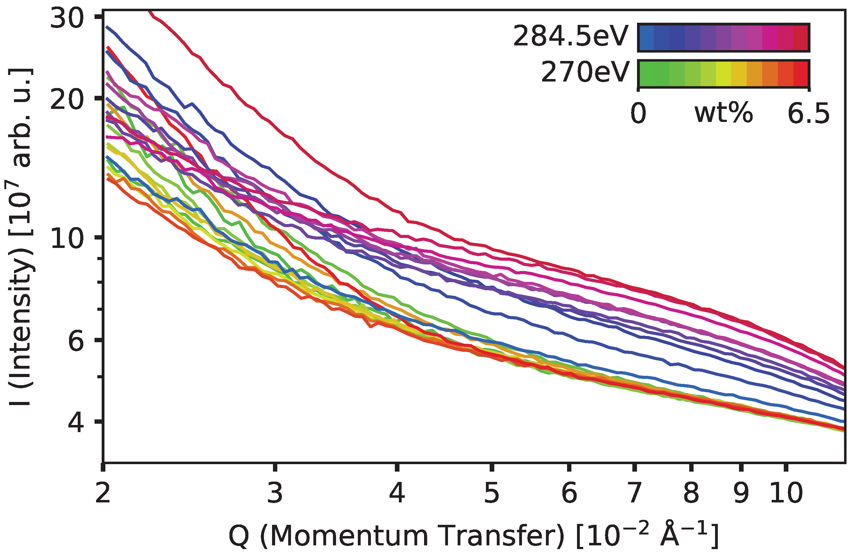
The 1D-reduced data—the scattering intensity I as a function of momentum transfer q—is shown in Figure 2. This data can also be found in the Supplementary Materials. The low-q region, corresponding to larger size scales of features such as roughness, shows no features or trends, with all samples showing a monotonically increasing intensity towards lower q, hence showing larger features. At high momentum transfer for non-resonant scattering at 270 eV, the intensity is consistent with background scattering, regardless of concentration. However, scattering at the resonant energy of 284.5 eV shows broad peaks between q = 0.05 Å 1 and 0.08 Å 1 that are superimposed onto the background scattering tails. This enhanced scattering intensity at the DO11 resonance energy over the non-resonant scattering background indicates heterogeneity emerging from fluctuations in DO11 concentration within the PMMA matrix. This scattering feature also increases in intensity with dye concentration. Such an increase indicates an enhanced amplitude of dye concentration. The q-range of this feature corresponds to a characteristic length l c = 2 π / q of l c [ 39.3 , 62.8 ] Å, which is roughly in agreement with the domain size that is predicted by the domain model [43] assuming domains form loosely packed spheres, which have a mode diameter of 56Å. Put together, these results provide the first direct evidence of the existence of dye domains in PMMA, the most basic proposition of the CCM. The subsequent steps amplify this difference by removing the background scattering.
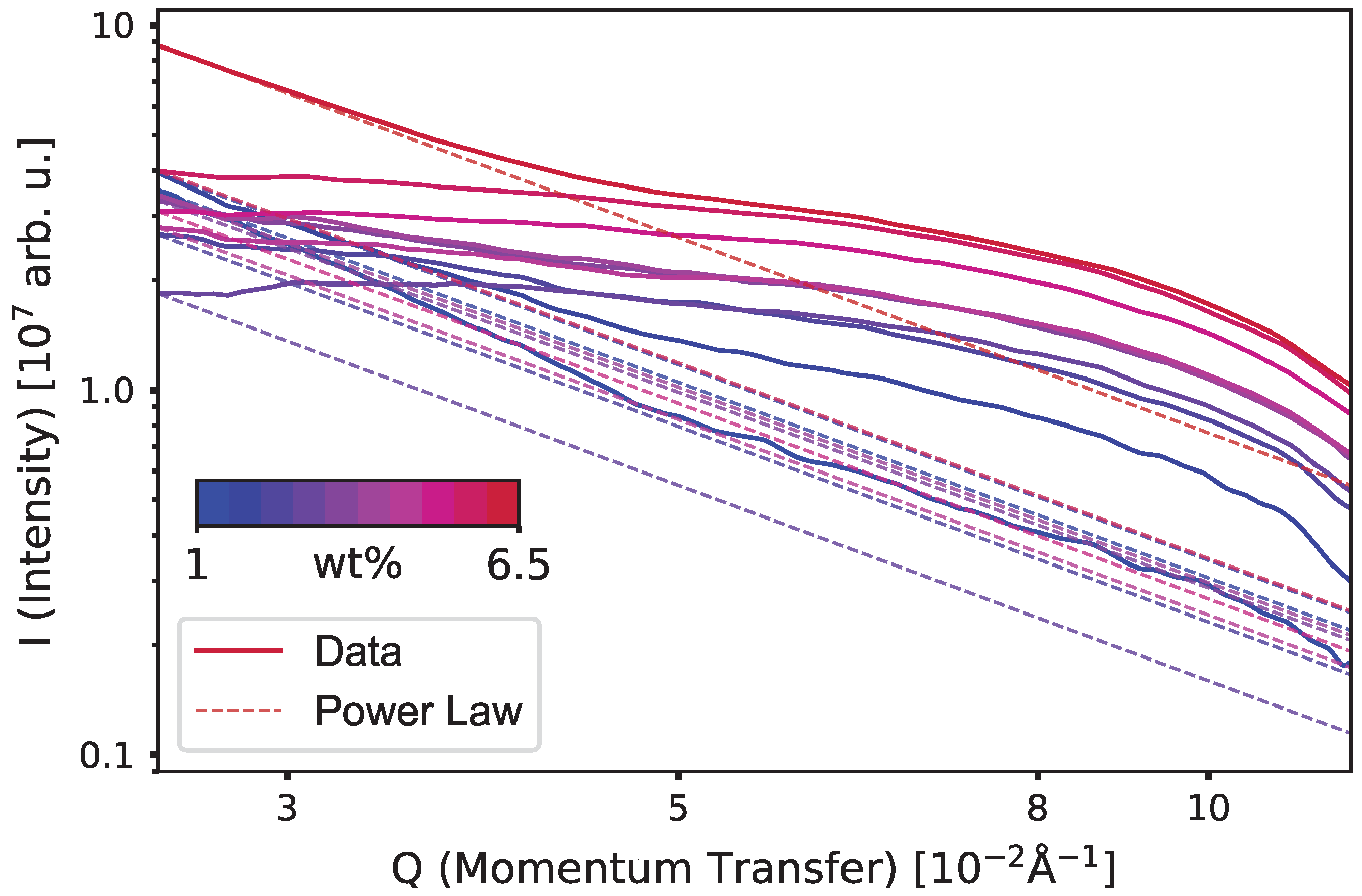
The raw data shown in Figure 2 is then boxcar smoothed using an 80-point averaging window, which removes small fluctuations—due to what we can consider as noise—to reduce the effect of compounding noise during background subtraction. The effect of this smoothing window is approximated with a standard error of the same 80-point window. Next, a two-step process is utilized, which is designed to remove all scattering that is unrelated to the DO11 molecules. The first subtraction step is background subtraction using neat PMMA (0% DO11 by weight) as the baseline. Here the scattering data from each dye-doped sample at one X-ray energy uses neat PMMA as the baseline, which standardizes all samples at both energies as compared to a plain PMMA sample. This subtraction is implemented for both the resonant and non-resonant X-ray energies. Then, the non-resonant scattering data at 270 eV is used as a reference for scattering due to the PMMA, which is removed from the DO11 scattering by subtracting it from the scattering data for the 284.5 eV X-rays for each individual dye concentration. This accounts for scattering artifacts from sources such as surface roughness and parasitic scattering. This data is referred to as “doubly subtracted,” meaning that both sources of background—system/experimental and non-resonant contributions from the sample—are considered. As an example, the doubly subtracted scattering data for 6 wt% is determined using
I ( Q ; 6 % at 284.5 eV ) I ( Q ; 0 % at 284.5 eV ) ( I ( Q ; 6 % at 270 eV ) I ( Q ; 0 % at 270 eV ) ) ,
where I ( Q ; wt % at E ) indicates the 1D scattering data I ( Q ) for the sample of concentration wt% at energy E. The results of the double subtraction are shown in Figure 3.
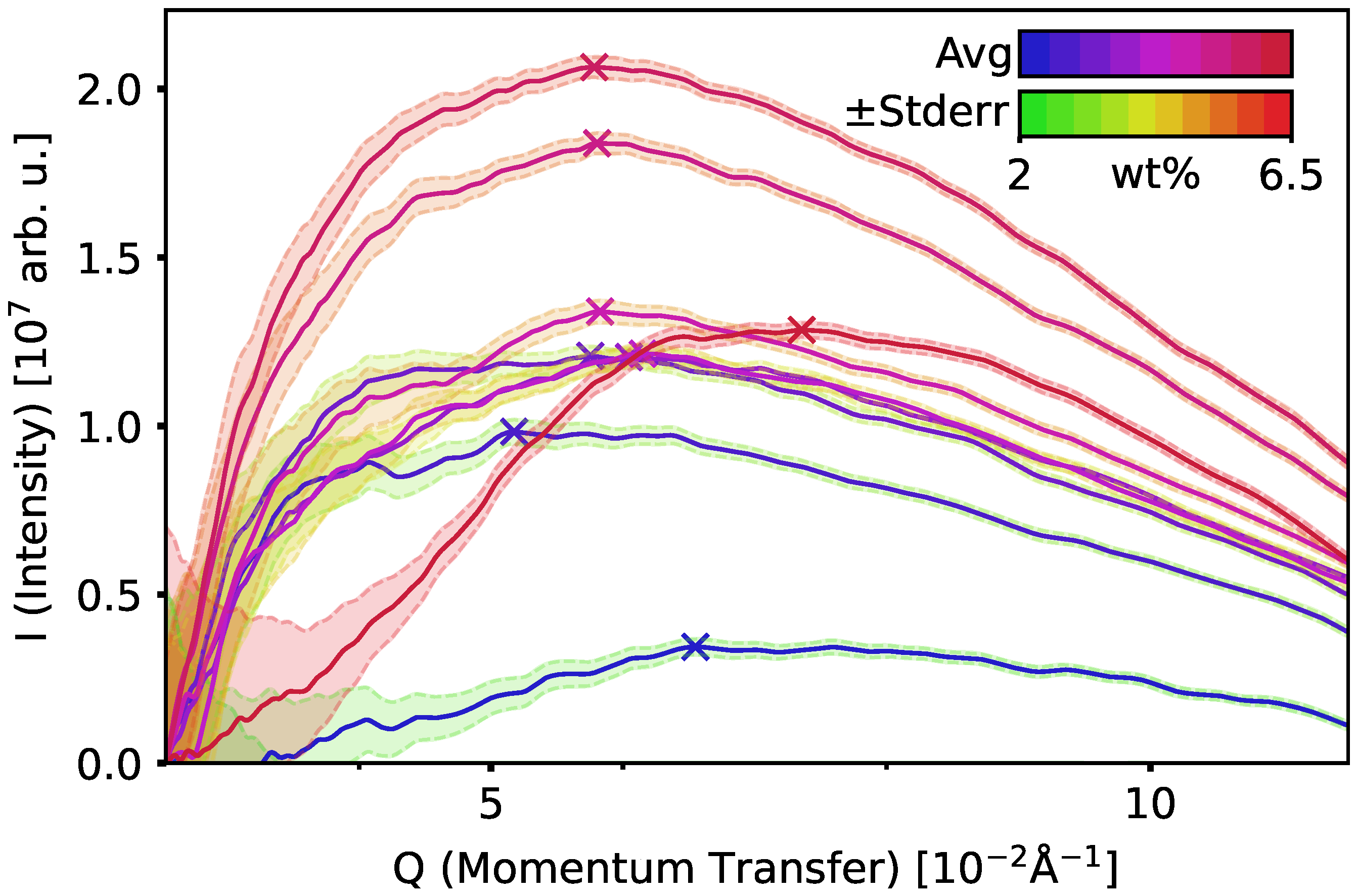
The low-q region of the scattering data was fit to a power law (shown by dot-dashed lines), which is motivated by fractal structures following power law scattering behavior. Furthermore, the low-q region is away from the DO11 resonance, so the fit represents scattering from what may be a fractal structure of the polymeric material. Such a power law fit on a logarithmic scale [78] appears linear to the eye and forms a true baseline for the data. Thus, the peaks above it are due to the resonant enhancement caused entirely by the DO11 and not explainable by the plain PMMA structure. The 1% sample data was linearized at low-q and fit using the slope as the dimensional parameter in the power law fit for the 1% sample and the other concentrations, as shown in Figure 3. This slope is also generally consistent with the slope provided by the 6.5% sample data; however, the 1% was used because of its consistency regardless of the lower bound—enforced by the smoothing region—and its relative uniformity for a larger low-q range, meaning it did not change with the choice of the smoothing window or linearization point, which were somewhat subjective. The result of subtracting the power law fits from the data is shown in Figure 4.
From this subtraction, it appears the domains due to the DO11 molecules may be affecting the fractal nature of the polymer. Further discussion of this interesting possibility is beyond the scope of this paper but will be addressed in later work. For now, the important point is that subtracting this power law baseline produces well-resolved peaked functions that are due solely to the DO11 molecules, where all other effects such as parasitic scattering, surface roughness, and polymer contributions have been nulled.
The resonant enhancement due to the DO11 molecules is characterized by a peak with a peak position, height, and width. The peak heights monotonically increase with increased concentration of DO11 dye, with the exception of the 6.5 wt% sample. The peak centers are approximately at the same q value. The CCM predicts the number of molecules in a domain but can be extended to predict physical sizes in the following way. If the simplification is made that the domains’ shapes remain the same, the mode of the size distribution will increase with an increasing number of available dye molecules per volume, so we expect the peak centers to shift towards lower q with increasing dye concentration. The fact that the X-ray data appears to be inconsistent with this scaling suggests that the domain morphologies may change with concentration, or the physical size of a domain observed by scattering is not the same as the scale over which molecules interact with each other, which is the central concern of the self-healing model. However, the peak amplitude increases for most dye concentrations as expected.
The fact that the 6.5 wt% sample is not consistent with either trend is most likely due to extrinsic sources of inhomogeneity. For example, a speck of dust, a poorly chosen or masked measurement spot, or miscibility limitations may be responsible. Such outliers are found in larger data sets as a small fraction of the total points. The peak height of the low-q data for 6.5% is also indicative of differences in characteristics of the overall sample rather than the dye morphology. This does present an issue with the background-subtraction protocol in the final step, but the data prior to that, shown in Figure 3, may also hint at a greater shift to larger domains, resulting in the observed overall enhancement at lower q values compared to other concentrations. This anomalous low-q scattering is demonstrable in Figure 2. The 6.5 wt% sample has a much higher scattering intensity at 270 eV in the low-q region, which influenced the generation of the following plot. However, the overall inconsistency in low-q data for all samples implies the low-q region may not have much use for making high-q comparisons in general. Figure 3 has the expected monotonicity with dye concentration in the mid- to high-q region, so it is possible the current method could be refined to place more weight on that region instead of on the low-q region.

4. Discussion

These results clearly show a resonant scattering enhancement between q = 0.05 Å 1 and 0.08 Å 1 corresponding to a size of between 62.8 Å and 39.3 Å. The intensity of scattering increased with the concentration of dopant dye, confirming that the dye molecule is responsible for the scattering enhancement. The peaks indicate a characteristic size scale of features that would not exist in a homogenous mixture. This is direct experimental evidence of the existence of physical domains that were postulated from indirect experimental evidence that applies the CCM to determine the domain size in terms of the number of molecular units and the chemical potential required when adding an additional molecule to the domain [44]. The results of these studies suggest a domain size in the range of 25–35 molecular units (note, not as a physical size) and a chemical potential of μ = 0.29 ( ± 0.01 ) eV [43]. These parameters can now be used to bridge the gap between the indirect predictions and our own direct observations.
Neither previous experiments nor the supported models require the physical arrangement of the molecules in a domain in absolute terms, though studies in the presence of an electric field suggest that the molecules form a 1D chain [42], which could be straight or tangled like an unfurled ball of yarn. Thus, the size range could span from a spherical ball whose size is on the order of N 1 / 3 to a straight segment of length N molecular units. The density of each domain may itself depend on N, however, and there is no guarantee all domains have the same shape. Proceeding as if they are the same shape for some bounding estimates, we will establish this simplistic constant scaling parameter using domain diameter D as a function of N, the number of dye molecules in the domain, D ( N ) = A N α . A is a factor determined by the arrangements of the molecule given the shape, i.e., for a line it will be the periodic connection distance between molecules. The dimensionality factor α [ 1 3 , 1 ] is a constant corresponding to shapes ranging between a sphere and a line, respectively. As a simple example, for a molecule of length 10 Å, 25 to 35 of the molecules end-to-end would lead to a length of 25–35nm. In a tightly packed ball, the diameter would be about 30 Å. If the molecules attach along the polymer chain and form a loose ball, given the extra 6–10 Å of space possible if the dye is attached to the monomer units, we increase A by this proportion, and the simple formula is scaled to give a diameter of 45–60 Å.
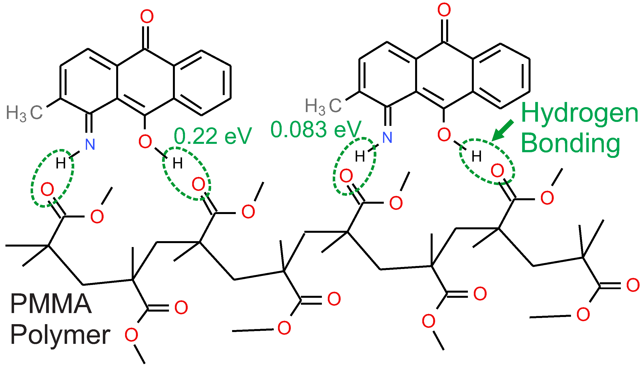
The chemical potential is another clue to possible domain shape. Figure 5 shows how DO11 molecules might attach to the polymer backbone through hydrogen bonding [79,80]. The cis tautomer has an NH and OH group, which can bind to the oxygens in the polymer chain. Both groups might hydrogen-bind to one segment of a chain as shown in the figure or across adjacent polymer chains. The proposed models together with the self-healing experiment suggest the former. In the configuration shown in Figure 5, the binding energy corresponds to 0.218eV due to the O-H · · · O hydrogen bond enthalpy and 0.0829eV due to the N-H · · · O hydrogen bond enthalpy, for a total of 0.30eV [81], which is within experimental uncertainty of the chemical potential μ = 0.29 ( ± 0.01 ) eV [43], the domain model fit parameter determined from self-healing experiments. Also note that the molecules are further apart than end-to-end by about 0.6 Å. This would bring the ball diameter up to as much as 56 Å, which is still in the range determined by the scattering data. This description is similar to one proposed by Hung et al. [82], but in that case unreacted monomer is proposed to be the mediator of healing.

5. Conclusions

We have used RSoXS to characterize the domain size of DO11 molecules doped into PMMA polymer. These domains were predicted to exist based on observations of self-healing and studies since then have provided robust support for the model. While this work provides strong evidence that such domains exist, and the basic prediction falls in the range that was measured with RSoXS, these results do not have any bearing on whether domains mediate self-healing. Neither does it show the effects of related factors such as light exposure and damage, which motivates further study for evidence connecting domain shape to healing. It would be interesting to characterize the self-healing process with time-resolved X-ray studies, as has been demonstrated in the study of photoisomerzation [83] of azobenzene molecules [84] to see if domain sizes change due to the process. At present, these capabilities are not available to us. Other avenues for exploration include further development of the morphological predictions of the domain model, applying principle component analysis [85], using spot variation in damaging and optical quenching [86], and applying simulations to the extended models [87].
The data presented here may suggest that the domain size does not change with dye concentration, but the number of domains does increase with concentration. However, this is not a contradiction since the domain model is based on the number of interacting molecules and not the physical size of the domain, so it is possible that the number of molecules that interact with each other within a domain changes with concentration due to unknown effects during domain formation. As such, the observations presented here suggest new hypotheses for testing.
If domains are the mediators of self-healing as proposed, this will give the materials scientist another tool for designing new materials that are more robust to damage from high light intensities, making possible new applications that require active materials that generate high-intensity light or control its flow.

Supplementary Materials

The following supporting information can be downloaded at https://www.mdpi.com/article/10.3390/app152212006/s1, File S1: Spincoated Data, and File S2: Peak Points.

Author Contributions

Conceptualization, M.G.K. and B.A.C.; data curation, E.S. and A.P.; formal analysis, E.S.; funding acquisition, M.G.K. and B.A.C.; investigation, E.S., A.P., C.B., M.S., M.G.K. and B.A.C.; methodology, E.S., A.P., M.G.K. and B.A.C.; project administration, M.G.K. and B.A.C.; resources, B.A.C.; software, E.S.; supervision, M.G.K. and B.A.C.; validation, E.S., A.P., M.G.K. and B.A.C.; visualization, E.S. and A.P.; writing—original draft, E.S. and M.G.K.; writing—review and editing, E.S., A.P., C.B., M.S., M.G.K. and B.A.C. All authors have read and agreed to the published version of the manuscript.

Funding

This research received no external funding.

Institutional Review Board Statement

Not applicable.

Informed Consent Statement

Not applicable.

Data Availability Statement

The original contributions presented in this study after averaging the intensity over the azimuthal angle for fixed momentum transfer are included in the article/Supplementary Material. The CCD data prior to the reduced form presented in this article/Supplementary Material are not readily available because of their size. Further inquiries or requests to access the datasets should be directed to the corresponding author.

Acknowledgments

Support for this work, including support for AP and BAC, is from the US National Science Foundation (NSF), Division of Materials Research, Electronic and Photonic Materials Program under grant #2247711. CB and MS were supported by the NSF Division of Physics Research Experience for Undergraduates under grant #2349426. This research used beamline SST-1 (7-ID-1) of the National Synchrotron Light Source II, a U.S. Department of Energy (DOE) Office of Science User Facility operated for the DOE Office of Science by Brookhaven National Laboratory under Contract No. DE-SC0012704.

Conflicts of Interest

The authors declare no conflicts of interest.

References

  1. Singer, K.D.; Kuzyk, M.G.; Sohn, J.E. Second-order Nonlinear-optical Processes in Orientationally Ordered Materials: Relationship Between Molecular and Macroscopic Properties. J. Opt. Soc. Am. B 1987, 4, 968–976. [Google Scholar] [CrossRef]
  2. Howell, B.; Kuzyk, M.G. Lasing Action and Photodegradation of Disperse Orange 11 Dye in Liquid Solution. Appl. Phys. Lett. 2004, 85, 1901–1903. [Google Scholar] [CrossRef]
  3. Dhakal, P.; Kuzyk, M.G. Molecular structure and reversible photodegradation in anthraquinone dyes. J. Photochem. Photobiol. A 2016, 328, 66–76. [Google Scholar] [CrossRef]
  4. Dhakal, P.P.; Kuzyk, M.G. Self-Healing Properties of 1-Amino,2,4-Dibromoanthraquinone Dye Doped in PMMA Polymer. J. Phys. Chem. C 2017, 121, 351. [Google Scholar] [CrossRef]
  5. Duarte, F.J. Organic Lasers and Organic Photonics, 2nd ed.; IOP Publishing: Bristol, UK, 2024. [Google Scholar]
  6. Sorokin, P.P.; Lankard, J.R.; Hammond, E.C.; Moruzzi, V.L. Laser-pumped Stimulated Emission from Organic Dyes: Experimental Studies and Analytical Comparisons. IBM J. Res. Dev. 1967, 11, 130–148. [Google Scholar] [CrossRef]
  7. Farmer, G.I.; Huth, B.G.; Taylor, L.M.; Kagan, M.R. Time-Resolved Stimulated Emission Spectra of an Organic Dye Laser. Appl. Phys. Lett. 1968, 12, 136–138. [Google Scholar] [CrossRef]
  8. Fletcher, A.N. FY 1980 Report on Dye Laser Materials; Defense Technical Information Center: Fort Belvoir, VA, USA, 1981. [CrossRef]
  9. Daughton, W.J.; Givens, F.L. An Investigation of the Thickness Variation of Spun-on Thin Films Commonly Associated with the Semiconductor Industry. J. Electrochem. Soc. 1982, 129, 173. [Google Scholar] [CrossRef]
  10. Peng, G.D.; Ji, P.; Chu, P. Electro-optic polymer optical fibers and their device applications. Proc. SPIE 2002, 4459, 101–117. [Google Scholar] [CrossRef]
  11. Lu, Y.; Lowther, Z.; Christianson, N.D.; Li, Z.; Baer, E.; Kuzyk, M.G.; Dawson, N.J. Demonstration of a self-healing all-polymer distributed Bragg reflector laser. Appl. Phys. Lett. 2020, 116, 103301. [Google Scholar] [CrossRef]
  12. Li, S.; Tu, Y.; Bai, H.; Hibi, Y.; Wiesner, L.W.; Pan, W.; Wang, K.; Giannelis, E.P.; Shepherd, R.F. Simple Synthesis of Elastomeric Photomechanical Switches That Self-Heal. Macromol. Rapid Commun. 2019, 40, 1800815. [Google Scholar] [CrossRef]
  13. Karothu, D.P.; Weston, J.; Desta, I.T.; Naumov, P. Shape-Memory and Self-Healing Effects in Mechanosalient Molecular Crystals. J. Am. Chem. Soc. 2016, 138, 13298–13306. [Google Scholar] [CrossRef]
  14. Habault, D.; Zhang, H.; Zhao, Y. Light-triggered self-healing and shape-memory polymers. Chem. Soc. Rev. 2013, 42, 7244–7256. [Google Scholar] [CrossRef]
  15. Stubbs, N.; Bridgewater, M.; Stubbs, M.; Crescimanno, M.; Kuzyk, M.G.; Dawson, N.J. Polylactic acid promotes healing of photodegraded disperse orange 11 molecules. Opt. Mater. 2018, 76, 11–15. [Google Scholar] [CrossRef]
  16. Zhang, J.; Huo, M.; Li, M.; Li, T.; Li, N.; Zhou, J.; Jiang, J. Shape memory and self-healing materials from supramolecular block polymers. Polymer 2018, 134, 35–43. [Google Scholar] [CrossRef]
  17. White, S.; Sottos, N.; Geubelle, P.; Moore, J.; Kessler, M.; Sriram, S.; Brown, E.; Viswanathan, S. Autonomic healing of polymer composites. Nature 2001, 409, 794–797. [Google Scholar] [CrossRef] [PubMed]
  18. Westfall, N.J.; Dirk, C.W. The photochemistry of the self-healing chromophore Disperse Orange 11. J. Phys. Organ. Chem. 2012, 25, 704–712. [Google Scholar] [CrossRef]
  19. Anderson, B.; Ramini, S.K.; Kuzyk, M.G. Imaging studies of photodamage and self-healing of anthraquinone derivative dye doped polymers. In SPIE Laser Damage Symposium Proceedings; Exarhos, G., Ed.; SPIE: Boulder, CO, USA, 2011; Number 16. [Google Scholar]
  20. Anderson, B.R.; Gunawidjaja, R.; Eilers, H. Photodegradation and self-healing in a Rhodamine 6G dye and Y2O3 nanoparticle-doped polyurethane random laser. Appl. Phys. B 2015, 120, 1–12. [Google Scholar] [CrossRef]
  21. Braunlich, P.; Schmid, A. Contributions of Multiphoton Absorption to Laser-Induced Intrinsic Damage in NaCl. Appl. Phys. Lett. 1975, 26, 150–153. [Google Scholar] [CrossRef]
  22. Brawer, S. Phenomenological Theory of Laser Damage in Insulators. Phys. Rev. B 1979, 20, 3422–3444. [Google Scholar] [CrossRef]
  23. Chan, C.L.; Mazumder, J. The Effect of Radiative Heat Lost on Laser Material Damage. J. Appl. Phys. 1988, 63, 5890–5892. [Google Scholar] [CrossRef]
  24. Chase, L.L.; Lee, H.W.H.; Hughes, R.S. Investigation of Laser-Surface Interactions and Optical Damage Mechanisms Using Excitation by Pairs of Picosecond Laser Pulses. Appl. Phys. Lett. 1990, 57, 443–455. [Google Scholar] [CrossRef]
  25. Chen, C.L.; Mazumder, J. One-dimensional Steady-state Model for Damage by Vaporization and Liquid Expulsion Due to Laser-material Interaction. J. Appl. Phys. 1987, 62, 4579–4586. [Google Scholar] [CrossRef]
  26. Estler, R.C.; Nogar, N.S. Chemical Precursor to Optical Damage Detected by Laser Ionization Mass Spectrometry. Appl. Phys. Lett. 1988, 52, 2205–2207. [Google Scholar] [CrossRef]
  27. Exarhos, G.J.; Guenther, A.H.; Kozlowski, M.R.; Soileau, M.J. Laser-Induced Damage in Optical Materials; SPIE: Bellevue, WA, USA, 1998. [Google Scholar]
  28. Felix, M.P.; Nachbar, W. A Closer Look at Laser Damage in PMMA. Appl. Phys. Lett. 1974, 25, 25–27. [Google Scholar] [CrossRef]
  29. Jones, S.C.; Braunlich, P.; Casper, R.T.; Shen, X.A. Recent Progress on Laser-Induced Modifications and Intrinsic Bulk Damage of Wide Gap Optical Materials. Opt. Eng. 1989, 28, 1039–1068. [Google Scholar] [CrossRef]
  30. Kim, H.; Postlwaite, J.C.; Zyung, T.; Dlott, D.D. Ultrafast Imaging of Optical Damage Dynamics and Laser-Produced Wave Propagation in Polymethyl Methacrylate. J. Appl. Phys. 1988, 64, 2955–2958. [Google Scholar] [CrossRef]
  31. Milam, D. Laser-Induced Damage at 1064 nm, 125 psec. Appl. Opt. 1977, 16, 1204–1213. [Google Scholar] [CrossRef]
  32. Nechitailo, S. Mechanisms of Laser Damage to Transparent Polymer Materials. Bull. Acad. Sci. USSR, Phys. Ser. 1982, 46, 159–163. [Google Scholar]
  33. O’Connell, R.M.; Deaton, T.F.; Saito, T.T. Single- and Multiple-shot Laser-damage Properties of Commercial Grade PMMA. Appl. Opt. 1984, 23, 682–688. [Google Scholar] [CrossRef]
  34. Petzoldt, S.; Elg, A.P.; Reichling, M.; Reif, J.; Matthias, E. Surface Laser Damage Thresholds Determined by photoacoustic Deflection. Appl. Phys. Lett. 1988, 53, 2005–2007. [Google Scholar] [CrossRef]
  35. Summers, A.M.; Ramm, A.S.; Paneru, G.; Kling, M.E.; Flander, B.N.; Trallero-Herrero. Optical damage threshold of Au nanowires in strong femtosecond laser fields. Opt. Express 2014, 22, 4235. [Google Scholar] [CrossRef]
  36. Wood, R.M. Laser-Induced Damage of Optical Materials; Series in Optics and Optoelectronics; Taylor & Francis: Boca Raton, FL, USA, 2003. [Google Scholar]
  37. Zhang, Q.; Canva, M.; Stegeman, G. Wavelength dependence of 4-dimethylamino-4-nitrostilbene polymer thin film photodegradation. Appl. Phys. Lett. 1998, 73, 912. [Google Scholar] [CrossRef]
  38. Corbertt, J.W. Radiation Damage, Defects and Surfaces. Surf. Sci. 1979, 90, 205–239. [Google Scholar] [CrossRef]
  39. Fersman, I.; Khazov, L.D. Mechanism of Damage of the Surface of a Transparent Dielectric during Illumination with Short Light Pulse. Sov. J. Quantum Electron. 1973, 2, 319–323. [Google Scholar] [CrossRef]
  40. Kuznetsova, N.A.; Kaliya, O.L.; Solodar, S.L. The role of singlet oxygen in photodegradation of aminophenalenones. J. Appl. Spectrosc. 1985, 43, 901–904. [Google Scholar] [CrossRef]
  41. Howell, B.; Kuzyk, M.G. Amplified Spontaneous Emission and Recoverable Photodegradation in Disperse-Orange-11-Doped-Polymer. J. Opt. Soc. Am. B 2002, 19, 1790. [Google Scholar] [CrossRef]
  42. Anderson, B.; Kuzyk, M.G. Generalizing the correlated chromophore domain model of reversible photodegradation to include the effects of an applied electric field. Phys. Rev. E 2014, 89, 032601. [Google Scholar] [CrossRef]
  43. Ramini, S.; Kuzyk, M. A self healing model based on polymer-mediated chromophore correlations. J. Chem. Phys. 2012, 137, 054705. [Google Scholar] [CrossRef]
  44. Ramini, S.K.; Anderson, B.; Hung, S.T.; Kuzyk, M.G. Experimental tests of a new correlated chromophore domain model of self-healing in a dye-doped polymer. Polym. Chem. 2013, 4, 4948–4954. [Google Scholar] [CrossRef]
  45. Anderson, B.; Ramini, S.; Kuzyk, M. Imaging studies of photodamage and self-healing in disperse orange 11 dye-doped PMMA. J. Opt. Soc. Am. B 2011, 28, 528–532. [Google Scholar] [CrossRef]
  46. Anderson, B.R.; Hung, S.T.; Kuzyk, M.G. Imaging studies of temperature dependent photodegradation and self-healing in disperse orange 11 dye-doped polymers. J. Chem. Phys. 2016, 145, 024901. [Google Scholar] [CrossRef]
  47. Anderson, B.; Bernhardt, E.; Kuzyk, M.G. A White Light Interferometric Microscope for Measuring Dose-Dependent Reversible Photodegradation. J. Appl. Phys. 2013, 114, 123103. [Google Scholar] [CrossRef]
  48. Zhang, M.Q.; Rong, M.Z. Extrinsic and Intrinsic Approaches to Self-Healing Polymers and Polymer Composites; Wiley: Hoboken, NJ, USA, 2022. [Google Scholar] [CrossRef]
  49. Mangialetto, J.; Cuvellier, A.; Verhelle, R.; Brancart, J.; Rahier, H.; Van Assche, G.; Van den Brande, N.; Van Mele, B. Diffusion- and Mobility-Controlled Self-Healing Polymer Networks with Dynamic Covalent Bonding. Macromolecules 2019, 52, 8440–8452. [Google Scholar] [CrossRef]
  50. Stukalin, E.B.; Cai, L.H.; Kumar, N.A.; Leibler, L.; Rubinstein, M. Self-Healing of Unentangled Polymer Networks with Reversible Bonds. Macromolecules 2013, 46, 7525–7541. [Google Scholar] [CrossRef] [PubMed]
  51. Wu, D.Y.; Meure, S.; Solomon, D. Self-healing polymeric materials: A review of recent developments. Prog. Polym. Sci. 2008, 33, 479–522. [Google Scholar] [CrossRef]
  52. Van Keuren, E.; Schrof, W. Fluorescence Recovery after Two-Photon Bleaching for the Study of Dye Diffusion in Polymer Systems. Macromolecules 2003, 36, 5002–5007. [Google Scholar] [CrossRef]
  53. Ramini, S.; Dawson, N.; Kuzyk, M. Testing the diffusion hypothesis as a mechanism of self-healing in Disperse Orange 11 doped in poly(methyl methacrylate). J. Opt. Soc. Am. B 2011, 28, 2408–2412. [Google Scholar] [CrossRef]
  54. Embaye, N.; Ramini, S.K.; Kuzyk, M.G. Mechanisms of reversible photodegradation in disperse orange 11 dye doped in PMMA polymer. J. Chem. Phys. 2008, 129, 054504. [Google Scholar] [CrossRef]
  55. Abbas, B.; Khalil, M.A. Dichroism of Poly(Methyl Methacrylate) Thin Films Doped with Disperse Orange 11 Molecules. Acta Phys. Pol. A 2009, 115, 857–863. [Google Scholar] [CrossRef]
  56. Hestand, N.J.; Spano, F.C. Expanded Theory of H- and J-Molecular Aggregates: The Effects of Vibronic Coupling and Intermolecular Charge Transfer. Chem. Rev. 2018, 118, 7069–7163. [Google Scholar] [CrossRef] [PubMed]
  57. Kobrin, P.; Fisher, R.; Gurrola, A. Reversible photodegradation of organic light-emitting diodes. Appl. Phys. Lett. 2004, 85, 2385–2387. [Google Scholar] [CrossRef]
  58. Howell, B. Transient Absorption and Stimulated Emission of the Organic Dye Disperse Orange 11. Ph.D. Thesis, Washington State University, Pullman, WA, USA, 2001. [Google Scholar]
  59. Anderson, B.R.; Gunawidjaja, R.; Eilers, H. Self-healing organic-dye-based random lasers. Opt. Lett. 2015, 40, 577–580. [Google Scholar] [CrossRef]
  60. Anderson, B.R.; Gunawidjaja, R.; Eilers, H. Random lasing and reversible photodegradation in disperse orange 11 dye-doped PMMA with dispersed ZrO2 nanoparticles. J. Opt. 2015, 18, 015403. [Google Scholar] [CrossRef]
  61. Christianson, N.D.; Lu, Y.; Dawson, N.J. Recovery of photodegraded rhodamine 6g in ester-containing polymer matrices. RSC Photochem. Photobiol. Sci. 2019, 18, 2865–2874. [Google Scholar] [CrossRef] [PubMed]
  62. Roe, R.J. Methods of X-Ray and Neutron Scattering in Polymer Science; Oxford University Press: Oxford, UK, 2000. [Google Scholar]
  63. Magnussen, O.M.; Drnec, J.; Qiu, C.; Martens, I.; Huang, J.J.; Chattot, R.; Singer, A. In Situ and Operando X-ray Scattering Methods in Electrochemistry and Electrocatalysis. Chem. Rev. 2024, 124, 629–721. [Google Scholar] [CrossRef] [PubMed]
  64. van Franeker, J.J.; Hermida-Merino, D.; Gommes, C.; Arapov, K.; Michels, J.J.; Janssen, R.A.J.; Portale, G. Sub-Micrometer Structure Formation during Spin Coating Revealed by Time-Resolved In Situ Laser and X-Ray Scattering. Adv. Funct. Mater. 2017, 27, 1702516. [Google Scholar] [CrossRef]
  65. Freychet, G.; Cordova, I.; McAfee, T.R.; Kumar, G.; Pandolfi, R.; Anderson, C.; Dhuey, S.; Naulleau, P.; Wang, C.; Hexemer, A. Reconstructing the three-dimensional latent image of extreme ultraviolet resists with resonant soft X-ray scattering. J. Micro 2019, 18, 024003. [Google Scholar] [CrossRef]
  66. Nakamoto, A.; Yamakawa, S.; Harada, T.; Watanabe, T. Grazing-Incidence Soft-X-ray Scattering for the Chemical Structure Size Distribution Analysis in EUV Resist. J. Photopolym. Sci. Technol. 2022, 35, 61–65. [Google Scholar] [CrossRef]
  67. Henke, B.L.; Gullikson, E.M.; Davis, J.C. X-Ray Interactions: Photoabsorption, Scattering, Transmission, and Reflection at E = 50–30,000 eV, Z = 1–92. At. Data Nucl. Data Tables 1993, 54, 181–342. [Google Scholar] [CrossRef]
  68. Center for X-ray Optics and Advanced Light Source. X-Ray Data Booklet. Available online: https://xdb.lbl.gov/ (accessed on 17 September 2025).
  69. Ingham, B.; Kirby, N.; Wang, C.; Brady, M.; Carr, A. Elemental fingerprinting of mineral species in iron-fortified milk: Anomalous small-angle X-ray scattering and resonant soft X-ray scattering studies. J. Synchrotron Radiat. 2018, 25, 1106–1112. [Google Scholar] [CrossRef]
  70. Collins, B.A.; Gann, E. Resonant soft X-ray scattering in polymer science. J. Polym. Sci. 2022, 60, 1199–1243. [Google Scholar] [CrossRef]
  71. Swaraj, S.; Wang, C.; Araki, T.; Mitchell, G.; Liu, L.; Gaynor, S.; Deshmukh, B.; Yan, H.; McNeill, C.R.; Ade, H. The utility of resonant soft x-ray scattering and reflectivity for the nanoscale characterization of polymers. Eur. Phys. J. Spec. Top. 2009, 167, 121–126. [Google Scholar] [CrossRef]
  72. Linder, V.; Gates, B.D.; Ryan, D.; Parviz, B.A.; Whitesides, G.M. Water-Soluble Sacrificial Layers for Surface Micromachining. Small 2005, 1, 730–736. [Google Scholar] [CrossRef] [PubMed]
  73. Barbour, A.; Cai, Y.Q.; Fluerasu, A.; Freychet, G.; Fukuto, M.; Gang, O.; Gann, E.; Laasch, R.; Li, R.; Ocko, B.M.; et al. X-Ray Scattering for Soft Matter Research at NSLS-II. Synchrotron Radiat. News 2023, 36, 24–30. [Google Scholar] [CrossRef] [PubMed]
  74. Ilavsky, J. Nika: Software for two-dimensional data reduction. J. Appl. Crystallogr. 2012, 45, 324–328. [Google Scholar] [CrossRef]
  75. Dudenas, P.J.; Flagg, L.Q.; Goetz, K.; Shapturenka, P.; Fagan, J.A.; Gann, E.; DeLongchamp, D.M. How to RSoXS. J. Chem. Phys. 2025, 163, 061501. [Google Scholar] [CrossRef]
  76. Ade, H.; Hitchcock, A.P. NEXAFS microscopy and resonant scattering: Composition and orientation probed in real and reciprocal space. Polymer 2008, 49, 643–675. [Google Scholar] [CrossRef]
  77. Ferri, F.; Bertolotti, F.; Guagliardi, A.; Masciocchi, N. Nanoparticle size distribution from inversion of wide angle X-ray total scattering data. Sci. Rep. 2020, 10, 12759. [Google Scholar] [CrossRef]
  78. Anitas, E.M. Small-Angle Scattering from Fractals: Differentiating between Various Types of Structures. Symmetry 2020, 12, 65. [Google Scholar] [CrossRef]
  79. Kim, B.; Kim, W.; Kim, T.; Ko, B.; Hong, S.; Lee, K.; Kim, J.; Song, S.; Lee, S. Hydrogen-Bonding-Mediated Molecular Vibrational Suppression for Enhancing the Fluorescence Quantum Yield Applicable for Visual Phenol Detection. ACS Appl. Mater. Interfaces 2021, 13, 54339–54347. [Google Scholar] [CrossRef]
  80. Kalogeras, I.M.; Vassilikou-Dova, A. Dielectric Probe of Intermolecular Interactions in Poly(methyl methacrylate) (PMMA) and PMMA + SiO2 Matrixes Doped with Luminescent Organics. J. Phys. Chem. B 2001, 105, 7651–7662. [Google Scholar] [CrossRef]
  81. Moldoveanu, S.C.; David, V. Selection of the HPLC Method in Chemical Analysis; Elsevier: Chantilly, VA, USA, 2016; pp. 510–511. [Google Scholar]
  82. Hung, S.T.; Bhuyan, A.; Schademan, K.; Steverlynck, J.; McCluskey, M.D.; Koeckelberghs, G.; Clays, K.; Kuzyk, M.G. Spectroscopic studies of the mechanism of reversible photodegradation of 1-substituted aminoanthraquinone-doped polymers. J. Chem. Phys. 2016, 144, 114902. [Google Scholar] [CrossRef]
  83. Sampedro, D.; Sampedro Ruiz, D. Photoisomerization: Causes, Behavior and Effects; Chemistry research and applications; Nova Science Publishers: New York, NY, USA, 2019. [Google Scholar]
  84. Keefer, D.; Aleotti, F.; Rouxel, J.R.; Segatta, F.; Gu, B.; Nenov, A.; Garavelli, M.; Mukamel, S. Imaging conical intersection dynamics during azobenzene photoisomerization by ultrafast X-ray diffraction. Proc. Natl. Acad. Sci. USA 2021, 118, e2022037118. [Google Scholar] [CrossRef] [PubMed]
  85. Zolgharnein, J.; Asanjrani, N.; Bagtash, M.; Azimi, G. Multi-response optimization using Taguchi design and principle component analysis for removing binary mixture of alizarin red and alizarin yellow from aqueous solution by nano γ-alumina. Spectrochim. Acta Part A Mol. Biomol. Spectrosc. 2014, 126, 291–300. [Google Scholar] [CrossRef] [PubMed]
  86. Wang, X.; Egelhaaf, H.; Mack, H.; Azimi, H.; Brabec, C.J.; Meixner, A.J.; Zhang, D. Morphology Related Photodegradation of Low-Band-Gap Polymer Blends. Adv. Energy Mater. 2014, 4, 1400497. [Google Scholar] [CrossRef]
  87. Saurabh, K.; Dudenas, P.J.; Gann, E.; Reynolds, V.G.; Mukherjee, S.; Sunday, D.; Martin, T.B.; Beaucage, P.A.; Chabinyc, M.L.; DeLongchamp, D.M.; et al. CyRSoXS: A GPU-accelerated virtual instrument for polarized resonant soft X-ray scattering. J. Appl. Crystallogr. 2023, 56, 868–883. [Google Scholar] [CrossRef]
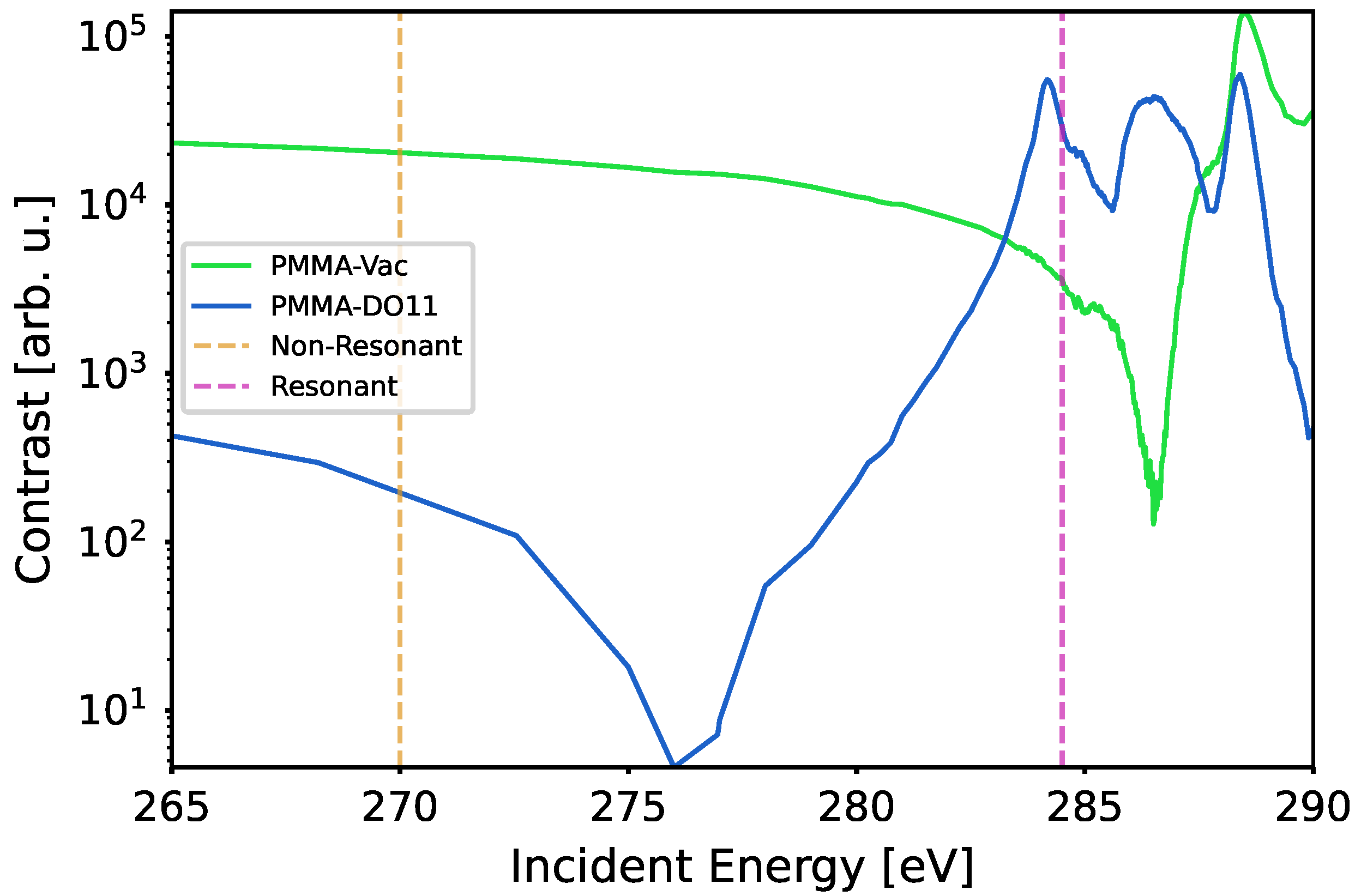
Figure 1. The contrast function | Δ n | 2 between PMMA and vacuum and between PMMA and DO11. Chosen RSoXS energies for resonant (284.5 eV) and non-resonant (270 eV) scattering are shown by the vertical lines.
Applsci 15 12006 g001
Figure 2. 1D-reduced scattering data: I (intensity) vs. Q (momentum transfer) for 270 eV (bottom scale, HLS color gradient from green to red with increasing wt%) and 284.5 eV (top scale, HLS color gradient from blue to hot pink with increasing wt%); wt% of samples are 0, 1, 2, 3, 3.5, 3.7, 3.9, 4, 5, 6, and 6.5. Data provided in Supplementary Material S1.
Applsci 15 12006 g002
Figure 3. The result of smoothing and doubly subtracting the scattering data corresponding to 1, 2, 3, 3.5, 3.7, 3.9, 4, 5, 6, 6.5 wt%. The parallel dashed lines show plots of power laws that are tangent to the doubly subtracted scattering data at low-q limit for 1%.
Applsci 15 12006 g003
Figure 4. Enhanced features due to DO11 inhomogeneity extracted by subtracting the material background power law fit from the data for 2, 3, 3.5, 3.7, 3.9, 4, 5, 6, 6.5 wt%. Standard errors computed over 80 point windows displayed as error bands. Maximum intensity marked on each trace with a × marker of the same color. Coordinates of marked points included in Supplementary Material S2.
Applsci 15 12006 g004
Figure 5. Binding energy between DO11 Tautomer and PMMA polymer yields 0.30 eV .
Applsci 15 12006 g005
Disclaimer/Publisher’s Note: The statements, opinions and data contained in all publications are solely those of the individual author(s) and contributor(s) and not of MDPI and/or the editor(s). MDPI and/or the editor(s) disclaim responsibility for any injury to people or property resulting from any ideas, methods, instructions or products referred to in the content.