Article

Effect of Heavy Metal Contamination on Caciotta Cheese Made from Buffalo Milk

Department of Agricultural, Forestry, Food and Environmental Sciences, University of Basilicata, Via dell’Ateneo Lucano 10, 85100 Potenza, Italy
*
Author to whom correspondence should be addressed.
Appl. Sci. 2025, 15(22), 11881; https://doi.org/10.3390/app152211881 (registering DOI)
Submission received: 12 September 2025 / Revised: 3 November 2025 / Accepted: 4 November 2025 / Published: 7 November 2025

Abstract

Cadmium (Cd) and lead (Pb) are environmental contaminants that induce oxidative damage in milk and dairy products. The aim of the study was to evaluate the effect of in vitro administration of Pb and Cd on the oxidative status and antioxidant capacity of buffalo cheese. Three batches (10 L each) of buffalo milk were divided: lead acetate (1 ppm) was added to the first batch, cadmium chloride (1 ppm) was added to the second batch and the last batch was control milk. Milk samples were stored at 4 °C for 24 h before the production of “caciotta” cheese (250 g each; n = 10 for each batch) and analyzed at the end of repining. The effect of the addition did not record significant differences in chemical composition (p > 0.05) compared to control. In contrast, oxidative stability assessed by primary (peroxide value—PV and acid degree value—ADV) and secondary (fatty acid oxidation products—TBARs assay) peroxidation showed significant differences (p < 0.05). Furthermore, Cd and Pb led to a significant increase in saturated fatty acids (SFAs) and monounsaturated fatty acids (MUFAs), while polyunsaturated fatty acids (PUFAs) were lower compared to the control (C; p < 0.05), especially in Pb cheese. The Cd and Pb cheese showed an increase in MDA value of 26 and 38% compared to the C. Metals also negatively influenced the antioxidant capacity of cheese (p < 0.05); specifically, Cd cheese showed lower ABTS, FRAP and thiol values. Finally, the activity of superoxide dismutase (SOD) was also strongly decreased with the addition of metals, especially Pb (p < 0.05).

1. Introduction

The increase in environmental pollution has led to the presence of some extremely harmful chemicals in food. Among these, heavy metals lead (Pb) and cadmium (Cd) are extremely harmful to health, so much so that the European Commission has published the new Regulations (EU) 2021/1323 and 2021/1317 which amend Regulation (EC) No. 1881/2006 as regards maximum levels of lead and cadmium in certain food products [1]. For milk and products made from cow milk protein or cow milk protein hydrolysates, the maximum permissible limit for cadmium is 0.005 mg/kg; for lead, the maximum limit is 0.020 mg/kg. The presence of toxic metals, including Cd and Pb, even in small amounts in dairy products is toxic to human health; in fact, prolonged exposure to heavy metals can lead to morphological abnormalities, neurological disorders, and malfunctions of the kidneys, liver, and prostate [2]. The presence of heavy metals in milk and milk-derived products depends on numerous factors: the farm’s geographic location, the types of fertilizers used for forage production, the quality of drinking water supplied to dairy cattle on the farm, etc. [3]. Lead is a contaminant present everywhere in the environment due to human activities: mining, metal processing, battery production [4]. The main sources of contamination (e.g., lead paints, organic compounds used in gasoline) have been eliminated or reduced since the 1970s following regulatory measures aimed at reducing human exposure to this toxic element [5]. Other measures have included the restriction of its use in drinking water pipes and leaded food cans. Overall, these measures have led to a documented reduction in exposure [6]. Cadmium is also widespread, especially in soil and water near industrial areas or hazardous waste dumps, and in areas with heavy car traffic or fertilized agricultural land [7]. Pb and Cd could be highly toxic if ingested in large quantities, causing irreversible damage to the body with long-term manifestations, which is why they are called “silent killers” [8,9]. There are numerous studies investigating the long-term toxic effect of heavy metals that accumulate in the human body through absorption, bioavailability, bioconcentration and biomagnification and negatively affect the functionality of the neurological, skeletal, reproductive, hematopoietic, renal and cardiovascular systems [10]. One of the main mechanisms underlying the toxicity of the two metals has been attributed to the induction of oxidative stress that occurs through very complex and not yet fully known systems; studies have shown that these metals have the ability to induce the generation of reactive radicals (ROS), resulting in cellular damage such as the exhaustion of enzymatic activities, damage to the lipid bilayer and DNA [11,12]. Generally, the first matrix to be attacked are lipids due to their high susceptibility [13] attributed to the high number of double bonds that favors the oxidation process [14]. Proteins can also be attacked by ROS that can cause oxidation in both amino acid side chains and main protein structures, resulting in protein fragmentation or protein–protein cross-links [15]. Oxidative modifications of proteins can alter their physical and chemical properties, including conformation, structure, solubility, susceptibility to proteolysis and enzymatic activities [15]. In a Polish study, high concentrations of lead and cadmium were detected in the blood of workers exposed to these toxic metals, generating a significant increase in SOD activity. The same researchers hypothesized that it is one of the body’s defense mechanisms against oxidative stress caused by environmental factors [16]. Superoxide dismutase, therefore, is a very resistant enzyme to the influence of environmental factors, but the mechanisms underlying these processes are not well understood. For the consumer, the main source of exposure to Pb and Cd is represented by the diet, in particular pasture/food for animals and milk and dairy products for humans [17]. Milk and its derivatives are the foods most consumed by humans and especially by children [18]. Among the various dairy products, buffalo cheese is enjoying great success for its beneficial properties, so much so that it is considered a functional food since each biological component present plays a functional and beneficial role for the health of the consumer [19,20]. Grassi et al. [21] highlighted the effect of species on the distribution of Cd and Pb in casein and fat fractions, observing that in buffalo milk, the affinity of these toxic metals for caseins was high, even though their levels in the fat fractions were also very high. Pb and Cd contamination in foods, particularly in cheese, negatively influences the oxidative stability and antioxidant capacity of cheese and its determination is of fundamental importance for consumer safety. The aim of our study was to evaluate the effect of the presence of Pb and Cd, by in vitro administration (direct addition to milk), on the oxidative state and antioxidant capacity of buffalo cheese pressed at the end of ripening. Therefore, the working hypothesis of this study was that the in vitro supplementation of buffalo milk with lead (Pb) and cadmium (Cd) would trigger oxidative stress in cheese by promoting lipid and protein oxidation, impairing non-enzymatic antioxidant defenses (ABTS, FRAP, thiols), and inhibiting the enzymatic antioxidant system (SOD), even in the absence of detectable changes in the basic chemical composition of the product.

2. Materials and Methods

2.1. Experimental Design and Cheese Manufacturing

This study was conducted on bulk milk of Italian buffalo, purchased from a supermarket in Potenza, Southern Italy (Basilicata), which also has a store selling buffalo milk and dairy products from a confined, free-range buffalo farm located in the province of Potenza, a low-environmental-impact zone. The concentration of 1 ppm for Pb and Cd was chosen as a representative experimental value, based on the following: realistic contamination levels found in milk and cheese in contaminated industrial or agricultural areas; previous scientific literature (e.g., Grassi et al. [21]); and the need to observe oxidative and antioxidant effects without compromising cheese processability.
Certainly, the added metals influence the cheeses produced from that milk, specifically by accelerating oxidative phenomena and modifying lipid and protein quality, as highlighted in the results and discussion. Furthermore, preliminary determinations of the heavy metals under study were performed on the bulk milk, which were below the detection limit. Bulk milk was collected and divided into three batches: the first batch (10 L) of control milk (C; without any addition); the second batch (10 L) of milk with the addition of lead acetate (Pb(CH3COO)2; Sigma Aldrich, Milan, Italy) to obtain a final concentration of 1 ppm (Pb); and the third batch (10 L) of milk with the addition of cadmium chloride (CdCl2; Sigma Aldrich, Milan, Italy) to obtain a final concentration of 1 ppm (Cd). Milk samples were stored at refrigeration temperature (4 °C) for 24 h (conditioning time) in glass containers. Bulk milk from buffalo had a higher percentage of fat (7.8) and protein (4.5%). After the conditioning time, C, Pb, and Cd were processed for the production of pressed cheese. Caciotta cheeses of approximately 250 g each were formed (n = 10 for each treatment). Subsequently, the cheeses were salted by immersion (20% NaCl) and maintained at a temperature of 4 °C for 15 min. The brine time was determined in relation to the size of the cheeses (1 day for each kg of 93 weight, minus one [22]). The maturation process was carried out in air-conditioned cells whose microclimatic characteristics were 10 °C and a relative humidity of 85% for 30 days. The chemical composition of each cheese was determined following official protocols [23].

2.2. Extraction, Identification, and Quantification of Fatty Acids by Gas Chromatography

2.2.1. Lipid Extraction in Cheese

The analysis was carried out by performing a series of three cold extractions of cheese lipids according to the modified method of Folch et al. [24]. The samples were homogenized by Ultra—Turrax T25 (Janke & Kunkel, Gmbh & Co, Staufen, Germany), sonicated for 10 min at 0 °C, and then centrifuged at 3350 rpm for 5 min at 0 °C. After centrifugation, the supernatant was recovered in a falcon containing anhydrous sodium sulfate and the extraction solution was added again to the precipitate and centrifuged. After the second centrifuge, the supernatant was put together with the previous one and the precipitate resuspended in ethyl ether/heptane was centrifuged again. After the third centrifuge, the supernatant was put together with the previous ones, the clear part was recovered and dried with a rotary evaporator (Heidolph, Laborota 4000, Heidolph Instruments GmbH & Co. KG, Schwabach, Germany).

2.2.2. Extraction and Derivatization of Free Fatty Acids

Cheese samples were extracted and derivatized by the method described by Kilcawley and Mannion, [25]. Free fatty acids (FFAs) were prepared on the extracted fat by adding 2 mL of ether per 100 mg of lipids. The mixture was placed inside pointed vials and vortexed. Methylation of FFAs was carried out by adding 200 µL of tetramethylammonium (TMA). After shaking the solution, the upper layer containing the methyl esters of the fatty acids was diluted with water, and finally used for the gas chromatography analysis.

2.2.3. Gas Chromatography (GC) Analysis

Gas chromatography analysis for the acid profile of the products was performed using a Varian 3400 gas chromatograph (Varian, Turin, Italy), equipped with a flame ionization detector (FID), split–splitless injector, capillary column (Omegawax 250 column 30 m × 0.25 µm × 0.25 µm; Thermo Fisher Scientific, Milan, Italy), and a Galaxie ™ Chromatography software version 2003–2007 (Varian, Inc., Walnut Creek, CA, USA) for data acquisition. Helium was used as carrier gas with a flow rate of 1 mL/min. The injector and detector temperatures were 250 and 220 °C, respectively. The operating conditions were as follows: initial temperature 120 °C for 1 min, 180 °C with an increase of 5 °C/min, and maintained at 180 °C for 18 min, 230 °C with an increase of 2 °C/min and maintained at 230 °C for 19 min. The identification of individual acids was obtained by comparing the retention times of the different peaks of the chromatographic trace with the retention times of known fatty acids, contained in a mixture of standards (Sigma-Aldrich, St. Louis, MO, USA and Supelco, Bellefonte, PA, USA). The results were expressed as % of the total methyl esters. The data on the fatty acid composition were processed and subsequently, the contents of saturated fatty acids (SFAs), monounsaturated fatty acids (MUFAs), and polyunsaturated fatty acids (PUFAs) were calculated.

2.3. Determination of Lipid Oxidation Products

2.3.1. Peroxide Value (PV)

Initial oxidation products were measured on lipid fraction by peroxide value (PV) method according to the ISO 3976:2006-IDF 74:2006 method [26]. The results were expressed as meq O2/kg−1 fat. Each determination was made in triplicate.

2.3.2. Acid Degree Value (ADV)

The lipolysis index was performed according to the modified procedure by Thomas and Pearce [27]. A cheese sample (10 g) was treated with anhydrous sodium sulfate (20–50 mL) until it became a homogeneous powder. The sample was then macerated with dichloromethane (50–70 mL) using a mortar to extract the fat. The mixture was filtered (Whatman n°42), and the extraction was repeated two more times to allow good fat extraction. Most of the solvent was recovered by evaporation using a refrigerated Heidolph rotavapor (Laborota 4000), while the residual solvent was removed using a thermostatic bath. Then a certain amount of fat (200–400 mg) was dissolved in 5 mL of hexane–propanol (4:1) and the resulting solution was titrated with 0.02 N methanolic KOH using phenolphthalein as an indicator. The blank was prepared with the hexane–propanol mixture without the fat sample. The lipolysis index was expressed as mg KOH 100 g−1 fat.

2.3.3. Fatty Acid Oxidation Products

The products of the secondary oxidation of fatty acids were determined by the quantification of thiobarbituric acid reactive substances (TBARS), as suggested by Grassi et al. [21]. Two grams of cheese were homogenized in a solution of hexane–isopropanol (4:1). The suspension was vortexed and sonicated for 10 min at room temperature. A total of 218 µL of sample, 382 µL of distilled H2O, and 900 µL of TBA reagent were added to the tube. The latter was placed in a water bath at 80 °C for 45 min with a solution for the development of the colorimetric reaction. After cooling, the sample was transferred into Eppendorf and centrifuged at 12,000 rpm for 10 min. Finally, the supernatant was filtered, and the absorbance was read on the spectrophotometer at 525 nm. Malonaldehyde quantification (MDA) was carried out using a calibration curve, and the data was expressed in ng of MDA/kg of cheese.

2.4. Pre-Treatment for Antioxidant Activity

The method of Sacchetti et al. [28] was followed for sample preparation with some modifications. An aliquot of sample (2 g) was added to 6 mL of distilled water and homogenized with the polytron (PT-MR 2100, Kinematica AG, Littau, Luzern, Switzerland). The mixture was then sonicated for 5 min at room temperature and centrifuged at 5000 rpm for 20 min at 4 °C. The supernatant was filtered and analyzed.

2.5. Protein Oxidation

2.5.1. Free Thiols Group

Thiols are organic compounds containing sulfhydryl groups able to counteract the action of ROS by neutralizing their toxicity. The Ellman’s assay was used (Ellman, [29]) to quantify the presence of sulfhydryl groups, with some modifications. A total of 250 μL of water-soluble extract of cheese samples was mixed with 2.5 mL of 0.1 M sodium phosphate buffer (containing 1 mM EDTA; pH 8.0, reaction buffer) and 50 μL of DTNB (5,5’-Dithio-bis(2-Nitrobenzoic acid) reagent solution (4 mg in 1 mL of sodium phosphate buffer). Subsequently, the solution was mixed and left at room temperature (25 °C) for 30 min. Finally, the reaction mixture was read at 412 nm. Reaction buffer was used instead of sample, as a reagent blank. A molar extinction coefficient of 14.150 M−1cm−1 was used to calculate moles of thiol groups.
C (mol/g) = ΔA/ε × b
where A is the absorbance at 412 nm and b is the optical path (cm). Each measurement was made in triplicate.

2.5.2. Radical Scavenging Assay (ABTS)

In order to evaluate the scavenging ability of cheese samples in a reaction with the ABTS radical, a modification of the original method of Re et al. [30] was used. An amount of 2 mL of ABTS•+ solution was added to 20 μL of sample. Absorbance was measured at 734 nm after 30 min of incubation at 30 °C against the blank. The measurement was performed in triplicate. The results were expressed as microgram of Trolox equivalent (TE) per gram of cheese (µg TE/g of cheese).

2.5.3. Activity by Ferric-Reducing Antioxidant Power

The assay of the antioxidant power ferric reduction (FRAP) was evaluated as suggested by Grassi et al. [21]. Aliquots of 100 μL of supernatant were mixed with 2.9 mL of FRAP reagent and incubated at 37 °C for 30 min. The increase in absorbance was measured at 593 nm against acetate buffer (pH 3.6). The blank reagent was prepared by adding distilled water instead of the sample. The results were expressed as microgram of TE per gram of cheese (µg TE/g of cheese).

2.5.4. Superoxide Dismutase (SOD) Activity

SOD activity was detected by measuring the inhibition of pyrogallol autoxidation following the method proposed by Grassi et al. [31]. Briefly, the reaction mixture was prepared by adding 1.9 mL of Tris-HCl 0.1 M, 50 μL of sample, and 50 μL pyrogallol 20 mM in HCl 1 mM. Inhibition of self-oxidation was followed spectrophotometrically at 325 nm every 30 s for 3 min. The results were expressed as percentage inhibition (I%) and were calculated by following the equation:
I(%) = [Ab − (As − At)/Ab] × 100
where Ab = absorbance of blank sample (t = 3 min); As = absorbance of sample (t = 3 min); At = absorbance of the test sample (pyrogallol was replaced by distilled water; t = 3 min).

2.6. Statistical Analysis

Statistical analyses were performed using the General Linear Model (GLM) procedure of SAS software (version 9.4, SAS Institute Inc., Cary, NC, USA) [30]. Data are expressed as mean ± standard deviation (SD). The normality of data distribution was verified using the Shapiro–Wilk test, and the homogeneity of variances was assessed using Levene’s test. When the assumptions of normality and homoscedasticity were met, a one-way analysis of variance (ANOVA) was carried out to evaluate the effect of treatment (Control, Cd, Pb) on the oxidative stability and antioxidant capacity of the cheese samples. Differences among means were considered significant at p < 0.05 and were further separated using Tukey’s post hoc test.

3. Results and Discussion

3.1. Chemical Composition of Cheese

The chemical composition of control cheese (C) and contaminated cheese (Cd, Pb) is shown in Table 1.
Statistical analysis showed that there is no significant change in the qualitative and quantitative nutritional profile of cheeses containing Cd and Pb compared to control cheese. These results are consistent with those of [32], who did not observe significant correlations between protein, fat, and heavy metals.
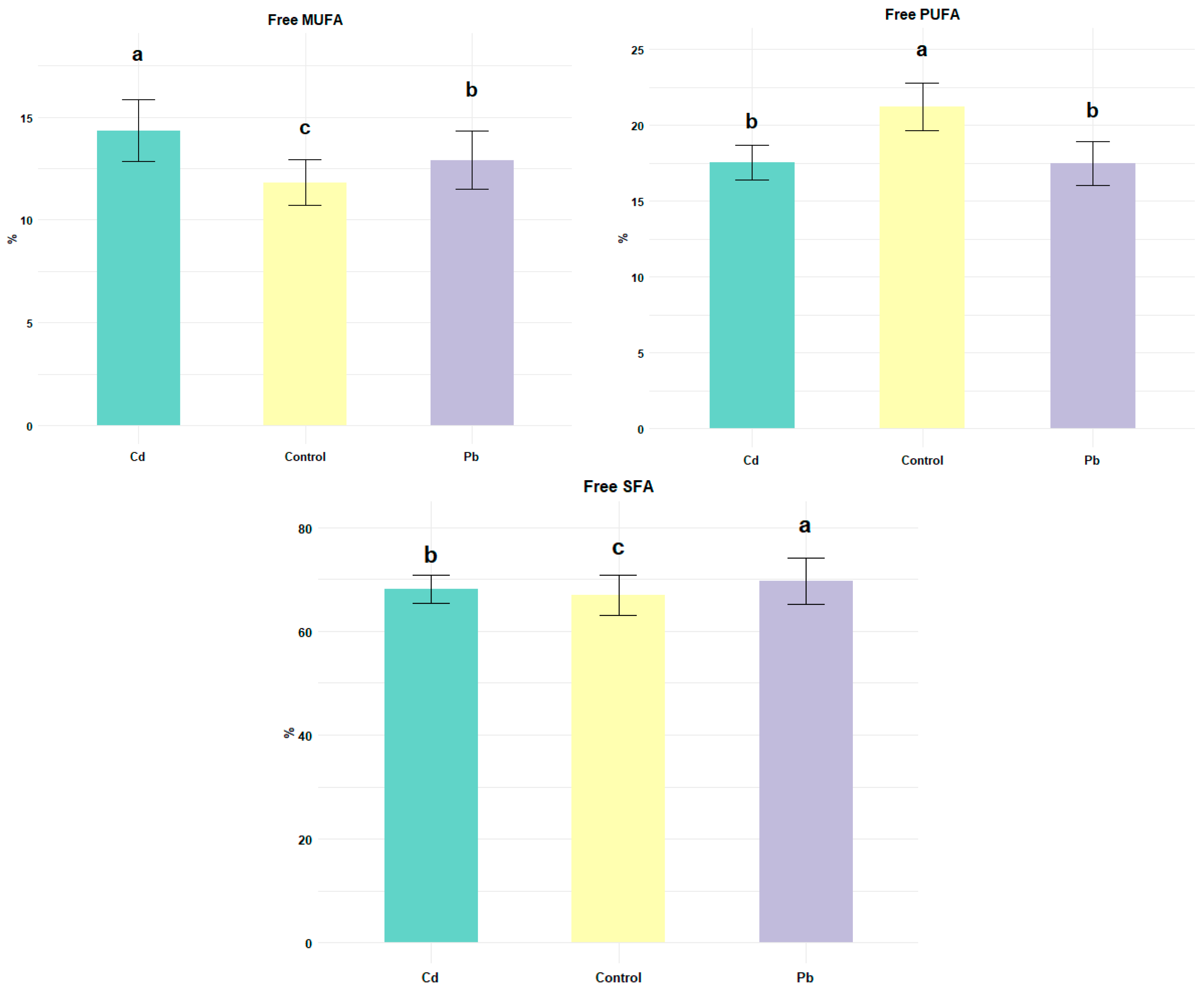
To evaluate the change in oxidative stability of cheese induced by the presence of Pb and Cd, the oxidation of lipid fractions was studied using acid degree value (ADV), peroxide value (PV), malonaldehyde assay (Figure 1), and percentage of free fatty acids (FFAs; Figure 2). The index of ADV is a measure of the amount of free fatty acids generated in a food sample during a given period by the hydrolysis of triglycerides. The index of PV (milliequivalents of peroxide per kilogram of fat sample) provides an indication of the formation of peroxide, a primary product of lipid oxidation (conjugated dienes and peroxides) that could have a negative impact on the health of the consumer if ingested [33].
The malonaldehyde assay is widely used to determine lipid peroxidation [13,34]. Malondialdehyde is one of the end products generated following the degradation of lipid peroxidation products that are positive for thiobarbituric acid (TBA). On average, the control cheese showed an ADV of 1.39 ± 0.15%, a PV of 1.17 ± 0.11 meq/kg fat, and an MDA concentration of 937.51 ± 90.01 ngMDA/kg cheese. These results are in line with those reported in the literature for late-ripening cheese [35]. The addition of heavy metals resulted in a significant increase in the values of each parameter studied (p < 0.05). The ability of Cd and Pb to induce ROS generation resulted in lipid peroxidation and changes in membrane structure, fatty acid composition and increased MDA levels [36]. Regarding the ADV, Pb and Cd cheeses showed a significantly higher percentage content (2.43 ± 0.19% and 2.17 ± 0.18%, respectively; p < 0.05) with an increase in the ADV equal to 1.7 and 1.6 times compared to the control. In support of our results, Quiroga-Roger et al. (2015) [37] indicated that there is a requirement of divalent cation for full milk lipase activity. It is known that ADV is the index of lipolysis. The result of lipolysis is the release of FFA. Figure 2 showed the percentage content of free fatty acid classes by product type.
Regarding free fatty acid classes, the presence of metals led to a significant increase in saturated fatty acids (SFAs) and monounsaturated fatty acids (MUFAs), while polyunsaturated fatty acids (PUFAs) were lower compared to the control (p < 0.05). In particular, Pb cheese had higher levels of SFAs and lower levels of MUFAs compared to Cd-contaminated cheese, while no significant differences were recorded for PUFAs (p > 0.05). Triglyceride hydrolysis mainly depends on moisture content, temperature, metal ion contamination, and lipase concentration. After lipolysis, the released free fatty acids contribute directly to the cheese flavor by acting as precursor molecules for a series of catabolic reactions leading to the production of flavor compounds, such as methyl ketones, lactones, esters, alkanes, and secondary alcohols [38]. Lipid oxidation of cheese intentionally contaminated with toxic heavy metals was determined by quantitative evaluation of peroxide and malondialdehyde formation (Figure 1). The PV of the studied cheeses was higher in the contaminated products (Cd and Pb) compared to the C cheese (p < 0.05); furthermore, the PV was significantly higher in the Pb-contaminated cheese compared to the Cd-contaminated cheese (1.79 ± 0.17 and 2.09 ± 0.20 meqO2/kg, respectively; p < 0.05). The results of this study showed that the formation of primary oxidation products could be immediately accelerated by toxic heavy metal contamination, also causing the promotion of secondary oxidation products expressed in ngMDA/Kg. The addition of Cd and Pb influenced the TBAR content in cheese (p < 0.05; Figure 1). Pb contamination revealed an MDA value of 1005.21 ± 80.12 ngMDA/Kg in cheese, with an increase of 8% compared to the control. The Cd-contaminated cheese presented a significantly higher MDA value (1290.28 ± 125.50 ngMDA/Kg (p < 0.05), which compared to the control, increased by 38%. The toxicity of Cd and Pb is mainly attributed to oxidative damage due to ROS generation [39]. Heavy metals such as Pb and Cd, although not redox active, caused alterations in the oxidation state of many biological macromolecules [9,40]. It is known that reactive oxygen species (ROS) are chemically aggressive species and the attack of free radicals on polyunsaturated fatty acid components of membrane lipids triggers lipid peroxidation, an autocatalytic process that modifies membrane structure and function [13]. In line with our findings, the authors of [38,41] found significant lipid peroxidation evidenced by increased malondialdehyde levels and changes in free fatty acid composition following oxidative stress induced by the presence of Pb and Cd in animal tissues.

3.2. Protein Oxidation

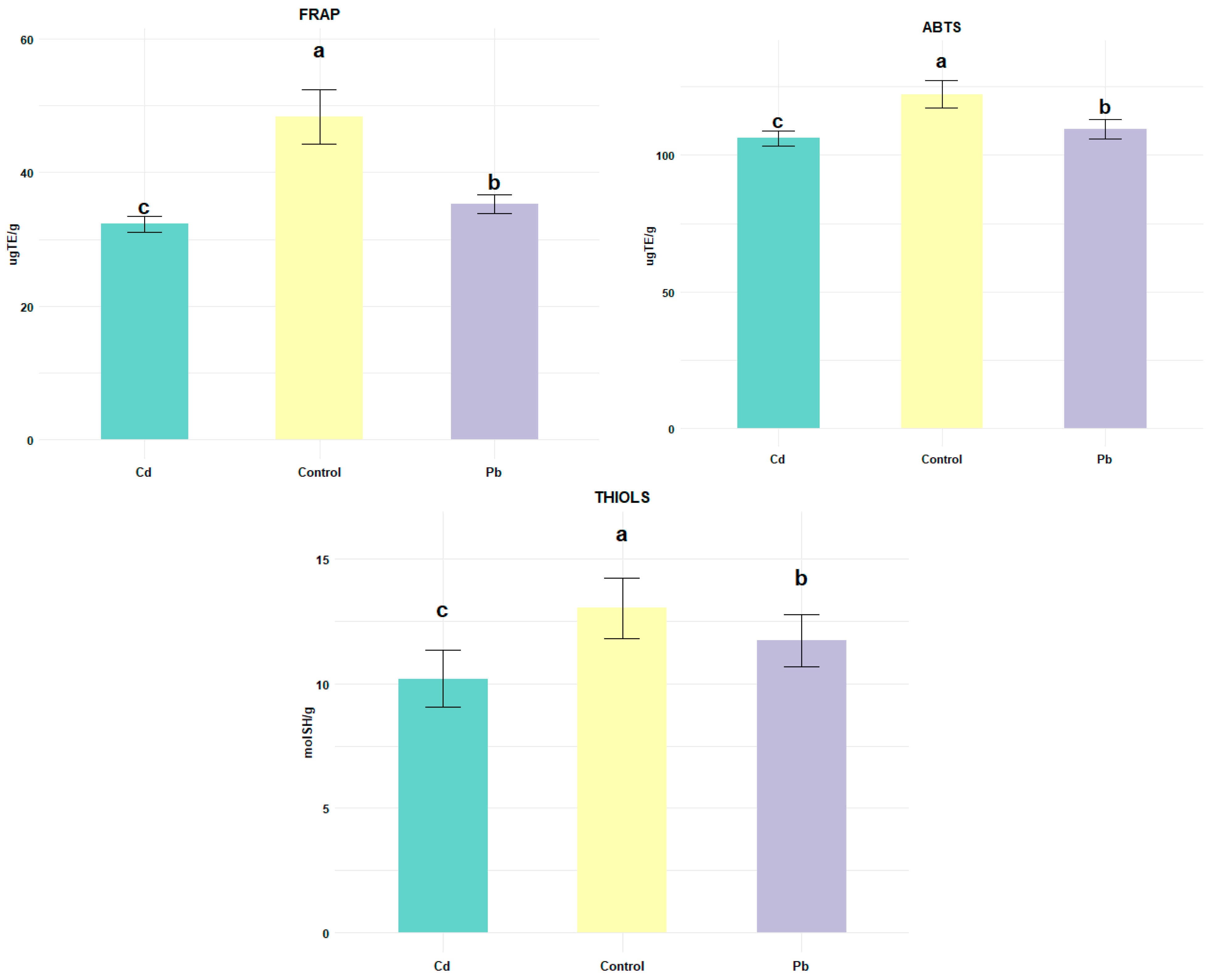
In Figure 3, the antioxidant capacities studied for the control, Cd- and Pb-contaminated cheeses are shown. The antioxidant capacity of the products represents a useful tool for evaluating the oxidative stability of the cheese [38].
In this study, the antioxidant status of cheese samples was assessed by ABTS spectrophotometric assay with 2,2’-azinobis (3-ethylbenzothiazoline-6-sulfonic acid) based on free radical scavenging; FRAP (ferric reducing antioxidant power) assay based on the reduction in ferric ion and thiol assay measuring sulfhydryl groups reacting with the thiol reagent 5-5′-dithiobis (2-nitrobenzoic acid) (DTNB), forming 5-thionitrobenzoic acid and disulfide bridges [42].
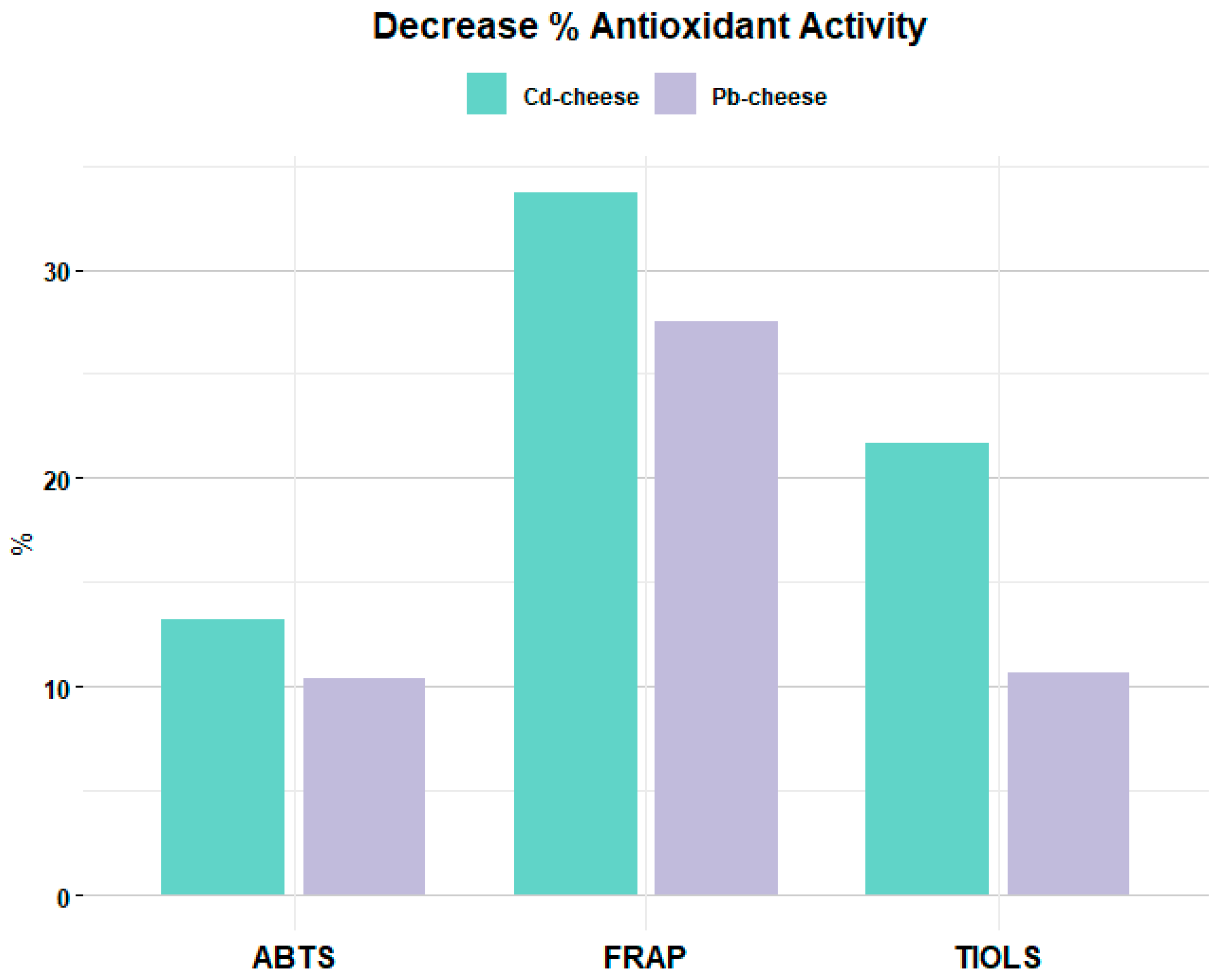
In general, all the analyzed samples showed antioxidant activity (Figure 3). However, the presence of Cd and Pb negatively affected the antioxidant capacity of cheese (p < 0.05). The values of ABTS, FRAP, and thiols were significantly lower in contaminated cheeses. In particular, Cd-contaminated cheese showed lower ABTS, FRAP, and thiol values (106.11 ± 2.82; 32.25 ± 1.17; and 10.19 ± 1.14, respectively; p < 0.05), highlighting a percentage decrease in antioxidant activity compared to the control of 13.19% for ABTS, 33.7% for FRAP, and 21.7% for thiols. Pb-contaminated cheese showed a smaller decrease of 10.4% for ABTS, 27.48% for FRAP, and 10.07% for thiols (Figure 4). These metals disrupt the functions of native proteins by binding to free thiols or other functional groups, catalyzing the oxidation of amino acid side groups, perturbing protein folding, and/or replacing essential metal ions in enzymes [43]. The oxidative alterations observed in buffalo cheese contaminated with Cd and Pb may be mechanistically linked to molecular signaling pathways commonly involved in heavy metal-induced oxidative stress [44]. Both Cd2+ and Pb2+ are known to generate a surplus of mitochondrial reactive oxygen species (ROS), leading to the activation of the Nrf2/Keap1–ARE pathway, which regulates the expression of antioxidant defense enzymes such as SOD, CAT, and GPx. The observed decrease in SOD activity and thiol content in contaminated cheese suggests an alteration of this adaptive response [45].
Concurrently, heavy metal exposure may stimulate MAPK signaling (ERK, JNK, p38), promoting/accelerating lipid peroxidation and protein carbonylation through increased ROS generation. Furthermore, the two heavy metals can inhibit glutathione metabolism and suppress the ascorbate–glutathione cycle, reducing the ability to scavenge free radicals [46]. These effects collectively contribute to redox imbalance and oxidative damage. Therefore, the biochemical findings reported here (increased PV, MDA, and decreased SOD/FRAP) can be interpreted as downstream consequences of disrupting Nrf2-mediated redox regulation and enhancing MAPK-driven oxidative signaling.
During cheese ripening, protein oxidation occurs naturally, leading to loss of sulfhydryl (thiol) group with formation of disulfide bridges, aided by lipid oxidation whose products such as superoxide radicals, hydroxyl radicals, and nitric oxide radicals could be generated indirectly. Wang and Wang [47] demonstrated the generation of non-radical hydrogen peroxide, which itself became a significant source of free radicals through Fenton chemistry. These free radicals directly affect proteins by creating cross-links and aggregations [48]. Furthermore, lipid oxidation products show greater interaction with proteins than their primary counterparts. The inclusion of specific toxic heavy metals, such as Cd and Pb, accelerated and increased these processes, negatively affecting the oxidative stability of cheese. To highlight the effect of contamination on the antioxidant stability of cheese, the percentage of decrease in SOD activity was evaluated. SOD is an important enzyme in the control of free radicals and, together with other enzymes such as glutathione peroxidase and catalase, modulates the effects of oxidative stress [49]. Its activity represents a valid index of oxidative stability. SOD activity, expressed as percentage of inhibition of pyrogallol autoxidation (I%), was significantly lower in contaminated cheeses (p < 0.05; Figure 5).
An activity of 78.18% was detected in the control cheese, 68.96% in the Cd-contaminated cheese, and 64.97% in the Pb-cheese, corresponding to a decrease of 11.79% in the presence of Cd and 16.90% with Pb compared to the control. A comparison between the contaminated cheeses revealed that Pb has a significantly lower I% than Cd; 64.97% and 68.97%, respectively (p < 0.05). SOD is a potential target for Pb toxicity because this antioxidant enzyme depends on various essential trace elements such as copper and zinc for its activity; copper ions play a functional role in the reaction by undergoing alternating oxidation, while zinc ions appear to stabilize the enzyme. Both of these biometals can be replaced by Pb and Cd in the active site of the enzyme, resulting in decreased SOD activity [48]. Elarabany and Bahnasawy [50] observed an accumulation of ROS in African catfish (Clarias gariepinus) exposed to Cd and Pb resulting in decreased SOD, demonstrated by the significant increase in MDA. These results could be related to the high electron sharing affinities of Pb and Cd, which can lead to the formation of covalent bonds mainly between heavy metals and sulfhydryl groups of proteins. Cangelosi et al. [51] showed that Pb(II) in proteins is coordinated by sulfur, oxygen, and nitrogen donors, although coordination via cysteine (Cys) is preferred over glutamic acid (Glu) or histidine (His). Jomova and Valko [52] reported that among milk proteins, α-casein (αs1 and αs2) has been identified as having the ability to bind many metal ions, including Pb. Another mechanism for cadmium- and lead-induced oxidative stress involves the antioxidant defense systems of cells. The binding affinity of Pb and Cd to sulfhydryl (-SH) groups influences many enzymes of the antioxidant defense system, such as SOD, catalase (CAT), glutathione peroxidase (GPx) and glucose-6-phosphate dehydrogenase (G6PD), subsequently inhibiting their activity [52,53]. Pb and Cd, as divalent cations, not only bind to -SH groups, but can also replace divalent bioelements that serve as important cofactors of antioxidant enzymes such as GPx, SOD, and CAT, causing their inactivation. Several components determine the antioxidant activity of cheese, such as ascorbate, ureate, and other low molecular weight compounds [54], including vitamins and phenolic compounds, together with enzymes such as glutathione peroxidase [55]. The presence of Cd and Pb, however, influences the ascorbate–glutathione pathway depending on the concentration of toxic metals; the greater their presence, the lower its activity [56]. The results obtained allow us to outline an integrated picture of the main mechanisms of oxidative stress induced by Cd and Pb in buffalo cheese. Both metals, although not redox-active, act as indirect pro-oxidants through three main and interconnected pathways: 1. Triggering of lipid peroxidation, resulting in a significant increase in PV, ADV, and MDA, demonstrating that the metals promote the formation of peroxyl and hydroxyl radicals responsible for the degradation of PUFAs. This phenomenon is confirmed by the reduction in PUFAs and the relative increase in SFAs and MUFAs, indicating a selective oxidation of unsaturated fatty acids. The effect is more pronounced for cadmium, likely due to its greater affinity for the sulfhydryl groups of membrane lipoproteins, which facilitates its interaction with redox systems. 2. Protein oxidation and loss of enzymatic functionality.
The decrease in thiol groups and the formation of disulfide bonds confirm the direct oxidation of proteins by free radicals and secondary products of lipid peroxidation (aldehydes, hydroperoxides). This modification of protein structure limits the activity of enzymes sensitive to –SH bonds, including SOD, whose activity is reduced by 12–17%. This loss is consistent with the competitive substitution of Cu2+ and Zn2+ in the enzyme’s active site by Pb2+ and Cd2+, which destabilize its quaternary structure. 3. Depression of non-enzymatic antioxidant systems. Significant reductions in ABTS and FRAP values indicate a depletion of the overall reducing capacity of the matrix, due both to the direct consumption of antioxidants (such as ascorbate and glutathione) and to the inhibition of the ascorbate–glutathione cycle documented for Cd and Pb. The cumulative effect of these alterations amplifies oxidative damage and accelerates the degradation of lipid and protein components. Overall, the correlation between the measured parameters suggests that lipid peroxidation represents the dominant mechanism, explaining approximately 60% of the overall variation in oxidative markers, while protein oxidation and antioxidant depletion contribute 17% and 23%, respectively. This quantitative model, albeit simplified, highlights the multifactorial nature of metal-induced oxidative stress: lipids serve as the primary target, generating reactive products that, in turn, amplify protein damage and reduce the matrix’s defensive capacity. Although the experiment was conducted by adding 1 ppm of each metal in vitro, such concentrations can be related to the food safety limits established by EFSA and WHO. Considering an average cheese consumption of 30 g/day, even relatively low concentrations in milk or cheese can significantly contribute to cumulative exposure. Since Cd and Pb are present in various food matrices (cereals, vegetables, fish, etc.), exposure from dairy products should be evaluated in a multi-food and cumulative context, as proposed by Lučić et al. [57]. EFSA has established a tolerable weekly intake (TWI) for cadmium of 2.5 µg/kg body weight and a lower confidence limit of the benchmark dose (BMDL01) for lead ranging from 0.5 to 1.5 µg/kg body weight per day based on neurotoxic effects.

4. Conclusions

This study showed that heavy metal contamination significantly alters the oxidative status of buffalo cheese without altering its basic chemical composition. Specifically, the increase in lipid peroxidation products (PV, ADV, and TBARs) and the change in the fatty acid profile, with an increase in SFAs and MUFAs and a reduction in PUFAs, indicated that both lead and cadmium accelerate lipid oxidation processes, with Pb showing a more pronounced effect.
Protein quality, the significant decrease in antioxidant activities (ABTS, FRAP, and thiol groups), and the reduction in SOD enzymatic activity confirmed that both metals compromise the antioxidant defense capacity of the cheese, likely through the inhibition or competitive substitution of the enzyme’s metal cofactors.
Overall, the data suggest that Pb and Cd act as promoters of oxidative stress in dairy products, influencing both lipid and protein oxidation mechanisms. These alterations, while not altering the apparent nutritional characteristics, can compromise the sensory quality and safety of the product.
Therefore, this study proposes the use of oxidative parameters (such as MDA, FRAP, and SOD) as indirect and rapid indices of heavy metal contamination, offering a potential alternative to traditional analytical methods, which are often expensive and complex. Future research should therefore focus on field studies involving milk from animals exposed to contaminated environments, in order to validate the biomarkers identified here (MDA, FRAP, SOD) under real-world production conditions and evaluate potential interactions between multiple contaminants, minerals, and milk components.

Author Contributions

Methodology, G.G.; Software, A.M.P.; Validation, G.G. and A.M.P.; Formal analysis, G.G.; Investigation, G.G. and A.M.P.; Data curation, A.M.P.; Writing—original draft, G.G. and A.M.P.; Writing—review and editing, G.G.; Project administration, A.M.P. All authors have read and agreed to the published version of the manuscript.

Funding

This research received no external funding.

Institutional Review Board Statement

Not applicable.

Informed Consent Statement

Not applicable.

Data Availability Statement

The original contributions presented in the study are included in the article, further inquiries can be directed to the corresponding author.

Conflicts of Interest

The authors declare no conflicts of interest.

References

  1. Commission Regulation (EU) 2021/1323 of 10 August 2021 Amending Regulation (EC) No 1881/2006 as Regards the Maximum Levels of Cadmium in Certain Foodstuffs. Available online: https://eur-lex.europa.eu/eli/reg/2021/1323/oj/eng (accessed on 3 November 2025).
  2. Amalfitano, N.; Stocco, G.; Maurmayr, A.; Pegolo, S.; Cecchinato, A.; Bittante, G. Quantitative and Qualitative Detailed Milk Protein Profiles of 6 Cattle Breeds: Sources of Variation and Contribution of Protein Genetic Variants. J. Dairy Sci. 2020, 103, 11190–11208. [Google Scholar] [CrossRef]
  3. Hassan, T.; Elarnaoutti, M.S. Heavy Metals Transfer from Milk into Milk Products. Turk. J. Agric.-Food Sci. Technol. 2025, 13, 900–906. [Google Scholar] [CrossRef]
  4. Raj, K.; Das, A.P. Lead Pollution: Impact on Environment and Human Health and Approach for a Sustainable Solution. Environ. Chem. Ecotoxicol. 2023, 5, 79–85. [Google Scholar] [CrossRef]
  5. Hettiarachchi, G.M.; Betts, A.R.; Chandima Wekumbura, W.G.; Lake, L.; Mayer, M.M.; Scheckel, K.G.; Basta, N.T. Chapter 6—Lead: The Most Extensively Spread Toxic Environmental Contaminant. In Inorganic Contaminants and Radionuclides; Naidu, R., Ed.; Elsevier: Amsterdam, The Netherlands, 2024; pp. 113–150. ISBN 978-0-323-90400-1. [Google Scholar]
  6. Jarvis, P.; Fawell, J. Lead in Drinking Water—An Ongoing Public Health Concern? Curr. Opin. Environ. Sci. Health 2021, 20, 100239. [Google Scholar] [CrossRef]
  7. Kubier, A.; Wilkin, R.T.; Pichler, T. Cadmium in Soils and Groundwater: A Review. Appl. Geochem. 2019, 108, 104388. [Google Scholar] [CrossRef]
  8. Banerjee, A.; Nandi, P.; Bhattacharya, C.; Kabir, Z.; Mukherjee, S.; Maji, B.K. Cadmium Acts as a Silent Killer of Liver by Inducing Oxidative Stress and Hepatocellular Injury and a Possible Amelioration by Vitamin B12 and Folic Acid in Rat Model. Prog. Health Sci. 2019, 1, 105–117. [Google Scholar] [CrossRef]
  9. Flora, G.; Gupta, D.; Tiwari, A. Toxicity of Lead: A Review with Recent Updates. Interdiscip. Toxicol. 2012, 5, 47–58. [Google Scholar] [CrossRef] [PubMed]
  10. Collin, F. Chemical Basis of Reactive Oxygen Species Reactivity and Involvement in Neurodegenerative Diseases. Int. J. Mol. Sci. 2019, 20, 2407. [Google Scholar] [CrossRef]
  11. Ali, H.; Khan, E.; Ilahi, I. Environmental Chemistry and Ecotoxicology of Hazardous Heavy Metals: Environmental Persistence, Toxicity, and Bioaccumulation. J. Chem. 2019, 2019, 6730305. [Google Scholar] [CrossRef]
  12. Briffa, J.; Sinagra, E.; Blundell, R. Heavy Metal Pollution in the Environment and Their Toxicological Effects on Humans. Heliyon 2020, 6, e04691. [Google Scholar] [CrossRef] [PubMed]
  13. Juan, C.A.; Pérez de la Lastra, J.M.; Plou, F.J.; Pérez-Lebeña, E. The Chemistry of Reactive Oxygen Species (ROS) Revisited: Outlining Their Role in Biological Macromolecules (DNA, Lipids and Proteins) and Induced Pathologies. Int. J. Mol. Sci. 2021, 22, 4642. [Google Scholar] [CrossRef] [PubMed]
  14. Geng, L.; Liu, K.; Zhang, H. Lipid Oxidation in Foods and Its Implications on Proteins. Front. Nutr. 2023, 10, 1192199. [Google Scholar] [CrossRef]
  15. Kehm, R.; Baldensperger, T.; Raupbach, J.; Höhn, A. Protein Oxidation—Formation Mechanisms, Detection and Relevance as Biomarkers in Human Diseases. Redox Biol. 2021, 42, 101901. [Google Scholar] [CrossRef]
  16. Yan, Y.; Jin, L.; Li, J.; Chen, G. Association of Cadmium and Lead Exposure with Mortality in Cancer Survivors: A Prospective Cohort Study. Ecotoxicol. Environ. Saf. 2025, 292, 117960. [Google Scholar] [CrossRef]
  17. Afzal, A.; Mahreen, N. Emerging Insights into the Impacts of Heavy Metals Exposure on Health, Reproductive and Productive Performance of Livestock. Front. Pharmacol. 2024, 15, 1375137. [Google Scholar] [CrossRef]
  18. Visioli, F.; Strata, A. Milk, Dairy Products, and Their Functional Effects in Humans: A Narrative Review of Recent Evidence. Adv. Nutr. 2014, 5, 131–143. [Google Scholar] [CrossRef]
  19. Basilicata, M.G.; Pepe, G.; Adesso, S.; Ostacolo, C.; Sala, M.; Sommella, E.; Scala, M.C.; Messore, A.; Autore, G.; Marzocco, S.; et al. Antioxidant Properties of Buffalo-Milk Dairy Products: A β-Lg Peptide Released after Gastrointestinal Digestion of Buffalo Ricotta Cheese Reduces Oxidative Stress in Intestinal Epithelial Cells. Int. J. Mol. Sci. 2018, 19, 1955. [Google Scholar] [CrossRef]
  20. Vargas-Ramella, M.; Pateiro, M.; Maggiolino, A.; Faccia, M.; Franco, D.; De Palo, P.; Lorenzo, J.M. Buffalo Milk as a Source of Probiotic Functional Products. Microorganisms 2021, 9, 2303. [Google Scholar] [CrossRef] [PubMed]
  21. Grassi, G.; Simonetti, A.; Gambacorta, E.; Perna, A. Effect of Species on the Distribution and Oxidative Stability of Milk Added of Lead and Cadmium. Ital. J. Anim. Sci. 2023, 22, 1162–1171. [Google Scholar] [CrossRef]
  22. Fucà, N.; McMahon, D.J.; Caccamo, M.; Tuminello, L.; La Terra, S.; Manenti, M.; Licitra, G. Effect of Brine Composition and Brining Temperature on Cheese Physical Properties in Ragusano Cheese. J. Dairy Sci. 2012, 95, 460–470. [Google Scholar] [CrossRef]
  23. Horwitz, W. (Ed.) AOAC Official Methods of Analysis of the Association of Official Analytical Chemists, 13th ed.; Association of Official Analytical Chemists (AOAC): Rockville, MA, USA, 1980. [Google Scholar]
  24. Folch, J.; Lees, M.; Sloane Stanley, G.H. A Simple Method for the Isolation and Purification of Total Lipides from Animal Tissues. J. Biol. Chem. 1957, 226, 497–509. [Google Scholar] [CrossRef] [PubMed]
  25. Kilcawley, K.N.; Mannion, D.T. Free Fatty Acids Quantification in Dairy Products. In Fatty Acids; Catala, A., Ed.; InTech: Rijeka, Croatia, 2017; ISBN 978-953-51-3301-8. [Google Scholar]
  26. ISO 3976:2006-IDF 74:2006; Milk Fat—Determination of Peroxide Value Published. International Organization for Standardization: Geneva, Switzerland, 2006.
  27. Thomas, T.D.; Pearce, K.N. Influence of Salt on Lactose Fermentation and Proteolysis in Cheddar Cheese. J. Dairy Sci. Technol. 1981, 16, 253–259. [Google Scholar]
  28. Sacchetti, G.; Di Mattia, C.; Pittia, P.; Martino, G. Application of a Radical Scavenging Activity Test to Measure the Total Antioxidant Activity of Poultry Meat. Meat Sci. 2008, 80, 1081–1085. [Google Scholar] [CrossRef]
  29. Ellman, G.L. Tissue Sulfhydryl Groups. Arch. Biochem. Biophys. 1959, 82, 70–77. [Google Scholar] [CrossRef]
  30. Re, R.; Pellegrini, N.; Proteggente, A.; Pannala, A.; Yang, M.; Rice-Evans, C. Antioxidant Activity Applying an Improved ABTS Radical Cation Decolorization Assay. Free. Radic. Biol. Med. 1999, 26, 1231–1237. [Google Scholar] [CrossRef]
  31. Grassi, G.; Simonetti, A.; Gambacorta, E.; Perna, A. Decrease of Activity of Antioxidant Enzymes, Lysozyme Content, and Protein Degradation in Milk Contaminated with Heavy Metals (Cadmium and Lead). JDS Commun. 2022, 3, 312–316. [Google Scholar] [CrossRef]
  32. SAS User’s Guide: Statistics; Cary, N.C., Ed.; SAS Institute: Cary, NC, USA, 1985; ISBN 978-0-917382-66-6. [Google Scholar]
  33. Morsy, M.K.; Elsabagh, R. Quality Parameters and Oxidative Stability of Functional Beef Burgers Fortified with Microencapsulated Cod Liver Oil. LWT 2021, 142, 110959. [Google Scholar] [CrossRef]
  34. Ruiz-Ojeda, F.J.; Olza, J.; Gil, Á.; Aguilera, C.M. Chapter 1—Oxidative Stress and Inflammation in Obesity and Metabolic Syndrome. In Obesity; del Moral, A.M., Aguilera García, C.M., Eds.; Academic Press: Cambridge, MA, USA, 2018; pp. 1–15. ISBN 978-0-12-812504-5. [Google Scholar]
  35. Semeniuc, C.A.; Mandrioli, M.; Socaci, B.S.; Socaciu, M.-I.; Fogarasi, M.; Podar, A.S.; Michiu, D.; Jimborean, A.M.; Mureşan, V.; Ionescu, S.R.; et al. Changes in Lipid Composition and Oxidative Status during Ripening of Gouda-Type Cheese as Influenced by Addition of Lavender Flower Powder. Int. Dairy J. 2022, 133, 105427. [Google Scholar] [CrossRef]
  36. Liu, X.; Liu, X.; Yao, Y.; Qu, X.; Chen, J.; Xie, K.; Wang, X.; Qi, Y.; Xiao, B.; He, C. Effects of Different Levels of Hermetia Illucens Larvae Meal on Performance, Egg Quality, Yolk Fatty Acid Composition and Oxidative Status of Laying Hens. Ital. J. Anim. Sci. 2021, 20, 256–266. [Google Scholar] [CrossRef]
  37. Quiroga-Roger, D.; Babul, J.; Guixé, V. Role of Monovalent and Divalent Metal Cations in Human Ribokinase Catalysis and Regulation. Biometals 2015, 28, 401–413. [Google Scholar] [CrossRef] [PubMed]
  38. Thierry, A.; Collins, Y.F.; Abeijón Mukdsi, M.C.; McSweeney, P.L.H.; Wilkinson, M.G.; Spinnler, H.E. Chapter 17—Lipolysis and Metabolism of Fatty Acids in Cheese. In Cheese, 4th ed.; McSweeney, P.L.H., Fox, P.F., Cotter, P.D., Everett, D.W., Eds.; Academic Press: San Diego, CA, USA, 2017; pp. 423–444. ISBN 978-0-12-417012-4. [Google Scholar]
  39. Charkiewicz, A.E.; Omeljaniuk, W.J.; Nowak, K.; Garley, M.; Nikliński, J. Cadmium Toxicity and Health Effects—A Brief Summary. Molecules 2023, 28, 6620. [Google Scholar] [CrossRef]
  40. Monteverde, V.; Camilleri, G.; Arfuso, F.; Pennisi, M.; Perillo, L.; Patitò, G.; Gioia, G.; Castronovo, C.; Piccione, G. Heavy Metal Levels in Milk and Serum of Dairy Cows from Different Farms Located near an Industrial Area. Animals 2022, 12, 2574. [Google Scholar] [CrossRef]
  41. Tsaturyan, V.; Poghosyan, A.; Toczyłowski, M.; Pepoyan, A. Evaluation of Malondialdehyde Levels, Oxidative Stress and Host–Bacteria Interactions: Escherichia Coli and Salmonella Derby. Cells 2022, 11, 2989. [Google Scholar] [CrossRef]
  42. Stapelfeldt, H.; Nielsen, B.R.; Skibsted, L.H. Effect of Heat Treatment, Water Activity and Storage Temperature on the Oxidative Stability of Whole Milk Powder. Int. Dairy J. 1997, 7, 331–339. [Google Scholar] [CrossRef]
  43. Witkowska, D.; Słowik, J.; Chilicka, K. Heavy Metals and Human Health: Possible Exposure Pathways and the Competition for Protein Binding Sites. Molecules 2021, 26, 6060. [Google Scholar] [CrossRef]
  44. Kasai, S.; Shimizu, S.; Tatara, Y.; Mimura, J.; Itoh, K. Regulation of Nrf2 by Mitochondrial Reactive Oxygen Species in Physiology and Pathology. Biomolecules 2020, 10, 320. [Google Scholar] [CrossRef]
  45. Snezhkina, A.V.; Kudryavtseva, A.V.; Kardymon, O.L.; Savvateeva, M.V.; Melnikova, N.V.; Krasnov, G.S.; Dmitriev, A.A. ROS Generation and Antioxidant Defense Systems in Normal and Malignant Cells. Oxid. Med. Cell Longev. 2019, 2019, 6175804. [Google Scholar] [CrossRef] [PubMed]
  46. Haidar, Z.; Fatema, K.; Shoily, S.S.; Sajib, A.A. Disease-Associated Metabolic Pathways Affected by Heavy Metals and Metalloid. Toxicol. Rep. 2023, 10, 554–570. [Google Scholar] [CrossRef] [PubMed]
  47. Wang, J.; Wang, S. Reactive Species in Advanced Oxidation Processes: Formation, Identification and Reaction Mechanism. Chem. Eng. J. 2020, 401, 126158. [Google Scholar] [CrossRef]
  48. Domínguez, R.; Pateiro, M.; Gagaoua, M.; Barba, F.J.; Zhang, W.; Lorenzo, J.M. A Comprehensive Review on Lipid Oxidation in Meat and Meat Products. Antioxidants 2019, 8, 429. [Google Scholar] [CrossRef] [PubMed]
  49. Ighodaro, O.M.; Akinloye, O.A. First Line Defence Antioxidants-Superoxide Dismutase (SOD), Catalase (CAT) and Glutathione Peroxidase (GPX): Their Fundamental Role in the Entire Antioxidant Defence Grid. Alex. J. Med. 2018, 54, 287–293. [Google Scholar] [CrossRef]
  50. Elarabany, N.; Bahnasawy, M. Comparative and Interactive Biochemical Effects of Sub-Lethal Concentrations of Cadmium and Lead on Some Tissues of the African Catfish (Clarias gariepinus). Toxicol. Res. 2019, 35, 249–255. [Google Scholar] [CrossRef]
  51. Cangelosi, V.; Ruckthong, L.; Pecoraro, V.L. Lead(II) Binding in Natural and Artificial Proteins. In Lead: Its Effects on Environment and Health; Sigel, A., Sigel, H., Sigel, R.K.O., Eds.; De Gruyter: Berlin, Germany; Boston, UK, 2017; Volume 17, pp. 271–318. [Google Scholar] [CrossRef]
  52. Jomova, K.; Valko, M. Advances in Metal-Induced Oxidative Stress and Human Disease. Toxicology 2011, 283, 65–87. [Google Scholar] [CrossRef]
  53. Nair, A.; DeGheselle, O.; Smeets, K.; Van Kerkhove, E.; Cuypers, A. Cadmium-Induced Pathologies: Where Is the Oxidative Balance Lost (or Not)? Int. J. Mol. Sci. 2013, 14, 6116–6143. [Google Scholar] [CrossRef]
  54. Clausen, M.R.; Connolly, C.; Skibsted, L.H.; Stagsted, J. Oxidative Stability of Bovine Milk Determined by Individual Variability in Herd Irrespective of Selenium Status. Int. Dairy J. 2010, 20, 507–513. [Google Scholar] [CrossRef]
  55. Revilla, I.; González-Martín, M.I.; Vivar-Quintana, A.M.; Blanco-López, M.A.; Lobos-Ortega, I.A.; Hernández-Hierro, J.M. Antioxidant Capacity of Different Cheeses: Affecting Factors and Prediction by near Infrared Spectroscopy. J. Dairy Sci. 2016, 99, 5074–5082. [Google Scholar] [CrossRef] [PubMed]
  56. Liu, Y.; Wang, X.; Zeng, G.; Qu, D.; Gu, J.; Zhou, M.; Chai, L. Cadmium-Induced Oxidative Stress and Response of the Ascorbate–Glutathione Cycle in Bechmeria Nivea (L.) Gaud. Chemosphere 2007, 69, 99–107. [Google Scholar] [CrossRef] [PubMed]
  57. Lučić, M.; Miletić, A.; Savić, A.; Lević, S.; Ignjatović, I.S.; Onjia, A. Dietary Intake and Health Risk Assessment of Essential and Toxic Elements in Pepper (Capsicum annuum). J. Food Compos. Anal. 2022, 111, 104598. [Google Scholar] [CrossRef]
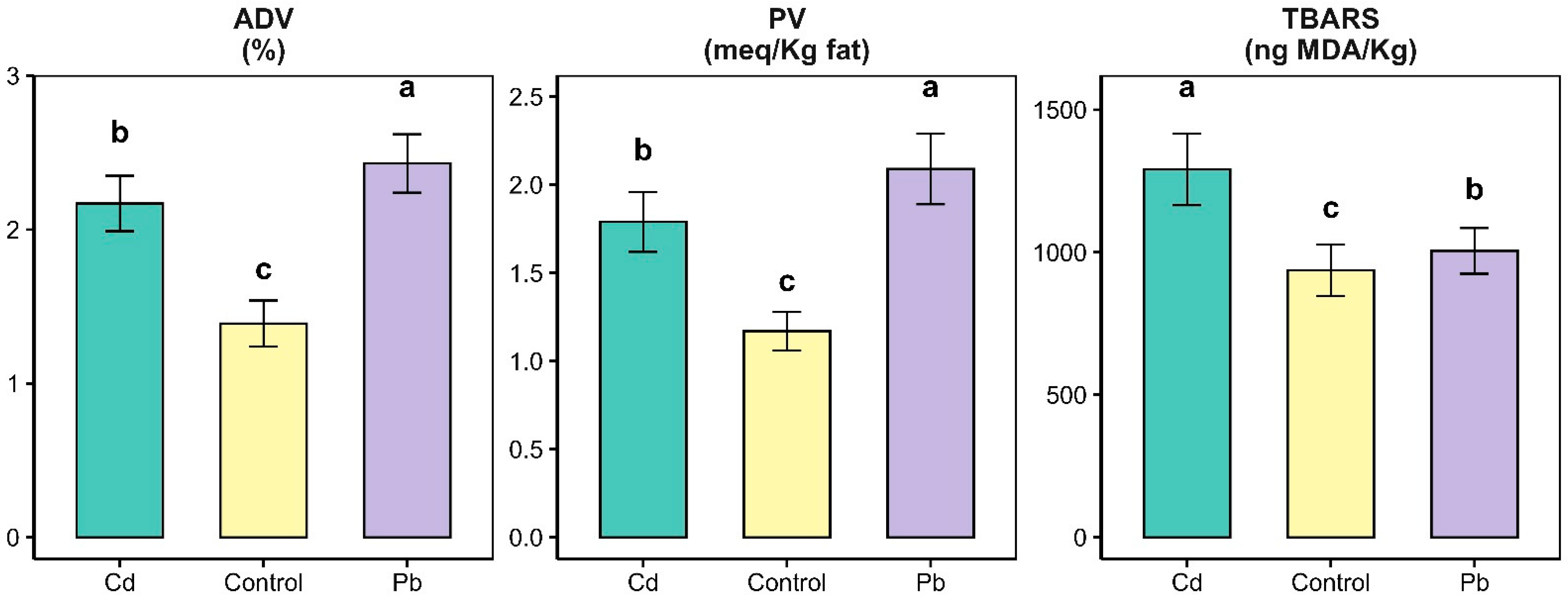
Figure 1. Acid degree value (ADV), peroxide value (PV), and content of TBARs in control cheese, cadmium-contaminated (Cd), and lead-contaminated (Pb) cheese. a,b,c p < 0.05: values with different letters are statistically different; error bars indicate standard deviation.
Applsci 15 11881 g001
Figure 2. Content of saturated fatty acids (SFAs), monounsaturated fatty acids (MUFAs) and polyunsaturated fatty acids (PUFAs) in control, cadmium-contaminated (Cd), and lead-contaminated (Pb) cheese. a,b,c p < 0.05: values with different letters are statistically different; error bars indicate standard deviation.
Applsci 15 11881 g002
Figure 3. The antioxidant capacities (Thiols, ABTS and FRAP) in control, cadmium (Cd) and lead (Pb) cheese. a,b,c p < 0.05: values with different letters are statistically different; error bars indicate standard deviation.
Applsci 15 11881 g003
Figure 4. Percentage decrease in antioxidant activity of contaminated cheese compared to control.
Applsci 15 11881 g004
Figure 5. Percentage of SOD activity of control, Cd-contaminated, and Pb-contaminated cheese. a,b,c p < 0.05: values with different letters are statistically different; error bars indicate standard deviation.
Applsci 15 11881 g005
Table 1. Chemical composition (%) of control cheese, cadmium-contaminated (Cd), and lead-conataminated (Pb) cheese.
ControlCdPb
µSDµSDµSD
Moisture (%)33.662.9033.351.9733.823.28
Total fat (%)35.822.7036.032.4635.702.16
Total protein (%)26.962.1726.961.6426.861.42
Ash (%)3.550.283.640.303.800.20
µ: mean; SD: standard deviation.
Disclaimer/Publisher’s Note: The statements, opinions and data contained in all publications are solely those of the individual author(s) and contributor(s) and not of MDPI and/or the editor(s). MDPI and/or the editor(s) disclaim responsibility for any injury to people or property resulting from any ideas, methods, instructions or products referred to in the content.