Article

Non-Redox-Based Electrochemical Detection of Adrenaline: A Simple and Reliable Approach Using Glass Nanopipets

Department of Chemistry and Chemical Engineering, Florida Institute of Technology, 150 W. University Blvd, Melbourne, FL 32901, USA
*
Author to whom correspondence should be addressed.
These authors contributed equally to this work.
Appl. Sci. 2025, 15(2), 869; https://doi.org/10.3390/app15020869
Submission received: 19 November 2024 / Revised: 14 January 2025 / Accepted: 15 January 2025 / Published: 17 January 2025

Abstract

:
The detection of adrenaline (Adr) is essential for monitoring physiological and clinical conditions, including stress response, cardiovascular health, and neurological disorders. We present a novel glass-nanopipet electrode sensor based on a non-redox ion-transfer approach using ion transfer across two immiscible electrolyte solutions (ITIES). Two ionophores, dibenzo-24-crown-8 ether (DB24C8) and dibenzo-18-crown-6 ether (DB18C6), were evaluated for their ability to facilitate Adr transfer across aqueous/dichloroethane interfaces. Among these, DB24C8 demonstrated superior stability, attributed to its larger ring size and stronger complexation with Adr. We systematically studied Adr transfer in various media, including KCl, DI water, Millipore DI water, and Tris buffer, and constructed calibration curves based on peak potential shifts that follow a power-law relationship with Adr concentration. The sensor achieved a detection limit of 5 pM in Tris buffer using DB24C8 and 50 pM with DB18C6, both significantly lower than the physiological concentration of Adr. Furthermore, the effects of pH and ionic strength on the peak shifts were analyzed, revealing that pH changes had a more substantial impact compared to ionic strength variations. Importantly, while DB24C8 and DB18C6 are known to facilitate the transfer of other cations, such as potassium and calcium, our findings confirm that these cation transfers do not interfere with Adr detection. This innovative ITIES-based sensing platform offers ease of fabrication, robustness, and excellent potential for real-time, in vivo applications. It represents a significant advancement in electrochemical detection technologies, paving the way for practical applications in clinical and physiological settings.

1. Introduction

Stress is a complex and evolving concept that describes the interactions between organisms and environmental stimuli perceived as threats to survival or well-being [1]. The dynamics of stress responses vary depending on the type, intensity, duration, context, and pre-existing health conditions of the organism [2]. Research into stress and stress-related disorders indicates that the chemical mediators of the stress response have biphasic effects, potentially leading to pathophysiological imbalances with long-term implications for physical and mental health [2,3,4,5,6,7]. Adrenaline (Adr), also known as epinephrine, is a key catecholamine and medullary hormone that acts as both a neurotransmitter and hormone modulator. It is secreted predominantly by chromaffin cells in the adrenal glands in response to acute stress, mediated by splanchnic nerves [1,8]. This hormone triggers rapid responses in the body, including vasopressor activation of α- and β-adrenergic receptors, leading to increased respiration, blood pressure, and heart rate [5,6,7]. Abnormal and persistently elevated levels of Adr are associated with chronic diseases such as pheochromocytoma [9], diabetes, heart disease, obesity [10], compromised immunity [1], chronic depression [4], schizophrenia [11], attention deficit hyperactivity disorder [12], and neurodegenerative diseases [2,13].
Traditional methods for determining and characterizing Adr levels involve the analysis of blood, plasma, urine, or cerebrospinal fluid samples using techniques like immunoassays [14], mass spectrometry [15], chromatography [16], fluorescence [17], and colorimetry [18]. While these methods are sensitive and offer low limits of detection, they often lack biological compatibility, require intensive sample pretreatment, and fail to provide real-time physiological information [19,20]. Electrochemical sensors have emerged as a promising alternative, offering real-time, in situ characterization of biomolecules with excellent selectivity, sensitivity, portability, and reduced pretreatment requirements [20]. However, the voltammetric determination of Adr at solid electrode surfaces is challenging due to the presence of high concentrations of ascorbic acid in biological samples [21]. The versatility of the electrodes in the method of ion transfer across two immiscible electrolyte solutions (ITIES), capable of monitoring a wide range of reactions and analytes at the nanoscale, makes them particularly well-suited for such analyses.
ITIES has gained attention as a unique electrochemical analysis method, sensitive to both redox-active and redox-inactive analytes such as catecholamines [22,23,24], metal ions [25], pharmaceuticals [26,27], and complex molecules like proteins [28]. The introduction of chemical ionophores by Koryta and colleagues provided a solution to detecting chemicals with an ion-transfer potential beyond the conventional window, enabling selective targeting based on functional groups and expanding the scope of ITIES [29]. In 2013, Ribeiro and colleagues reported the transfer of dopamine and noradrenaline cations across a water/1,6-Dichlorohexane interface with the ionophore dibenzo-18-crown-6 ether (DB18C6); however, the transfer of Adr cations was not observed, likely due to the low affinity of the crown ether cavity for the sterically hindered secondary amine on the Adr side chain. The authors suggested that a larger crown ether ring, such as dibenzo-24-crown-8 ether DB24C8, could increase complexation affinity and facilitate Adr cation transfer [22].
Despite the potential of ITIES, there is a lack of extensive research on Adr cation transfer using this technique. In this study, we propose a new approach for characterizing ion transfer for Adr across the water/1,2-Dichloroethane (DCE) interface using both DB18C6 and a larger crown ether ring, DB24C8 ionophores, using glass nanopipets. Though no significant change in the maximum current was observed upon changing the concentration of Adr in our buffer solution, we noticed a significant potential shift in the resulting voltammograms. This observation led to the construction of a calibration curve based on the potential shift and Adr concentration, a novel approach compared to the existing literature. We defined the detection limit of our method using this calibration curve and investigated the effects of pH, ionic strength, and ion interference in the solution matrix on the sensor. Additionally, our electrode fabrication is simple and fast, requires no sample pretreatment, and no selective specific ionophores, making measurements more straightforward. Our electrode fabrication is simple, fast, and requires no sample pretreatment or highly selective ionophores, making it more practical for real-time measurements in clinical or point-of-care settings where sensors are typically handled by non-experts. To the best of our knowledge, this is the first report using ITIES to quantitatively characterize Adr with glass nanopipets, offering promising potential for future in vivo sensor development.

2. Materials and Methods

2.1. Reagents

D-/L-Adrenaline hydrochloride (Adr HCl) was used as the Adr source, and all chemicals were obtained from Sigma-Aldrich (St. Louis, MO, USA) unless otherwise stated. Adr solutions were prepared in various matrices, including DI water, HCl, KCl, CaCl2, Millipore DI water, and Tris buffer. The Tris buffer composition (pH 7.4) included 15 mM Tris HCl, 140 mM NaCl, 3.25 mM KCl, 1.2 mM CaCl2, 1.25 mM NaH2PO4, 1.2 mM MgCl2, and 2 mM Na2SO4.

2.2. Fabrication of Nanopipets

Borosilicate glass capillaries (O.D. 1.0 mm, I.D. 0.58 mm, Sutter Instruments, Novato, CA, USA) were pulled using a P-2000 laser puller to produce nanopipets with a tip diameter of approximately 600 nm. The inner walls of the nanopipets were silanized with chlorotrimethylsilane. Silanization was performed by placing up to 14 pipets on a custom tray inside a desiccator connected to a vacuum pump. After achieving a vacuum, 300–500 μL of chlorotrimethylsilane was introduced into the desiccator for 30–60 min, with the exact time dependent on the ambient humidity and temperature. Following silanization, the nanopipets were filled with the organic phase, consisting of 1,2-dichloroethane (DCE), 10 mM DB24C8 or DB18C6 as the ionophore, and 0.1 M tetradodecylammonium tetrakis(pentafluorophenyl)borate (TDDATFAB) as the electrolyte. TDDATFAB was synthesized according to established protocols [30]. Between testing, the electrodes were stored at 19 °F in a refrigerator to minimize the loss of the organic phase from the nanopipets.

2.3. Electrochemical Measurements

All electrochemical experiments were performed using a CHI853D Electrochemical Workstation (CH Instruments, Bee Cave, TX, USA) in a three-electrode configuration. A lab-built Ag/AgCl electrode served as the reference electrode, while a platinum wire (Alfa Aesar, Haverhill, MA, USA) was used as the counter electrode. Each measurement was conducted with at least four independently fabricated nanopipets, and each nanopipet was tested in triplicate, resulting in a minimum of 12 runs per experimental condition to ensure reproducibility.

2.4. Mathematical Determination of Potential Shift

The potential shift was determined as the difference between the E1/2 of the background CV and the E1/2 of the CV corresponding to a specific concentration of Adr. The E1/2 was calculated as the inflection point of the steady-state voltammogram, which corresponds to the voltage at the maximum derivative of current with respect to voltage. A simple numerical differentiation was performed on the raw CV data to locate the voltage associated with the maximum derivative of the current, allowing for accurate determination of the potential shift.

3. Results and Discussion

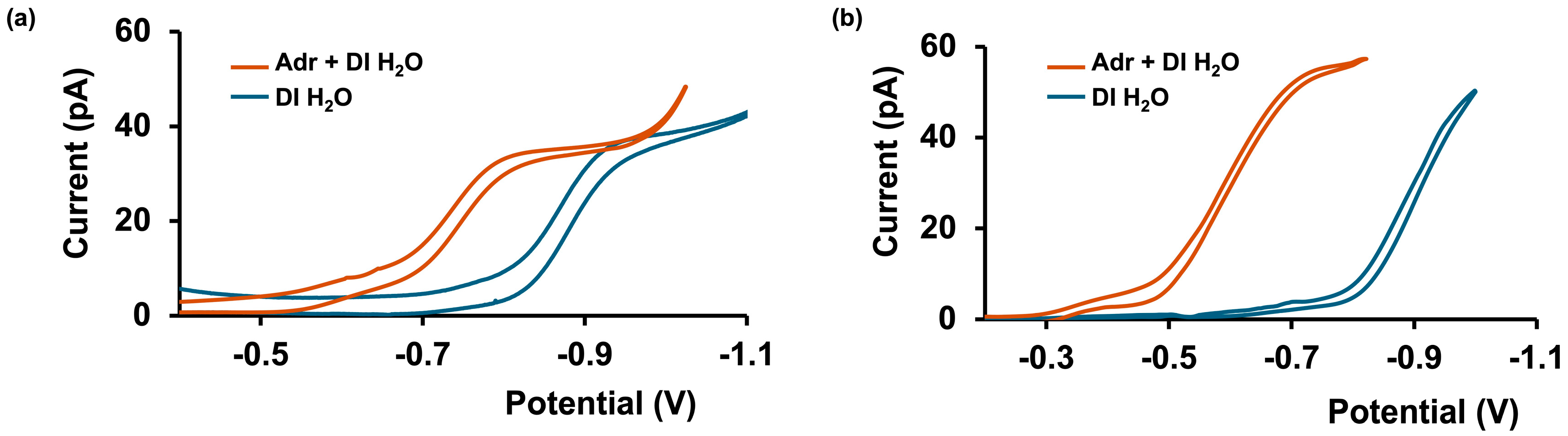
3.1. Adrenaline Transfer Across Aqueous/DCE Interface in Simple Matrices

The ITIES technique has proven to be a valuable electrochemical approach for detecting non-redox active molecules. It works by facilitating the transfer of ions (with a Z+ charge) across two immiscible phases: in this case, an aqueous solution and DCE. The transfer of ions between these phases (described in Equation (1)) is driven by the Gibbs free energy, which can be supplied in the form of a voltage using a potentiostat. By reversing the applied potential, ions are returned across the interface, creating an ion-transfer cycle that can be visualized as a cyclic voltammogram (CV). These CVs offer crucial information, with the half-wave potential (E1/2)—the potential at which half of the maximum current occurs—serving as a fingerprint to distinguish different analytes.
I a q z + I org     z +
I ( a q ) z + + L ( o r g ) [ I L ] ( o r g ) z +
In cases where the energy required for transfer is high, it can be reduced by introducing a suitable ionophore, L, which facilitates the transfer process by interacting selectively with specific ions (Equation (2)). These facilitated ion transfers produce characteristic CVs and steady-state currents at micro- or nano-ITIES, which are often formed at the tips of nanopipets, and the steady-state current (id) can be described using the relationship shown in Equation (3). In this equation, z represents the charge of the analyte; C is its concentration; D is the diffusion coefficient in the originating phase; r is the pipet tip radius; x accounts for the thickness of the pipet’s glass wall [25]; and F is the Faraday constant.
i d = 4 x z F D C r
In this study, the ionophore DB24C8 was chosen to facilitate the transfer of Adr across the aqueous/DCE interface, using a glass-nanopipet electrode. The selection of DB24C8 was based on its proposed ability to form a stable complex with Adr [22], which enhances ion transfer efficiency and improves the sensitivity of the electrochemical response. Unlike previously reported electrodes for Adr detection [31,32,33,34], the fabrication of these glass nanopipets offers several advantages. The process is not only straightforward and rapid but also cost-effective, making it an attractive alternative to more complex or expensive electrode fabrication methods. Furthermore, these nanopipet electrodes are highly versatile and can be tailored for specific applications, making them ideal for the development of portable yet robust electrochemical sensors. The simplicity of the fabrication process, combined with their high performance, paves the way for the deployment of these sensors in field-based or point-of-care settings, where cost and ease of use are critical. This approach represents a significant advancement in the development of practical, real-time detection systems for Adr and potentially other analytes in various environments.
Initially, the experiments were conducted in a simple matrix of 3 M KCl before moving on to more complex matrices. At an Adr concentration of 100 µM, steady-state current and CV data were collected at two distinct E1/2 values in KCl. Increasing the concentration of Adr allowed us to explore the impact of concentration changes on the steady-state current. However, unlike previous studies and the predictions based on Equation (3) [25,35], we observed no corresponding increase in steady-state current with higher Adr concentrations (Figure 1). Instead, CVs exhibited a shift toward more positive potentials as the Adr concentration increased, while the shape of Adr CVs closely resembled that of the background signal. This similarity in shape suggests that the Adr transfer process may not be the sole contributor to the CVs, with the observed signal potentially affected by additional factors within the KCl matrix. Small fluctuations in the steady-state current across the different Adr concentrations (shown in Figure 1) are likely due to minor variations in pipet size between experimental setups.
To determine if the observed peak shift (the difference in the E1/2 values of background and Adr CVs) was linked to KCl or the ionophore DB24C8, we conducted further tests using an alternative ionophore, DB18C6 (Figure S1). Interestingly, similar to our initial experiments with KCl and DB24C8, we observed no increase in steady-state current with increasing Adr concentrations. Instead, the CVs demonstrated a similar E1/2 shift, indicating that the potential shift may be influenced by intrinsic factors rather than the matrix or ionophore composition alone.
Given the potential for multiple factors to influence the observed peak shift, we next examined the role of pH as initial experiments suggested that Adr addition altered the pH of the solution, likely due to the amine groups in Adr. To isolate the pH effect, Adr was tested in deionized (DI) water, eliminating additional positive ions from the matrix. As shown in Figure 2b, the presence of Adr in DI water resulted in a pH of approximately 7.8, accompanied by a potential shift of around 0.3 V. This solution was taken to represent a basic condition. When the pH was reduced to ~4 by adding hydrochloric acid, the potential shift decreased to ~0.1 V, as seen in Figure 2a. This decrease in potential shift at lower pH values is likely due to a lower concentration of the Adr cation, which is less prevalent in acidic conditions. This observation confirms that the potential shift may indeed be due to Adr cation presence and its interaction with the interface, which varies with pH.
Having established that pH influences the potential shift, we next investigated whether ions in the DI water could contribute to the observed CVs and affect the potential shift. To explore this, we prepared Adr solutions using high-purity Millipore water (18.2 MΩ) and initially analyzed them with DB24C8. As shown in Figure 3a, the CVs obtained for Millipore DI water and Adr display distinct shapes and E1/2 values, in contrast to the similar CV profiles observed for background and Adr in regular DI water. This difference suggests that the CVs obtained with regular DI water (Figure 2) are likely influenced by one or more ions in the matrix, or by combined transfer processes involving both Adr and an interference ion.
Interestingly, when Adr solutions prepared in Millipore DI water were tested with DB18C6 as the ionophore, no significant current response was detected. This lack of response can be attributed to two primary factors. First, the stability of the Adr complex when paired with DB18C6 is likely lower than that formed with the larger ring structure of DB24C8. The smaller cavity of DB18C6 may not provide sufficient coordination to stabilize the Adr complex as effectively as DB24C8, which could hinder the transfer of Adr in the presence of DB18C6. This is further supported by Figure 3b, which shows a more stable complex between Adr and DB24C8, likely facilitated by the secondary amine groups proposed by Späth and König [36]. These groups, present in Adr, appear to form a more stable interaction with the larger 24C8 crown ether, enabling better ion transfer and current response. Second, the CVs obtained in regular DI water using both ionophores might indicate the presence of interfering ions in the matrix, whose effects are absent in the purified Millipore DI water. This observation emphasizes the critical role of water purity in reducing interference in ion transfer studies.
While Millipore DI water provides a clearer signal for investigating Adr transfer, it also highlights a limitation. Pure water systems, though useful for isolating specific transfer behavior, oversimplify conditions compared to biological matrices, which are more complex and contain various ions, proteins, and other components that could influence ion-transfer dynamics. Consequently, while Millipore DI water is valuable for establishing baseline behavior, findings from such a pure system may be less applicable to real biological contexts, where additional interferences are likely to affect ion transfer.

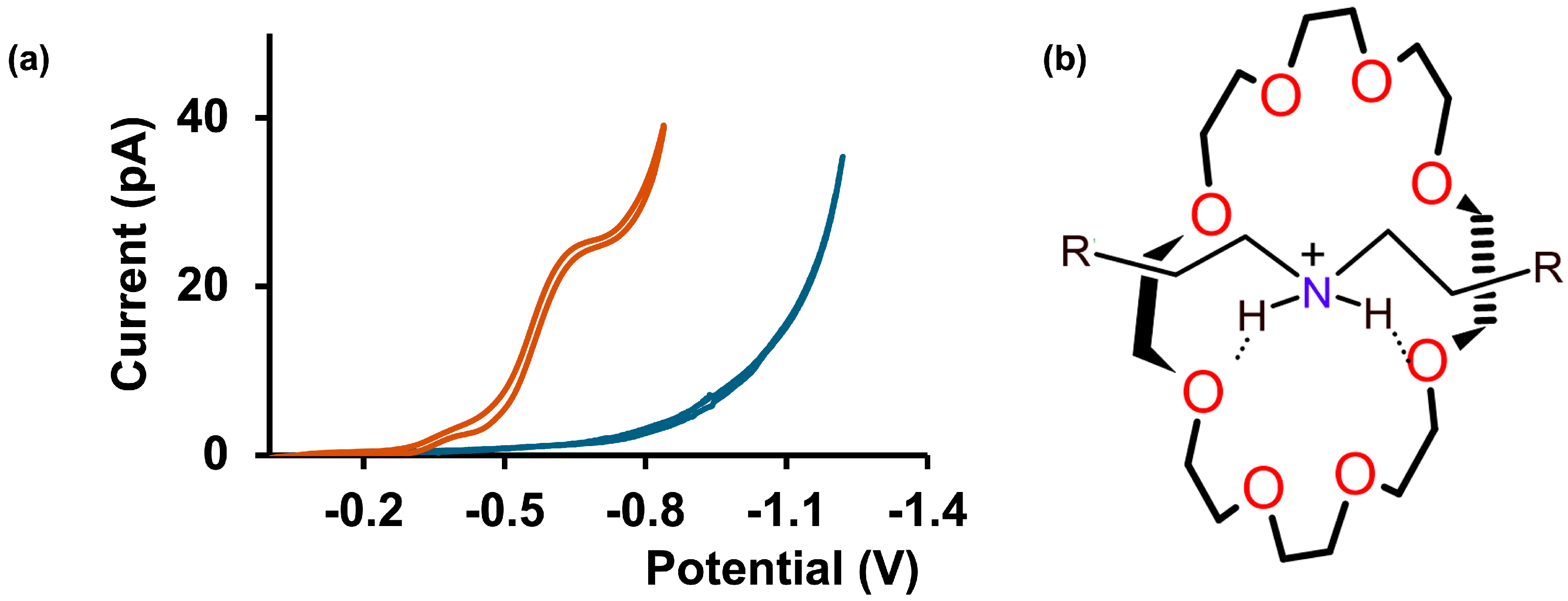
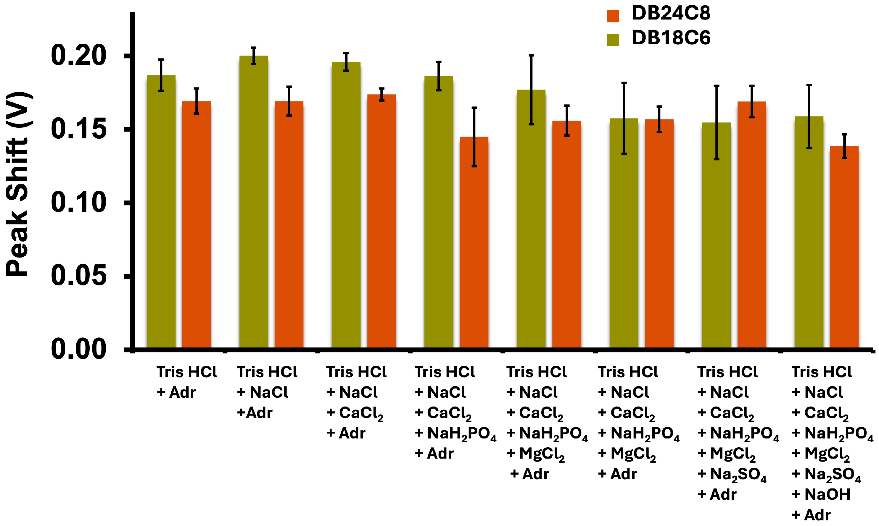
3.2. Adrenaline Transfer in Complex Matrices

To achieve our goal of developing a sensor capable of detecting Adr in cerebellum fluid, we extended our experiments to a more complex matrix: a Tris buffer solution made with Tris hydrochloride (Tris HCl), which closely mimics the composition of artificial cerebrospinal fluid. This buffer system is widely employed in neurochemical studies [37,38,39] due to its similarity to the ionic environment of biological matrices. In addition to Tris HCl, the buffer contains a mixture of salts, including NaCl, KCl, CaCl2, NaH2PO4, MgCl2, and Na2SO4, with the pH carefully adjusted to 7.4 using NaOH. This composition creates an environment that approximates the ionic strength and complexity of natural cerebrospinal fluid, making it an ideal matrix for studying Adr transfer and evaluating the performance of our sensor in physiologically relevant conditions.
Given our earlier observation of potential shifts in CVs for Adr in Millipore DI water, we hypothesized that similar shifts might occur due to the individual components of the Tris buffer solution as ionic strength increased (Table S1). Such shifts could arise from interactions between Adr and the ions in the matrix, potentially influencing the Adr transfer behavior. To systematically evaluate this, we performed a stepwise analysis where we began with Tris HCl alone and sequentially added each salt to the solution. For each matrix composition, Adr was introduced, and CVs were recorded using both ionophores, DB24C8 and DB18C6, to investigate their behavior under these conditions.
The stepwise addition of salts allowed us to isolate the contribution of each ionic species to the observed CV response. After each salt addition, the CVs corresponding to Adr transfer were analyzed, and any shifts in E1/2 values were quantified. Figure 4 illustrates those shifts in CVs upon adding individual salts and Adr to the matrix. We also recorded the pH of each solution as we performed the stepwise addition (Table S1). While shifts in the E1/2 values were observed for both ionophores after the addition of salts, the magnitude of these shifts was relatively minor compared to the pronounced shifts we observed during earlier experiments with varying pH values. These results suggest that while the ionic components of the Tris buffer can influence Adr transfer, their impact is less significant than that of pH changes.
This finding is critical because it validates the use of the Tris buffer for further studies. Despite the minor shifts observed with individual salt additions, the buffer system remains robust and reflective of the physiological environment. Moreover, the Tris buffer’s composition ensures stability and reproducibility for calibration studies, making it a suitable medium for investigating the Adr transfer process and optimizing sensor performance.

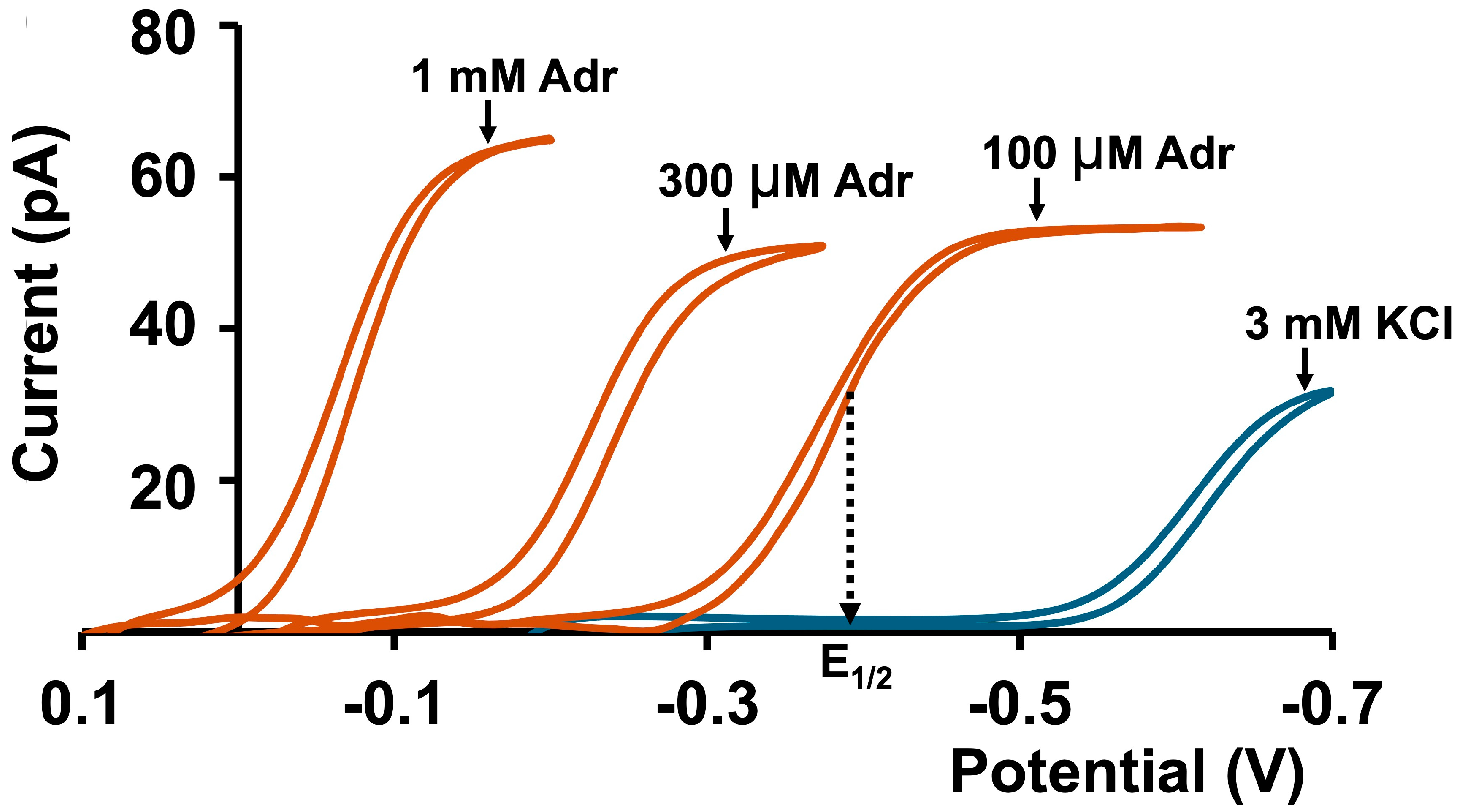
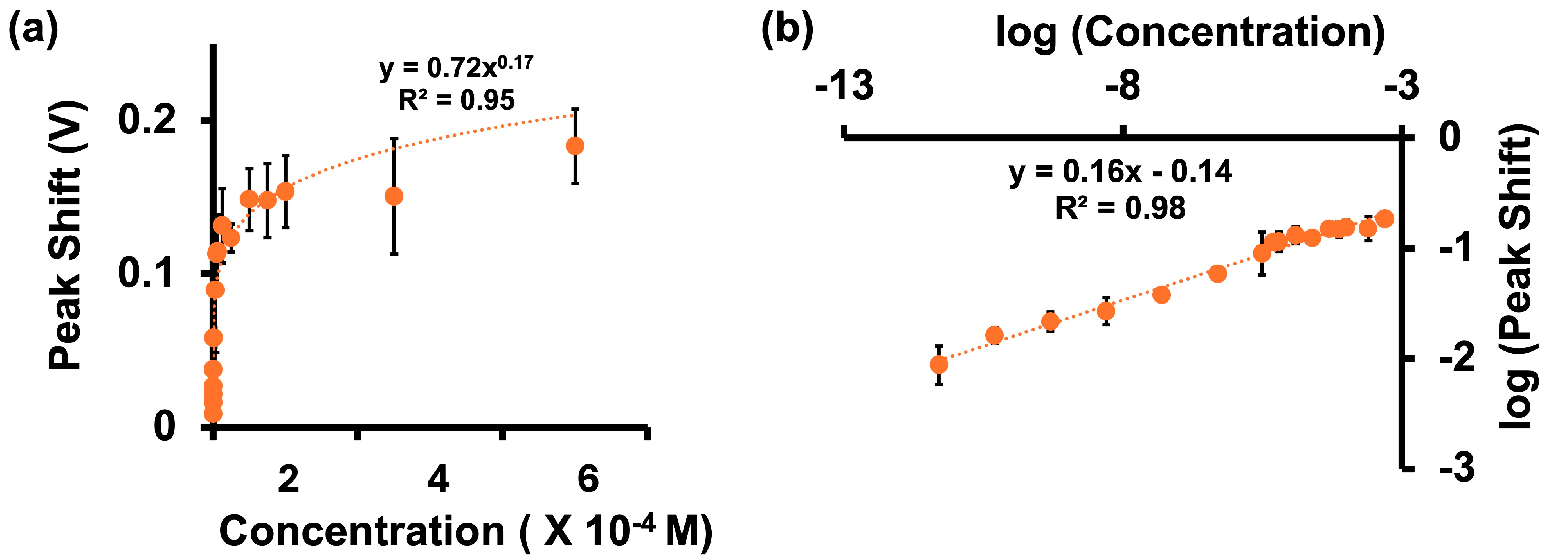
3.3. Calibration Curves for Adr in Tris Buffer

After confirming that the shifts observed in the CVs due to individual components of the Tris buffer matrix were negligible, we proceeded to construct calibration curves for Adr in the complete Tris buffer system. This step was critical for evaluating the performance of our electrochemical sensor in detecting Adr under conditions that mimic the ionic and chemical complexity of artificial cerebrospinal fluid.
To construct the calibration curves, we systematically varied the concentration of Adr in the Tris buffer and recorded CVs for Adr transfer using both ionophores, DB24C8 and DB18C6. The CVs were analyzed to determine the relationship between Adr concentration and the observed electrochemical signal. Interestingly, although there was no significant increase in the current with increasing Adr concentration, a clear and reproducible increase was observed in the shifts between the half-wave potential for Adr and the Tris buffer as the concentration of Adr increased (Figure 5).
With DB24C8, the shift in E1/2 exhibited a well-defined trend, increasing with Adr concentration and eventually plateauing, resembling a classical calibration curve. This behavior highlights the high specificity and sensitivity of DB24C8 toward Adr. A similar trend was observed with DB18C6 (Figure S2), although the regression value of the calibration curve was slightly lower compared to DB24C8. Despite this difference, both ionophores demonstrated their capability to detect Adr reliably. Additionally, the log-transformed data revealed an excellent linear relationship between the potential shift and the Adr concentration (Figure 5b). For DB24C8, the calibration curve yielded an R2 value of 0.98, with a calculated limit of detection (LOD) of 5 pM. Similarly, DB18C6 demonstrated an R2 value of 0.95, with an LOD of 50 pM (Figure S2). These results indicate that our sensor achieves high sensitivity and precision in detecting Adr, even in the complex Tris buffer matrix.
Notably, the LODs reported here are well within the physiologically relevant concentration range of Adr, making these ionophores highly suitable for neurochemical applications. Furthermore, while DB24C8 and DB18C6 are known to facilitate the transfer of other cations such as potassium and calcium, our findings confirm that these transfers do not interfere with Adr detection. This selectivity is a crucial advantage of our approach, ensuring accurate Adr measurement in matrices with competing ions.
The importance of this calibration approach lies in its ability to provide reliable Adr detection in a highly complex environment. Unlike conventional methods, which often rely on direct current measurements, our technique leverages shifts in E1/2 to quantify the Adr concentration. This novel method not only enhances sensitivity but also offers robustness against potential interference in complicated biological matrices. The successful calibration of our sensor in Tris buffer sets the stage for further testing in real biological samples, ultimately advancing its applicability for in vivo and in vitro neurochemical studies.
While our approach is less intricate than redox-based methods, it is best suited for charged molecules. Extending this technique to neutral analytes would require additional mediators or sensor modifications to facilitate their detection. Furthermore, when used with real biological samples such as blood or urine, the interface may become blocked by matrix components, potentially compromising stability over time. To mitigate this, filtration of the sample prior to analysis or the use of larger pipets might be necessary. Additionally, the complexity of biological matrices can cause variations in the observed potential shifts, necessitating the development of new calibration curves for each sample type. However, the challenge of requiring matrix-specific calibration curves can be addressed by integrating artificial intelligence (AI)-based tools. AI algorithms can be trained to analyze and interpret CVs in complex media, eliminating the need for external calibration. This integration could significantly enhance the practicality of the sensor, enabling seamless detection across different sample types without additional recalibration. These considerations are essential for transitioning this promising approach from laboratory testing to real-world applications, particularly for clinical and point-of-care settings.

4. Conclusions

In this study, we developed a novel glass-nanopipet electrode sensor leveraging a non-redox ion-transfer approach via ITIES for the detection of Adr. By utilizing ionophores DB24C8 and DB18C6, we demonstrated that DB24C8 offers superior stability possibly due to its larger ring size and stronger complexation with Adr. Our findings revealed that Adr transfer across the aqueous/DCE interface generates significant peak potential shifts, enabling the construction of a calibration curve with a detection limit as low as 5 pM in Tris buffer, far below physiological Adr concentrations. Notably, the sensor performed robustly across various media, including KCl, DI water, and Tris buffer, and we established that pH variations exerted a more substantial influence on peak potential shifts compared to ionic strength changes. Additionally, our results confirmed that the presence of other cations such as potassium and calcium does not interfere with Adr detection, ensuring reliable performance in complex matrices.
The practical implications of this work are substantial. The sensor is easy to fabricate, requires no sample pretreatment, and relies on peak potential shifts rather than specific ionophores or current measurements, making it user-friendly and accessible for non-experts in clinical or point-of-care settings. Its robustness and ability to function in real time position this approach as a promising tool for in vivo and in vitro neurochemical studies. However, challenges such as matrix-induced blocking of the interface and the need for recalibration for different biological samples highlight areas for future improvement. Integrating AI-based tools could further enhance the sensor’s applicability by eliminating external calibration steps and improving performance in complex biological environments. This work not only advances the understanding of ion transfer for Adr detection but also establishes a framework for future developments in electrochemical sensing. By addressing limitations such as interface stability and expanding the approach to other analytes, this platform can inspire the design of next-generation sensors for diverse applications in health diagnostics and beyond.

Supplementary Materials

The following supporting information can be downloaded at https://www.mdpi.com/article/10.3390/app15020869/s1, Figure S1: Detection of Adr in KCl with DB18C6; Figure S2: Calibration of Adr in Tris Buffer with DB18C6; Table S1: Tris Buffer Analysis; Sample output of MATLAB (R2022b) code used to generate peak shift.

Author Contributions

Conceptualization, R.J.P. and G.K.; methodology, R.J.P. and G.K.; software, R.J.P. and G.K.; validation, R.J.P. and G.K.; formal analysis, R.J.P., P.P. and G.K.; investigation, R.J.P. and G.K.; resources, P.P.; data curation, R.J.P. and G.K.; writing—original draft preparation, R.J.P. and G.K.; writing—review and editing, R.J.P., G.K., N.M., J.L.S. and P.P.; visualization, R.J.P. and G.K.; supervision, P.P.; project administration, P.P.; funding acquisition, P.P. All authors have read and agreed to the published version of the manuscript.

Funding

This research received no external funding.

Institutional Review Board Statement

Not applicable.

Informed Consent Statement

Not applicable.

Data Availability Statement

All the necessary data are available in the article.

Conflicts of Interest

The authors declare no conflicts of interest.

References

  1. Smith, S.M.; Vale, W.W. The Role of the Hypothalamic-Pituitary-Adrenal Axis in Neuroendocrine Responses to Stress. Dialogues Clin. Neurosci. 2006, 8, 383–395. [Google Scholar] [CrossRef] [PubMed]
  2. Berger, I.; Werdermann, M.; Bornstein, S.R.; Steenblock, C. The Adrenal Gland in Stress–Adaptation on a Cellular Level. J. Steroid Biochem. Mol. Biol. 2019, 190, 198–206. [Google Scholar] [CrossRef]
  3. Schmidt, M.V.; Sterlemann, V.; Müller, M.B. Chronic Stress and Individual Vulnerability. Ann. N. Y. Acad. Sci. 2008, 1148, 174–183. [Google Scholar] [CrossRef] [PubMed]
  4. McEwen, B.S. Neurobiological and Systemic Effects of Chronic Stress. Chronic Stress 2017, 1, 2470547017692328. [Google Scholar] [CrossRef]
  5. Bornstein, S.R.; Berger, I.; Scriba, L.; Santambrogio, A.; Steenblock, C. Adrenal Cortex–Medulla Interactions in Adaptation to Stress and Disease. Curr. Opin. Endocr. Metab. Res. 2019, 8, 9–14. [Google Scholar] [CrossRef]
  6. Dayas, C.V.; Buller, K.M.; Crane, J.W.; Xu, Y.; Day, T.A. Stressor Categorization: Acute Physical and Psychological Stressors Elicit Distinctive Recruitment Patterns in the Amygdala and in Medullary Noradrenergic Cell Groups. Eur. J. Neurosci. 2001, 14, 1143–1152. [Google Scholar] [CrossRef] [PubMed]
  7. Aich, P.; Potter, A.A.; Griebel, P.J. Modern Approaches to Understanding Stress and Disease Susceptibility: A Review with Special Emphasis on Respiratory Disease. Int. J. Gen. Med. 2009, 2, 19–32. [Google Scholar] [CrossRef] [PubMed]
  8. Dalal, R.; Grujic, D. Epinephrine; StatPearls Publishing: Treasure Island, FL, USA, 2023. [Google Scholar]
  9. Farrugia, F.; Martikos, G.; Tzanetis, P.; Charalampopoulos, A.; Misiakos, E.; Zavras, N.; Sotiropoulos, D. Pheochromocytoma, Diagnosis and Treatment: Review of the Literature. Endocr. Regul. 2017, 51, 168–181. [Google Scholar] [CrossRef] [PubMed]
  10. ESLER, M. Sympathetic Nervous System and Insulin Resistance: From Obesity to Diabetes. Am. J. Hypertens 2001, 14, S304–S309. [Google Scholar] [CrossRef]
  11. Guest, F.L.; Martins-de-Souza, D.; Rahmoune, H.; Bahn, S.; Guest, P.C. The Effects of Stress on Hypothalamic-Pituitary-Adrenal (HPA) Axis Function in Subjects with Schizophrenia. Arch. Clin. Psychiatry (São Paulo) 2012, 40, 20–27. [Google Scholar] [CrossRef]
  12. Rahi, V.; Kumar, P. Animal Models of Attention-deficit Hyperactivity Disorder (ADHD). Int. J. Dev. Neurosci. 2021, 81, 107–124. [Google Scholar] [CrossRef] [PubMed]
  13. Diego, A.M.G.; Ortega-Cruz, D.; García, A.G. Disruption of Exocytosis in Sympathoadrenal Chromaffin Cells from Mouse Models of Neurodegenerative Diseases. Int. J. Mol. Sci. 2020, 21, 1946. [Google Scholar] [CrossRef] [PubMed]
  14. Ghazal, K.; Brabant, S.; Prie, D.; Piketty, M.-L. Hormone Immunoassay Interference: A 2021 Update. Ann. Lab. Med. 2022, 42, 3–23. [Google Scholar] [CrossRef]
  15. Gorbunova, M.V.; Gutorova, S.V.; Berseneva, D.A.; Apyari, V.V.; Zaitsev, V.D.; Dmitrienko, S.G.; Zolotov, Y.A. Spectroscopic Methods for Determination of Catecholamines: A Mini-Review. Appl. Spectrosc. Rev. 2019, 54, 631–652. [Google Scholar] [CrossRef]
  16. Kirkpatrick, D.; Yang, J.; Trehy, M. Determination of the Enantiomeric Purity of Epinephrine by HPLC with Circular Dichroism Detection. J. Liq. Chromatogr. Relat. Technol. 2017, 40, 556–563. [Google Scholar] [CrossRef]
  17. Bertler, Å.; Carlsson, A.; Rosengren, E. A Method for the Fluorimetric Determination of Adrenaline and Noradrenaline in Tissues. Acta Physiol. Scand. 1958, 44, 273–292. [Google Scholar] [CrossRef] [PubMed]
  18. Saraf, N.; Bosak, A.; Willenberg, A.; Das, S.; Willenberg, B.J.; Seal, S. Colorimetric Detection of Epinephrine Using an Optimized Paper-Based Aptasensor. RSC Adv. 2017, 7, 49133–49143. [Google Scholar] [CrossRef]
  19. Bucher, E.S.; Wightman, R.M. Electrochemical Analysis of Neurotransmitters. Annu. Rev. Anal. Chem. 2015, 8, 239–261. [Google Scholar] [CrossRef] [PubMed]
  20. Anik, Ü. Electrochemical Medical Biosensors for POC Applications. In Medical Biosensors for Point of Care (POC) Applications; Elsevier: Amsterdam, The Netherlands, 2017; pp. 275–292. [Google Scholar]
  21. Zhang, H. Studies of the Electrochemical Behavior of Epinephrine at a Homocysteine Self-Assembled Electrode. Talanta 2002, 56, 1081–1088. [Google Scholar] [CrossRef] [PubMed]
  22. Ribeiro, J.A.; Silva, F.; Pereira, C.M. Electrochemical Sensing of Catecholamines at the Water/1,6-Dichlorohexane Interface. Electroanalysis 2013, 25, 2331–2338. [Google Scholar] [CrossRef]
  23. Ribeiro, J.A.; Miranda, I.M.; Silva, F.; Pereira, C.M. Electrochemical Study of Dopamine and Noradrenaline at the Water/1,6-Dichlorohexane Interface. Phys. Chem. Chem. Phys. 2010, 12, 15190. [Google Scholar] [CrossRef] [PubMed]
  24. Ou, Y.; Buchanan, A.M.; Witt, C.E.; Hashemi, P. Frontiers in Electrochemical Sensors for Neurotransmitter Detection: Towards Measuring Neurotransmitters as Chemical Diagnostics for Brain Disorders. Anal. Methods 2019, 11, 2738–2755. [Google Scholar] [CrossRef]
  25. Ahmed, M.M.N.; Bodowara, F.S.; Zhou, W.; Penteado, J.F.; Smeltz, J.L.; Pathirathna, P. Electrochemical Detection of Cd(II) Ions in Complex Matrices with Nanopipets. RSC Adv. 2022, 12, 1077–1083. [Google Scholar] [CrossRef] [PubMed]
  26. You, Y.; Bai, S.; Ma, Y.; Liu, C.; Wang, L. A Nanopipette Supported Oil/Water Interface Sensor for the Kinetics Analysis and Determination of Phenothiazine Derivatives. Electrochim. Acta 2022, 423, 140568. [Google Scholar] [CrossRef]
  27. Gupta, V.K.; Jain, R.; Radhapyari, K.; Jadon, N.; Agarwal, S. Voltammetric Techniques for the Assay of Pharmaceuticals—A Review. Anal. Biochem. 2011, 408, 179–196. [Google Scholar] [CrossRef]
  28. Pathirathna, P.; Balla, R.J.; Jantz, D.T.; Kurapati, N.; Gramm, E.R.; Leonard, K.C.; Amemiya, S. Probing High Permeability of Nuclear Pore Complexes by Scanning Electrochemical Microscopy: Ca 2+ Effects on Transport Barriers. Anal. Chem. 2019, 91, 5446–5454. [Google Scholar] [CrossRef]
  29. Koryta, J. Electrochemical Polarization Phenomena at the Interface of Two Immiscible Electrolyte Solutions. Electrochim. Acta 1979, 24, 293–300. [Google Scholar] [CrossRef]
  30. Guo, J.; Amemiya, S. Voltammetric Heparin-Selective Electrode Based on Thin Liquid Membrane with Conducting Polymer-Modified Solid Support. Anal. Chem. 2006, 78, 6893–6902. [Google Scholar] [CrossRef]
  31. Sochr, J.; Švorc, Ľ.; Rievaj, M.; Bustin, D. Electrochemical Determination of Adrenaline in Human Urine Using a Boron-Doped Diamond Film Electrode. Diam. Relat. Mater. 2014, 43, 5–11. [Google Scholar] [CrossRef]
  32. Sukanya, S.D.; Swamy, B.E.K.; Shashikumara, J.K.; Sharma, S.C.; Hariprasad, S.A. A Novel, Extreme Low-Cost Poly (Erythrosine) Modified Pencil Graphite Electrode for Determination of Adrenaline. Sci. Rep. 2023, 13, 4523. [Google Scholar] [CrossRef] [PubMed]
  33. Charithra, M.M.; Manjunatha, J.G. Electrochemical Sensing of Adrenaline Using Surface Modified Carbon Nanotube Paste Electrode. Mater. Chem. Phys. 2021, 262, 124293. [Google Scholar] [CrossRef]
  34. Elugoke, S.E.; Fayemi, O.E.; Adekunle, A.S.; Sherif, E.-S.M.; Ebenso, E.E. Electrochemical Sensor for the Detection of Adrenaline at Poly(Crystal Violet) Modified Electrode: Optimization and Voltammetric Studies. Heliyon 2022, 8, e10835. [Google Scholar] [CrossRef] [PubMed]
  35. Ribeiro, J.A.; Pereira, C.M. Applications of Electrochemistry at the ITIES in Drug Discovery and Development—A Review. ChemElectroChem 2024, 11, e202400134. [Google Scholar] [CrossRef]
  36. Späth, A.; König, B. Molecular Recognition of Organic Ammonium Ions in Solution Using Synthetic Receptors. Beilstein J. Org. Chem. 2010, 6, 32. [Google Scholar] [CrossRef]
  37. Vizi, E.S.; Orsó, E.; Osipenko, O.N.; Haskó, G.; Elenkov, I.J. Neurochemical, Electrophysiological and Immunocytochemical Evidence for a Noradrenergic Link between the Sympathetic Nervous System and Thymocytes. Neuroscience 1995, 68, 1263–1276. [Google Scholar] [CrossRef] [PubMed]
  38. Volpicelli-Daley, L.A.; Levey, A. Immunohistochemical Localization of Proteins in the Nervous System. Curr. Protoc. Neurosci. 2003, 25, 1–2. [Google Scholar] [CrossRef] [PubMed]
  39. Magnone, M.C.; Rossolini, G.; Piantanelli, L.; Migani, P. Neurochemical Parameters of the Main Neurotransmission Systems in Aging Mice. Arch. Gerontol. Geriatr. 2000, 30, 269–279. [Google Scholar] [CrossRef] [PubMed]
Figure 1. Representative CVs illustrating the transfer of Adr across the interface between a 3 mM KCl aqueous solution and DCE using a glass nanopipet sensor. CVs were recorded for Adr solutions at concentrations of 100 µM, 300 µM, and 1 mM, prepared in 3 mM KCl (orange), and in the presence of DB24C8 as the facilitating ionophore at a scan rate of 10 mV/s. The CV shown in blue represents the background signal obtained for 3 mM KCl without Adr.
Applsci 15 00869 g001
Figure 2. Representative CVs illustrating Adr (50 µM) transfer across the interface of DI water and DCE in the presence of DB24C8 as the ionophore at (a) pH 4.0 and (b) pH 7.8. The CVs were recorded at a scan rate of 10 mV/s. The blue CVs represent the background CVs obtained in DI water. The peak shift in the acidic medium (pH 4.0) is significantly smaller compared to that observed in the basic medium (pH 7.8), highlighting the influence of pH on the transfer process.
Applsci 15 00869 g002
Figure 3. (a) A representative CV for Adr transfer across the interface between Millipore DI water and DCE in the presence of DB24C8 (orange). The background CV (blue), recorded in Millipore DI water, highlights the distinction between Adr and the background matrix, as evidenced by their differing CV shapes. These CVs were recorded at a scan rate of 10 mV/s. (b) A proposed model for secondary amine complexation with the DB24C8 ether, redrawn from [36].
Applsci 15 00869 g003
Figure 4. Peak shifts observed for Adr transfer in different solution matrices in the presence of DB24C8 (orange) and DB18C6 (yellow) upon stepwise addition of Adr.
Applsci 15 00869 g004
Figure 5. (a) Calibration plot illustrating the relationship between the peak shift and the concentration of Adr (5 pM to 500 µM) in Tris buffer in the presence of DB24C8. (b) Log–log plot of peak shift versus Adr concentration, showing a power-law relationship. This highlights the dynamic range and scalability of the nanopipet sensor. Together, these plots validate the robustness and accuracy of the electrochemical approach for Adr quantification in Tris buffer.
Applsci 15 00869 g005
Disclaimer/Publisher’s Note: The statements, opinions and data contained in all publications are solely those of the individual author(s) and contributor(s) and not of MDPI and/or the editor(s). MDPI and/or the editor(s) disclaim responsibility for any injury to people or property resulting from any ideas, methods, instructions or products referred to in the content.

