Article

Study on Hematological and Biochemical Index of Blood and Vitreous Humor in the Celestial-Eye Goldfish

Fisheries Science Institute, Beijing Academy of Agriculture and Forestry Sciences, Beijing 100068, China
*
Authors to whom correspondence should be addressed.
Appl. Sci. 2025, 15(2), 774; https://doi.org/10.3390/app15020774
Submission received: 8 November 2024 / Revised: 12 January 2025 / Accepted: 13 January 2025 / Published: 14 January 2025

Abstract

:
In this study, we conducted an exploration of blood collection, hematological and biochemical comparative analysis of the unique vitreous humor and blood of the celestial-eye goldfish (CE) (Carassius auratus), and obtained hematological and biochemical indicators for healthy celestial goldfish blood. After exploring the optimal method for collecting blood and ocular fluid, blood and vitreous humor samples were drawn from healthy CE, and an automatic hematology analyzer was used to measure 17 physiological indicators in the blood and vitreous humor. Additionally, 20 biochemical indices were measured by an automatic biochemical analyzer. Experiments were also conductedto evaluate the effect of blood and vitreous humor collection on the subsequent survival rate of the CE. The results showed that the blood of CE exhibited values for 17 hematological indicators, including red blood cells (mean value: 2.19 × 10¹²/L), white blood cells (mean value: 62.21 × 10⁹/L), hemoglobin (mean value:138.25 g/L), and no eosinophils or basophils. We also found that the levels of aspartate amino transferase (AST) and glutamyl transpeptidase (GGT) in the vitreous humor of CE were significantly higher than those in the serum (p < 0.01). No cells were detected in the vitreous humor of CE, and there was no significant difference in survival rate compared to normal goldfish after extracting blood and vitreous humor from CE (p > 0.05). This study provides fundamental data for the cultivation of healthy goldfish, the development of goldfish hematology, and further research on the development and formation of CE’s unique eye traits.

1. Introduction

The goldfish, scientifically known as Carassius auratus (Linnaeus, 1758), belongs to the Percoiformes, Sparidae, Carassius [1]. One particular strain of goldfish, known as the celestial-eye goldfish (CE), is highly valued for its unique eye traits. CE is commonly known as ‘the sky-watching dragon’ or ‘sky-fazing’. The eyeball of CE resembles that of a telescope goldfish (Carassius auratus) and extends beyond the eye socket. When swimming in the water, the pupils of CE are brighter and more powerful than those of the telescope goldfish. The beauty of the fish lies in the fact that its eyes are always facing the sky, giving the impression that it is looking at you from any angle. During the Qing dynasty, CE was a favorite in the imperial palace, as its name originally meant ‘looking upward to the Son of Heaven’ [2]. Despite the popularity of CE, relatively few studies have been conducted on it. Most of the existing research focused on their genetics and body color [3,4,5]. Somereports suggested that CE had a degenerated retina and weak vision [6,7], making them suitable as model animals for studying human eye diseases [8]. Yisai Wang [9] speculated on the activation of a necrotic apoptosis program in the photoreceptor cells and vitreous humor following the detachment of the human retinal layer. This phenomenon was attributed to the close proximity of the vitreous humor to the retina, with retinal vessels playing a nutritive role in the vitreous humor. The research on human eye diseasesnoted that the composition of the vitreous humor was influenced by the physiological and pathological states of both the retina and its blood vessels [10]. CE, with itsunique eye featuring a large malformed vitreous humor, can serve as an excellent model for studying human-related ocular diseases. A comparative study of the physiological and biochemical characteristics of the vitreous humor and blood in CE would offer valuable insights into the pathogenesis of human vitreous humor disorders. Additionally, considering the enlargement of the posterior chamber in the CE eyeball, which is filled with vitreous humor and relies on retinal blood vessels for nourishment, a comparative analysis of the component levels in both the vitreous humor and blood would provide some data for understanding the mechanisms underlying the development of CE’s distinctive eye structure.
Blood is a vital component of the fish organism and immune system [11], and blood indicators have long been emphasized by fish production practitioners and researchers, as they are frequently used to assess the health, nutrition, and environmental adaptation of fish [12]. Additionally, blood indicators are used in water quality monitoring [13]. Our study, similar to some literature describing the blood of goldfish (Carassius auratus) [14,15], may contribute additional reference data to the field of goldfish blood research. This study’s primary objective was to measure and analyze specific hematological and biochemical indicators present in the blood and vitreous humor of CE. The intended outcome was to contribute base data that would enrich the comparative study of goldfish hematology. Moreover, the findings of this study were anticipated to serve as a foundational reference, not only for understanding the intricacies of CE physiology but also for guiding practices in their healthy cultivation. Thus, this study is positioned to contribute meaningfully to the broader understanding of CE in the context of existing goldfish strains.

2. Methods

2.1. Experimental Animal

CE was obtained from the Fisheries Science Institute of the Beijing Academy of Agriculture and Forestry Sciences. The goldfish were bred for 16 months in cement pools with a water depth of 35 cm, and they had an average body length of 12 cm. The specific culture method followed the protocol described by Li R. [4]. In September, a total of 72 individuals were selected for the study based on the criterion that their eyeballs were upward at a standard 90° anglefrom the horizontal plane, as depicted in Figure 1. All experiments were conducted in accordance with the Guidelines for Experimental Animals established by the Ministry of Science and Technology (Beijing, China).

2.2. Experimental Design and Procedures

2.2.1. Preparation of EDTA-K2 and Heparin Sodium Anticoagulant Solution

Ethylenediaminetetraacetic acid dipotassium salt (EDTA-K2), weighing 3 g on an electronic balance, was added to 200 mL of distilled water. The mixture was thoroughly stirred to prepare a 15 g/L solution of EDTA-K2 for future use.
The sodium heparin was weighed and added to distilled water to make 200 mL of a 1% sodium heparin solution for future use.

2.2.2. Effect of Three Methods of Pretreatment Syringe on Blood Sampling of Tail Vein of Goldfish and Preparation of Anticoagulant

To facilitate blood collection, each fish was securely swaddled in sterile medical gauze, and the phlebotomy was swiftly performed to minimize stress and injury. Subsequently, the fish were promptly returned to their aquatic environment, with iodine applied to the puncture site to prevent infection and ensure their survival. We evaluated three syringe pretreatment methods to determine the most effective for tail vein blood sampling in goldfish: utilizing a 1 mL syringe pre-moistened with 1% heparin sodium to draw blood, a 1 mL syringe moistened with the prepared EDTA-K2 solution, and a 1mL syringe with no solutionto draw blood from caudal lateral scale vein of the celestial goldfish to assess the ease of collection and the presence of clotting within the syringe. Each method was labeled as A, B, and C, respectively. This experiment used a total of 15 fish, with each blood collection method employing five fish. The 1mL syringes were with needles, approximately 1.5 cm in length.
The method resulting in the most efficient blood draw and the absence of clots was selected for the formal blood collection from goldfish, and a total of three fish were used. After blood collection, the samples were carefully transferred into a centrifuge tube preloaded with 0.01 mL of prepared EDTA-K2 anticoagulant, mixed thoroughly to produce anticoagulated blood, and then stored at 4 °C until further use.

2.2.3. Preparation of Serum from CE

For serum preparation, we utilized the pretreatment approach from step 2.2.2 that yielded the smoothest, non-coagulated blood. The blood was gently deposited into a centrifuge tube immediately after collection from the quiescent pulse of the caudal fin in three fish. Without anticoagulants, the blood was centrifuged at a speed of 8000 rpm/min (RPM) for 10 min. After centrifugation, the supernatant serum was carefully extracted and refrigerated at 4 °C until further use.

2.2.4. Collection of Vitreous Humor in CE

For vitreous humor collection, the three goldfish were immobilized with only their eyes exposed and wrapped in sterile, damp medical gauze. Using a 2 mL syringe, the needle was carefully inserted into the posterior segment of the eyeball to aspirate the vitreous humor. After removal, the vitreous humor was slowly injected into a centrifuge tube containing 0.01 mL of EDTA-K2 anticoagulant, and the sample was stored at 4 °C. Each sample contained approximately 1–2 mL of vitreous fluid.
The vitreous humor was collected from another three fish and preserved at 4 °C following the above procedure. At the same time, the syringe needle was withdrawn, and the fluid was slowly injected into the centrifuge tube without using an anticoagulant.
Notably, during vitreous humor aspiration, careful needle placement was crucial to avoid the ocular blood vessels, thus preventing blood admixture and minimizing injury to the fish. Controlling the depth of needle insertion was vital to avoid penetrating the eyeball. Through extensive experimentation, the extraction technique was refined to prevent accidental vascular or ocular hemorrhage. Following the procedure, the goldfish were promptly returned to their habitat to ensure their survival.

2.2.5. Analysis of Hematological Indexes of Blood and Vitreous Humor of CE

Approximately 1mL of blood was collected from each fish for analysis. Three replicate samples of whole blood with anticoagulant (prepared as described in Section 2.2.2) and vitreous humor from the same three replicate samples with anticoagulant (prepared as described in Section 2.2.4) were taken. The blood routine indexes were measured using an automatic blood analyzer (Manufacturer: Mindray, Model: BC-5180 CRP, Shenzhen, China). The measured indicators included white blood cell count (WBC), red blood cell count (RBC), hemoglobin (HGB), hematocrit (HCT), mean corpuscular volume (MCV), average erythrocyte hemoglobin content (MCH), mean erythrocyte hemoglobin concentration (MCHC), platelet count (PLT), erythrocyte distribution width (RDW-CV), neutrophil count (NEU), lymphocyte count (LYM), monocyte count (MON), eosinophil count or percentage (EOS), basophil count or percentage (BAS), neutrophil percentage (NEU), lymphocyte percentage (LYM), and monocyte percentage (MON). A total of 17 indicators were measured.

2.2.6. Analysis of Biochemical Indexes of Blood and Vitreous Humor of CE

Both the upper vitreous humor from three fish and CE serum in three fish that were made in 2.2.3 and 2.2.4 in triplicates (three fish, with about 0.5mL from each fish) had their respective biochemical indexes measured by a fully automatic biochemical analyzer (Manufacturer: Hitachi, Model: 3100, Equipment ID: IE-3652, Tokyo, Japan). The 16 measured indicators included alanine aminotransferase (ALT), aspartate amino transferase (AST), total protein (TP), albumin (ALB), alkaline phosphate (ALP), glutamyl transpeptidase (GGT), total bile acid (TBA), glucose (GLU), urea (UREA), uric acid (UA), creatinine (Cre), total cholesterol (CHOL), triglyceride (TG), high-density lipoprotein cholesterol (HDL-C), low-density lipoprotein cholesterol (LDL-C), and cholinesterase (CHE). Meanwhile, the four indexes, including K+, Na+, Cl, and Ca2+, were measured by an electrolyte analyzer (Manufacturer: Mindray, Model: AL9800, Equipment ID: IE-5116 Shenzhen, China).
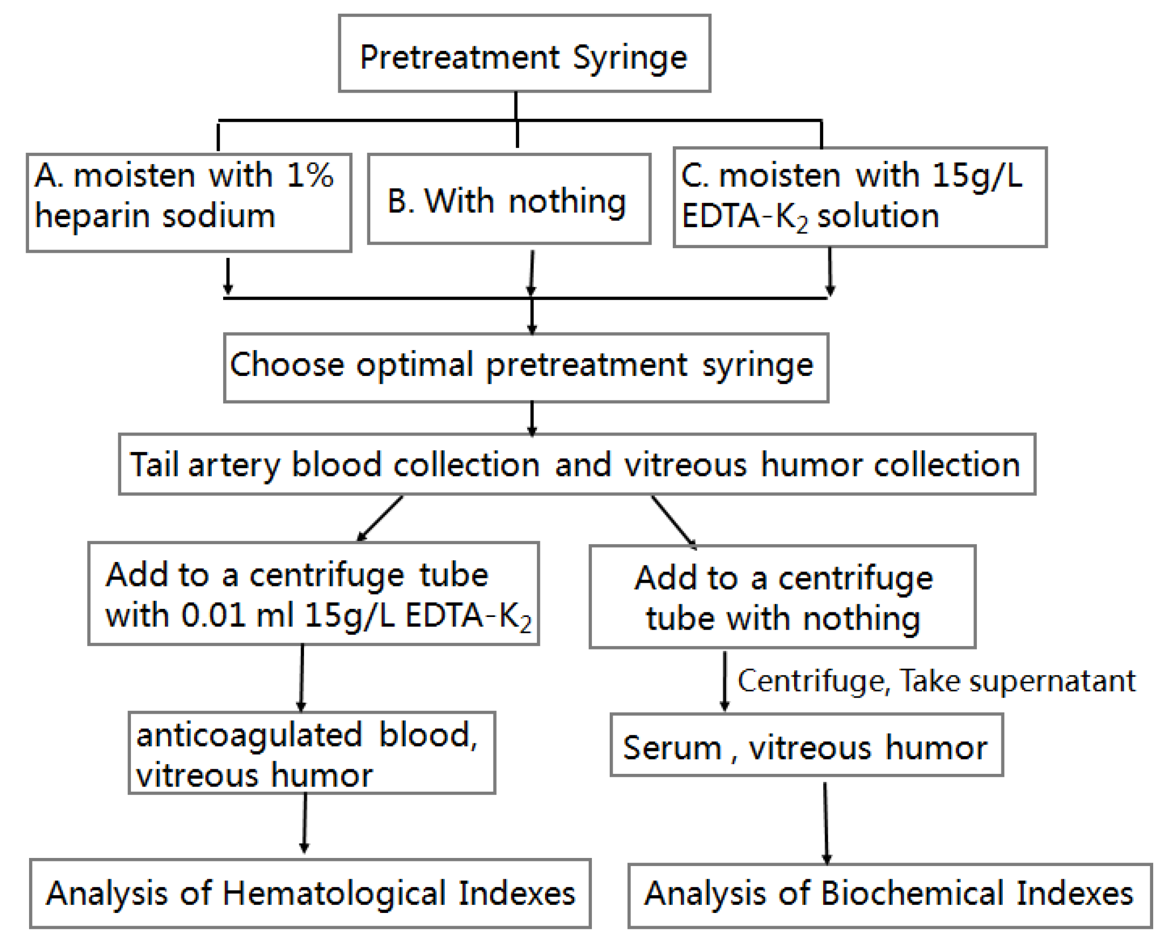
For ease of understanding, the experimental process and procedures for Section 2.2.2, Section 2.2.3, Section 2.2.4, Section 2.2.5 and Section 2.2.6 are shown in Figure 2.

2.2.7. Effect of Collecting Blood and Vitreous Humor on the Follow-Up Survival Rate of CE

To evaluate the influence of blood and vitreous humor collection on CE survival, we designed three experimental groups for observation and comparison, and this experiment used a total of 45 fish, with each group employing 15 fish. For the first experimental condition, we distributed 15 goldfish, post-vitreous humor extraction, into three subgroups with five individuals each. These groups were isolated and maintained in their original habitat for a period of two months. In the second experimental setup, another set of 15 goldfish, following caudal vein phlebotomy, were equally divided into three subgroups. They were secluded in the initial concrete pond habitat for two months. The third experimental assembly comprised 15 goldfish from the original stock, which were randomly allocated into three control subgroups. These fish underwent a mock procedure where they were restrained with fresh, damp gauze for approximately 30 s—the typical duration for blood and vitreous humor extraction—before being released back into their native environment. After two months of separate cultivation, the ocular health of three experimental groups was examined and survival rates were recorded.

2.3. Calculations and Statistical Analysis

The data obtained from the test were statistically presented as mean ± standard error (mean ± SE) using Excel 2007 software. For the success rates of blood collection in the three syringe pretreatment methods and the survival rates of CE under three different treatments, Duncan’s multiple comparison test was used. For the analysis of blood and ocular humor biochemical indicators in the CE, a t-test was applied. The significance of differences was considered at a significance level of p < 0.05, and a highly significant level was defined as p < 0.01.

3. Results

3.1. Comparison of Three Methods of Pretreatment Syringe Collecting Blood from Arterial (Static) Pulse in the Tail of CE

Through this experimentation, it was determined that pre-moistening a 1mL syringe with a 1% sodium heparin solution (A) facilitated the smoothest blood collection from the tail vein, significantly reducing the likelihood of coagulation during the procedureand the success rate of blood collection reached 80%. Alternatives, such as using a 15 g/L EDTA-K2 solution to prepare 1mL syringes (B) or directly drawing blood (C) with them, frequently resulted in needle blockages due to clot formation, thwarting successful blood collection from CE. In this experiment, blood collection attempts using both methods B and C were unsuccessful. Consequently, for optimal results, our methodology incorporated the use of 1% sodium heparin to condition 1mL syringes prior to tail vein blood collection. The statistical results are presented in Table 1.

3.2. Survival Statistics of CE After Blood and Vitreous Humor Collection

Following the collection of blood and vitreous humor samples from CE, these subjects were subsequently cultured for an additional two months. This period allowed for careful monitoring of their health status and survival rate. Statistical analysis of the survival rate revealed no significant difference when compared to a control group of CE (Table 2). However, the appearance symptoms varied among the groups: the CE undergoing blood collection recovered within one month; the CE undergoing vitreous humor collection showed red conjunctival congestion in the eyeballs within one month, and after two months, their eyeballs exhibited white opacity; the control CE remained normal.

3.3. Analysis of Hematological Indexes of Blood and Vitreous Humor of CE

The hematological blood indicators in CE were detected using a fully automated hematology analyzer, which provided detailed data results. The specific results are presented in Table 3 and Table 4. However, data for the indicators measured in the vitreous humor were not available.

3.4. Analysis of Biochemical Indexes of Blood and Vitreous Humor of CE

After analysis using a fully automated biochemical analyzer, 20 biochemical components were detected in the vitreous humor and serum of CE. Among these, 11 indicators showed lower concentrations in the vitreous humor compared to the serum: AST, TP, ALB, GLU, UA, K+, Na+, CHOL, TG, HDL-C, and LDL-C. Conversely, ALT and GGT concentrations were significantly higher in the vitreous humor than in the serum. The concentrations of the remaining indicators did not show significant differences between the vitreous humor and serum. The results of the biochemical indexes in both the blood and vitreous humor of CE are presented in Table 5 and Table 6.

4. Discussion

4.1. Exploration of Blood Collection Methods for CE

The technique of drawing blood from the fish’s artery using a syringe is widely utilized due to its speed and simplicity [16]. However, practical challenges were encountered during its application in this study. Firstly, difficulties arose in locating blood vessels, leading to repeated lancing and causing excessive injury to the fish. Secondly, the needle often slipped out when the fish struggled, making it difficult to draw blood. Lastly, the use of syringes for blood collection was prone to clotting, hindering subsequent experiments. To mitigate these issues, exploratory pre-experiments were conducted before the main experiment. Initially, experimental goldfish were dissected to examine the position and tissue structure of the tail arterial (static) pulse. The investigation revealed that the arterial (static) pulse of the CE tail was distributed in the muscles under the lateral line and on the tailbone. Consequently, the needle was positioned obliquely below the lateral line scales of the goldfish tail stalk. To minimize movement during blood collection, the fish were restrained using medical gauze moistened with water from the goldfish pond. The gauze was wrapped around the fish, covering its eyes and leaving only the tail and caudal fin exposed. Compared to using tap or distilled water, the goldfish’s pool water effectively reduced stress and potential harm to the fish during the blood collection procedure. The osmotic pressure and oxygen content of the goldfish’s pool water were found to be more suitable, providing a familiar and secure environment for the fish. Stabilizing the goldfish in this manner resulted in minimal struggle and minimal interference during blood sampling, in accordance with the recommendation by Haipeng Zhao [16], which suggested wrapping the fish with a wet towel during blood sampling. Finally, to tackle the issue of coagulation associated with syringe blood draws, a comparative experiment was conducted. Through the experiment, it was determined that the most effective approach involved moistening a 1mL syringe with 1% sodium heparin and then drawing blood from the tail vein. Although anesthesia was commonly employed to prevent fish from struggling, there were reports indicating that the use of anesthetics caused rapid changes in certain blood parameters in fish [17]. The goldfish selected for this study were medium-sized individuals, approximately 12 cm in length, and by wrapping them in moist medical gauze, they could be easily controlled without the need for anesthesia. In conclusion, these adjustments successfully addressed the challenges associated with the syringe method, offering a more effective and humane approach to blood sampling from CE.
In hematological research, particularly for the treatment of blood samples or the lining of blood storage containers, anticoagulants are crucial for preventing premature coagulation. The selection of anticoagulants is pivotal, as various agents like heparin, EDTA-Na2, EDTA-K2, and sodium citrate are traditionally employed in fish hematology due to their unique compositions and coagulation inhibition mechanisms [18]. Ping Li’s research highlighted that among these, EDTA-K2 was superior for blood cell analysis [19]. Consequently, for the assessment of celestial goldfish blood hematological parameters in our study, EDTA-K2 was the chosen anticoagulant, integrated into the blood collection process to ensure the integrity of the samples.

4.2. Effect on Subsequent Survival of CE After Extraction of Blood and Vitreous Humor

CE were valued for their esthetic appeal, economic worth, and research potential [8]. Maintaining their survival is beneficial for further research and conservational efforts, as well as their economic exploitation. In our study, CE underwent blood sampling followed by a two-month observational period to assess their health and survival rates. The findings indicated that the sampled fish’s survival rates did not significantly differ from those of the non-sampled group, suggesting that the blood collection method employed did not adversely affect their subsequent survival. Analogous to human medical practices where blood withdrawal is deemed safe within certain limits, ensuring minimal stress and trauma during blood collection in celestial goldfish is crucial for preserving their life post-procedure.
Drawing parallels with human ocular research, where aqueous humor extraction was a routine diagnostic tool [20], our study extended this practice to CE by employing syringe extraction of vitreous humor. Although surgical vitrectomy in humans could lead to postoperative complications such as infectious endophthalmitis [21], our study observed signs of intraocular congestion and cloudiness in fish post-extraction, possibly indicating infection and inflammation. Nevertheless, the comparative survival analysis showed no significant difference, supporting the viability of vitreous humor extraction in CE. These results provided a reassuring basis for advanced research into CE health and disease management.

4.3. Analysis of the Number and Composition of Hemoglobin and Erythrocyte in CE

Red blood cells are pivotal constituents of fish peripheral blood and play an essential role in physiological functions [22]. The red blood cell count in CE was found to be closely aligned with the count reported by Watson for goldfish (Carassius auratus) [14]. As a member of the Sparidae family, Carassius auratus, celestial goldfish exhibited red blood cell numbers in line with those of Xiangjiang wild carp (Cyprinus carpio xiangjiangnenesis) [23] and common carp (Cyprinus carpio) [24]. However, variations were noted compared to other species like carp (Cyprinus carpio) [25], furong crucian carp (Cyprinus capio Furong) [26], crucian carp (Carassius auratus) [27], and koi carp (Cyprinus carpio) [28], suggesting that species and environmental factors influenced red blood cell counts. By comparing with carps, the findings indicated that healthy CE typically possessed red blood cell counts in the order of 1012/L.
Hemoglobin primarily functions in oxygen transport and storage, while also regulating CO levels, pH balance, and immune responses [29]. Hematocrit (HCT) plays a crucial role in O2 transportation in blood [30]. In this study, the hemoglobin concentration in CE surpassed that of carp [25] and Furong crucian carp [26]. The HCT in these goldfish was comparable to that of crucian carp [27]. Goldfish demonstrated remarkable adaptability, including survival in low-O2 environments, which might be attributed to their hemoglobin’s O2-carrying capacity and the erythrocytes’ efficiency in O2 utilization.

4.4. Analysis of the Number and Composition of White Blood Cells andPlatelets in CE

White blood cells serve as a crucial component of the immune defense system in fish, and their variation in peripheral blood is often used to assess the immune system [31]. CE had a higher white blood cell count than that of goldfish [14] and carp [25], though all three species shared a similar order of magnitude (109/L). In contrast, koi carp [28] and Furong crucian carp [26] showed a significantly higher range, both in the 1011/L range. These discrepancies may arise from factors such as varying fish species, different environmental conditions, and measurement methodologies [16].
The primary types of white blood cells in fish include neutrophils, monocytes, lymphocytes, eosinophils, and basophils [16]. Neutrophils, crucial for non-specific immune responses and inflammation [32], were found in CE, comparable to goldfish [14]. Monocytes, also involved in non-specific immunity through phagocytosis of senescent cells and foreign materials [33], were measured at 2.05 × 10⁹/L in CE. Lymphocytes, the most prevalent white blood cell type [16], showed in CE, higher than in goldfish [14]. Eosinophils and basophils, less commonly found in fish peripheral blood, were only observed in certain species, with some fish lacking both [34]. In the study, conducted by Watson L.J. [14], eosinophils and basophils in goldfish were identified at 0.499 × 10⁹/L and 0.04 × 10⁹/L, respectively. However, our investigation did not detect eosinophils and basophils in the peripheral blood of CE. Similar findings were reported in various studies. For example, Qiao Liu [35] observed the absence of these cells in the blood of various ploidy crucian carps (Carassius auratus). H.W. Zhu [36] also did not detect basophils in crucian carp (Carassius auratus), and X.J. Zhou [37] found no eosinophils or basophils in three Pu’an crucian carp varieties (Carassius auratus). Furthermore, H.P. Zhao [16] observed the absence of these cells in several economically significant fish species in the middle and upper reaches of the Yangtze River. Eosinophils and basophils in fish are primarily located in tissues such as the head, kidney, spleen, and liver, rather than in the peripheral blood [16]. This could explain why these cells were not detected in the peripheral blood of CE in our study, possibly due to their low presence or near absence in this blood type.
Platelets, in addition to participating in defense through phagocytosis, arealso required for hemostatic plug or fibrin clot formation [16]. PLT measured in this study for CE was (6.00 ± 0.94) × 10⁹/L, which was significantly lower than the (51.55 ± 3.32) × 10⁹/L reported by Watson L.J. et al. [14]. This discrepancy might be due to differences in the sampling and counting methods. Watson L.J. et al. [14] used tail amputation for blood collection and manual counting under a microscope, while we employed tail artery blood collection and automated counting with a blood analyzer using a transmission microscope. The tail amputation might have caused higher stress in the goldfish, leading to elevated PLT levels. Additionally, under a light microscope, PLT and small lymphocytes appeared similar and were difficult to distinguish, potentially resulting in an overestimation of PLT. In contrast, with a transmission electron microscope, PLT could be distinguished by their boundary, vascular bundles, and vacuoles—structures absent in small lymphocytes [16]—thus reducing the error.

4.5. Analysis of the Characteristics of Blood Biochemical Indexes in CE

The stability of various inorganic components in fish blood plays a pivotal role in maintaining homeostasis, with Na+ and Cl being particularly crucial. These elements constitute over 95% of the blood’s cations and anions, respectively, and are essential for regulating plasma osmolality and maintaining acid–base balance [38]. Hisao Ozaki [30] provided a range ofserum inorganic constituents in bony fishes. In our study, the concentrations of K+, Na+, CL, and Ca2+ in CE blood were within this established range. Organic components of fish blood include triglycerides, proteins, cholesterol, creatinine, and glucose. Blood glucose levels are indicative of a fish’s metabolic rate [39]. Triglycerides play a central role in lipid metabolism, and cholesterol is a precursor for certain hormones and enzymes, vital for normal fish physiology [40]. Creatinine levels are associated with muscle activity [41]. In our analysis, the GLU level in CE was 3.75 mg/L, which was lower compared to grass carp (Mylopharyngodon piceus) [42], carp [25], Furong crucian carp [26], bighead carp (Aristichthys nobilis) [42], and long-snout catfish (Leiocassis longirostris) [42]. However, it was higher than the GLU level measured by Jiaxi Li in goldfish (Carassius auratus) [15]. The levels of TG in CE were higher than those in Furong carp [26] and bighead carp [42], but lower than long-snout catfish [42], grass carp [32], and the goldfish measured by Jiaxi Li [15]. The HDL and LDL levels in CE were similar to those in Furong crucian carp [26]. The levels of TC in CE were higher than those in Furong carp [26], bighead carp [42], and long-snout catfish [42], but lower than the goldfish concentration measured by Jiaxi Li [15]. CE’s Cre level was lower than that of grass carp and bighead carp [42], but higher than long-snout catfish [42] and the goldfish measured by Jiaxi Li [15]. Given that goldfish are ornamental fish with relatively easy access to feed and less physical activity, they exhibit lower GLU and Cre levels. This suggests a reduced energy expenditure and an increase in stored fats, leading to higher TC and TG.
Blood proteins are crucial for assessing the health, nutrition, and disease status of goldfish [43]. As outlined by Hisao Oizaki [30], most bony fish exhibit serum protein concentrations ranging between 30 and 50 g/L. In our study, the TP level in CE was closely aligned with that of allogenic silver crucian carp (Carassius auratus) [44] and fell within the determined range for bony fish [30]. However, it was higher compared to the concentrations in Furong crucian carp [26] and the goldfish measured by Jiaxi Li [15].
The blood’s organic component profile also includes various enzymes such as ALP, ALT, AST, and CHE. Notably, ALP, ALT, and AST are present in both blood and tissues like the liver, heart, gills, kidney, and muscle in fish [45]. In this study, the ALP and AST levels in CE were comparable to those found in Furong carp and grass goldfish (Carassius auratus), respectively [26,46]. The ALT level in CE fell between the levels observed in Furong carp [26] and grass goldfish [46]. The data for this experiment were derived from healthy CE, and their biochemical indicators would serve as foundational blood index parameters for goldfish.

4.6. Biochemical Components in Vitreous Humor of CE

The human eye contains vitreous humor, which is 80% of the eyeball’s volume and comprises 99% water. The residual 1% consists of proteins, inorganic salts, carbohydrates, lactic acid, urea, vitamin C, amino acids, and lipids, among other substances. The structural macromolecules of the vitreous include hyaluronic acid, proteoglycan, collagen, and non-collagen proteins [47]. Vitreous cells are sparse and predominantly located at the vitreous base and cortex [47]. In our study, the vitreous humor was extracted from the central region of CE’s vitreous body, which is typically devoid of cells.
Chen C.H. [48] indicated that while the vitreous humor might contain most serum components, their concentrations were usually lower than those in the serum. Our findings confirmed this, showing that the concentrations of 11 indices in the vitreous humor of CE were lower than those in the serum, in agreement with the research by Chen C.H. [48]. However, there was no significant difference in the levels of ALP, TBA, urea, Cl, Ca2+, Cre, and CHE between the vitreous humor and the serum. Notably, the serum levels of ALT and GGT were considerably lower than in the vitreous humor. The synthesis of alanine is linked to ALT activity [49], while GGT was associated with the transmembrane transport of amino acids and the regulation of glutathione (GSH) levels [50]. These disparities might be indicative of abnormal metabolism in the vitreous humor of CE.
A trace amount of ALB present in the vitreous humor might serve as an indicator of the integrity of the blood–ocular barrier [51]. Research by Yew [51] suggested that in celestial goldfish, ALB levels in the vitreous humor rose in correlation with ocular enlargement in larger fish. The largest group of telescope goldfish demonstrated an ALB concentration of 2.41 g/L in the vitreous humor, only slightly lower than that found in our CE, which was 2.50 g/L.
Both celestial and telescope goldfish had suffered from visual function deficit, but the eye diseases were different between them. CE, along with telescope goldfish, exhibited eye abnormalities; however, CE development was often accompanied by varying degrees of retinal degeneration [6], leading to impaired vision and sensitivity to light. In contrast, despite the protruding eyes of telescope goldfish, they predominantly suffered from severe myopia without retinal degeneration [52]. Through comparative analysis, we noted that the ALB concentration in the vitreous humor of our CE was higher than that in David’s telescope goldfish [51]. This could imply that CE might experience more pronounced alterations in the blood–retinal barrier compared to telescope goldfish.
In CE, levels of GLU, Cre, and UREA were found to be lower in the vitreous humor compared to those in deceased humans [51]. This difference might stem from increased membrane permeability of the blood–eye barrier postmortem, allowing blood constituents to permeate into the vitreous humor. Notably, the vitreous humor samples from the CE in this study were collected in vivo. Furthermore, no significant variations were observed in the concentrations of Cre and UREA between the vitreous humor and blood of CE. This contrasted with findings in cattle [53] and humans [51], where Cre and UREA levels in vitreous humor were lower than in blood, suggesting a unique permeability of the blood–retinal barrier in CE eyes regarding these substances.
Regarding Ca2+ content, CE’s vitreous humor exhibited levels similar to those in humans (2.45 mmol/L) [54]. However, the concentrations of Ca2+ and Cl in the vitreous humor and blood of CE did not display noticeable differences, unlike the disparity observed in humans [51]. This implies that the mechanisms of ion transport and metabolism in the vitreous humor of CE might diverge from those in the human vitreous body.

4.7. The Limiting Factors in the Study

Our current experimental study also had some limiting factors. We did not establish reference intervals for the various indicators in CE here, because creating reference intervals required a larger sample size and more complex statistical analysis. Due to limitations in experimental conditions and budget, we were currently unable to conduct experiments with a sufficiently large sample size. However, we have provided a goodbase for further in-depth research on CE.

5. Conclusions

  • There was no significant difference in survival rates between celestial goldfish subjected to blood and vitreous humor extraction and those not involved in the experiment (p > 0.05). This provided a data reference for evaluating whether the experimental samples could be used for subsequent research and whether their ornamental and economic value was affected by the sampling process;
  • In the celestial-eye goldfish tested, the mean value of the red blood cell count was 2.19 × 10¹²/L, the mean value of the white blood cell count was 62.21 × 10⁹/L, and the mean value of hemoglobin concentration was 138.25 g/L. Neither eosinophils nor basophils were detected in the blood samples;
  • The analysis of biochemical markers in the blood and vitreous humor of CE revealed that the levels of ALP, TBA, UREA, Cl, Ca2⁺, Cre, and CHE were lower in the vitreous humor compared to serum, in contrast to the increased levels of ALT and GGT. Seven indicators showed no significant difference between the vitreous humor and serum.
The data were collected from constitutionally healthy CE, and the mean values of hematological and biochemical indicators could provide a good basis for understanding the blood index parameters of this species. The study of these indicators in the blood and vitreous humor of CE could offer a bit of reference material for further insights into their biological characteristics, disease prevention, control strategies, and the unique eye formation of CE.

Author Contributions

Conceptualization and supervision, W.L. and X.Z.; data analysis: R.L. and Y.S.; writing—original draft preparation, review, and editing, R.L. All authors have read and agreed to the published version of the manuscript.

Funding

This research was funded by the National Natural Science Foundation of China, Youth Fund Project grant number 32302996; Beijing Natural Science Foundation for Young Science grant number 6234045; BAAF Innovation Capacity Building Foundation of Research on Innovation and Biotechnology of Fish Germplasm Resources grant number KJCX20230122 and National Freshwater Aquatic Germplasm Resource Bank grant number FGRC: 18537.

Institutional Review Board Statement

This study was reported in accordance with the ARRIVE guidelines. All experiments were conducted according to the Guidelines for Experimental Animals established by the Ministry of Science and Technology (Beijing, China). Animal experiments were approved by The Science Ethics Review Committee of the Beijing Academy of Agriculture and Forestry Sciences (Beijing, China) (approval number: Baafs20240901, approval date: 1 September 2024).

Informed Consent Statement

Not applicable.

Data Availability Statement

The original contributions presented in this study are included in the article. Further inquiries can be directed to the corresponding author.

Conflicts of Interest

The authors declare no conflicts of interest.

References

  1. Wang, C. Studies on the Karyotype of Goldfish (Carassius auratus) I. A Comparative Study of the Chromosomes in Crucian and Red Dragon-eye Goldfish. J. Genet. Genom. 1982, 9, 238–242. [Google Scholar]
  2. Wang, H. Atlas of Chinese Goldfish; Culture and Art Publishing House: Beijing, China, 2000. [Google Scholar]
  3. Li, R.; Sun, Y.; Cui, R.; Zhang, X. Comprehensive Transcriptome Analysis of Different Skin Colors to Evaluate Genes Related to the Production of Pigment in Celestial Goldfish. Biology 2023, 12, 7. [Google Scholar] [CrossRef] [PubMed]
  4. Li, R.N.; Sun, Y.S.; Tian, Z.; Wang, S.S.; Zhang, X. Effects of genetic and environmental factors on the celestial eye in Carassius auratus. Freshw. Fish. 2022, 52, 66–73. [Google Scholar]
  5. Li, R.N.; Sun, Y.S.; Tian, Z.; Wang, S.S.; Zhang, X. Comparative transcriptome analysis of body color change in red celestial goldfish Carassius auratus. J. Dalian Ocean. Univ. 2022, 37, 191–201. [Google Scholar]
  6. Sakaue, H.; Negi, A.; Matsumura, M.; Ohkuma, M.; Honda, Y. The developmental changes of ERGs on spontaneous retinal degeneration of Celestial goldfish. Adv. Ophthalmol. 1988, 70, 97–101. [Google Scholar] [CrossRef] [PubMed]
  7. Matsumura, M.; Ohkuma, M.; Honda, Y. Retinal degeneration in celestial goldfish. Developmental study. Ophthalmic Res. 1982, 14, 344–353. [Google Scholar] [CrossRef] [PubMed]
  8. Yoshihiro, O.; Tetsuo, K. Goldfish: An old and new model system to study vertebrate development, evolution and human disease. J. Biochem. 2019, 165, 209–218. [Google Scholar]
  9. Wang, Y. Expression and Significance of Necroptotic Proteins in Vitreous After Rhegmatogenous Retinal Detachment. Ph.D. Thesis, Anhui Medical University, Hefei, China, 2021. [Google Scholar]
  10. Rocha, A.S.; Santos, F.M.; Monteiro, J.P.; Castro-De-Sousa, J.P.; Queiroz, J.A.; Tomaz, C.T.; Passarinha, L.A. Trends in proteomic analysis of human vitreous humor samples. Electrophoresis 2014, 35, 2495–2508. [Google Scholar] [CrossRef]
  11. Zhou, Y.; Xing, Y.; Feng, Q. Reserch advance in haemocytes of fisher. J. Hainan Univ. Nat. Sci. Ed. 2003, 21, 171–176. [Google Scholar]
  12. Bahmani, M.; Oryan, S.; Pourkazemi, M.; Vosoughi, G. Effects of Ecophysiological Stress on Cellular Immunity System of Persian Sturgeon Acipenser persicus. In Proceedings of the 14th Iranian Congress of Physiology and Pharmacology, Tehran, Iran, 16–20 May 1999; pp. 16–20. [Google Scholar]
  13. Ren, P.; Zhang, Y.; Geng, G.; Qi, Y. Changes in morphology and quantity of peripheral blood cells in Carassius auratus collected from polluted water area. Chin. J. Zool. 2008, 43, 37–42. [Google Scholar]
  14. Watson, L.J.; Shechmeister, I.L.; Jackson, L.L. The hematology of goldfish Carassius auratus. Cytologia 1963, 28, 118–130. [Google Scholar] [CrossRef]
  15. Li, J. Effects of Different Stocking Density on Goldfish Growth, Physiological Parameters and Expression of Appetite Regulatory Factors. Ph.D. Thesis, Foshan University, Foshan, China, 2020. [Google Scholar]
  16. Zhao, H. Studies on Haematology of Several Economic Fish in Middle and Upper Reaches of Yangtze River. Ph.D. Thesis, Southwest Unversity, Chongqing, China, 2008. [Google Scholar]
  17. Mcknight, I.M. A Hematological study on the mountain whitefish, Prosopium williamsoni. South. Fish Reb. 1966, 23, 45–64. [Google Scholar] [CrossRef]
  18. Ning, D.; Li, X.; Wu, K. Influence of anticoagulant selection on whole blood cell analysis. Acta Med. Sin. 2004, 17, 550–551. [Google Scholar]
  19. Li, P. The value of heparin, sodium citrate and EDTA-K2 in blood cell analysis. Med. Lab. Sci. Clin. 2021, 32, 63–65. [Google Scholar]
  20. Zhang, T.; Li, K.; Li, L.; Song, T.; Ren, Z.; Fu, C.; Lan, Y.; Jiang, L. Proteome Characterization of Primary Angle-Closure Glaucoma Aqueous Humor. J. Chengdu Med. Coll. 2023, 18, 180–186. [Google Scholar]
  21. Huang, S.; Lu, S.; Lin, M.; Wu, S. Factors Associated with Postvitrectomy Endophthalmitis. Chin. J. Nosocomiol. 2009, 4, 401–403. [Google Scholar]
  22. Gao, Z.; Wang, W. Research progress on peripheral blood red blood cells of fish. Reserv. Fish. 2008, 28, 1–3. [Google Scholar]
  23. Wang, Y.; Liu, S.; Wang, G. Comparative Hematological Studies in Cyprinus carpio Xiangyunnensis and Cyprinus carpio Xiangjiangnensis. J. Nat. Sci. Hunan Norm. Univ. 1988, 1, 71–75. [Google Scholar]
  24. Mi, R. Determination of hematological indexes of grass carp, carp and abalone. Freshw. Fish. 1982, 8, 10–16. [Google Scholar]
  25. Cheng, C.; Ming, Q. Study on the blood physiobiochemic and hemorheologic properties of carp in Weishan Lake. Jiangsu Agric. Sci. 2005, 5, 95–97. [Google Scholar]
  26. Cheng, X.F.; Jiang, G.M.; Xiang, J.; Song, R.; Li, S.M.; Wu, Y.A.; Liu, L.; Wang, Z.M. Effects of dietary fishmeal repiacement with meat and bone meal on the growth performance, blood physiological and biochemical indices, muscle chemical composition and texture characteristics in juvenile furong crucian carp (fueong♀ × red crucian carp). Acta Hydrobiol. Sin. 2022, 44, 85–94. [Google Scholar]
  27. Lin, G. Study on Carassius auratus blood. Curr. Zool. 1979, 25, 210–219. [Google Scholar]
  28. Wang, D.; Deng, S.; Zou, L.; Li, S.; Jiang, G. Studied on the hematological indices of different Cyprinus carpio Koi. J. Aquac. 2016, 37, 1–5. [Google Scholar] [CrossRef]
  29. Yu, L.N.; Yang, D.; Liu, H.Y.; Zhang, F.R. Correlation between Hemoglobin and Asphyxiation Point in Twelve Species of Freshwater Fish. Chin. J. Zool. 2017, 52, 478–484. [Google Scholar]
  30. Oizaki, H. Fish Hematology and Circulatory Physiology; Shanghai Scientific and Technical Publishers: Shanghai, China, 1982; pp. 6–96. [Google Scholar]
  31. Tierney, K.B.; Farrell, A.P.; Kennedy, C.J. The differential leucocyte landscape of four teleosts: Juvenile Oncorhynchus kisutch, Clupea pallasi, Culaea inconstans and Pimephales promelas. J. Fish Biol. 2004, 65, 906–919. [Google Scholar] [CrossRef]
  32. Chen, F. Hematopathological studies in proliferative kidney disease of red grouper. Trop. Oceanol. 1997, 16, 49–53. [Google Scholar]
  33. Zhang, Y.; Sun, B.; Nie, P. Immune tissues and cells of fish: A review. Acta Hydrobiol. Sin. 2000, 24, 648–654. [Google Scholar]
  34. Chen, G.; Zhou, H.; Ye, F.; Wu, Z. A hematological study and observation on development of blood cells in American red fish (Sciaenops ocellatus). J. Trop. Oceanogr. 2006, 25, 59–66. [Google Scholar]
  35. Liu, Q.; Wang, Y.Q.; Liu, S.J.; Guo, X.H.; Luo, K.K.; Zhang, C.; Liu, Y. Comparison of blood and blood cells in crucian carp with different ploidy. Adv. Nat. Sci. 2004, 14, 1111–1117. [Google Scholar]
  36. Zhu, H.; Wang, H.; Qin, G. Studies on the blood cell morphology of crucian carp (Carassius auratus L.). Zool. Res. 1985, 6, 147–153. [Google Scholar]
  37. Zhou, X.; An, M.; Huang, L.; Hu, F.; Shao, Y. Hematologic Studies of Three Types of Carassius auratus. Guizhou Agric. Sci. 2012, 40, 133–138. [Google Scholar]
  38. Zhao, H.; Zhao, H.; Jin, L.; Zhang, Y. Microscopic structures of peripheral hematocytes in sinilabeo rendahli. Fish. Sci. 2005, 24, 24–27. [Google Scholar]
  39. Chen, X. The Fishes Blood. J. Chongqing Teach. Coll. 2000, 19, 70–73. [Google Scholar]
  40. Cheng, C. The Study of Blood Physiological and Biochemeical Parameters of Monopterus Albus. Ph.D. Thesis, Jiangxi Agricultural University, Nanchang, China, 2014. [Google Scholar]
  41. Han, N.; Shi, C. The Application of Blood Indexes in Ichthyological Research. J. Anhui Agric. Sci. 2010, 38, 18877–18878. [Google Scholar]
  42. Dong, S.; Miao, J.; Zhao, K. Study on Physiological and Biochemical Index of Blood in Leiocassis longirostri. Hubei Agric. Sci. 2016, 55, 3690–3693. [Google Scholar]
  43. Qian, Y.; Chen, H.; Sun, J. Effects of starvation on hematological and blood biochemical indices in cultured Lateolabrax japonicus. J. Fish. Sci. China 2002, 2, 133–137. [Google Scholar]
  44. Gu, L.F.; Hou, Y.Q.; Ding, B.Y. Effects of Several Plant Extracts on Growth Performance and Blood Biochemical Indices in Carassius auratus gibelio. Freshw. Fish. 2008, 28, 23–26. [Google Scholar]
  45. Asadi, F.; Masoudifard, M.; Vajhi, A.; Lee, K.; Pourkabir, M.; Khazraeinia, P. Serum biochemical parameters of Acipenser persicus. Fish Physiol. Biochem. 2006, 32, 43–47. [Google Scholar] [CrossRef]
  46. Rahimikia, E. Analysis of antioxidants and serum biochemical responses in goldfish under nickel exposure by sub-chronic test. J. Appl. Anim. Res. 2017, 45, 320–325. [Google Scholar] [CrossRef]
  47. Fan, H. Study on the Relationship Between Postmortem change of Chemical Composition in Vitreous Humor and Time of Death. Ph.D. Thesis, Sichuan University, Chengdu, China, 2005. [Google Scholar]
  48. Chen, C.H.; Chen, S.C. Studies on soluble proteins of vitreous in experimental animals. Exp. Eye Res. 1981, 32, 381. [Google Scholar] [CrossRef]
  49. Li, Y.; Wu, P.; Wang, F.; Yan, H.; Xu, C.; Wang, Y. Effects of methoxyfenozide on transaminase and alanine aminotransferase in the silkworm. Guangdong Seric. 2019, 53, 1–7. [Google Scholar]
  50. Liu, R. Research progress of gamma-glutamyltranspeptidase activity in mammals. Shanghai J. Prev. Med. 2002, 14, 24–25. [Google Scholar]
  51. Yew, D.T.; Lai, H.W.; Zhou, L.; Lam, K.W. Chromatographic identification of a biochemical alteration in the aqueous humor of megalophthalmic Black Moor goldfish. J. Chromatogr. B Biomed. Sci. Appl. 2001, 751, 349–355. [Google Scholar] [CrossRef] [PubMed]
  52. Easter, S.S.; Hitchcock, P.F. The myopic eye of the Black Moor goldfish. Vis. Res. 1986, 26, 1831–1833. [Google Scholar] [CrossRef] [PubMed]
  53. Lane, V.M.; Lincoln, S.D. Changes in urea nitrogen and creatinine concentrations in the vitreous humor of cattle after death. Am. J. Vet. Res. 1985, 46, 1550–1552. [Google Scholar]
  54. Coe, J.I. postmortem chemistries of human Vitreous humor. Am. J. Clin. Pathol. 1969, 51, 741–750. [Google Scholar] [CrossRef] [PubMed]
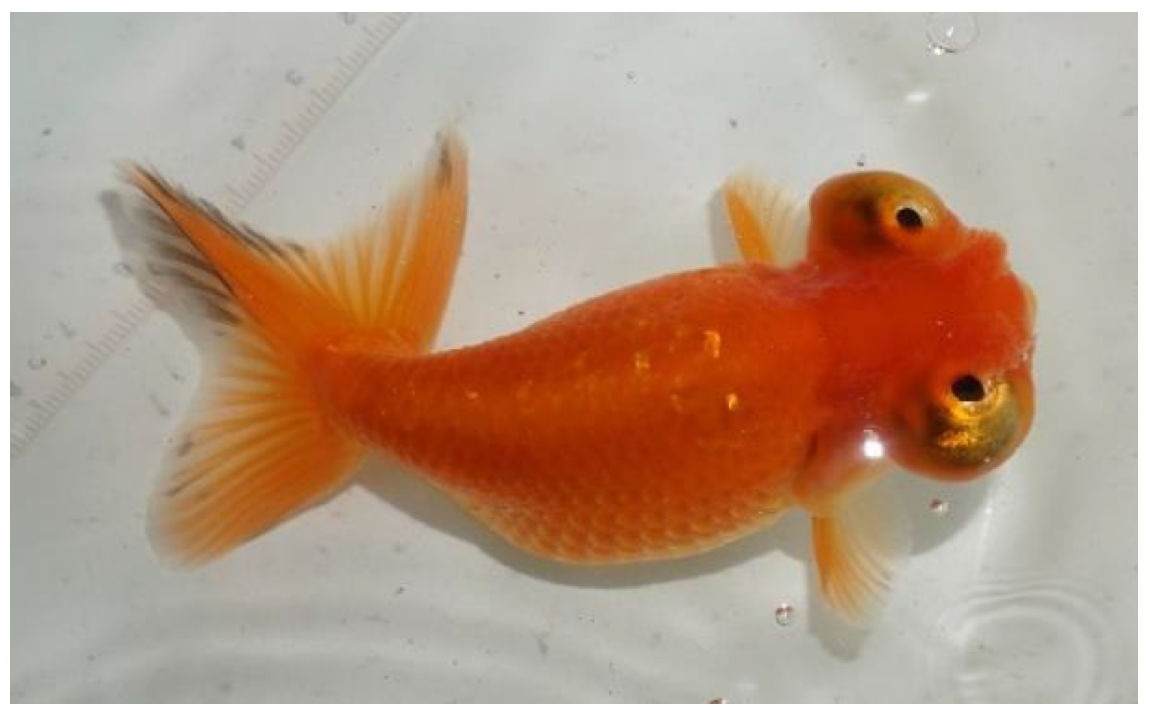
Figure 1. The celestial-eye goldfish (cultured and photographed by the Institute of Fisheries Sciences, Beijing Academy of Agriculture and Forestry Sciences).
Applsci 15 00774 g001
Figure 2. Section 2.2.2, Section 2.2.3, Section 2.2.4, Section 2.2.5 and Section 2.2.6 experimental procedure diagram.
Applsci 15 00774 g002
Table 1. Statistics on the success rate of blood drawing in three syringe pretreatment methods.
Syringe Pretreatment MethodsSuccess Rate of Blood Collection
A80.00% A
B0.00% B
C0.00% B
Note: Significant differences (p < 0.01) are highly evident when the shoulder labels have different letters, while no significant differences (p > 0.05) are observed when the shoulder labels are the same.
Table 2. Statistics on the survival rate of CE in three groups.
GroupsSurvival Rate%
Blood collection groups80 ± 5.44 a
Vitreous humor collection groups90 ± 5.44 a
Normal CE groups90 ± 5.44 a
Note: Significant differences (p < 0.05) are evident when the shoulder labels have different letters, while no significant differences (p > 0.05) are observed when the shoulder labels are the same.
Table 3. Results of the erythrogram indicators in CE.
SampleWBC 109/LRBC 1012/LHGB g/LHCT %MCV flMCH PgMCHC g/LRDW-CV %
blood62.21 ± 8.542.19 ± 0.09138.25 ± 14.7441.70 ± 1.58187.10 ± 1.4475.75 ± 1.16410.50 ± 2.0521.70 ± 0.95
Table 4. Results of thrombogram and the leukogram indicators in CE.
SamplePLT 109/LNEU 109/LLYM 109/LMON 109/LEOS 109/LBAS 109/LNEU %LYM %MON %
blood6.00 ± 0.941.37 ± 0.1059.53 ± 14.322.05 ± 0.19002.05 ± 0.0394.75 ± 0.262.50 ± 0.28
Table 5. Results of blood and ocular humor biochemical indicators in CE.
SampleTP g/LALB g/LALT U/LAST U/LALP U/LGGT U/LTBA μmol/LGLU mmol/LUREA μmol/L
serum31.63 ± 0.52 **14.93 ± 0.07 **26.33 ± 1.09235.33 ± 3.81 **30.67 ± 1.191.76 ± 0.320.70 ± 0.003.75 ± 0.16 **2.34 ± 0.08
vitreous humor2.05 ± 0.052.50 ± 0.0387.00 ± 4.03 **114.00 ± 1.0926.00 ± 0.5414.89 ± 0.47 **0.40 ± 0.110.70 ± 0.021.86 ± 0.02
Note: ** indicates a very significant difference (p < 0.01).
Table 6. Results of electrolytes and lipid status indices of blood and ocular humor in CE.
SampleK+ mmol/LNa+ mmol/LClmmol/LCa2+ mmol/LCre μmol/LCHOLmmol/LTGmmol/LHDL-C mmol/LLDL-C mmol/L CHE mmol/L
serum5.17 ± 0.07 **149.40 ± 0.95 **162.47 ± 19.152.62 ± 0.0729.67 ± 0.726.37 ± 0.16 **2.42 ± 0.04 **2.37 ± 0.13 **1.41 ± 0.01 **345.00 ± 11.43
vitreous humor2.24 ± 0.05132.85 ± 0.08151.20 ± 15.052.61 ± 0.0530.00 ± 2.051.13 ± 0.050.10 ± 0.020.39 ± 0.020.17 ± 0.00323.00 ± 8.50
Note: ** indicates a very significant difference (p < 0.01).
Disclaimer/Publisher’s Note: The statements, opinions and data contained in all publications are solely those of the individual author(s) and contributor(s) and not of MDPI and/or the editor(s). MDPI and/or the editor(s) disclaim responsibility for any injury to people or property resulting from any ideas, methods, instructions or products referred to in the content.