Article

Production, Characterization, and In Vitro Antifungal Evaluation of Itraconazole-Loaded Fibrous Sheets Prepared by Electrospinning with a Factorial Design

by
Emőke-Margit Rédai
1,†,
Orsolya Péterfi
1,†,
Attila Levente Gergely
2,*,
Enikő Barabás
3,
Andrada Pintea
1,
Paula Antonoaea
1,
Robert-Alexandru Vlad
1,
Nicoleta Todoran
1,
Cornelia-Titiana Cotoi
1,
Adriana Ciurba
1 and
Emese Sipos
4
1
Department of Pharmaceutical Technology and Cosmetology, Faculty of Pharmacy, George Emil Palade University of Medicine, Pharmacy, Science, and Technology of Targu Mures, 540142 Targu Mures, Romania
2
Department of Mechanical Engineering, Faculty of Technical and Human Sciences, Sapientia Hungarian University of Transylvania, Calea Sighisoarei nr. 2., 540485 Targu Mures, Romania
3
Department of Cellular Biology and Microbiology, Faculty of Pharmacy, George Emil Palade University of Medicine, Pharmacy, Science, and Technology of Targu Mures, 540142 Targu Mures, Romania
4
Department of Industrial Pharmacy and Management, Faculty of Pharmacy, George Emil Palade University of Medicine, Pharmacy, Science, and Technology of Targu Mures, 540142 Targu Mures, Romania
*
Author to whom correspondence should be addressed.
These authors contributed equally to this work.
Appl. Sci. 2025, 15(2), 710; https://doi.org/10.3390/app15020710
Submission received: 2 December 2024 / Revised: 31 December 2024 / Accepted: 8 January 2025 / Published: 13 January 2025

Abstract

:
Itraconazole (ITZ) is a broad-spectrum triazole antifungal agent suitable for the treatment of superficial and systemic mycoses. This study aimed to formulate, characterize, and evaluate the in vitro antifungal performance of single-jet electrospun itraconazole-loaded polyvinylpyrrolidone-based fibers. Fibrous mats were prepared under the following experimental conditions: 10, 12.5, and 15 cm needle–collector distance, 20 kV tension, and 1, 1.5, and 2 mL/hour flow rate. The fibers were characterized by SEM, DSC, FTIR, assays, disintegration tests, dissolution tests, and in vitro antifungal activity. Using a 22 factorial design, the effects of preparation variables on the characteristics of the fibrous sheets were described. The electrospinning process led to smooth-surfaced, randomly oriented, and bead-free fibers. The average fiber diameter ranged from 887 nm to 1175 nm. The scanning calorimetry of pure ITZ revealed a sharp endothermic melting point at a temperature of 170 °C, not present in the curves of the fibers. After 60 min, between 70 and 100% of ITZ was released. The antifungal assay revealed that the fibers inhibited the growth of Candida albicans and Candida parapyilosis. The obtained fiber mats prepared from the hydrophilic polymer presented almost instantaneous disintegration, with potential applications for rapid antifungal delivery in oral or topical pharmaceutical form.

1. Introduction

Polymeric drug delivery systems can be defined as formulations that enable the delivery of active substances into the human body. These systems are characterized by well-known place, time, and rate of drug delivery, improving the safety and efficacy of the treatment [1]. A wide range of biodegradable and non-biodegradable polymers have been explored in the formulation of delivery systems [2,3]. Polyvinylpyrrolidone (PVP) is a white, bulky, non-toxic, and non-irritating, water-soluble polymer, containing a strong hydrophilic pyrrolidone moiety and a hydrophobic alkyl group [4,5]. PVP is stable over a wide pH range, from 3.7 to 11.5 [6].
Itraconazole (ITZ) is a broad-spectrum triazole antifungal agent suitable for the treatment of a variety of superficial and systemic mycoses, including aspergillosis, blastomycosis, cryptococcosis, candidiasis, and histoplasmosis [7]. Imidazole derivatives interfere with lanosterol-14-α-demethylase, the enzyme responsible for converting lanosterol into ergosterol. This leads to the depletion of ergosterol in the fungal cell membrane and increases membrane permeability, leading to fungal growth inhibition [8]. ITZ is available in capsules, oral solutions, and intravenous formulations [9]; however low solubility and dissolution rate limit its bioavailability after oral administration [10]. ITZ capsules display a large variability in plasma concentrations and low bioavailability; thus, monitoring plasma concentrations is recommended during therapy [11]. ITZ presents significant drug interactions and hepatotoxicity with cyclophosphamide. ITZ is a highly lipophilic but poorly water-soluble weak base (pKa = 3.7), and, thus, it is ionized only at a low pH, such as in gastric secretion. The solubility of ITZ (Figure 1) is estimated at 1 ng/mL at pH 7.0 and 4 ng/mL at pH 1.0 in water [12,13]. Given the fact that its permeability is adequate, ITZ is included among class II drugs in the Biopharmaceutical Classification System (BCS). To overcome this problem, solubility-enhancing techniques can be used to improve the bioavailability of BCS class II drugs [14].
Micro- and nanotechnology-based approaches are one of the most suitable methods to increase the solubility and dissolution rate of an active pharmaceutical ingredient (API). Nanofibers are micro- and nano-scale structures with high porosity and a high surface area-to-volume ratio, which contribute to their dissolution-improving effects [15]. Electrospinning is a simple, economical, and versatile method for fiber production, during which an electric force is used to stretch and elongate charged threads from polymer solutions or melts, forming polymer nanofibers [16]. Numerous factors influence the electrospinning process. These parameters may be grouped in three classes: electrospinning factors (electric field, needle–collector distance, flow rate, and needle diameter), solution factors (solvent, polymer concentration, viscosity, and solution conductivity), and environmental factors (humidity and temperature) [17,18]. Therefore, a thorough understanding of these parameters is required to produce electrospun nanofibers with favorable characteristics [19]. Experimental designs, such as fractional designs and full factorial designs, can be employed to identify the process variables and/or their coupled effects that have a significant influence on ultimately aiding production process optimization [20,21,22].
Itraconazole nanofibers can be prepared via electrostatic spinning using water-soluble hydroxypropyl methylcellulose as a polymer for immediate release in oral applications [22], while, for topical application, segmented polyurethane, a non-biodegradable water-insoluble polymer, is used to ensure controlled drug delivery. Three solvent-based technologies (spray-drying, electrospinning, and air-assisted electrospinning) are similarly applicable for improving the dissolution rate of ITZ from Eudragit E-based solid dispersions; however, only the electrospinning and air-assisted electrospinning methods result in a homogenous distribution of ITZ in the polymer matrix [23]. ITZ-loaded polymer nanofibers with PVPVA64 have been previously prepared on a larger scale using high-speed electrospinning, which is a pharma-compatible scaled-up technology. The dissolution results of these electrospun fibers showed significant improvement when compared to crystalline ITZ [24]. In a follow-up study, the authors demonstrated the feasibility of the scaled-up tableting of an electrospun material with a rotary press [25]. In a recent study, nanofibrous ocular inserts were developed by electrospinning. Polymeric nanofibers of ITZ were prepared using polyvinyl alcohol–cellulose acetate and polycaprolactone–polyethylene glycol 12 000 polymeric blends. The results indicated that the nanofibers could be considered suitable for prolonged delivery of ITZ [26].
ITZ is authorized on the Romanian market only as 100 mg capsules. ITZ microfibers as an intermediary pharmaceutical form may be incorporated in gelatine capsules or compressed into tablets for their finite form.
Novel antifungal drug delivery systems offer the benefits of improving antifungal pharmacokinetics with targeted delivery, thus combating drug resistance [27,28]. Solubility enhancement can mend insufficient and variable bioavailability [29].
This study aimed to fabricate, characterize, and evaluate the in vitro antifungal performance of ITZ-loaded hydrophilic polymer-based single-jet electrospun fibers. In the current study for antifungal fiber formation, we chose polyvinylpyrrolidone, a polymer not previously studied for ITZ. A factorial design with two independent variables (flow rate and needle–collector distance) was employed for a better understanding of the preparation conditions and their influence on the characteristics of the fibrous sheets.

2. Materials and Methods

2.1. Materials

Itraconazole (ITZ, 99.64% purity) was purchased from Acros (Geel, Belgium). Polyvinylpyrrolidone (PVP, Plasdone K29/32) was obtained from ISP Technologies (Wayne, NJ, USA). Dichloromethane, chlorohydric acid, and ethanol 96° of analytical grade were purchased from Chemical Company (Iași, Romania). Chlorohydric acid was obtained from Silver Chemicals (Bucharest, Romania).

2.2. Methods

2.2.1. Fiber Preparation by Electrospinning

ITZ was dissolved at room temperature under continuous stirring (at a speed of 500 rpm) on a JK SMS HS magnetic stirrer (JKI, Shanghai, China) in a dichloromethane and ethanol mixture (1:1 volume ratio). After the solution was formed, PVP was added and stirred with the MS-H280-Pro DLAB agitator (DLAB, La Mirada, CA, USA) for 40 min at 1000 rpm at room temperature. ITZ-loaded mats were prepared with an eSpin Tube system (SpinSplit, Budapest, Hungary) under the following conditions: 10 and 15 cm needle-to-collector distance (controlled mechanically), 20 kV tension, and 1 and 2 mL/hour flow rate using a G21 needle with ID = 0.8 mm. The electrospinning was conducted at ambient temperature (25 ± 2 °C) and humidity (30–60%). Empty fibers were also prepared.

2.2.2. Fiber Morphology—Scanning Electron Microscopy (SEM)

The fiber diameter and morphology were determined using a JOEL JSM-5200 (JOEL, Tokyo, Japan) instrument with a 10 kV accelerating voltage. The fibrous sheets were examined in their original state, without sputter coating. The average fiber diameter was calculated using 100 fibers measured randomly from three SEM photos of different parts of each sample, with a magnification of 5000×. The open-source ImageJ version 1.45 (National Institution of Health, Bethesda, MD, USA) software was used to perform the measurements.

2.2.3. Differential Scanning Calorimetry (DSC)

Calorimetry was performed by a Shimadzu DSC 60 (Shimadzu, Kyoto, Japan) apparatus, using a temperature range of 30 to 300 °C, at a 5 °C/min linear heating rate, under air flow. Accurately weighted samples of 5 mg were sealed in 40 µL aluminum pans at room temperature. Aluminum oxide was used as a reference.

2.2.4. Fourier-Transform Infrared Spectroscopy (FTIR)

The infrared spectra were recorded with ATR at room temperature using a Thermo Nicolet 380 spectrometer (Thermo Electron Corporation, Madison, WI, USA), with a scan range of 4000–1000 cm−1, performing 32 scans at a resolution of 4 cm−1. The samples were positioned in thin layers on the Zn-Se crystal surface.

2.2.5. Determination of Drug Content

Fibrous sheets containing 10 mg of ITZ were weighed and dissolved in a 20 mL volumetric flask in chlorohydric acid 0.1 N. The ITZ content of the sheets was measured spectrophotometrically at a wavelength of 268 nm using a 10 mm diameter cuvette (apparatus UV-1800 240V IVDD, SHIMADZU UV-VIS, Kyoto, Japan) based on a calibration line of solutions with known concentrations (10–60 µg/mL, R2 = 0.9991, and y = 0.0184x − 0.0183).

2.2.6. Disintegration Test

The disintegration time was measured with the TFUT3 Tablet Four Usage Tester (Biobase, Jinan, China) at 37 ± 1 °C. The tests were performed using disks in water medium (900 mL) on six fibrous sheets with 10 mg of each sample. The device oscillated with a frequency rate of 28 and 32 cycles/min.

2.2.7. In Vitro Dissolution Test

The dissolution of the fibrous mats was measured with an ERWEKA DT rotating basket apparatus (Erweka Gmbh, Germany). Samples containing 10 mg of ITZ were weighed and immersed in 500 mL of 0.1 N chlorohydric acid at 37 ± 0.5 °C, with a rotation speed of 50 rpm. After 5, 10, 15, 30, and 60 min, 5 mL of samples was collected and replaced with fresh 0.1 N chlorohydric acid. The ITZ content was measured in UV at a wavelength of 268 nm (apparatus UV-1800 240V IVDD, SHIMADZU UV-VIS, Kyoto, Japan).

2.2.8. In Vitro Antifungal Activity

For the antifungal assay, two yeast strains (Candida albicans ATCC 90028 and Candida parapsylosis ATCC 22019) were used. Each strain was inoculated in a sterile saline solution to make a suspension equal to 0.5 McFarland (1.5 × 10⁸ CFU/mL) and plated on Sabouraud dextrose agar medium, which facilitates fungal growth. A total of 10 mg of the fibrous sheets was accurately weighted, folded, and placed on the medium. The inoculated plates were kept at room temperature for 30 min to allow for the diffusion of the agent into the agar; this was followed by 48 h of incubation at human body temperature (37 °C). After incubation, the zone of inhibition was measured in millimeters.

2.2.9. Factorial Design

The influence of the flow rate and needle-to-collector distance was studied using a 22 factorial design with a triplicate of the central points for estimating the experimental error. The data were analyzed using Modde 12.1 Software (Unmetric, Umea, Sweden). The parameters of the seven fibrous sheets (four plus three central points) and the independent variables are presented in Table 1.
The evaluated measures (dependent parameters) were fiber diameter (nm), disintegration time (seconds), and ITZ release after 5, 10, 15, 30, and 60 min. The parameters for the evaluation of the model were the coefficient of determination R2 (which indicates the variance in the response variable as explained by the model), Q2 (which indicates the variation in the response as predicted by the model), model validity, and reproducibility. The software (Modde version12.1) offered an equation for the evaluation of the influence of the independent parameters [30,31].

2.2.10. Statistical Analysis

All measurements were performed in triplicate and are hereby presented as the means ± standard deviation. A one-way analysis of variance (ANOVA) was used to determine differences between the groups (Data Analysis Toolpack, Microsoft Excel 365). The significance level was set to 0.05.

3. Results and Discussion

3.1. Fiber Preparation by Electrospinning

The first challenge of fiber preparation is the solubilization of poorly soluble active pharmaceutical ingredients. In this study, the BCS class II drug ITZ, with a water solubility of less than 5 ng/mL at an acidic pH, was dissolved in a dichloromethane and ethanol mixture (1:1 volume ratio) under magnetic stirring at 500 rpm, at room temperature. The next step was the addition of a polymer, also under continuous stirring. Scouting experiments were performed to adequately adjust the concentration of PVP. The viscoelasticity of the polymer’s dispersion, the charge density carried by the jet, and the surface tension were the main parameters influencing the formation of beaded fibers [32,33,34,35]. Increasing the PVP concentration resulted in smooth-surfaced bead-free fibers.
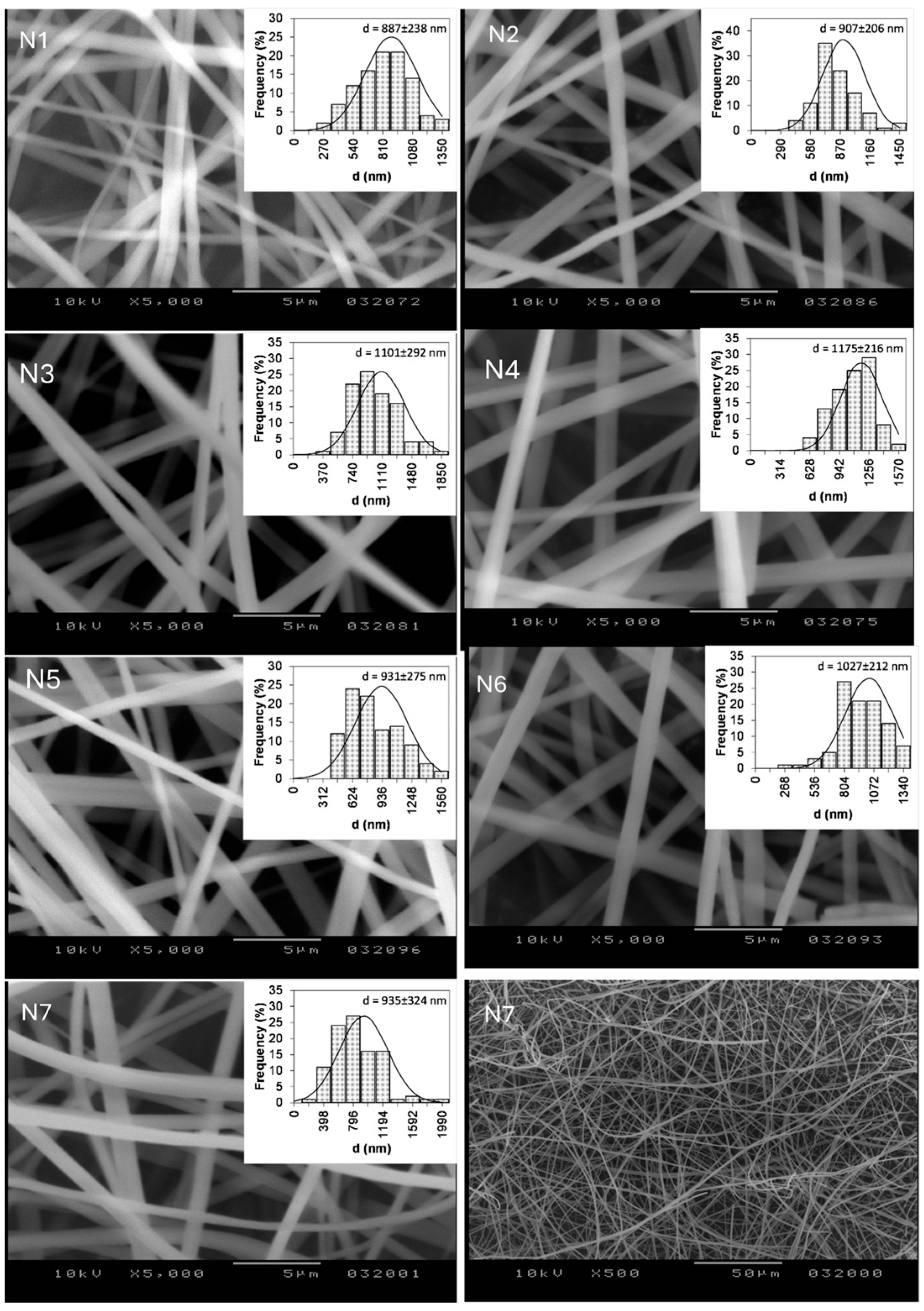
Microfiber-based nonwoven fabrics were obtained using drug–polymer dispersions in a dichloromethane and ethanol mixture. The electrospinning resulted in randomly oriented smooth fibers at all the investigated parameters. The average fiber size ranged from 887 nm to 1175 nm, depending on the electrospinning parameters (Figure 2).
Empty fibers were prepared at a 1.5 mL flow rate and a 12.5 cm needle–collector distance, resulting in smooth and bead-free fibers with a diameter of 796 ± 157 nm.

3.2. Fiber Morphology—Scanning Electron Microscopy (SEM)

The SEM images indicated no crystallized ITZ on the surface of the microfibers, suggesting that the drug was in a non-crystalline state after electrospinning [36]. N1 and N3 samples prepared at a 1 mL/h flow rate compared to N2 and N4 at a 2 mL/h flow rate led to smaller-diameter fibers. The needle-to-collector distance of 10 cm resulted in a lower fiber diameter compared to 15 cm, specifically for samples N1 (887 ± 238 nm) and N2 (907 ± 206 nm) compared to N3 (1101 ± 292 nm) and N4 (1175 ± 226 nm).
All fiber histograms were unimodal, N1 appeared to be symmetric, N2, N3, N5, N6, and N7 were right-skewed, and N4 was left-skewed (with the highest values for fiber diameter, flow rate, and needle–collector distance). The lowest fiber diameter of 887 ± 238 nm was associated with the lowest flow rate and needle-to-collector distance. The needle-to-collector distance influenced the diameter of the fibers in a statistically significant manner (p < 0.001, when comparing N1 to N3 and N2 to N4).

3.3. Differential Scanning Calorimetry (DSC)

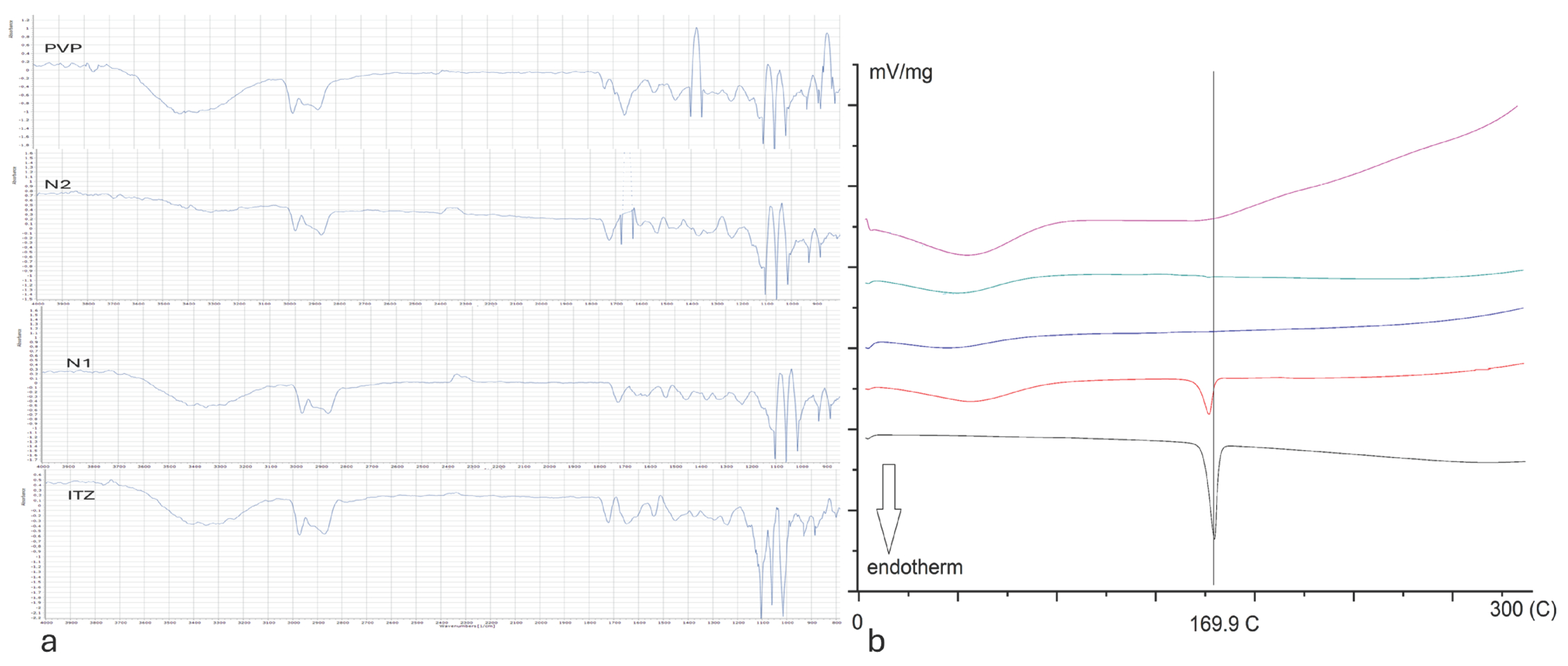
The scanning calorimetry of pure ITZ revealed a sharp endothermic melting point at a temperature of 170 °C with a starting temperature 163.6 °C, an end temperature of 173.9 °C, and 69.4 J/g enthalpy (Figure 3), which is comparable to the results obtained in the literature [22]. The PVP curve showed a larger peak under 100 °C, which was related to water loss.
The DSC curve of PM at a 1:1 weight ratio presented a well-detectable melting peak of ITZ at 169.6 °C (starting temperature 161.9 °C and end temperature 172.6 °C), with an enthalpy value (24.1 J/g) which was half of that of pure ITZ. Differential scanning calorimetry revealed that the DSC curves of the fibrous samples resembled the thermogram of PVP, not presenting melting endotherm peaks. This suggests the incorporation of ITZ into the fibers in an amorphous state [24].

3.4. Fourier-Transform Infrared Spectroscopy (FTIR)

Our results showed ITZ peaks at 1180 cm−1 (C–N stretching) and 1138 cm−1 (C–O–C ring cyclic ether stretching). Peaks attributable to alkyl C–H stretch vibrations were detected at 2800–3100 cm−1 [29]. The FTIR spectrum of fiber-forming PVP showed a broad band at 3500–3200 cm−1 assigned to O-H stretching of the hydroxyl groups and at 1647 cm−1 for C=O stretching. The peak at 1437 cm−1 was assigned to the C-H deformation of the CH2 group [6,35].
The fibrous sheets showed peaks at 1179 cm−1 and 1123 cm−1, as well as a broad band at 3500–3200 cm−1 assigned to the O-H stretching of the hydroxyl groups (from PVP).
The peaks were very similar to those of pure ITZ, presenting a shift to lower wave numbers and weaker peak intensities. Shifts in FTIR spectrum peaks suggested, in the case of drug–polymer interactions, hydrogen bonding [34].

3.5. Determination of Drug Content

The determined drug content (Table 2) of the fibers corresponded well to the theoretical value of 3.08 mg/10 mg fibers (calculated from the composition, considering full solvent evaporation). Differences may be attributed to incomplete solvent evaporation.

3.6. Disintegration

Disintegration times between 1.47 and 2.12 s (Table 3) were well below the stipulations of the currently in-force European and United States Pharmacopeia. Lower disintegration times were observed for the 10 cm needle-to-collector distance (N1 compared to N3 and N2 compared to N4), with an increasing flow rate leading to slightly higher disintegration time values (N1 compared to N2 and N3 to N4).
The highly hydrophilic nature of PVP combined with the porous three-dimensional structure and the high surface area of the fibrous webs explain the instantaneous disintegration of the samples. The 1090 nm diameter PVP fibers measured a disintegration time under 3 s [37].

3.7. In Vitro Dissolution Studies

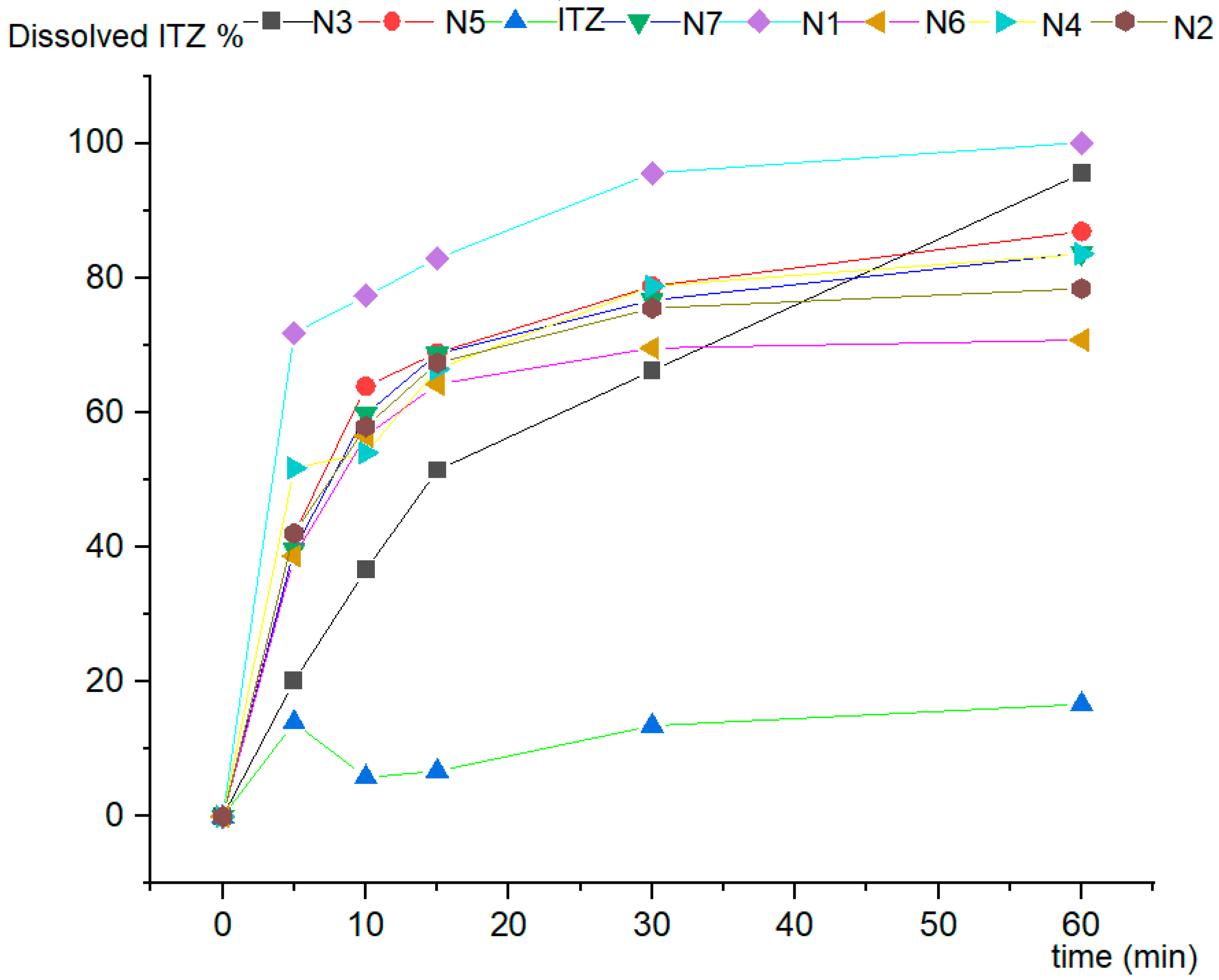
The performance of the fibrous sheets was evaluated by dissolution in 0.1 N HCl (Figure 4) compared to pure ITZ. The dissolution curve of pure crystalline ITZ showed a dissolved amount of 16.7% after 60 min. All the fibrous sheets proved to have a superior dissolution rate, as the dissolved amount of ITZ was between 78 and 100% after 60 min. For N1, ITZ was fully dissolved during this test. One speculation is that differences in the dissolution rates may be related to solvent accessibility between fibrous sheets [38].
Solid dispersions of drugs with low water solubility and highly hydrophilic polymers present a hidrophobization effect, making dissolution study results highly variable and poorly reproductible, as described by Nagy et al. for ITZ and PVPVA64 [24]. In our case, the rotating basket method ensured an acceptable sample variability. An almost complete dissolution in 0.1 N HCl medium after 60 min, comparable with our results for N1, was also obtained with PVPVA64-based high-speed-electrospun ITZ fibers and single-needle electrospun ITZ fibers [24]. In another study, optimizing the electrospinning parameters offered a tailored drug release of ITZ from HPMC-based fibers [22].

3.8. Antifungal Activity

The antifungal effect of the fibers (Table 4) was demonstrated on Sabouraud dextrose agar.
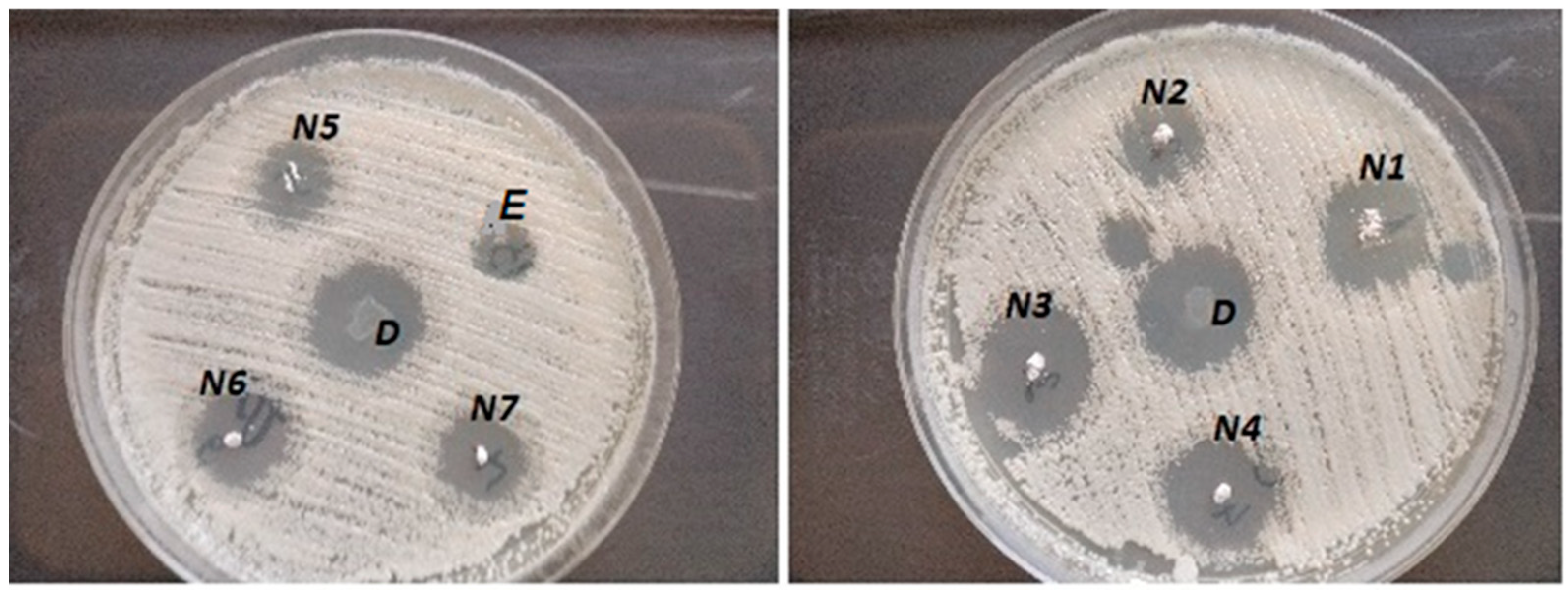
The antifungal assay revealed that the fibers inhibited the growth of Candida albicans (Figure 5) and Candida parapsylosis. The inhibition zone of blank fibers could be explained by the alcohol content of the fibers [39,40]. The diameter of the inhibition ring was only 3 mms for the blank fibers, compared to the itraconazole-containing fibers, which had diameters above 6 mm. The inhibition zone of the fibers could not be attributed only to the alcohol content [41], because, during fiber formation in the electrostatic field, the solvent evaporated. A flow rate of 2 mL/h led to a smaller inhibition zone, while increasing the needle-to-collector distance showed wider inhibition zones. Another study showed a similar result, where 3.8 mm inhibition zones were measured for empty fibers of PVA [42].
The effectiveness of the solid dispersions was also demonstrated by Qiu et al. in the case of nanoparticles against Candida albicans via in vitro and in vivo experiments on mice [43,44]. In the study, the inhibition zones were equal in diameter for both the solid dispersions and the microfibers, suggesting the diffusion of ITZ into Sabouraud dextrose agar.

3.9. Factorial Design

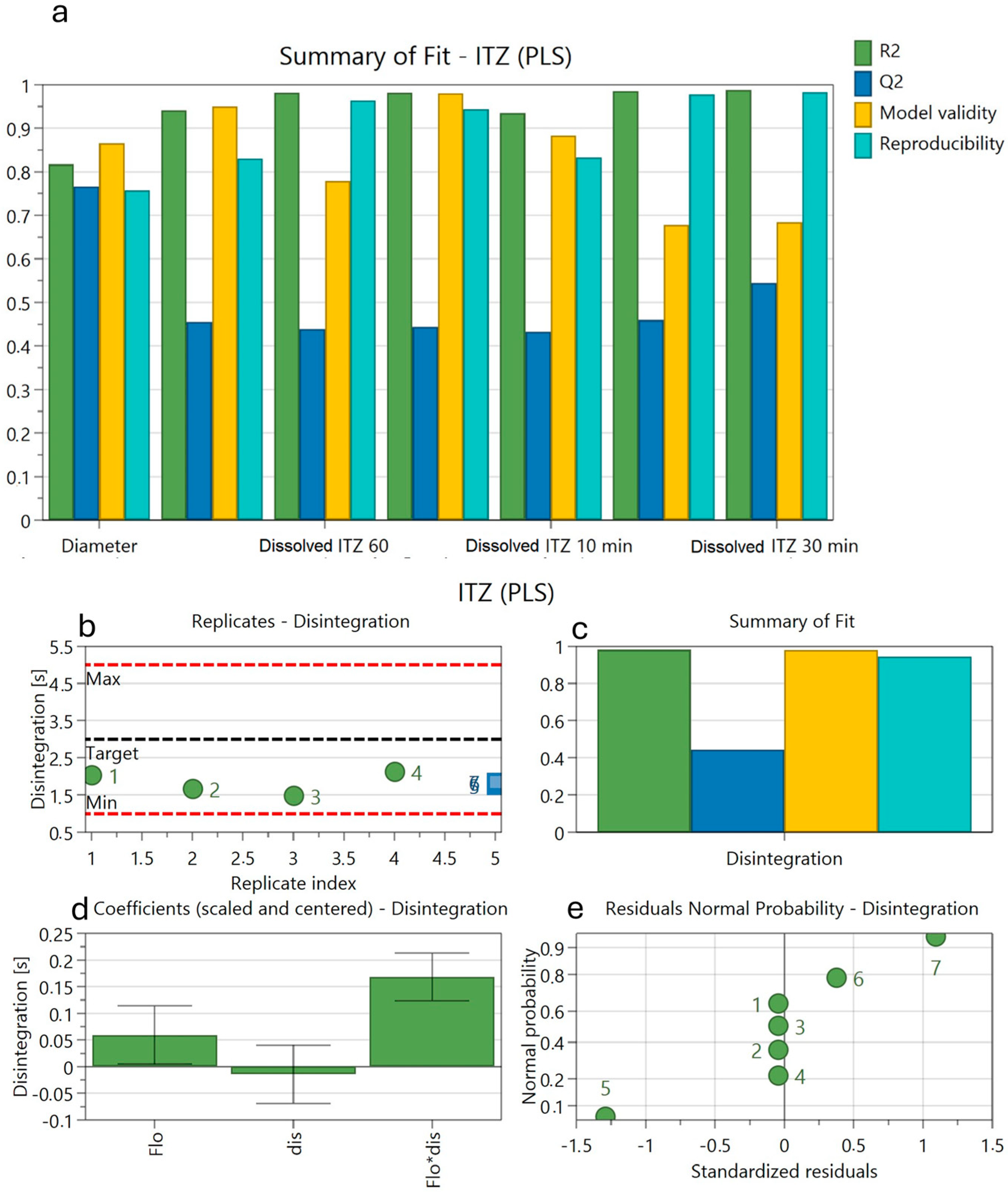
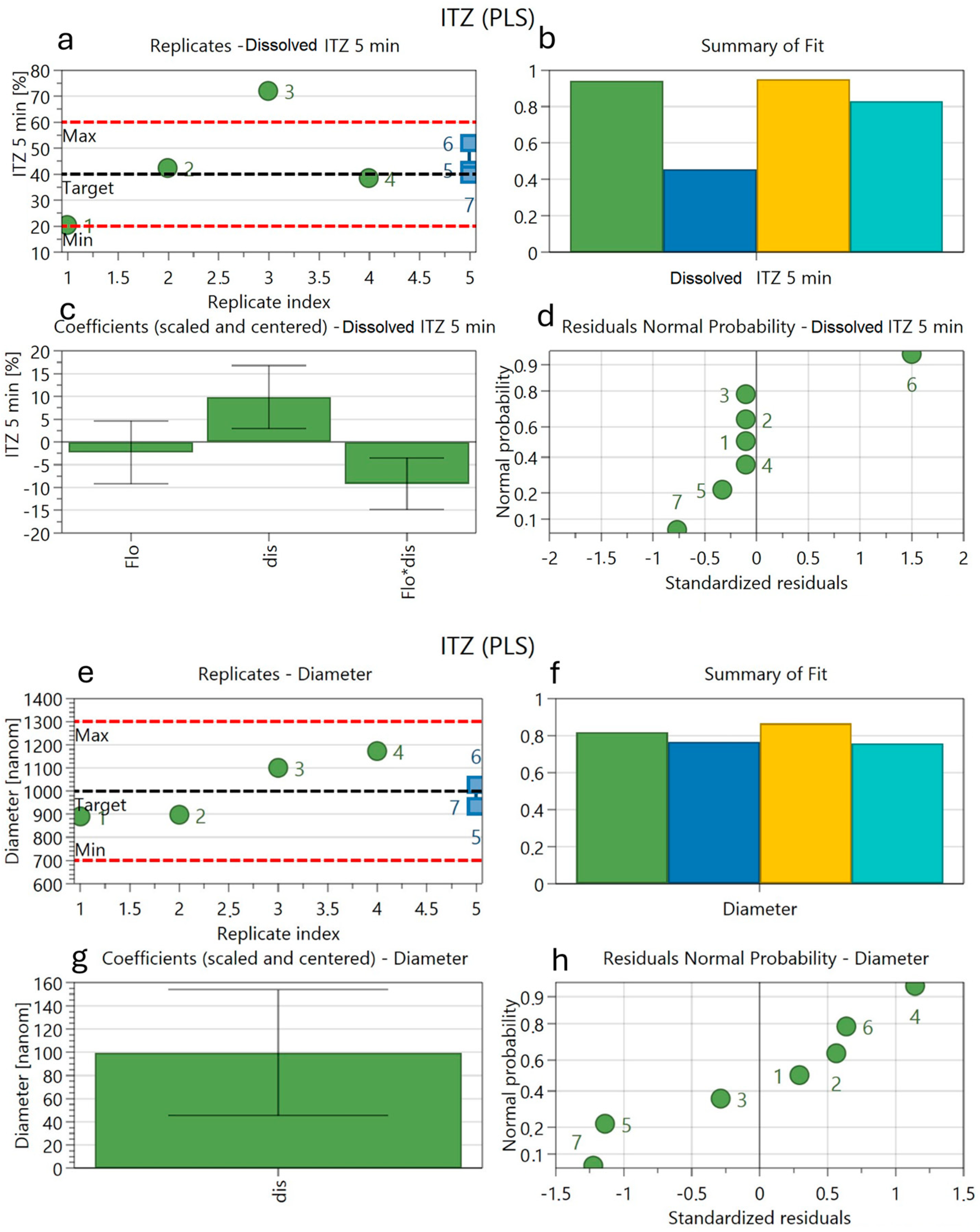
The results of the evaluated dependent parameters are presented in Table 3 and Figure 6 and Figure 7, including R2, Q2, validity, and reproducibility.
Screening for influencing factors offered additional data (Figure 6).
The experimental data were analyzed from a statistical point of view. R2 (Figure 6) presented values between 0.82 and 0.99 and Q2 (Figure 6) showed values between 0.44 and 0.76. None of the validity values were under 0.25. The values for reproducibility were over 0.75 in all cases.
The needle-to-collector distance was the significantly influencing parameter for fiber diameter (Figure 7). The disintegration times were positively influenced by the needle-to-collector distance and highly influenced by the interactions between the needle-to-collector distance and the flow rate.
The dissolved amounts of ITZ after 5 min were negatively influenced by the flow rate, positively by the distance, and negatively by the combination of the two factors. The dissolved amounts of ITZ were dependent on the distance and flow rate.
Our study had some limitations, such as the lack of in vivo testing to confirm antifungal efficacy and the limited strain variability (there were only two Candida species tested). Future studies will be required to prove the antifungal effect of these fibrous sheets on mice infected with Candida albicans and Candida parapsylosis via susceptibility and safety testing of the drug delivery system. Scaling up this process may lead to the development of an oral or topical delivery system.

4. Conclusions

PVP-based microfibrous sheets loaded with ITZ were prepared successfully by single-jet electrospinning. The spinning process resulted in bead-free, smooth-surfaced fibers. The FTIR and DSC investigations proved that the ITZ was homogeneously incorporated into the PVP fibers in an amorphous state. Rapid fiber disintegration allowed the fast dissolution of the ITZ. Solubility was 4.2- to 6-fold enhanced compared to crystalline ITZ. All the fibrous sheets inhibited fungal growth on the Sabouraud dextrose agar medium. The electrospinning parameter of the needle-to-collector distance significantly influenced the fibers’ morphology. Optimizing the electrospinning parameters improved the antifungal performance of the drug delivery system. An optimized delivery system may lead to further potential clinical applications.

Author Contributions

Conceptualization, E.-M.R., A.C., E.S., A.L.G. and P.A.; methodology, E.-M.R., O.P., R.-A.V., E.B. and P.A.; software, N.T., A.L.G. and P.A.; writing—original draft preparation, E.-M.R., O.P., A.L.G., C.-T.C. and N.T.; writing—review and editing, A.P., R.-A.V., E.S., P.A. and A.C.; visualization, E.-M.R., N.T., A.L.G., C.-T.C. and A.C.; and supervision, A.C. and E.S. All authors have read and agreed to the published version of the manuscript.

Funding

This research received no external funding.

Data Availability Statement

The original contributions presented in the study are included in the article, further inquiries can be directed to the corresponding author.

Acknowledgments

Orsolya Péterfi is grateful to the Federation Székely Előfutár Scholarship program for making this research possible.

Conflicts of Interest

The authors declare no conflicts of interest.

References

  1. Sung, Y.K.; Kim, S.W. Recent advances in polymeric drug delivery systems. Biomater. Res. 2020, 24, 12. [Google Scholar] [CrossRef] [PubMed]
  2. Kim, S.W.; Petersen, R.V.; Feijen, J. Polymeric drug delivery systems. Drug Des. 2016, 10, 193–250. [Google Scholar]
  3. De Souza, R.; Zahedi, P.; Allen, C.J.; Piquette-Miller, M. Polymeric drug delivery systems for localized cancer chemotherapy. Drug Deliv. 2010, 17, 365–375. [Google Scholar] [CrossRef] [PubMed]
  4. Koczkur, K.M.; Mourdikoudis, S.; Polavarapu, L.; Skrabalak, S.E. Polyvinylpyrrolidone (PVP) in nanoparticle synthesis. Dalton Trans. 2015, 44, 17883–17905. [Google Scholar] [CrossRef]
  5. Luo, Y.; Hong, Y.; Shen, L.; Wu, F.; Lin, X. Multifunctional Role of Polyvinylpyrrolidone in Pharmaceutical Formulations. AAPS PharmSciTech 2021, 22, 34. [Google Scholar] [CrossRef]
  6. Kedia, A.; Kumar, P.S. Solvent-adaptable poly (vinylpyrrolidone) binding induced anisotropic shape control of gold nanostructures. J. Phys. Chem. C 2012, 116, 23721–23728. [Google Scholar] [CrossRef]
  7. Maertens, J.; Boogaerts, M. The place for itraconazole in treatment. J. Antimicrob. Chemother. 2005, 56, i33–i38. [Google Scholar] [CrossRef]
  8. Lestner, J.; Hope, W.W. Itraconazole: An update on pharmacology and clinical use for treatment of invasive and allergic fungal infections. Expert Opin. Drug Metab. Toxicol. 2013, 9, 911–926. [Google Scholar] [CrossRef]
  9. De Beule, K.; Van Gestel, J. Pharmacology of itraconazole. Drugs 2001, 61, 27–37. [Google Scholar] [CrossRef]
  10. Aungst, B.J. Optimizing Oral Bioavailability in Drug Discovery: An Overview of Design and Testing Strategies and Formulation Options. J. Pharm. Sci. 2017, 106, 921–929. [Google Scholar] [CrossRef]
  11. Thompson, G.R.; Lewis, P.; Mudge, S.; Patterson, T.F.; Burnett, B.P. Open-Label Crossover Oral Bioequivalence Pharmacokinetics Comparison for a 3-Day Loading Dose Regimen and 15-Day Steady-State Administration of SUBA-Itraconazole and Conventional Itraconazole Capsules in Healthy Adults. Antimicrob. Agents Chemother. 2020, 64, 00400-20. [Google Scholar] [CrossRef] [PubMed]
  12. Bae, S.K.; Park, S.J.; Shim, E.J.; Mun, J.H.; Kim, E.Y.; Shin, J.G.; Shon, J.H. Increased oral bioavailability of itraconazole and its active metabolite, 7-hydroxyitraconazole, when coadministered with a vitamin C beverage in healthy participants. J. Clin. Pharmacol. 2011, 51, 444–451. [Google Scholar] [CrossRef] [PubMed]
  13. Domínguez-Gil Hurlé, A.; Sánchez Navarro, A.; Garcia Sanchez, M. Therapeutic drug monitoring of itraconazole and the relevance of pharmacokinetic interactions. Clin. Microbiol. Infect. 2006, 12, 97–106. [Google Scholar] [CrossRef]
  14. Wu, C.-Y.; Benet, L.Z. Predicting drug disposition via application of BCS: Transport/absorption/elimination interplay and development of a biopharmaceutics drug disposition classification system. Pharm. Res. 2005, 22, 11–23. [Google Scholar] [CrossRef]
  15. Akhgari, A.; Shakib, Z.; Sanati, S. A review on electrospun nanofibers for oral drug delivery. Nanomed. J. 2017, 4, 197–207. [Google Scholar]
  16. Kajdič, S.; Planinšek, O.; Gašperlin, M.; Kocbek, P. Electrospun nanofibers for customized drug-delivery systems. J. Drug Deliv. Sci. Technol. 2019, 51, 672–681. [Google Scholar] [CrossRef]
  17. Haider, A.; Haider, S.; Kang, I.-K. A comprehensive review summarizing the effect of electrospinning parameters and potential applications of nanofibers in biomedical and biotechnology. Arab. J. Chem. 2018, 11, 1165–1188. [Google Scholar] [CrossRef]
  18. Ibrahim, H.M.; Klingner, A. A review on electrospun polymeric nanofibers: Production parameters and potential applications. Polym. Test. 2020, 90, 106647. [Google Scholar] [CrossRef]
  19. Amiri, N.; Moradi, A.; Tabasi, S.A.S.; Movaffagh, J. Modeling and process optimization of electrospinning of chitosan-collagen nanofiber by response surface methodology. Mater. Res. Express 2018, 5, 045404. [Google Scholar] [CrossRef]
  20. Jacobs, V.; Patanaik, A.; Anandjiwala, R.D.; Maaza, M. Optimization of electrospinning parameters for chitosan nanofibres. Curr. Nanosci. 2011, 7, 396–401. [Google Scholar] [CrossRef]
  21. Akhgari, A.; Heshmati, Z.; Makhmalzadeh, B.S. Indomethacin electrospun nanofibers for colonic drug delivery: Preparation and characterization. Adv. Pharm. Bull. 2013, 3, 85. [Google Scholar] [PubMed]
  22. Verreck, G.; Chun, I.; Peeters, J.; Rosenblatt, J.; Brewster, M.E. Preparation and characterization of nanofibers containing amorphous drug dispersions generated by electrostatic spinning. Pharm. Res. 2003, 20, 810–817. [Google Scholar] [CrossRef] [PubMed]
  23. Sóti, P.L.; Bocz, K.; Pataki, H.; Eke, Z.; Farkas, A.; Verreck, G.; Kiss, É.; Fekete, P.; Vigh, T.; Wagner, I.; et al. Comparison of spray drying, electroblowing and electrospinning for preparation of Eudragit E and itraconazole solid dispersions. Int. J. Pharm. 2015, 494, 23–30. [Google Scholar] [CrossRef] [PubMed]
  24. Nagy, Z.K.; Balogh, A.; Démuth, B.; Pataki, H.; Vigh, T.; Szabó, B.; Molnár, K.; Schmidt, B.T.; Horák, P.; Marosi, G. High speed electrospinning for scaled-up production of amorphous solid dispersion of itraconazole. Int. J. Pharm. 2015, 480, 137–142. [Google Scholar] [CrossRef] [PubMed]
  25. Démuth, B.; Farkas, A.; Balogh, A.; Bartosiewicz, K.; Kállai-Szabó, B.; Bertels, J.; Vigh, T.; Mensch, J.; Verreck, G.; Van Assche, I.; et al. Lubricant-Induced Crystallization of Itraconazole From Tablets Made of Electrospun Amorphous Solid Dispersion. J. Pharm. Sci. 2016, 105, 2982–2988. [Google Scholar] [CrossRef]
  26. Mehrandish, S.; Mohammadi, G.; Mirzaeei, S. Preparation and functional evaluation of electrospun polymeric nanofibers as a new system for sustained topical ocular delivery of itraconazole. Pharm. Dev. Technol. 2022, 27, 25–39. [Google Scholar] [CrossRef]
  27. Wu, S.; Song, R.; Liu, T.; Li, T. Antifungal therapy: Novel drug delivery strategies driven by new targets. Adv. Drug Del. Rev. 2023, 199, 114967. [Google Scholar] [CrossRef]
  28. Yang, W.; Wiederhold, N.P.; Williams, R.O. Drug delivery strategies for improved azole antifungal action. Exp. Op. Drug Del. 2008, 5, 1199–1216. [Google Scholar] [CrossRef]
  29. Pawar, J.N.; Ali, T.M.; Moravkar, K.K.; Patole, R.K.; Jaiswar, D.R.; Amin, P.D. Recent development and achievements in solubility and dissolution enhancement of itraconazole: A review. Int. J. Pharm. Sci. Res. 2014, 5, 3096. [Google Scholar]
  30. Pintea, A.; Vlad, R.-A.; Antonoaea, P.; Rédai, E.M.; Todoran, N.; Barabás, E.-C.; Ciurba, A. Structural Characterization and Optimization of a Miconazole Oral Gel. Polymers 2022, 14, 5011. [Google Scholar] [CrossRef]
  31. Vlad, R.-A.; Antonoaea, P.; Todoran, N.; Rédai, E.-M.; Bîrsan, M.; Muntean, D.-L.; Imre, S.; Hancu, G.; Farczádi, L.; Ciurba, A. Development and evaluation of cannabidiol orodispersible tablets using a 23-factorial design. Pharmaceutics 2022, 14, 1467. [Google Scholar] [CrossRef] [PubMed]
  32. Fadke, J.; Desai, J.; Thakkar, H. Formulation Development of Spherical Crystal Agglomerates of Itraconazole for Preparation of Directly Compressible Tablets with Enhanced Bioavailability. AAPS PharmSciTech 2015, 16, 1434–1444. [Google Scholar] [CrossRef] [PubMed]
  33. Reksamunandar, R.P.; Edikresnha, D.; Munir, M.M.; Damayanti, S.; Khairurrijal. Encapsulation of β-carotene in poly(vinylpyrrolidone) (PVP) by Electrospinning Technique. Procedia Eng. 2017, 170, 19–23. [Google Scholar] [CrossRef]
  34. Fong, H.; Chun, I.; Reneker, D.H. Beaded nanofibers formed during electrospinning. Polymer 1999, 40, 4585–4592. [Google Scholar] [CrossRef]
  35. Uyar, T.; Besenbacher, F. Electrospinning of uniform polystyrene fibers: The effect of solvent conductivity. Polymer 2008, 49, 5336–5343. [Google Scholar] [CrossRef]
  36. Paaver, U.; Heinämäki, J.; Laidmäe, I.; Lust, A.; Kozlova, J.; Sillaste, E.; Kirsimäe, K.; Veski, P.; Kogermann, K. Electrospun nanofibers as a potential controlled-release solid dispersion system for poorly water-soluble drugs. Int. J. Pharm. 2015, 479, 252–260. [Google Scholar] [CrossRef]
  37. Celebioglu, A.; Uyar, T. Electrospun formulation of acyclovir/cyclodextrin nanofibers for fast-dissolving antiviral drug delivery. Mater. Sci. Eng. C 2021, 118, 111514. [Google Scholar] [CrossRef]
  38. Yu, D.-G.; Branford-White, C.; White, K.; Li, X.-L.; Zhu, L.-M. Dissolution Improvement of Electrospun Nanofiber-Based Solid Dispersions for Acetaminophen. AAPS PharmSciTech 2010, 11, 809–817. [Google Scholar] [CrossRef]
  39. Rane, H.S.; Bernardo, S.M.; Walraven, C.J.; Lee, S.A. In vitro analyses of ethanol activity against Candida albicans biofilms. Antimicrob. Agents Chemother. 2012, 56, 4487–4489. [Google Scholar] [CrossRef]
  40. Sequeira, S.O.; Phillips, A.J.; Cabrita, E.J.; Macedo, M.F. Ethanol as an antifungal treatment for paper: Short-term and long-term effects. Stud. Conservation. 2017, 62, 33–42. [Google Scholar] [CrossRef]
  41. Giller, C.B.; Chase, D.B.; Rabolt, J.F.; Snively, C.M. Effect of solvent evaporation rate on the crystalline state of electrospun Nylon 6. Polymer 2010, 51, 4225–4230. [Google Scholar] [CrossRef]
  42. Santos, V.A.D.; Viera, P.V.A.; Oliveira, A.M.D.; Zanin, M.H.A.; Borsatti, M.A. Antifungal effect of electrospun nanofibers containing cetylpyridinium chloride against Candida albicans. Braz. Oral Res. 2014, 28, 1–6. [Google Scholar] [CrossRef] [PubMed]
  43. Maha, H. Formula comparison of nanoemulsion and cream containing miconazole nitrate: Penetration test using franz diffusion cells and antifungal activity test on Tricophyton mentagrophytes, Microsporum canis and Candida albicans. Rasayan J. Chem. 2020, 13, 529–534. [Google Scholar] [CrossRef]
  44. Qiu, L.; Hu, B.; Chen, H.; Li, S.; Hu, Y.; Zheng, Y.; Wu, X. Antifungal efficacy of itraconazole-loaded TPGS-b-(PCL-ran-PGA) nanoparticles. Int. J. Nanomed. 2015, 10, 1415–1423. [Google Scholar] [CrossRef]
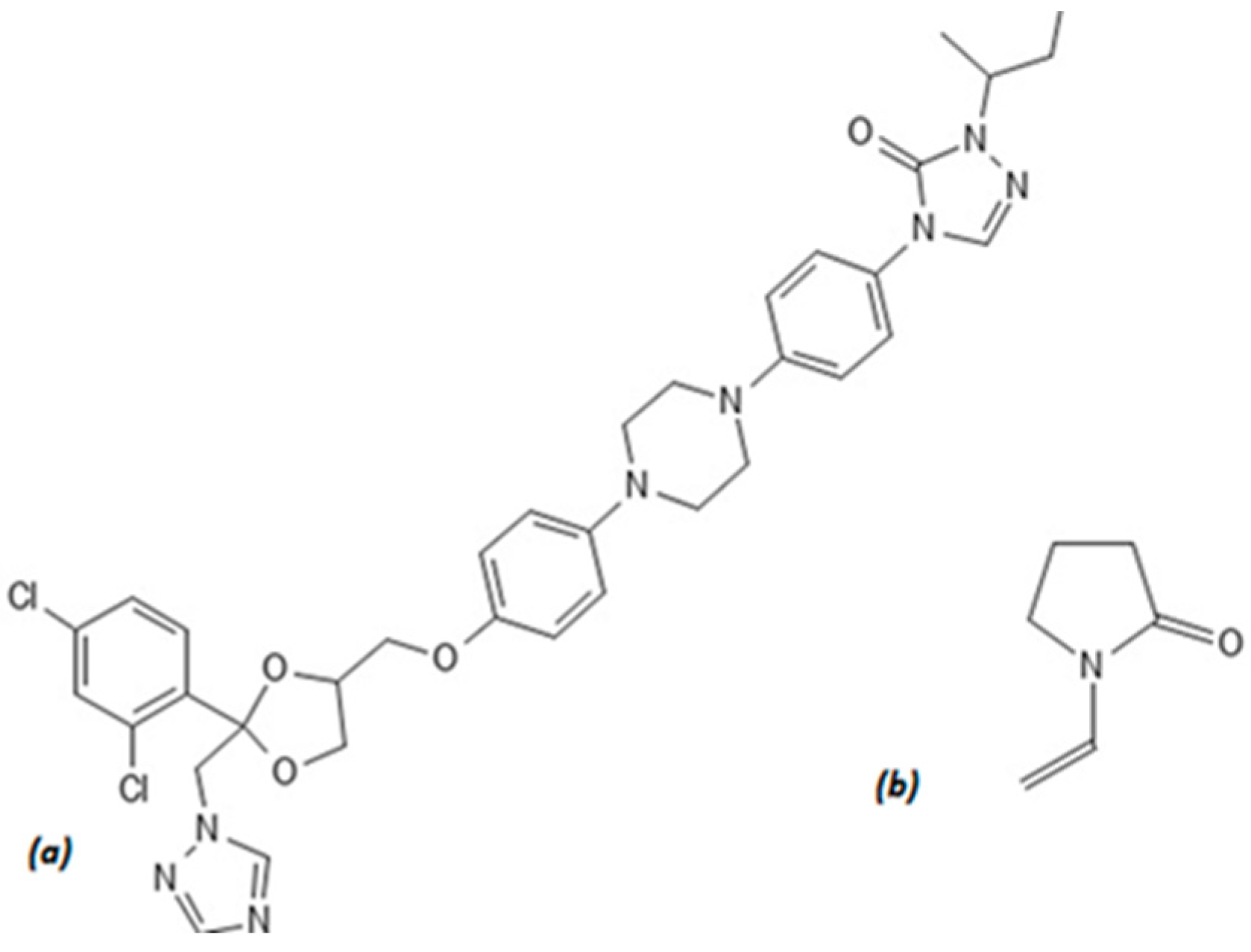
Figure 1. Chemical structures of itraconazole (a) and polyvinylpyrrolidone (b).
Applsci 15 00710 g001
Figure 2. SEM images at 5000× magnification of samples N1 to N7 and 500× magnification of sample N7.
Applsci 15 00710 g002
Figure 3. FTIR spectra (a) and DSC curves (b) of ITZ, physical mixture (PM), N1 and N2 fibers, and PVP.
Applsci 15 00710 g003
Figure 4. Comparative dissolution curves of the samples and pure ITZ.
Applsci 15 00710 g004
Figure 5. Inhibition zones for Candida albicans for samples (N1–N7), empty fibers (E), and dispersion of ITZ (D).
Applsci 15 00710 g005
Figure 6. Summary plot of the factorial design (a) and factors influencing disintegration, including (b) replicates, (c) summary of fit, (d) coefficients, and (e) residuals.
Applsci 15 00710 g006
Figure 7. Influence of factors on dissolved amounts of ITZ after 5 min ((a) replicates, (b) summary of fit, (c) coefficients, and (d) residuals) and fiber diameter ((e) replicates, (f) summary of fit, (g) coefficients, and (h) residuals).
Applsci 15 00710 g007
Table 1. Electrospinning parameters and independent factor level of ITZ sheets.
CodeFlow Rate (mL/hour)LevelNeedle–Collector Distance (cm)
N11−110
N22+110
N31−115
N42+115
N51.5012.5
N61.5012.5
N71.5012.5
Table 2. Drug content of ITZ sheets.
CodesN1N2N3N4N5N6N7
Drug content mg/10 mg
fibers ± SD
2.73 ± 0.212.71 ± 0.263.27 ± 0.282.89 ± 0.232.88 ± 0.172.75 ± 0.192.83 ± 0.18
Table 3. Dependent parameters for the samples.
CodeFiber
Diameter (nm) ± SD
Disintegration Time (s)ITZ Released
5 min (%)10 min (%)15 min (%)30 min (%)60 min (%)
N1887 ± 2381.47 ± 3.171.88 ± 4.6577.37 ± 3.8782.96 ± 6.6395.65 ± 2.36100.20 ± 1.99
N2907 ± 2061.65 ± 1.242.09 ± 2.2057.98 ± 3.8067.45 ± 6.0075.55 ± 4.3078.49 ± 2.14
N31101 ± 2922.01 ± 0.920.17 ± 2.0036.66 ± 3.0151.47 ± 4.3266.29 ± 5.1195.60 ± 6.2
N41175 ± 2262.12 ± 0.638.70 ± 8.3256.57 ± 2.0964.28 ± 0.8969.66 ± 1.1170.83 ± 1.06
N5931 ± 2751.76 ± 0.742.03 ± 3.6163.95 ± 5.6068.94 ± 5.2778.84 ± 6.3886.98 ± 5.02
N61027 ± 2121.83 ± 0.751.77 ± 5.2554.07 ± 3.9566.51 ± 2.8878.85 ± 2.8883.65 ± 1.77
N7935 ± 3241.86 ± 1.439.64 ± 4.8659.85 ± 3.8768.77 ± 5.1076.73 ± 2.9983.79 ± 0.06
Table 4. Inhibition zones of the antifungal assay.
CodesEDN1N2N3N4N5N6N7
Inhibition zone diameter (mm)3.07.17.16.97.57.06.66.56.2
E (empty fibers) and D (drug-containing dispersion) for samples N1 to N7.
Disclaimer/Publisher’s Note: The statements, opinions and data contained in all publications are solely those of the individual author(s) and contributor(s) and not of MDPI and/or the editor(s). MDPI and/or the editor(s) disclaim responsibility for any injury to people or property resulting from any ideas, methods, instructions or products referred to in the content.