Article

Elevated BP180 ELISA at Diagnosis Correlates with Disease Severity and Relapse in Oral Mucous Membrane Pemphigoid: Preliminary Results from a Retrospective Monocentric Italian Study

1
Section of Oral Sciences, Department of Biomedical and Neuromotor Sciences, University of Bologna, 40125 Bologna, Italy
2
Division of Dermatology, Department of Medical and Surgical Sciences, Alma Mater Studiorum University, 40126 Bologna, Italy
3
Division of Dermatology, IRCCS Policlinico S. Orsola-Malpighi Hospital, 40138 Bologna, Italy
*
Author to whom correspondence should be addressed.
Appl. Sci. 2025, 15(2), 689; https://doi.org/10.3390/app15020689
Submission received: 19 December 2024 / Revised: 4 January 2025 / Accepted: 10 January 2025 / Published: 12 January 2025
(This article belongs to the Special Issue Oral Diseases: Diagnosis and Therapy)

Abstract

:
Background: Little is known about the relevance of BP180 ELISA for the clinical management of oral mucous membrane pemphigoid (OMMP). The aim of the present study was to investigate if the levels of anti-BP180 antibodies at diagnosis could be correlated with clinical severity and relapse. Methods: The present study included 44 OMMP patients with positive direct immunofluorescence (DIF). Circulating anti-BP180 IgG was measured using the same available ELISA kit (Euroimmun cut-off 20 U/mL). Clinical severity at diagnosis was measured using the oral disease severity score (ODSS). Only patients who achieved clinical remission (CR) were included in the analysis of variables related to relapse. Relapse was calculated as the interval between the date of the best type of clinical remission achieved and the date of relapse. Results: Values of BP180 > 20 U/mL significantly correlated with higher ODSSs in both univariate (p < 0.05) and multivariate analyses (p < 0.05). Among 39/44 patients who achieved CR, 17/39 relapsed. Kaplan–Meier analysis revealed that patients with BP180 > 20 U/mL displayed worse clinical behavior in terms of relapse (p < 0.05). Conclusion: BP180 ELISA at diagnosis appears to be a useful parameter to stratify OMMP patients with more severe disease and worse clinical outcomes after clinical remission.

1. Introduction

Mucous membrane pemphigoid (MMP) is a rare autoimmune subepithelial blistering disease that frequently affects the oral cavity [1]. Oral MMP (OMMP) manifests as erythematous, erosive, or bullous lesions that easily tend to burst, leading to the formation of painful ulcerations [2]. Several antigens have been identified as targets for autoantibodies, which, in association with complement (C3) activation, promote immune cell recruitment and contribute to the inflammatory reaction that leads to the development of intraoral lesions. These include bullous pemphigoid (BP) antigen 180/collagen type XVII (particularly its C- terminal and NC16A domains), BP 230, integrin α6β4, laminin 332 (formerly known as laminin 5), and type VII collagen (COLVII) [3]. BP180 is a transmembrane collagenous protein of the plakin family and represents the main target antigen of OMMP. Oyama et al., using immunoblotting, found that 63 of 124 studied patients with MMP (51%) showed reactivity to BP180 (including its soluble ectodomains, 120-kDa LAD-1 and 97-kDa LABD97 antigens) [4]. Shmidt et al., using Western blotting analysis with the combined use of the soluble BP180 ectodomain (LAD-1) and recombinant BP180 NC16A, found that 16 of the 19 MMP patients’ sera (84%) targeted BP180 [5].
More specifically for OMMP, Hayakawa et al. found that 56% of OMMP patients reacted to BP180 using a combination of indirect immunofluorescence with 1 M NaCl-split skin, immunoblot analysis, and ELISAs [3]. ELISA testing for circulating autoantibodies detection is an easy-to-perform, moderately expensive, and minimally invasive serological technique that has emerged as an optimal diagnostic biomarker for BP with excellent values of sensitivity and specificity [6]. By contrast, in MMP, the titer of circulating pathogenetic antibodies is well known to be lower than in BP, thus limiting the diagnostic utility of ELISA techniques for the diagnosis of OMMP [7]. Indeed, Petruzzi et al., evaluating BP180 NC16A ELISA in OMMP, found a sensitivity of only 50% (using commercially available MBL Kit cut-off 9 U/mL) [8]. However, in the same study, the authors also found a linear correlation between the titer of BP180 and oral disease severity score (ODSS). Other studies failed to demonstrate an association between BP180 and greater clinical severity. Cozzani et al., analyzing the profiles of 78 patients with MMP by means of indirect immunofluorescence (IIF), immunoblotting (IB), and ELISA, found that BP180 reactivity was inversely correlated with disease severity [9]. Similarly, Endo et al., analyzing 22 MMP patients by means of ELISA, found that BP180NC16a reactivity was associated with the low-risk clinical phenotype but referred to patients characterized by only oral mucosal lesions or combined oral mucosal and skin lesions and not involving the eyes, upper respiratory tract, or esophagus [10]. Despite the fact that it is to be expected that high levels of this protein might influence the outcome of the disease, like in Pemphigus Vulgaris, another autoimmune blistering disease of the oral cavity [11,12], there is not enough evidence that BP180 levels can predict disease severity and prognosis in OMMP. Conflicting results may depend on several factors such as the sample size, the different techniques or kits used for BP180 analysis, or differences among clinical phenotypes involving different sites. And yet, a prognostic biomarker able to stratify OMMP patients at risk of more severe disease and prognosis would be of great benefit in clinical practice. The purpose of our study was to assess whether the levels of anti-BP180 antibodies at diagnosis may be correlated with clinical severity and relapse in patients with OMMP.

2. Materials and Methods

The medical records of all patients diagnosed and treated for OMMP (from July 2017 to April 2024) were reviewed after the approval by the local ethics committee: Comitato Etico Area vasta Emilia Centro with code OBLI01 (CE 22206). All patients or their legal guardians provided informed consent for the investigations required for the diagnostic work-up, in accordance with good clinical practice. The study was conducted in compliance with the principles of the Declaration of Helsinki. To be included, patients had to display (1) a histological diagnosis of OMMP based on direct immunofluorescence (DIF) displaying linear deposits of IgG and/or C3 in the epithelial basal membrane zone (Figure 1a,b); (2) levels of circulating anti-BP180 IgG measured at diagnosis using the same available ELISA kit (Euroimmun, Lubeck, Germany cut-off 20 U/mL); and (3) the absence of extraoral involvement.
Histopathological diagnoses and DIF were performed by a pathologist experienced in blistering diseases (CM) in the Section of Dermatopathology, Division of Dermatology, IRCCS Policlinico S. Orsola-Malpighi Hospital. Additional serological investigations, such as IIF on salt-split skin, were not performed because they were not available in our institution.
Therapy followed local and international guidelines: bethametasone 0.01% mouthwashes 3 times a day and/or clobetasol 0.05 in Orafix 50% were prescribed as local therapy, whereas systemic therapy included prednisone at a dosage of 0.5–1 mg/kg in a tapering regime and 50 mg of dapsone twice a day [7].
More in detail, patients prescribed local therapy were asked to perform bethametasone 0.01% mouthwashes 3 times a day if lesions involved areas difficult to reach or less appropriate for topical application (i.e., the soft palate). By contrast, if lesions involved the gingival area, hard palate, lips, and buccal mucosa areas, clobetasol 0.05 in Orafix 50% was prescribed. Therapy was scheduled until disease control was reached (defined as the time at which new inflammatory lesions cease to form and established lesions begin to heal). The decision to reduce or withdraw local therapy was based on clinician judgment considering clinical severity at follow-up and the frequency of transient new lesions.
In the case of non-responsiveness to local therapy or the involvement of areas at risk of scarring, systemic therapy was also added. The tapering of steroid dosage was scheduled over the next 8–12 weeks, preferably after the achievement of disease control with weekly/biweekly reductions. Patients prescribed dapsone were periodically checked for methemoglobine levels. In the case of hemolytic anemia, skin rash, malaise, or gastrointestinal problems, temporal or permanent dapsone discontinuation was considered. As before, the decision to reduce or withdraw systemic therapy was based on clinician judgment.
The type of clinical remission was defined according to standard criteria: complete remission off therapy (CRoT): no new lesions for 2 or more months off systemic and topical therapies; partial remission off therapy (PRoT): transient new lesions that healed within 1 week without treatment and while off all systemic and topical therapies for 2 or more months; complete remission on minimal therapy (CRmT): no new lesions for 2 or more months while receiving minimal doses of systemic and/or topical therapy; and partial remission during minimal therapy (PRmT): transient new lesions that healed within 1 week during minimal therapy, including topical corticosteroids. The criteria adopted followed the recommendations of Murrel et al., which are the results of the consensus of an international panel of experts organized in 2011 [13]. The aim of this consensus statement was to provide accurate and reproducible definitions for disease extent, activity, outcome measures, endpoints, and therapeutic response for mucous membrane pemphigoid. The final consensus is the product of many meetings, discussions, and agreements. Universal agreement in the committee was achieved about the definitions of endpoints, therapeutic responses, and treatment failures, including relapse. Minimal therapy was defined as dapsone ≤ 1.0 mg/kg/d, prednisone ≤ 0.1 mg/kg/d, and topical corticosteroids once a day.
For each patient, the following clinical and histological variables were recorded: sex (M/F), age (>65 years), type of therapy (local or systemic), the presence/absence of eosinophils, the presence/absence of a band-like infiltrate, and the presence/absence deposits of IgA, IgM, and C3.
Clinical severity at diagnosis was measured using the oral disease severity score (ODSS). The ODSS records the presence of lesions and the degree of disease activity at multiple oral sites. In the ODSS, the oral cavity is divided into 17 sites weighted 0–2 according to the area of possible involvement. These sites are the outer/inner lips, right/left buccal mucosae, right/left soft palate, right/left hard palate, right/left dorsum of the tongue, right/left ventrolateral tongue, right/left floor of the mouth, right/left oropharynx, and the gingivae (divided into 6 segments: 3 for upper maxillary gingiva and 3 for mandibular gingiva). A score of 2 corresponds to >50% of the buccal mucosa on one side being affected or bilateral involvement of the dorsum of the tongue, the floor of the mouth, the hard and soft palate, or the oropharynx. Each unit of the site is then allocated an activity score, which ranges from 0 to 3 (no activity = 0; mild inflammation—erythema or healing areas = 1; prominent erythema = 2; and blistering or ulceration = 3). For a specific area, the activity for each unit of the site is added together. The ODSS was validated for use in OMMP by Ormond et al. [14], analyzing inter- and intra-observer reliability and comparing the results with similar scores adopted in clinical practice for OMMP (Mucous Membrane Pemphigoid Disease Area Index, MMPDAI; Autoimmune Bullous Skin Disorder Intensity Score, ABSIS; and Physician’s Global Assessment, PGA). In terms of reliability, the inter-observer ODSS total score intraclass correlation coefficient (ICC) was 0.97 and the intra-observer ICCs (two observers) for ODSS, in total, were 0.97 and 0.93. Similarly, convergent validity between ODSS and MMPDAI was good (correlation coefficient 0.88), indicating a superimposable performance and validating ODSS for use in OMMP. Following institutional guidelines, in the present study, the visits, the assessment of sites involved, and their severity degrees were always performed by a board of stomatologists and dermatologists attending a joint oral medicine–dermatology clinic (A.G., D.B.G., R.R., F.F., C.L., G.C., M.L., F.B.). Clinical photographs were taken at the first visit and periodically during the follow-up and were used to discuss clinical cases until a consensus was reached.
Only patients who achieved clinical remission (any type) were included to calculate the influence of anti-BP180 ELISA on relapse. MMP institutional guidelines recommend waiting at least 6–12 months before withdrawing minimal therapy to avoid early relapse. Hence, a composite outcome for clinical remission (the best type of clinical remission achieved) was adopted for analysis. Relapse was defined as the appearance of ≥3 new lesions a month (blisters, erosions) that do not heal within 1 week or the extension of established lesions in a patient who has achieved disease control [13]. Time to relapse was calculated in months as the interval between the best type of clinical remission achieved and relapse.

2.1. Sample Size Calculation

The sample size was preliminarily planned on our primary outcome (the relationship between ODSS and BP180 ELISA). Based on a previous study that analyzed both ODSS and BP180NC16A [8], we assumed that the difference between the mean ODSS value in BP180 < 20 and the mean ODSS value in BP180 > 20 could be approximately 15–17 points. Based on these values, we calculated that to test the null hypothesis between groups at α = 0.05 with 80% power, approximately 40 patients would be sufficient.

2.2. Statistical Analysis

Descriptive statistics were computed for patient and treatment characteristics, as well as the rates of clinical outcomes. A multilevel mixed-effects ordered logistic regression with stepwise selection was fitted to evaluate the relationships between ODSSs and the following variables: gender (male vs. female), age (<65 vs. >65 years), type of therapy (local vs. systemic), eosinophils (presence vs. absence), band-like infiltrate (presence vs. absence), deposits of IgA, IgM, and C3 (presence vs. absence), and BP180 ELISA (<20 vs. >20).
Survival analysis was also performed to identify predictors of relapse. The survival rate was estimated using the Kaplan–Meier method. Statistical significance was evaluated using the log-rank test. Time was defined as the period between clinical remission and the appearance of relapse or the last follow-up visit. Patient’s age, gender, presence of eosinophils and band-like infiltrate in histological analysis, deposits of IgG, IgA, C3 in DIF, ODSS (<20 or >20), type of therapy (local or systemic), type of remission (PRmT, CRmT, PRoT, CRoT), and BP180 ELISA at diagnosis (<20 or >20) were analyzed for their relationship with the outcome of interest in the study. For those variables found to be statistically significant at univariate analysis with a significance level of p < 0.25, the Cox proportional hazards method with stepwise bidirectional selection (significance level of entry/removal = 0.05) was used for further evaluation by multivariate survival analysis. Only values of p ≤ 0.05 were considered statistically significant in all analyses.

3. Results

3.1. Descriptive Analysis

A total of 44 patients followed for 26.65 months ± 21.03 SD (range 3–97) were included.
In total, 34/44 (77%) patients were females and 23/44 (52%) were >65 years old (mean age 64.36 ± 15.44 SD range 15–90 years). At histology, 31/44 (70%) displayed detectable eosinophils in the biopsy specimen, whereas 4/44 (9%) showed a band-like infiltrate. At DIF, 9/44 (20%) showed linear deposits of IgA at the BMZ, 4/44 (9%) showed linear deposits of IgM, and 40/44 (90%) had C3 deposits. The mean BP180 level was 25.81 U/mL ± 29.29 SD (range < 2 U/mL–112 U/mL). A total of 17/44 (38%) had BP180 levels above the recommended cut-off (20 U/mL). See Table 1 for details.

3.2. ODSS Analysis

The mean ODSS was 15.15 ± 6.81 (range 4–29) and 13/44 (29%) patients had ODSS values above 20 points, indicating moderate–severe oral involvement.
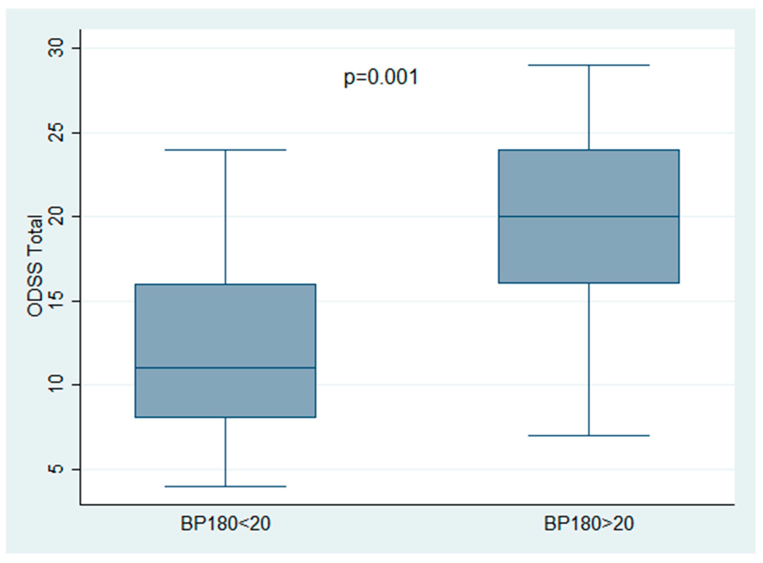
The ODSS in patients with values of BP180 ELISA > 20 U/mL was found to be statistically greater compared to patients with lower titers of circulating anti-BP180 (19.47 ± 6.70 vs. 12.44 ± 5.40 p = 0.001). (Figure 2).
By contrast, the presence of deposits of IgA was associated with lower values of ODSS (10 ± 2.39 p = 0.001).
BP180 ELISA > 20 and IgA both emerged in the multivariable analysis as variables statistically related to the ODSS (p = 0.002 and 0.0035 respectively) (see Table 1).

3.3. Therapy and Analysis of Predictors of Relapse

Twenty out of forty-four (46%) patients were treated by means of local steroids, whereas 24/44 (54%) patients were treated with a combination of local and systemic therapy (prednisone + dapsone). Chi-square analysis showed a non-significantly different distribution between patients treated with local therapy with respect to patients treated with local and systemic therapy (p = ns).
Thirty-nine out of forty-four (88%) patients achieved clinical remission (defined as composite endpoint PRmT or better) and were included in the analysis of relapse. In particular, 16/39 (41%) patients achieved PRmT (nine with local therapy and seven with local and systemic), 21/39 (53%) achieved CRmT (6 treated with local therapy and 15 with local and systemic therapy), 2/39 (5%) achieved PRoT (2 treated with local therapy), and 0/39 (0%) patients achieved CRoT. Specifically, in all cases of CRmT where the withdrawal of therapy was attempted, it was soon (within less than 2 months) followed by the onset of new lesions that required the re-introduction of minimal therapy (including local therapy) to be resolved. The mean time to achieve clinical remission (composite outcome) was 11.20 ± 11.10 months (see Supplementary Table S1 for details).
Seventeen out of thirty-nine (43%) patients experienced relapse in a median time of 11.23 ± 9.14 months.
Cox proportional hazard model analysis showed that BP180 ELISA > 20 U/mL was the only variable associated with relapse in the multivariate analysis (HR 3.4; p = 0.01). In particular, 9/17 patients with BP180 ELISA > 20 U/mL experienced relapse in a mean time of 9.66 ± 9.20 months compared to 8/17 patients below the cut-off (mean time to relapse: 13.0 ± 9.09 months) (Figure 3). See Table 2 for details.
Cox proportional hazard model analysis was also performed in the subgroup of patients treated with local therapy and in the subgroup of patients with local and systemic therapy. The results disclosed an association between BP180 > 20 and relapse only in the group treated with local and systemic therapy (HR 5.2 p = 0.01). In particular, of the 11 relapses that occurred in the group treated with local and systemic therapy, 4 had BP180 < 20, whereas 7 had BP180 > 20.

4. Discussion

The aim of this study was to investigate if BP180 ELISA may influence clinical severity and prognosis in patients with OMMP.
Greater BP180 ELISA values (>20 U/mL) emerged as correlating with both clinical severity (ODSS) and relapse.
Seventeen out of forty-four patients (38%) had BP180 levels above the recommended cut-off (20 U/mL) and values of ODSS greater than patients with BP180 levels below the cut-off (19.47 ± 6.70 vs. 12.44 ± 5.40 p = 0.001). This suggests that despite the diagnostic limits due to low sensitivity related to low levels of circulating antibodies, OMMP BP180 ELISA may serve as a biomarker of clinical severity.
At the same time, BP180 was the only variable associated with relapse, as OMMP patients with BP180 ELISA > 20 U/mL tended to relapse earlier after the achievement of clinical remission. Therefore, BP180 ELISA at diagnosis could be helpful to intercept OMMP patients who will experience worse clinical behavior in terms of relapse.
More in detail, the Kaplan–Meier analysis suggests that patients with different levels of BP180 might benefit from different clinical management and treatment regimens to delay relapse. Indeed, closer follow-up schedules in patients with BP180 > 20 may potentially allow clinicians to treat relapses at early stages, shortening the interval for an additional increase in steroids and steroids-sparing agents to achieve disease control. At the same time, BP180 ELISA may help identify those patients with a reduced risk of relapse who may benefit from therapy withdrawal or a decrease in minimal therapy dosage.
A correlation between circulating anti-BP180 and clinical severity is well established for cutaneous BP. Muhammed et al. found that BP180 ELISA correlated with ABSIS in a cohort of 42 treatment-naïve BP patients [15].
Liu et al., in a recent review, focused on the factors associated with the activity and severity of bullous pemphigoid and also included IgG autoantibodies against the NC16A domain of BP180 as closely correlated with the activity and severity of BP [16].
Concerning OMMP patients, the present results are in agreement with Petruzzi et al., who found a linear correlation between BP180 NC16A ELISA and ODSS [8]. Of note, Petruzzi et al. investigated the same immunodominant region NC16A of BP180 but used a different commercially available kit (MBL, Nagoya, Japan) which, according to the manufacturer’s recommendations, adopts a different cutoff value for BP180-NC16A (9.0 U/mL vs. 20 U/mL). Indeed, the rate of OMMP patients that authors found positive for BP180 NC16A ELISA was 50% (15/30), which is moderately greater than the rate of 34% (17/44) found in the present study using a cut-off of 20 U/mL. Despite the fact that the heterogeneity in the adopted cut-off may generate different diagnostic performances, the relationship between BP180 levels and ODSS was confirmed by independent laboratories using different commercial kits. This strengthens the findings of the present study and suggests that ELISA is a good technique to be investigated as a biomarker of clinical severity. Moreover, ELISA was reported to be superior to IIF on salt-split skin (SSS), another serological technique employed in the diagnosis of subepidermal autoimmune blistering diseases, which permits the differentiation of circulating antibodies targeting dermal antigens from those targeting subepidermal regions [17]. Other serological methods like immunoprecipitation and immunoblotting are highly sensitive but are non-quantitative and thus difficult to correlate with the severity of OMMP [18]. A major limitation of BP180-NC16A is that it recognizes only a single portion of BP180. However, Balding et al. demonstrated that regions other than NC16A may be recognized by MMP autoantibodies, especially against the C-terminal portion of BP180 [19]. It would be interesting in the future to also investigate if the presence of IgG directed against other regions of BP180 may influence clinical severity in OMMP. Izumi et al. developed an experimental full-length BP180 ELISA, but commercially available ELISA kits for similar purposes are still lacking [20]. At the same time, since other antigens such as integrin α6, β4, and laminin 332 have been reported to be targeted by MMP autoantibodies [3], and some of these targets are covered by commercially available ELISA Kits, future efforts should be made to disclose potential relations between circulating antibodies targeting α6, β4 integrin, and/or laminin 332 and ODSS in OMMP. Only a few other studies have investigated the relationship between circulating IgG anti-BP180 and clinical severity in OMMP.
Oyama et al. found that the combination of IgG and IgA against BP180 with immunoblotting was associated with a more severe phenotype [4]. In the present study, IgA deposits at DIF were negatively associated with ODSS, and the association was maintained in multivariate analysis. DIF is not able to discriminate the target antigen of IgA deposits. It is thus difficult to draw conclusions about the role of IgA and clinical severity in OMMP.
In MMP, IgA antibodies have been found to react with a number of antigens, some of which proved to be different from those recognized by IgG, including a 45-kDa keratin, which was recognized only by IgA antibodies described in ocular cicatricial pemphigoid [21,22]. Setterfield et al., in a retrospective study of 67 patients with MMP, found circulating IgA in 61.2%, in all cases binding the roof of the salt-split skin. Moreover, unlike the present results, patients with combined IgG and IgA-anti-BMZ reactivity had a more severe and persistent disease than those lacking IgA autoantibodies [23]. In the oral cavity, Carrozzo et al. found that in a cohort of 28 consecutive patients with MMP predominantly affecting the gingiva, the IgA of three sera (11%) reacted with BPAg2 and three (11%) reacted with BPAg1, but the frequency of positive immunoblotting among patients with exclusive oral lesions and MMP patients not restricted to the oral cavity was not significant [24]. Unlike Setterfield, Cozzani et al. failed to confirm the importance of IgA antibodies as a prognostic indicator and found that the IgA antibodies in a cohort of 28 MMP patients were directed to BPAg1 alone [25]. Interestingly, He et al. reported that the persistence of IgA-secreting plasmablasts/plasma cells could be responsible for unresponsiveness to treatment in refractory mucous membrane pemphigoid [26]. The antigenic specificity of IgA in MMP is not clearly understood, and whether or not IgA plays a pathogenetic role remains unclear. Noteworthily, many studies include, in the same analysis, MMP patients with oral and other sites involved, but differences may be involved among clinical phenotypes. For example, reactivity to the α6 subunit in the ocular MMP sera was found in only 23.3% for IgG and 18.6% for IgA, although the presence of anti-α6β4 integrin antibodies in MMP must be validated by independent laboratories [27]. In addition, van Beek et al. found that in MMP, ocular disease was inversely associated with oral involvement, suggesting site-preferred targeted antigens [28]. Speculatively, IgA could recognize different epitopes and might be responsible for the persistence of the inflammatory process, but their role in determining clinical severity might be exerted in sites different from the oral cavity. In future studies, it could be interesting to investigate IgA reactivity to different antigens in OMMP and their potential relationship with clinical severity and the risk of spreading to high-risk sites such as ocular mucous membranes.
Cozzani et al., in a large cohort of 78 Italian patients with MMP, found that BP180 reactivity presented a significant inverse association with disease severity. More in detail, Cozzani et al. conducted a well-designed study investigating the serological profiles of a large cohort of MMP patients using the most well-established diagnostic procedure, such as ELISAs based on BP180, BP230, and Col VII and immunoblotting on affinity-purified laminin 332. Clinical severity was measured using the Setterfield score, a score similar to ODSS but extended to regions beyond the oral cavity such as the skin, ocular mucosae, genital area, nasopharynx region, larynx, and esophagus. It emerged that besides the inverse relationship between BP180 reactivity and clinical severity, BP180 positivity was also related to oral and cutaneous localization of the lesions. By contrast, Col VII-positive patients presented a mixed form with the involvement of multiple mucosal sites, also showing a significant association with severity score [9]. Such apparent contradiction may be explained by the inclusion of patients with ocular, cutaneous, or mixed forms of MMP where other target antigens are at play.
Indeed, in a recent study, Endo et al. [10] found that BP180NC16a ELISA reactivity was associated with the clinical phenotype mainly characterized by only oral mucosal lesions. The authors concluded that BP180NC16a autoantibody might be useful as a serum marker to predict low-risk OMMP.
It may be speculated that in cases restricted to the oral cavity, reactivity to BP180 may indicate a phenotype with more aggressive oral involvement.
In cutaneous BP, Koga et al. [29] demonstrated that a high index value of BP180 ELISA at baseline may predict relapse.
To the best of our knowledge, we could not find studies investigating the prognostic significance of BP180 ELISA at diagnosis predicting relapse in OMMP. The present study thus represents a novelty in the field. Nevertheless, given the retrospective nature of the study and the limited population, the results should be confirmed by future prospective studies with greater numerosity.
Indeed, the results of the present study refer to a retrospective analysis of 44 patients diagnosed and treated for OMMP (from July 2017 to April 2024) in our institution. Retrospective analyses are well known to be more easily exposed to potential biases since study operations and data collection were not planned ahead of time. To mitigate selection bias, we included all patients among those who fulfilled diagnostic criteria without further restrictions. Problems deriving from data collection were overcome by institutional clinical protocols that included DIF (IgG, IgM, IgG, and C3) and a standard serological panel (anti-BP180 Ab, anti-BP230 Ab, anti-Dsg1 Ab, and anti-Dsg3 Ab ELISA Euroimmun) for each patient undergoing diagnostic work-up for autoimmune blistering diseases. Similarly, as part of clinical protocol, ODSS analysis was calculated at diagnosis and further confirmed using clinical pictures taken at the time of the first visit. To mitigate recall bias, patients were followed for a mean of 26.65 months (range 3–97).
Furthermore, results from prospective studies are lacking, but the relapse rates found in the present study (17/39 45%) are superimposable with previous retrospective studies investigating the clinical outcomes of MMP. In particular, Bohelay et al., in a cohort of 109 MMP patients treated with rituximab, reported that 38.7% of subjects experienced relapse [30]. Similarly, Lamberts et al., in a retrospective analysis of 28 patients with pemphigoid diseases who were treated with RTX, found that during follow-up, 66.7% of patients relapsed [31].
In addition, regarding the limitations of the present study, it is important to underline that despite the numerosity of the studied population allowing us to reach adequate statistical power to support results concerning ODSS, it was not for relapse analysis. Thus, the limited sample size, especially for subgroup analyses, suggests taking caution in making statistical conclusions on relapse analysis. Indeed, the value of BP180 ELISA at diagnosis as a prognostic predictor of relapse should be validated in prospective studies with adequate sample size before considering its utility as a clinical biomarker.
In conclusion, the association of BP180 with clinical severity and relapse is intriguing and seems to suggest that BP180 ELISA could be a good candidate to be further investigated not only as a diagnostic biomarker but also as a prognostic indicator for OMMP patients associated with more severe disease and worse clinical behavior. If confirmed, this may be of help to clinicians treating OMMP patients. In the future, it could be interesting to validate our results prospectively and monitor BP180-NC16A ELISA during follow-up to assess if the persistence of high levels is associated with relapse. Similarly, the role of other antigens targeted in OMMP (i.e., laminin 332) and their relationship with clinical severity and relapse should be investigated.

Supplementary Materials

The following supporting information can be downloaded at: https://www.mdpi.com/article/10.3390/app15020689/s1. Table S1: Remission rates and type of clinical remission achieved according to studied variables, Figure S1: Boxplot showing levels of ODSS in patients with and without the presence of IgA deposits at direct immunofluorescence.

Author Contributions

Conceptualization, A.G.; methodology, A.G., D.B.G. and F.B.; validation, A.G., D.B.G. and F.B.; formal analysis, A.G. and D.B.G.; investigation, R.R., F.F., C.L., G.C., C.M., M.L.P. and A.G.; data curation, A.G., D.B.G. and G.C.; writing—original draft preparation, A.G.; writing—review and editing, A.G., F.F., F.B., D.B.G. and M.L.P.; supervision D.B.G. and F.B.; project administration, D.B.G., A.G. and F.B. All authors have read and agreed to the published version of the manuscript.

Funding

This research received no external funding.

Institutional Review Board Statement

The study was conducted according to the guidelines of the Declaration of Helsinki and approved by the Institutional Ethics Committee of the University of Bologna CE AVEC (protocol code OBLI01 date of approval 18 January 2023).

Informed Consent Statement

Informed consent was obtained from all subjects involved in the study.

Data Availability Statement

The data that support the findings of this study are available from the corresponding author upon reasonable request.

Conflicts of Interest

The authors declare no conflicts of interest.

References

  1. Xu, H.-H.; Werth, V.P.; Parisi, E.; Sollecito, T.P. Mucous Membrane Pemphigoid. Dent. Clin. N. Am. 2013, 57, 611–630. [Google Scholar] [CrossRef] [PubMed]
  2. Carey, B.; Setterfield, J. Mucous Membrane Pemphigoid and Oral Blistering Diseases. Clin. Exp. Dermatol. 2019, 44, 732–739. [Google Scholar] [CrossRef] [PubMed]
  3. Hayakawa, T.; Furumura, M.; Fukano, H.; Li, X.; Ishii, N.; Hamada, T.; Ohata, C.; Tsuruta, D.; Shimozato, K.; Hashimoto, T. Diagnosis of Oral Mucous Membrane Pemphigoid by Means of Combined Serologic Testing. Oral Surg. Oral Med. Oral Pathol. Oral Radiol. 2014, 117, 483–496. [Google Scholar] [CrossRef] [PubMed]
  4. Oyama, N.; Setterfield, J.F.; Powell, A.M.; Sakuma-Oyama, Y.; Albert, S.; Bhogal, B.S.; Vaughan, R.W.; Kaneko, F.; Challacombe, S.J.; Black, M.M. Bullous Pemphigoid Antigen II (BP180) and Its Soluble Extracellular Domains Are Major Autoantigens in Mucous Membrane Pemphigoid: The Pathogenic Relevance to HLA Class II Alleles and Disease Severity. Br. J. Dermatol. 2006, 154, 90–98. [Google Scholar] [CrossRef]
  5. Schmidt, E.; Skrobek, C.; Kromminga, A.; Hashimoto, T.; Messer, G.; Bröcker, E.B.; Yancey, K.B.; Zillikens, D. Cicatricial Pemphigoid: IgA and IgG Autoantibodies Target Epitopes on Both Intra- and Extracellular Domains of Bullous Pemphigoid Antigen 180. Br. J. Dermatol. 2001, 145, 778–783. [Google Scholar] [CrossRef] [PubMed]
  6. Tampoia, M.; Giavarina, D.; Di Giorgio, C.; Bizzaro, N. Diagnostic Accuracy of Enzyme-Linked Immunosorbent Assays (ELISA) to Detect Anti-Skin Autoantibodies in Autoimmune Blistering Skin Diseases: A Systematic Review and Meta-Analysis. Autoimmun. Rev. 2012, 12, 121–126. [Google Scholar] [CrossRef]
  7. Schmidt, E.; Rashid, H.; Marzano, A.V.; Lamberts, A.; Di Zenzo, G.; Diercks, G.F.H.; Alberti-Violetti, S.; Barry, R.J.; Borradori, L.; Caproni, M.; et al. European Guidelines (S3) on Diagnosis and Management of Mucous Membrane Pemphigoid, Initiated by the European Academy of Dermatology and Venereology—Part II. J. Eur. Acad. Dermatol. Venereol. 2021, 35, 1926–1948. [Google Scholar] [CrossRef] [PubMed]
  8. Petruzzi, M.; Tampoia, M.; Serpico, R.; Lauritano, D.; Lajolo, C.; Lucchese, A.; Della Vella, F. Evaluation of BP180-NC16A ELISA in Exclusive Oral Pemphigoid Diagnosis. A Comparative Study. Oral Dis. 2020, 27, 525–531. [Google Scholar] [CrossRef] [PubMed]
  9. Cozzani, E.; Di Zenzo, G.; Calabresi, V.; Carrozzo, M.; Burlando, M.; Longanesi, L.; Cerri, A.; Caproni, M.; Sera, F.; Antiga, E.; et al. Autoantibody Profile of a Cohort of 78 Italian Patients with Mucous Membrane Pemphigoid: Correlation Between Reactivity Profile and Clinical Involvement. Acta Derm. Venereol. 2016, 96, 768–773. [Google Scholar] [CrossRef] [PubMed]
  10. Endo, H. BP180NC16a Autoantibody Positivity May Predict Low-Risk Mucous Membrane Pemphigoid. Oral Dis. 2023, 29, 2224–2229. [Google Scholar] [CrossRef]
  11. Joly, P.; Horvath, B.; Patsatsi, A.; Uzun, S.; Bech, R.; Beissert, S.; Bergman, R.; Bernard, P.; Borradori, L.; Caproni, M.; et al. Updated S2K Guidelines on the Management of Pemphigus Vulgaris and Foliaceus Initiated by the European Academy of Dermatology and Venereology (EADV). J. Eur. Acad. Dermatol. Venereol. 2020, 34, 1900–1913. [Google Scholar] [CrossRef]
  12. Delavarian, Z.; Layegh, P.; Pakfetrat, A.; Zarghi, N.; Khorashadizadeh, M.; Ghazi, A. Evaluation of Desmoglein 1 and 3 Autoantibodies in Pemphigus Vulgaris: Correlation with Disease Severity. J. Clin. Exp. Dent. 2020, 12, e440–e445. [Google Scholar] [CrossRef]
  13. Murrell, D.F.; Marinovic, B.; Caux, F.; Prost, C.; Ahmed, R.; Wozniak, K.; Amagai, M.; Bauer, J.; Beissert, S.; Borradori, L.; et al. Definitions and Outcome Measures for Mucous Membrane Pemphigoid: Recommendations of an International Panel of Experts. J. Am. Acad. Dermatol. 2015, 72, 168–174. [Google Scholar] [CrossRef]
  14. Ormond, M.; McParland, H.; Thakrar, P.; Donaldson, A.N.A.; Andiappan, M.; Cook, R.J.; Escudier, M.E.; Higham, J.; Hullah, E.; McMillan, R.; et al. Validation of an Oral Disease Severity Score (ODSS) Tool for Use in Oral Mucous Membrane Pemphigoid. Br. J. Dermatol. 2020, 183, 78–85. [Google Scholar] [CrossRef] [PubMed]
  15. Muhammed, N.; Korgaonkar, S.; Pradhan, V.; Khopkar, U.S. A Cross-Sectional Study to Correlate Disease Severity in Bullous Pemphigoid Patients with Serum Levels of Autoantibodies Against BP180 and BP230. Indian Dermatol. Online J. 2021, 12, 696–700. [Google Scholar] [CrossRef]
  16. Liu, Y.; Wang, Y.; Chen, X.; Jin, H.; Li, L. Factors Associated with the Activity and Severity of Bullous Pemphigoid: A Review. Ann. Med. 2020, 52, 55–62. [Google Scholar] [CrossRef]
  17. Bernard, P.; Antonicelli, F.; Bedane, C.; Joly, P.; Le Roux-Villet, C.; Duvert-Lehembre, S.; Rousselle, P.; Prost-Squarcioni, C. Prevalence and Clinical Significance of Anti-Laminin 332 Autoantibodies Detected by a Novel Enzyme-Linked Immunosorbent Assay in Mucous Membrane Pemphigoid. JAMA Dermatol. 2013, 149, 533–540. [Google Scholar] [CrossRef] [PubMed]
  18. Chan, Y.-C.; Sun, Y.-J.; Ng, P.P.-L.; Tan, S.-H. Comparison of Immunofluorescence Microscopy, Immunoblotting and Enzyme-Linked Immunosorbent Assay Methods in the Laboratory Diagnosis of Bullous Pemphigoid. Clin. Exp. Dermatol. 2003, 28, 651–656. [Google Scholar] [CrossRef] [PubMed]
  19. Balding, S.D.; Prost, C.; Diaz, L.A.; Bernard, P.; Bedane, C.; Aberdam, D.; Giudice, G.J. Cicatricial Pemphigoid Autoantibodies React with Multiple Sites on the BP180 Extracellular Domain. J. Investig. Dermatol. 1996, 106, 141–146. [Google Scholar] [CrossRef] [PubMed]
  20. Izumi, K.; Nishie, W.; Mai, Y.; Ujiie, H.; Iwata, H.; Natsuga, K.; Shimizu, H. Detection of Mucous Membrane Pemphigoid Autoantibodies by Full-Length BP180 Enzyme-Linked Immunosorbent Assay. J. Dermatol. Sci. 2017, 88, 247–248. [Google Scholar] [CrossRef]
  21. Smith, E.P.; Taylor, T.B.; Meyer, L.J.; Zone, J.J. Identification of a Basement Membrane Zone Antigen Reactive with Circulating IgA Antibody in Ocular Cicatricial Pemphigoid. J. Investig. Dermatol. 1993, 101, 619–623. [Google Scholar] [CrossRef] [PubMed]
  22. Zone, J.J.; Pazderka Smith, E.; Powell, D.; Taylor, T.B.; Smith, J.B.; Meyer, L.J. Antigenic Specificity of Antibodies from Patients with Linear Basement Membrane Deposition of IgA. Dermatology 1994, 189 (Suppl. S1), 64–66. [Google Scholar] [CrossRef] [PubMed]
  23. Setterfield, J.; Shirlaw, P.J.; Kerr-Muir, M.; Neill, S.; Bhogal, B.S.; Morgan, P.; Tilling, K.; Challacombe, S.J.; Black, M.M. Mucous Membrane Pemphigoid: A Dual Circulating Antibody Response with IgG and IgA Signifies a More Severe and Persistent Disease. Br. J. Dermatol. 1998, 138, 602–610. [Google Scholar] [CrossRef] [PubMed]
  24. Carrozzo, M.; Cozzani, E.; Broccoletti, R.; Carbone, M.; Pentenero, M.; Arduino, P.; Parodi, A.; Gandolfo, S. Analysis of Antigens Targeted by Circulating IgG and IgA Antibodies in Patients with Mucous Membrane Pemphigoid Predominantly Affecting the Oral Cavity. J. Periodontol. 2004, 75, 1302–1308. [Google Scholar] [CrossRef]
  25. Cozzani, E.; Drosera, M.; Parodi, A.; Carrozzo, M.; Gandolfo, S.; Rebora, A. Frequency of IgA Antibodies in Pemphigus, Bullous Pemphigoid and Mucous Membrane Pemphigoid. Acta Derm. Venereol. 2004, 84, 381–384. [Google Scholar] [CrossRef] [PubMed]
  26. He, Y.; Shimoda, M.; Ono, Y.; Villalobos, I.B.; Mitra, A.; Konia, T.; Grando, S.A.; Zone, J.J.; Maverakis, E. Persistence of Autoreactive IgA-Secreting B Cells Despite Multiple Immunosuppressive Medications Including Rituximab. JAMA Dermatol. 2015, 151, 646–650. [Google Scholar] [CrossRef]
  27. Li, X.; Qian, H.; Sogame, R.; Hirako, Y.; Tsuruta, D.; Ishii, N.; Koga, H.; Tsuchisaka, A.; Jin, Z.; Tsubota, K.; et al. Integrin Β4 Is a Major Target Antigen in Pure Ocular Mucous Membrane Pemphigoid. Eur. J. Dermatol. 2016, 26, 247–253. [Google Scholar] [CrossRef] [PubMed]
  28. van Beek, N.; Kridin, K.; Bühler, E.; Kochan, A.S.; Ständer, S.; Ludwig, R.J.; Zillikens, D.; Schmidt, E.; Günther, C. Evaluation of Site- and Autoantigen-Specific Characteristics of Mucous Membrane Pemphigoid. JAMA Dermatol. 2022, 158, 84–89. [Google Scholar] [CrossRef]
  29. Koga, H.; Teye, K.; Ishii, N.; Ohata, C.; Nakama, T. High Index Values of Enzyme-Linked Immunosorbent Assay for BP180 at Baseline Predict Relapse in Patients with Bullous Pemphigoid. Front. Med. 2018, 5, 139. [Google Scholar] [CrossRef]
  30. Bohelay, G.; Alexandre, M.; Le Roux-Villet, C.; Sitbon, I.; Doan, S.; Soued, I.; Shourick, J.; Rousset, L.; Mellottee, B.; Heller, M.; et al. Rituximab Therapy for Mucous Membrane Pemphigoid: A Retrospective Monocentric Study with Long-Term Follow-Up in 109 Patients. Front. Immunol. 2022, 13, 915205. [Google Scholar] [CrossRef] [PubMed]
  31. Lamberts, A.; Euverman, H.I.; Terra, J.B.; Jonkman, M.F.; Horváth, B. Effectiveness and Safety of Rituximab in Recalcitrant Pemphigoid Diseases. Front. Immunol. 2018, 9, 248. [Google Scholar] [CrossRef]
Figure 1. (a,b) Clinical and histological features of oral mucous membrane pemphigoid. Blisters and erosions in the hard palate (a). Linear deposits of IgG along the basement membrane zone (b).
Applsci 15 00689 g001
Figure 2. Boxplot showing levels of ODSS in patients with circulating levels of anti-BP180 NC16A ELISA below and above the cut-off of 20 U/mL.
Applsci 15 00689 g002
Figure 3. Kaplan–Meier curve showing relapses after clinical remission (composite outcome partial clinical remission or better, PRoM or better) in patients with levels of BP180 NC16A ELISA below and above the cut-off of 20 U/mL.
Applsci 15 00689 g003
Table 1. ODSS values according to the studied clinical variables in the studied population.
Clinical Variable ODSSUnivariate AnalysisMultivariable Analysis
N.(Mean ± SD)p-Valuep-Value
Sex 0.86
Male10/4414.5 ± 6.16
Female34/4415.35 ± 7.06
Age (mean 64.36 ± 15.44 yrs) 0.28
<65 yrs21/4416.28 ± 7.05
>65 yrs23/4414.13 ± 6.56
Eosinophils 0.74
Absent31/4415.41 ± 6.98
Detectable13/4414.53 ± 6.60
Band-like infiltrate 0.120.17
Absent40/4414.62 ± 6.44
Detectable4/4420.5 ± 9.11
IgA deposit 0.001 *0.035 *
Absent35/4416.48 ± 6.96
Detectable9/4410 ± 2.39
IgM deposit
Absent40/4415.61 ± 7.050.30
Detectable4/4411.5 ± 3.41
C3 deposit
Absent4/4416.5 ± 5.740.55
Detectable40/4415.02 ± 6.95
BP180 ELISA (mean 25.81 ± 29.29 U/mL) 0.001 *0.002 *
BP180 < 2027/4412.44 ± 5.40
BP180 > 2017/4419.47 ± 6.70
Total4415.15 ± 6.81
ODSS—oral disease severity score; SD—standard deviation. An asterisk (*) denotes a statistically significant association.
Table 2. Relapse rates in patients achieving clinical remission according to studied clinical variables.
Clinical VariableIn Remission
(Any Type)
Time to
Remission (Months)
RelapseTime to
Relapse
p-Value
nMean ± SD Mean ± SD
Sex 0.89
F30/4411.6 ± 11.5313/3910.84 ± 9.27
M9/449.88 ± 10.044/3912.5 ± 9.94
Age 0.38
<6519/4411.94 ± 13.468/3913.87 ± 10.57
>6520/4410.5 ± 8.589/398.88 ± 7.49
Eosinophils 0.22
Absent30/4412.66 ± 11.8516/3911.18 ± 9.43
Detectable9/446.33 ± 6.421/3912
Band-like infiltrate 0.77
Absent36/4411.11 ± 11.0916/3911.31 ± 9.43
Detectable3/4412.33 ± 13.651/3910
IgA deposit 0.40
Absent31/449.77 ± 8.4914/3910.35 ± 8.69
Detectable8/4416.75 ± 17.773/3915.33 ± 12.09
IgM deposit 0.60
Absent35/4411.11 ± 11.5915/3910.4 ± 9.34
Detectable4/4413.75 ± 7.52/3917.5 ± 4.94
C3 deposit 0.91
Absent4/4416.25 ± 8.422/399.0 ± 7.07
Detectable35/4410.62 ± 11.3215/3911.53 ± 9.54
BP180 ELISA 0.009 *
BP180 < 2024/449.04 ± 8.458/3913.0 ± 9.35
BP180 > 2015/4411.64 ± 7.979/399.66 ± 9.20
ODSS 0.13
ODSS < 2028/4411.39 ± 12.0411/3913.72 ± 10.36
ODSS > 2011/4414.66 ± 14.06/396.66 ± 3.72
Therapy 0.51
Local therapy17/447.76 ± 7.616/398.0 ± 7.07
Local and Systemic22/4413.86 ± 12.7211/3913.0 ± 9.94
Type of Clinical
remission
0.35
PRmT16/445.31 ± 4.895/3910.00 ± 12.02
CRmT21/4416.42 ± 12.4012/3911.75 ± 8.24
PRoT2/443.50 ± 0.700/39NA
CRoT0/44NANANA
Total39/4411.20 ± 11.1017/3911.23 ± 9.14
ODSS—oral disease severity score; SD—standard deviation. An asterisk (*) denotes a statistically significant association.
Disclaimer/Publisher’s Note: The statements, opinions and data contained in all publications are solely those of the individual author(s) and contributor(s) and not of MDPI and/or the editor(s). MDPI and/or the editor(s) disclaim responsibility for any injury to people or property resulting from any ideas, methods, instructions or products referred to in the content.