Quantitative Modeling of High-Energy Electron Scattering in Thick Samples Using Monte Carlo Techniques
Abstract
1. Introduction
2. Methods
2.1. Interaction Cross-Sections
2.2. Mean Energy Loss
2.3. Monte Carlo Simulation
2.4. Beer–Lambert Law
3. Results
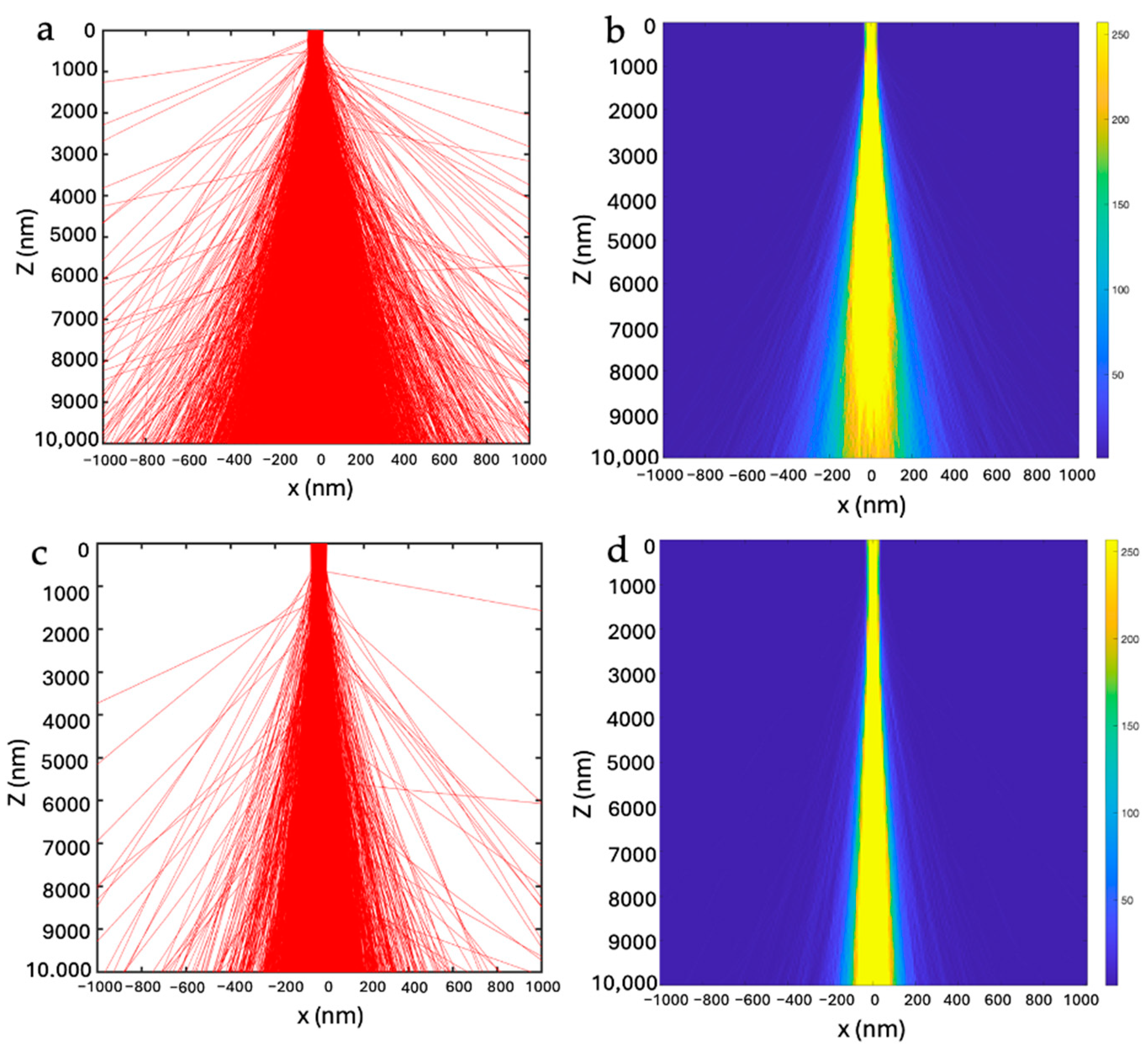
3.1. Electron Beam Profiles in Materials
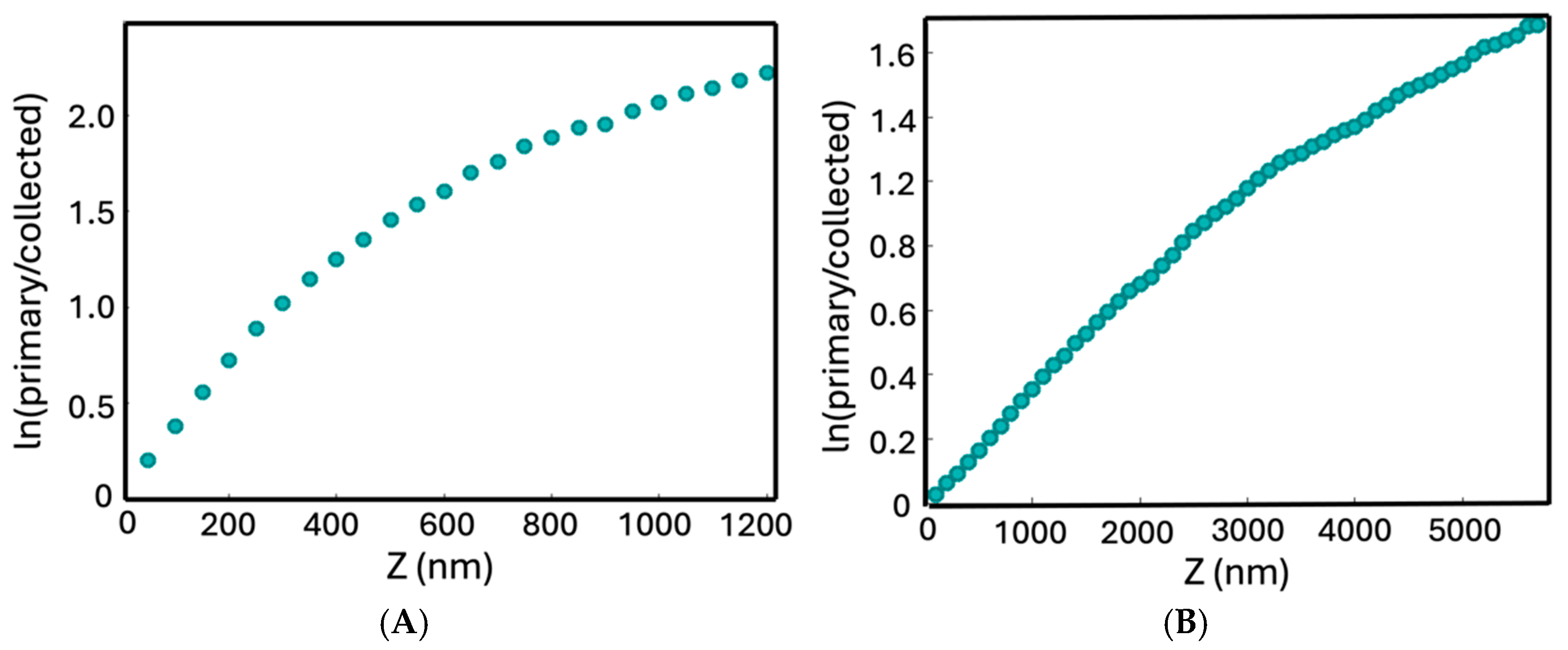
3.2. Beer–Lambert Law at Low and High Energies
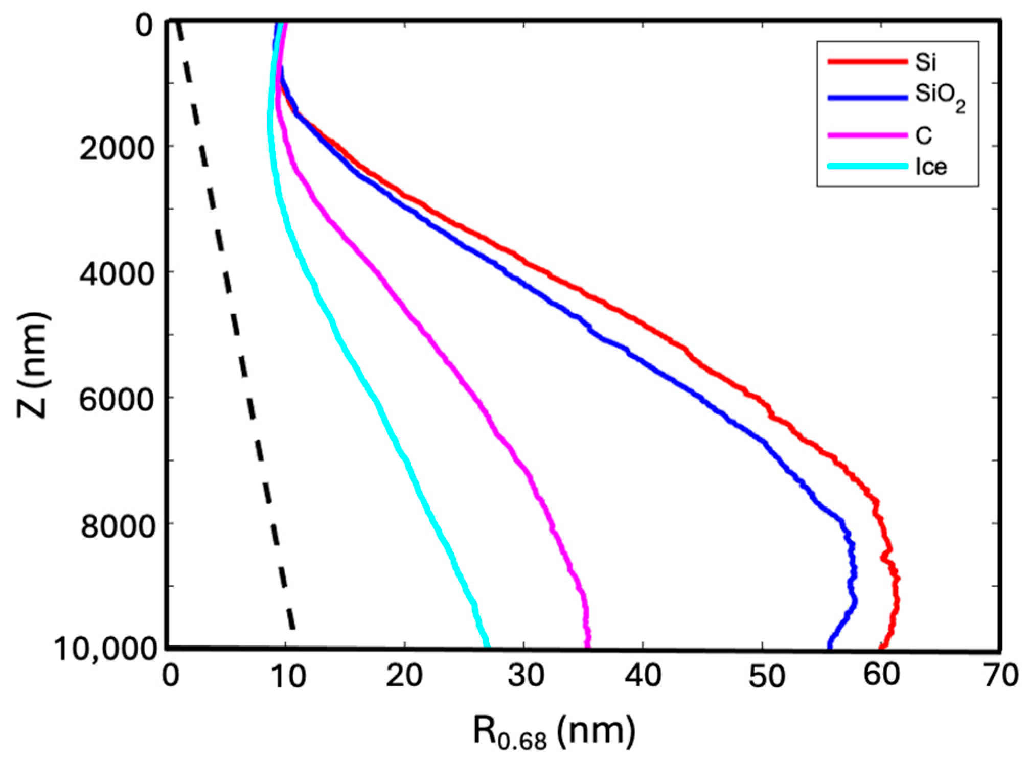
3.3. Effect of Sample Material on Beam Profile and Decay
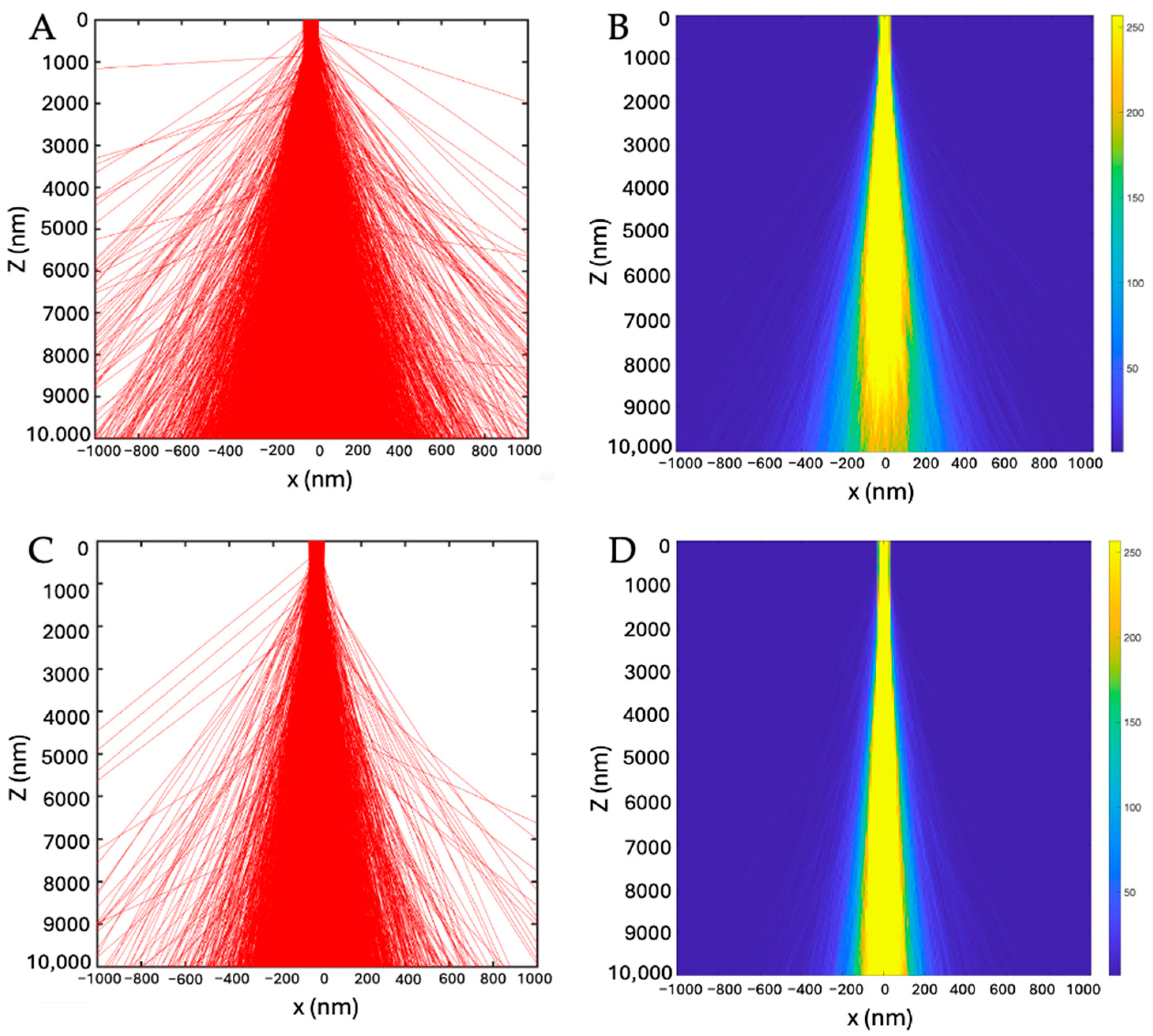
3.4. Effects of Convergence Semi-Angle and Imaging Modes
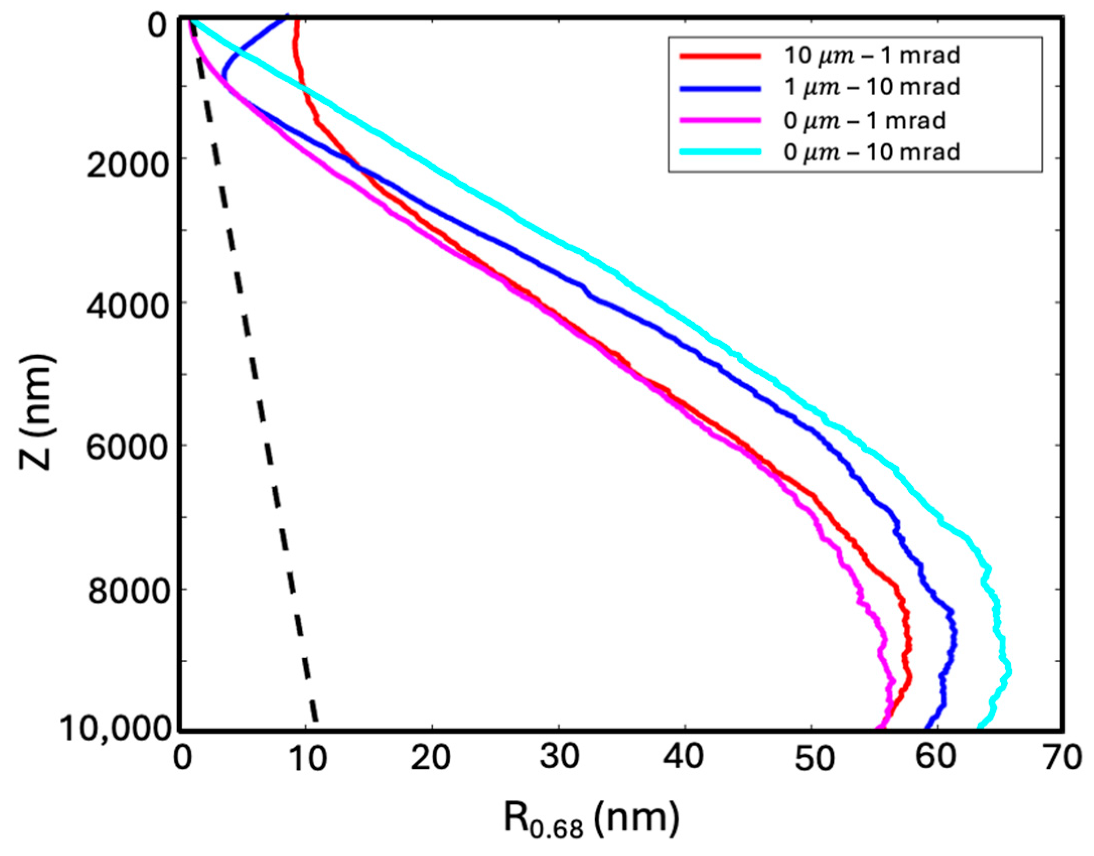
- When the beam is focused on the top surface, a larger convergence semi-angle produces wider beams, thus larger beam width: top-focused (at 0 µm) with α = 1 mrad was 55.0 nm, and top-focused with α = 10 mrad was 63.1 nm (pink and cyan curves in Figure 4).
- When the beam convergence semi-angle is set at 1 mrad, a deeper focus produces a wider beam in the top region of the sample, but the beam width is similar at locations deeper than 3 µm (pink and red curves in Figure 4). This observation is similar to what has been observed for a thinner sample [15].
- When the beam width is fixed at the top surface by either focusing the α = 10 mrad beam at 1 µm or focusing the α = 1 mrad beam at 10 µm (blue and red curves in Figure 4), the more diverged beam produces a narrower beam up to 2 µm in depth. This might be because the α = 10 mrad beam continues to focus after entering the sample. After that, the more diverged beam becomes wider than the less diverged beam as expected.
3.5. Beer–Lambert Law for Varying Materials at 3 MeV
4. Discussion
Author Contributions
Funding
Data Availability Statement
Conflicts of Interest
Appendix A
References
- Cooper, C.; Thompson, R.C.A.; Clode, P.L. Investigating parasites in three dimensions: Trends in volume microscopy. Trends Parasitol. 2023, 39, 668–681. [Google Scholar] [CrossRef]
- Collinson, L.M.; Bosch, C.; Bullen, A.; Burden, J.J.; Carzaniga, R.; Cheng, C.; Darrow, M.C.; Fletcher, G.; Johnson, E.; Narayan, K.; et al. Volume EM: A quiet revolution takes shape. Nat. Methods 2023, 20, 777–782. [Google Scholar] [CrossRef] [PubMed]
- Smith, D.; Starborg, T. Serial block face scanning electron microscopy in cell biology: Applications and technology. Tissue Cell 2019, 57, 111–122. [Google Scholar] [CrossRef] [PubMed]
- Xu, C.S.; Hayworth, K.J.; Lu, Z.; Grob, P.; Hassan, A.M.; García-Cerdán, J.G.; Niyogi, K.K.; Nogales, E.; Weinberg, R.J.; Hess, H.F. Enhanced FIB-SEM systems for large-volume 3D imaging. elife 2017, 6, e25916. [Google Scholar] [CrossRef]
- Micheva, K.D.; Smith, S.J. Array tomography: A new tool for imaging the molecular architecture and ultrastructure of neural circuits. Neuron 2007, 55, 25–36. [Google Scholar] [CrossRef]
- Capua-Shenkar, J.; Varsano, N.; Itzhak, N.-R.; Kaplan-Ashiri, I.; Rechav, K.; Jin, X.; Niimi, M.; Fan, J.; Kruth, H.S.; Addadi, L. Examining atherosclerotic lesions in three dimensions at the nanometer scale with cryo-FIB-SEM. Proc. Natl. Acad. Sci. USA 2022, 119, e2205475119. [Google Scholar] [CrossRef] [PubMed]
- Schertel, A.; Snaidero, N.; Han, H.-M.; Ruhwedel, T.; Laue, M.; Grabenbauer, M.; Möbius, W. Cryo FIB-SEM: Volume imaging of cellular ultrastructure in native frozen specimens. J. Struct. Biol. 2013, 184, 355–360. [Google Scholar] [CrossRef] [PubMed]
- Spehner, D.; Steyer, A.M.; Bertinetti, L.; Orlov, I.; Benoit, L.; Pernet-Gallay, K.; Schertel, A.; Schultz, P. Cryo-FIB-SEM as a promising tool for localizing proteins in 3D. J. Struct. Biol. 2020, 211, 107528. [Google Scholar] [CrossRef]
- Vidavsky, N.; Akiva, A.; Kaplan-Ashiri, I.; Rechav, K.; Addadi, L.; Weiner, S.; Schertel, A. Cryo-FIB-SEM serial milling and block face imaging: Large volume structural analysis of biological tissues preserved close to their native state. J. Struct. Biol. 2016, 196, 487–495. [Google Scholar] [CrossRef]
- Raguin, E.; Weinkamer, R.; Schmitt, C.; Curcuraci, L.; Fratzl, P. Logistics of Bone Mineralization in the Chick Embryo Studied by 3D Cryo FIB-SEM Imaging. Adv. Sci. 2023, 10, e2301231. [Google Scholar] [CrossRef]
- Turk, M.; Baumeister, W. The promise and the challenges of cryo-electron tomography. FEBS Lett. 2020, 594, 3243–3261. [Google Scholar] [CrossRef] [PubMed]
- Yang, X.; Wang, L.; Maxson, J.; Bartnik, A.C.; Kaemingk, M.; Wan, W.; Cultrera, L.; Wu, L.; Smaluk, V.; Shaftan, T.; et al. Towards Construction of a Novel Nanometer-Resolution MeV-STEM for Imaging Thick Frozen Biological Samples. Photonics 2024, 11, 252. [Google Scholar] [CrossRef]
- Wolf, S.G.; Shimoni, E.; Elbaum, M.; Houben, L. STEM Tomography in Biology. In Cellular Imaging: Electron Tomography and Related Techniques; Hanssen, E., Ed.; Springer International Publishing: Cham, Switzerland, 2018; pp. 33–60. [Google Scholar]
- Wolf, S.G.; Houben, L.; Elbaum, M. Cryo-scanning transmission electron tomography of vitrified cells. Nat. Methods 2014, 11, 423–428. [Google Scholar] [CrossRef] [PubMed]
- Wang, L.; Yang, X. Monte Carlo Simulation of Electron Interactions in an MeV-STEM for Thick Frozen Biological Sample Imaging. Appl. Sci. 2024, 14, 1888. [Google Scholar] [CrossRef]
- Langmore, J.P.; Smith, M.F. Quantitative energy-filtered electron microscopy of biological molecules in ice. Ultramicroscopy 1992, 46, 349–373. [Google Scholar] [CrossRef] [PubMed][Green Version]
- Composition of AMORPHOUS CARBON. Available online: https://physics.nist.gov/cgi-bin/Star/compos.pl?mode=text&matno=006 (accessed on 7 August 2024).
- Henins, I. Precision Density Measurement of Silicon. J. Res. Natl. Bur. Stand. A Phys. Chem. 1964, 68a, 529–533. [Google Scholar] [CrossRef] [PubMed]
- Available online: https://pubchem.ncbi.nlm.nih.gov/compound/24261 (accessed on 7 August 2024).
- Du, M.; Jacobsen, C. Relative merits and limiting factors for x-ray and electron microscopy of thick, hydrated organic materials. Ultramicroscopy 2018, 184 Pt A, 293–309. [Google Scholar] [CrossRef]
- Reimer, L.; Kohl, H. Transmission Electron Microscopy: Physics of Image Formation, 5th ed.; Springer: New York, NY, USA, 2008. [Google Scholar]
- Hayashida, M.; Malac, M. High-Energy Electron Scattering in Thick Samples Evaluated by Bright-Field Transmission Electron Microscopy, Energy-Filtering Transmission Electron Microscopy, and Electron Tomography. Microsc. Microanal. 2022, 28, 659–671. [Google Scholar] [CrossRef] [PubMed]
| Material | Collection Angle (0–10 mrad) | Collection Angle (10–50 mrad) |
|---|---|---|
| Ice | 41.43% | 53.18% |
| Carbon (a) | 27.87% | 58.96% |
| Silicon Dioxide | 15.67% | 51.14% |
| Silicon | 14.08% | 48.49% |
| Defocus Depth and Convergence Semi-Angle | 0 µm | 1 µm | 2 µm | 10 µm |
|---|---|---|---|---|
| 10/1 | 10,000 | 7565 | 5667 | 1567 |
| 1/10 | 7411 | 5513 | 4299 | 1533 |
| 0/1 | 10,000 | 7493 | 5559 | 1575 |
| 0/10 | 6928 | 5174 | 4124 | 1428 |
| Material | Linear Depth (µm) |
|---|---|
| Ice | 17.9 ± 0.9 |
| Carbon (a) | 9.1 ± 0.5 |
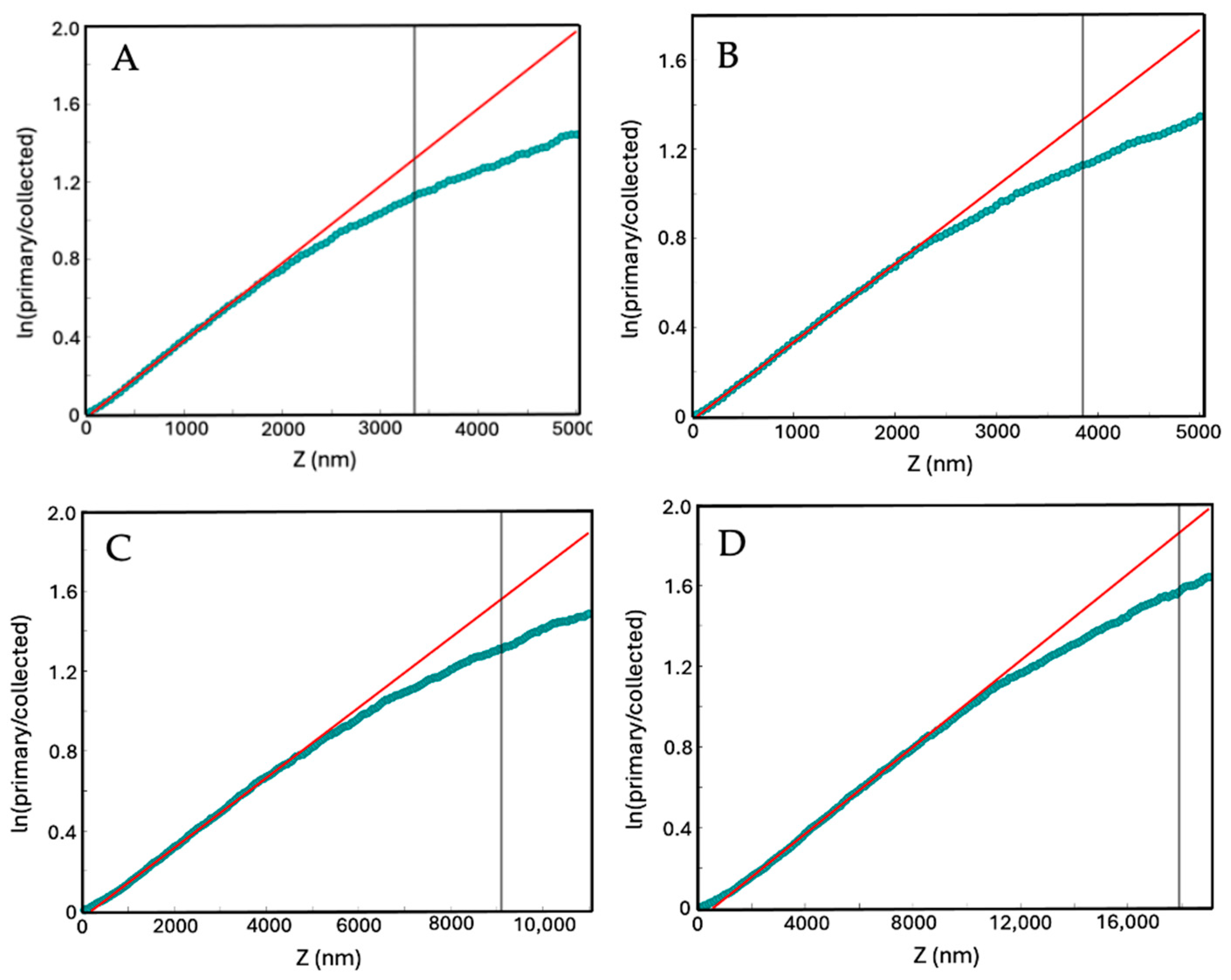
| Silicon Dioxide | 3.85 ± 0.19 |
| Silicon | 3.35 ± 0.17 |
Disclaimer/Publisher’s Note: The statements, opinions and data contained in all publications are solely those of the individual author(s) and contributor(s) and not of MDPI and/or the editor(s). MDPI and/or the editor(s) disclaim responsibility for any injury to people or property resulting from any ideas, methods, instructions or products referred to in the content. |
© 2025 by the authors. Licensee MDPI, Basel, Switzerland. This article is an open access article distributed under the terms and conditions of the Creative Commons Attribution (CC BY) license (https://creativecommons.org/licenses/by/4.0/).
Share and Cite
Quintard, B.; Yang, X.; Wang, L. Quantitative Modeling of High-Energy Electron Scattering in Thick Samples Using Monte Carlo Techniques. Appl. Sci. 2025, 15, 565. https://doi.org/10.3390/app15020565
Quintard B, Yang X, Wang L. Quantitative Modeling of High-Energy Electron Scattering in Thick Samples Using Monte Carlo Techniques. Applied Sciences. 2025; 15(2):565. https://doi.org/10.3390/app15020565
Chicago/Turabian StyleQuintard, Bradyn, Xi Yang, and Liguo Wang. 2025. "Quantitative Modeling of High-Energy Electron Scattering in Thick Samples Using Monte Carlo Techniques" Applied Sciences 15, no. 2: 565. https://doi.org/10.3390/app15020565
APA StyleQuintard, B., Yang, X., & Wang, L. (2025). Quantitative Modeling of High-Energy Electron Scattering in Thick Samples Using Monte Carlo Techniques. Applied Sciences, 15(2), 565. https://doi.org/10.3390/app15020565


