Article

“Dissolving the Evidence”: A Forensic Experimental Study on Tissue Destruction and Trace Detection

by
Larisa Adela Udriștioiu
1,2,*,
Ioana Dincă
3 and
George Cristian Curcă
1,2
1
Department of Legal Medicine and Bioethics, Faculty of Medicine, University of Medicine and Pharmacy “Carol Davila”, 050474 Bucharest, Romania
2
National Institute of Legal Medicine Mina Minovici, 042122 Bucharest, Romania
3
National Institute for Laser Plasma and Radiation Physics, 077125 Magurele, Romania
*
Author to whom correspondence should be addressed.
Appl. Sci. 2025, 15(19), 10347; https://doi.org/10.3390/app151910347
Submission received: 20 August 2025 / Revised: 16 September 2025 / Accepted: 22 September 2025 / Published: 24 September 2025

Abstract

This study presents a multidisciplinary forensic experiment evaluating the destructive effects of household chemical agents on animal bone and soft tissue analogues, with a particular focus on traumatic lesion persistence and trace evidence detection. A total of 59 domestic pig rib fragments, subjected to standardized lesions inflicted with either an axe or a ceramic knife, were immersed in acidic, basic, and oxidizing solutions for over two months. Samples were monitored through macroscopic scoring, serological species identification, and X-ray fluorescence (XRF) analysis. Results showed marked differences in tissue degradation depending on the chemical, with lesion persistence ranging from rapid obliteration to prolonged detectability. Axe-induced wounds generally remained visible longer than ceramic knife injuries, which tended to be erased earlier. XRF analysis revealed differential residue detection, with metallic traces persisting only under certain conditions, while serological testing demonstrated varying levels of protein preservation despite advanced tissue degradation. These findings underscore the forensic relevance of recognizing lesion persistence and chemical-specific degradation patterns, contributing to the assessment of chemical body disposal attempts and to the development of experimental training models for forensic practice.

1. Introduction

The deliberate dissolution of human remains using corrosive chemical agents has been sporadically reported in the forensic literature and often sensationalized in the media. The concept of “body disposal by dissolution” has evolved from anecdotal accounts to real-world forensic scenarios [1,2]. Methods involving acids, alkalis, or oxidizing substances have been associated with attempts to obscure identity, remove trauma evidence or hinder postmortem interval estimation [3,4,5]. Although a few scientifically documented cases exist [6,7,8], most available information still derives from media reports rather than systematic forensic investigations [9,10], making the true prevalence in forensic practice difficult to establish. This rarity of documented cases underscores the importance of experimental approaches to understand the phenomenon.
A further point of relevance is that many of the substances employed in reported cases are widely accesible as commercial products, such as household cleaning agents or industrial chemicals. Their ready availability increases the likelihood of misuse in crime concealment and justifies their inclusion in experimental settings.
Despite the recurring presence of such cases in criminal investigations, controlled experimental data on the destructive effects of corrosives remain scarce [11]. The scarcity arises from ethical and practical restrictions in using human material, variability in the types and concentrations of applied substances, and the inherent challenges of replicating clandestine disposal scenarios under standardized laboratory conditions. This study aims to fill part of that gap by evaluating the macroscopic evolution of soft and hard tissues subjected to immersion in widely accessible acidic, basic and oxidizing agents. Particular attention is given to the persistence of traumatic lesions and their detectability after prolonged exposure, as well as the utility of complementary methods—such as species identification via serological testing and X-ray fluorescence (XRF) spectrometry—to analyze and characterize the residues. To our knowledge, this is the first multidisciplinary experimental approach focusing on trauma–chemical interaction under these specific conditions.

2. Materials and Methods

2.1. Study Material

Rib fragments from domestic pigs (Sus scrofa domesticus) were selected due to their anatomical and biochemical similarity to human tissues, particularly in forensic decomposition and trauma analysis [12]. Rib samples were obtained as St. Louis–style ribs, consistently taken from the same anatomical portion (i.e., mid–lower rib segments without sternum or costal cartilages). All biological material used in this study was obtained from commercially available sources intended for food consumption, so that, for the specific purpose of this study, no animals were slaughtered or euthanized and no live animals were used. Therefore, ethical approval for animal research was not required. After acquisition, the samples were handled exclusively with plastic and glass instruments.

2.2. Study Solutions

We used commercially available cleaning solutions (detailed in Table 1). Substances were selected based on their accessibility in domestic settings and their possible application in criminal cases involving attempted cadaver disposal.
All chemical agents were handled in compliance with standard laboratory safety protocols, using personal protective equipment (PPE) and well-ventilated areas.

2.3. Tool Selection

Before immersion, on each rib fragment traumatic injuries were inflicted using a metallic object to simulate perimortem impact lesions. Tool selection was guided by forensic practice and literature on homicidal trauma homicides [13,14], initially including various cutting and impact instruments such as forged axe with a wooden handle, orthopedic saw, universal manual saw, bow saw, Tefal® kitchen knife (Groupe SEB, Lyon, France; manufactured in China), ceramic kitchen knife, and metal wire.

2.4. Documentation Protocol—Photography

Samples were photographed daily during the first week and every three days thereafter to document morphological changes and sharp force marks. A standardized photographic protocol using a Sony Alpha A7C (Sony Corporation, Tokyo, Japan; manufactured in Thailand) with a 90 mm macro lens was employed, consistent with methods used in previous sharp force trauma studies [15,16].

2.5. Serological Species Identification

For species identification purposes [17], anti-pig serum was used following the manufacturer’s protocol based on the Uhlenhuth precipitation reaction. This method detects antigen–antibody interactions through the formation of a visible precipitin ring. Each test was performed in triplicate to ensure reproducibility.

2.6. XRF Analysis Protocol

The XRF and radiographies analyses were conducted at the National Institute for Laser, Plasma & Radiation Physics. XRF measurements were conducted using an in-house-built Tomo-Analytic instrument [18] that integrates the micro-XRF method. The experimental configuration includes an X-ray source whit Mo target and energies up to 50 keV, a polycapillary X-ray lens that ensures a focal beam spot for excitation of ~50 µm, Si-PIN energy selective detector and a high precision XYZ motorized manipulator for sample mapping. The XRF spectra were acquired at an integration time of 120 s, X-ray source voltage of 40 kV and 200 µA current. To enable a reliable comparison between spectra obtained from different samples, the data were normalized relative to Ca peak-centered at 3.69 keV. The energy calibration procedure that was used is described in [19]. Before XRF measurement, each sample was examined first by the radiography in order to identify possible macroscopic contaminant from the weapon and to facilitate the sample positioning during the XRF measurements. If no contaminant were observed in radiographies, the entire wound section of the sample was scanned by XRF at an incremental step of 0.1 mm on the X-Y direction. The radiographies were acquired with a high-resolution nanofocus X-ray (YXLON FXE transmission X-ray tube) and a large 4k × 4k flat panel PerkinElmer detector that were incorporated into the customized sub-micron tomograph [20].

2.7. Pilot Study

A two-week pilot study on 12 pig rib fragments and the selected tools presented in Section 2.3. was conducted with two main objectives: first, to observe whether the tested substances were capable of altering intact samples, through macroscopic monitoring, in order to determine which agents would be further included in the main experiment; and second, to analyze by XRF both the composition of the selected instruments and their capacity to leave metallic traces on unaltered tissues, thereby identifying the most appropriate instruments. The pilot study conclusions are presented here, prior to the Section 3, to clarify the methodological decisions that follow: all solutions were included in the main experiment, while the forged axe and the ceramic knife were selected as the standard instruments for lesion infliction.

2.8. Conducting the Actual Research—Experimental Design

A total of 59 rib fragments domestic pigs were included in the study, given that the protocol initially comprised 60 (10 samples for each solution plus tap water for control, with 5 assigned to the ceramic knife and 5 to the axe), but one control sample was excluded prior to analysis due to accidental contamination during preparation. Soft tissues were preserved exactly as purchased, and the only procedure performed before immersion in the solutions was the infliction of standardized traumatic lesions using either the axe or the ceramic knife.
All lesions were inflicted by the same operator under standardized conditions. Each rib fragment was positioned on a stable wooden support in a uniform orientation. The point of application, cutting angle, and applied force were maintained as consistently as possible. Knife lesions consisted of five incisions per sample, producing narrow, linear cuts with sharp margins, whereas axe lesions consisted of three strikes per sample, resulting in deeper and wider chop wounds that reflected the distinct wounding capacity of each instrument.
The allocation of the samples to chemical groups was randomized and immersed samples were kept at 20 °C and monitored for over 2 months. Macroscopic changes were documented photographically at defined time points, and when relevant, samples were removed, air-dried at a constant temperature of 20 °C, and subsequently analyzed by XRF spectrometry and serological testing. Each condition was tested in duplicate to ensure reproducibility. An overview of the experimental workflow is provided below (Figure 1).
All macroscopic assessments were performed in a blinded manner by two independent evaluators. Discrepancies were resolved by consensus. Two semi-quantitative scoring systems (0–3) were applied.
Macroscopic evolution of the samples was evaluated using four descriptors: soft tissue evolution, bone modifications, general appearance, and final observations. For each descriptor, scores were assigned as: 0 = absent/no change; 1 = minimal change; 2 = moderate change; 3 = severe change. Soft tissue evolution included volume loss, color changes, and detachment. Bone modifications referred to cortical integrity, presence of cracks, and marrow extrusion. General appearance summarized overall visibility and preservation of the sample. Final observations described the endpoint status of the fragment after exposure.
Lesion detectability was determined based on macroscopic visibility, morphological clarity (edges, walls, and floor), and the ability to classify the lesion as traumatic rather than chemical/erosive. Scores ranged from 0 to 3: 0 = no detectable lesion; 1 = detectable but faint/unclear trace (vague discontinuity, no clear margins); 2 = detectable with partial morphological details preserved; 3 = fully detectable, sharp, intact lesion.

3. Results

3.1. Pilot Study Results (Only the Aspects Considered Relevant to the Actual Research)

For the first objective of the pilot study, macroscopic observations showed that all tested solutions produced alterations on the rib fragments, consistent with the results detailed in the following section. For the second objective, the XRF results for each instrument are presented in Table 2. We also considered the wear of the instruments, which we believe influenced the subsequent examination of metallic traces. In this regard, only the ceramic knife and the forged axe showed reliable results, with relevant intensity clearly above possible environmental contamination. This justified their selection as the standard instruments for lesion infliction in the main experiment.
Based on the findings of this pilot study, all corrosive solutions were included in the final experimental design, while only the forged axe and the ceramic kitchen knife were retained as lesion-producing instruments.

3.2. Actual Research Study Results

3.2.1. Macroscopic Evolution of Samples Immersed in Chemical Solutions

Significant chronological observations for each solution, in comparison with the samples kept in tap water, are summarized below.
Samples Freshly Extracted from Solution
  • Caustic Soda
The reaction was strongly exothermic from the outset, accompanied by rapid turbidity, gas release and fat dissolution (soft tissue evolution: 2). Early signs of saponification were evident by day 1, progressing to gelatinous transformation of the surface and initial cortical bone degradation by day 2 (bone modifications: 1–2; general appearance: 1). Between days 4 and 9, soft tissue detachment advanced, the bone marrow became exposed and discolored and superficial greenish hues appeared due to aerial exposure (soft tissue evolution: 3; bone modifications: 2; general appearance: 2). By day 21, the samples were friable but still retained partial lesion contours (lesion detectability: 2; final observations: 2). These changes occurred more rapidly in the samples with knife-inflicted lesions (Figure 2). Near-complete disintegration occurred between days 34 and 45 leaving only sub centimetric fragments and faint outlines of deeper, axe-inflicted, injuries (Figure 3) (lesion detectability: 0–1; bone modifications: 3; final observations: 3).
  • Promax—ACB
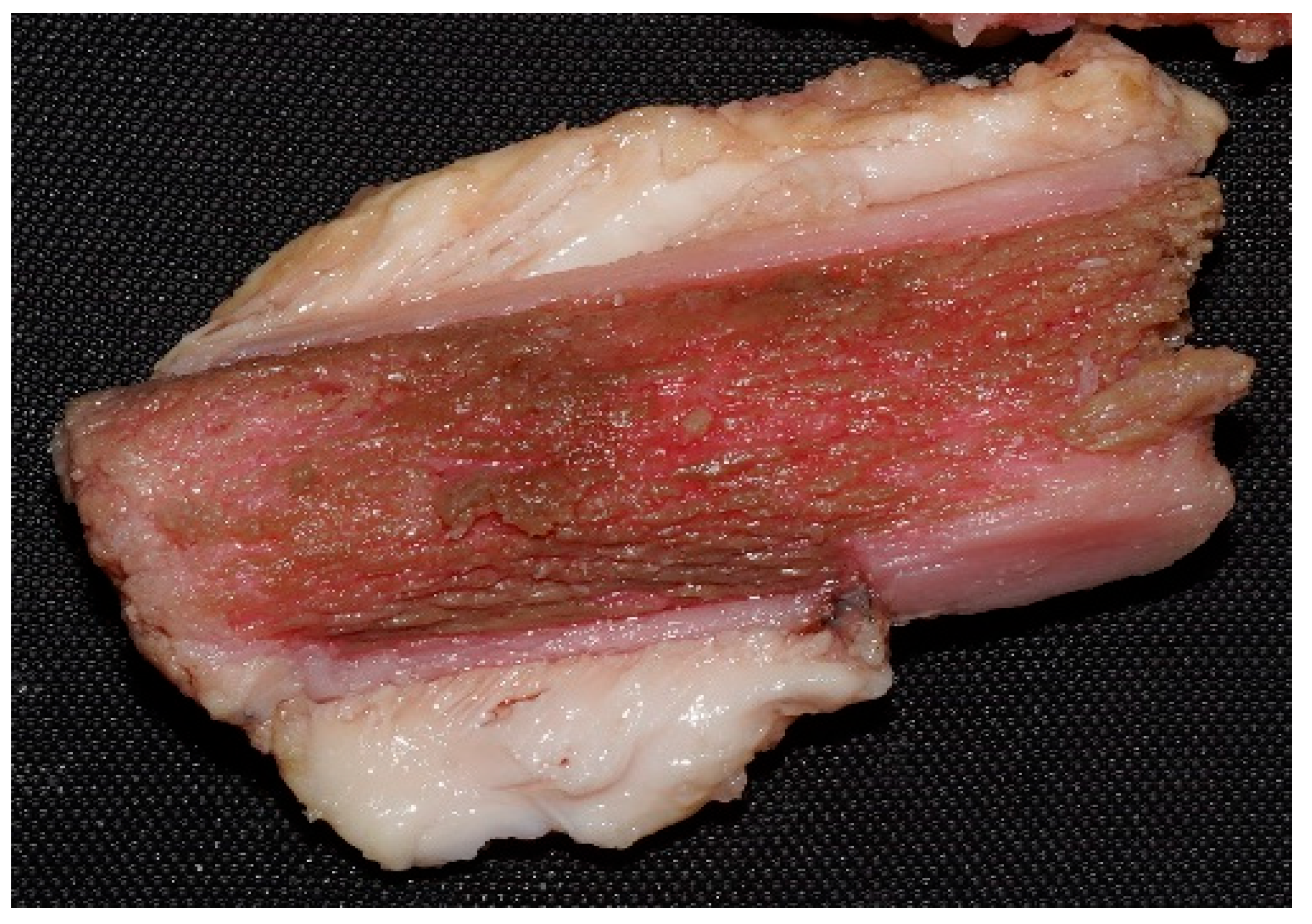
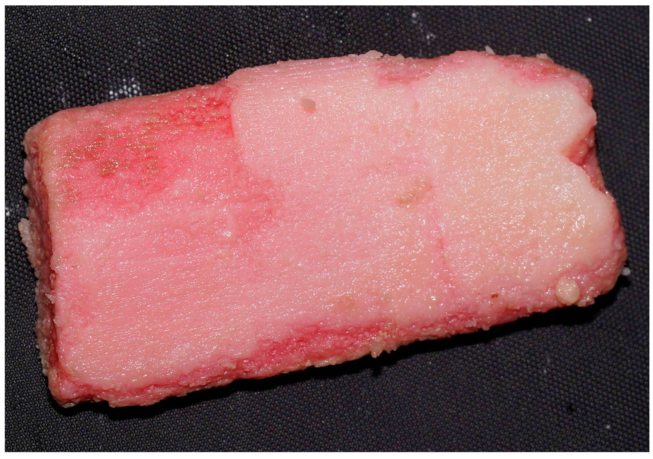
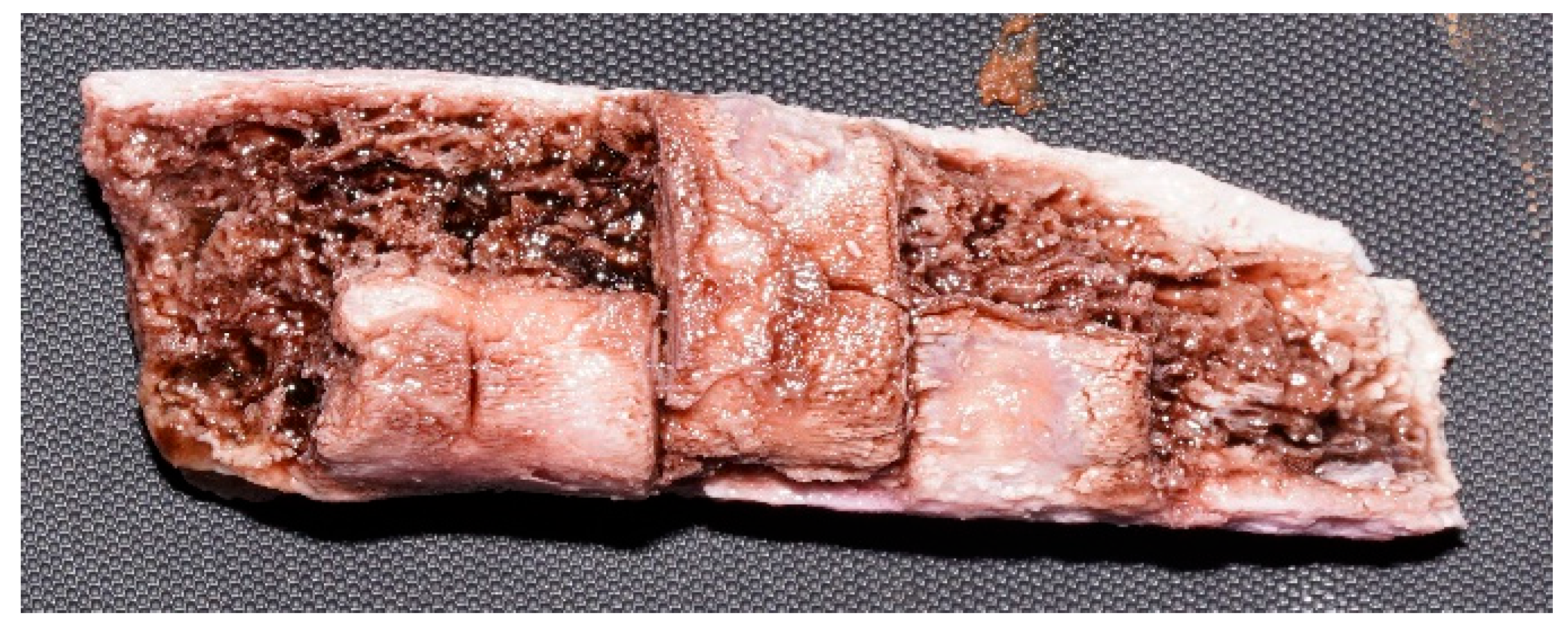
By day 1, soft tissues appeared matte and “salmon-like,” with whitening of the fat (soft tissue evolution: 1; general appearance: 1). Between days 3 and 4, bone marrow was lost through the lesions, exposing the bone (bone modifications: 1; lesion detectability: 3). By day 10, soft tissues were largely detached, bone contours were well defined and consistency increased (Figure 4) (soft tissue evolution: 3; bone modifications: 1–2; lesion detectability: 3; general appearance: 2). Between days 26 and 34, spongy bone whitening and cortical exfoliation were observed (Figure 5) (bone modifications: 1–2; general appearance: 2). At day 64, samples were nearly devoid of soft tissues, revealing firm, white bone with a cleaned appearance (soft tissue evolution: 3; bone modifications: 1–2; final observations: 2). No differences were observed between the evolution of samples with lesions inflicted by the ceramic knife and those inflicted by the axe.
  • Promax—FL
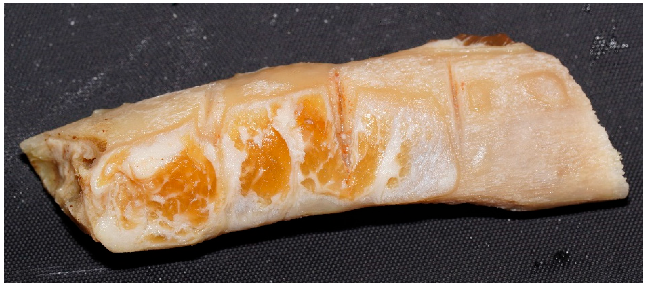
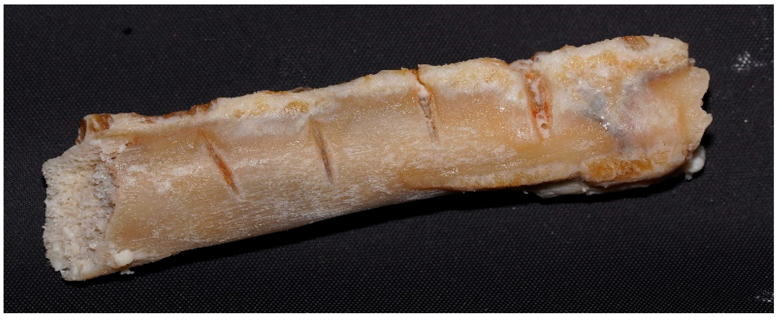
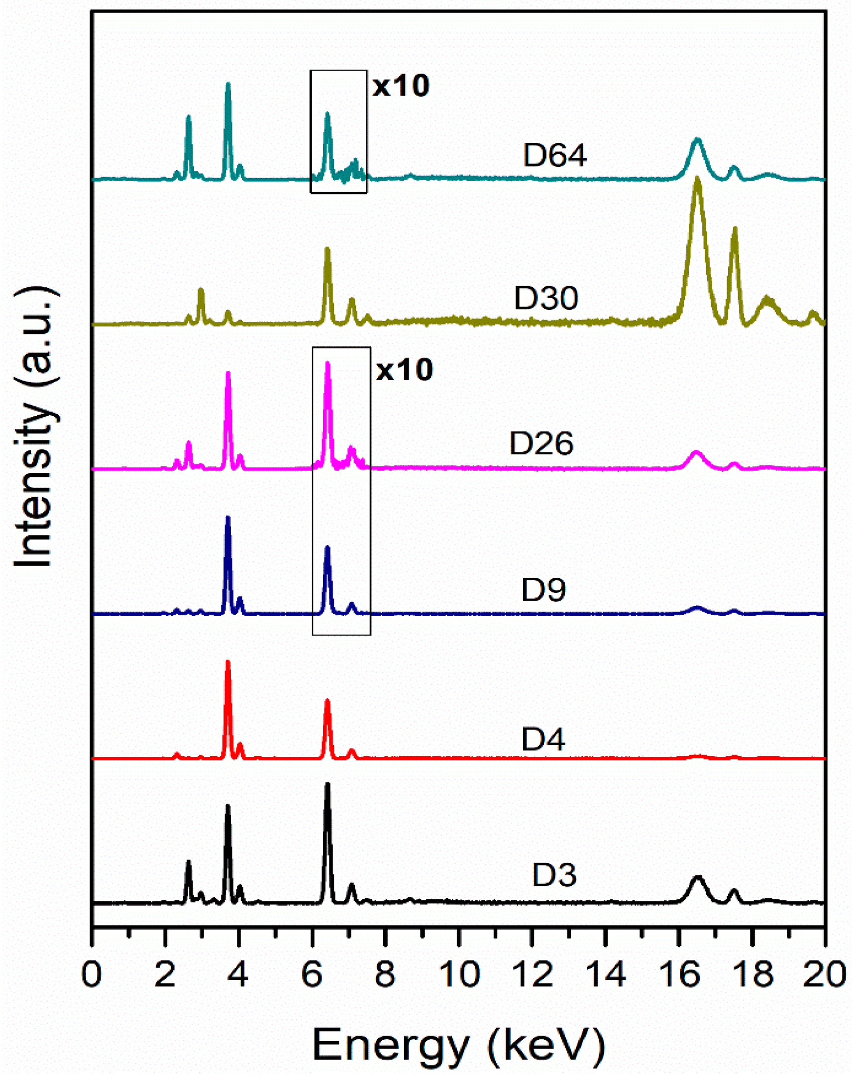
By day 1, samples exhibited a brown, waterlogged appearance with pale muscle tissue and dark bone marrow extruding through lesions (soft tissue evolution: 1–2; bone modifications: 1; lesion detectability: 3; general appearance: 1). Between days 2 and 4, soft tissue ruptures and longitudinal fine cracks developed, with wound margins retracting (soft tissue evolution: 2; bone modifications: 1–2; lesion detectability: 3; general appearance: 2). From day 5 onward, white surface deposits coalesced, and muscles acquired a “boiled meat” appearance (Figure 6) (soft tissue evolution: 2; general appearance: 2). At day 10, the samples became drier and elastic (soft tissue evolution: 2; general appearance: 2). Between days 26 and 45, lesions remained visible, but consistency declined, with deep fissures and initial fragmentation (Figure 7) (bone modifications: 3; lesion detectability: 2; final observations: 2). These changes were slightly faster and pronounced in samples with knife-inflicted lesions. By day 64, partial fragmentation was observed, although the general anatomical structure was still preserved (bone modifications: 3; general appearance: 2; final observations: 2).
  • Misavan
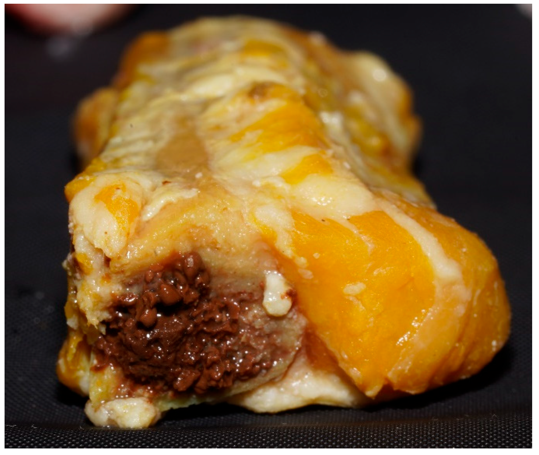
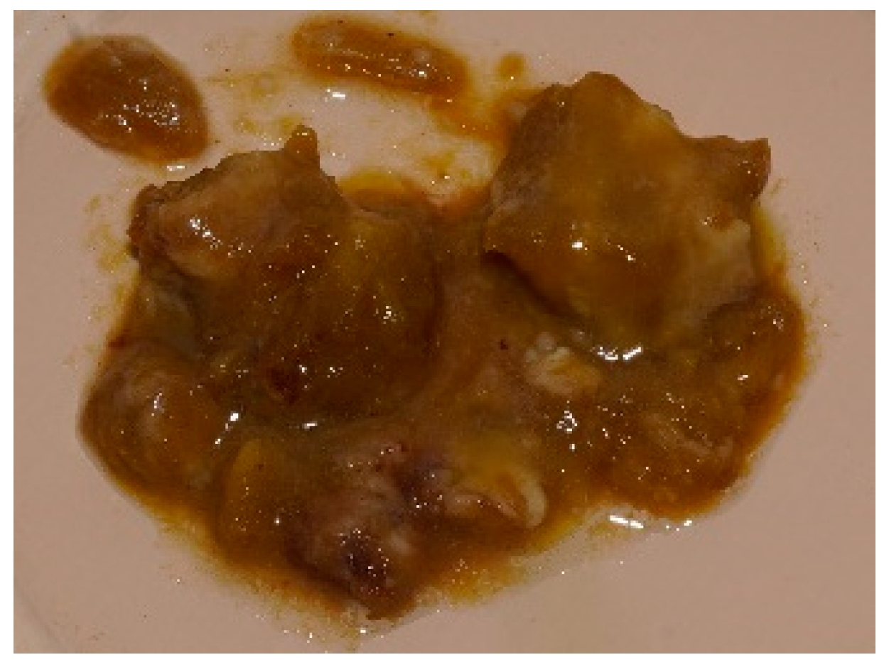
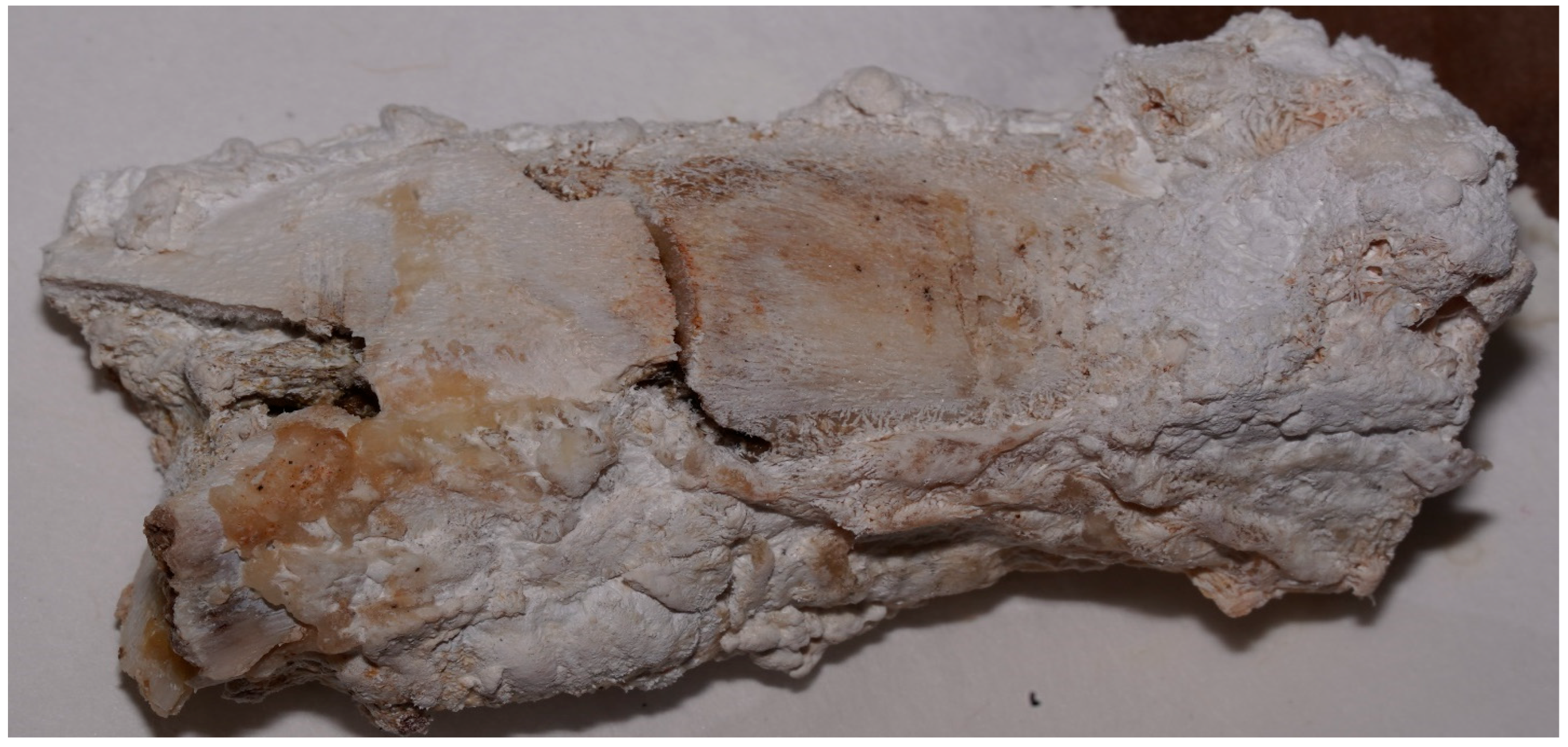
From day 1, samples showed an intense yellow-citrine coloration, with visible lesions and granular brown marrow extruding (Figure 8) (soft tissue evolution: 1; bone modifications: 1; lesion detectability: 3; general appearance: 1–2). By day 4, a soaked appearance developed (soft tissue evolution: 2; general appearance: 2), followed by increasing viscosity and irregular tissue consistency by days 9–10 (soft tissue evolution: 2–3; general appearance: 2). From day 18 onward, tissue integrity progressively declined (soft tissue evolution: 3; general appearance: 2–3). By day 34, even though the lesions were still visible, consistency was markedly reduced, leading to full softening and central collapse by day 36 (lesion detectability: 2; soft tissue evolution: 3; bone modifications: 2; general appearance: 3; final observations: 2). These changes were more marked in the samples with knife-inflicted lesions. At day 42, samples acquired a pasty texture (soft tissue evolution: 3; general appearance: 3), and by day 59, they had transformed into an amorphous paste (Figure 9) (soft tissue evolution: 3; bone modifications: 3; lesion detectability: 0; final observations: 3).
  • Dizol
By day 1, soft tissues were largely absent, and the sample had a soft, brown-violet appearance, visible lesions (soft tissue evolution: 3; general appearance: 2; lesion detectability: 2). By day 2, tissue remnants were liquefied, particularly in the marrow area, and lesion margins became indistinct (soft tissue evolution: 3; bone modifications: 2; lesion detectability: 1–2; general appearance: 2). At day 4, substantial tissue loss occurred (soft tissue evolution: 3; general appearance: 3) and by day 6, only partial traces of traumatic lesions remained (Figure 10) (lesion detectability: 1; final observations: 2). At day 9, all anatomical structures were lost, and bone was no longer macroscopically identifiable, prompting early termination of the experiment (soft tissue evolution: 3; bone modifications: 3; lesion detectability: 0; general appearance: 3; final observations: 3). These changes did not depend on the type of lesions present.
Descriptive aspects outlined above have been comparatively summarized for each substance in Table 3.
Samples Exposed to Air
At defined time points, samples were briefly removed from the solutions for photographic documentation and immediately returned to their containers, but when relevant macroscopic changes were observed, selected samples were instead permanently removed, air-dried in the same laboratory environment, maintained at a constant temperature of 20 °C, and subsequently subjected to XRF spectrometry and serological testing. This step also enabled the assessment of post-immersion changes under ambient conditions.
  • Caustic Soda
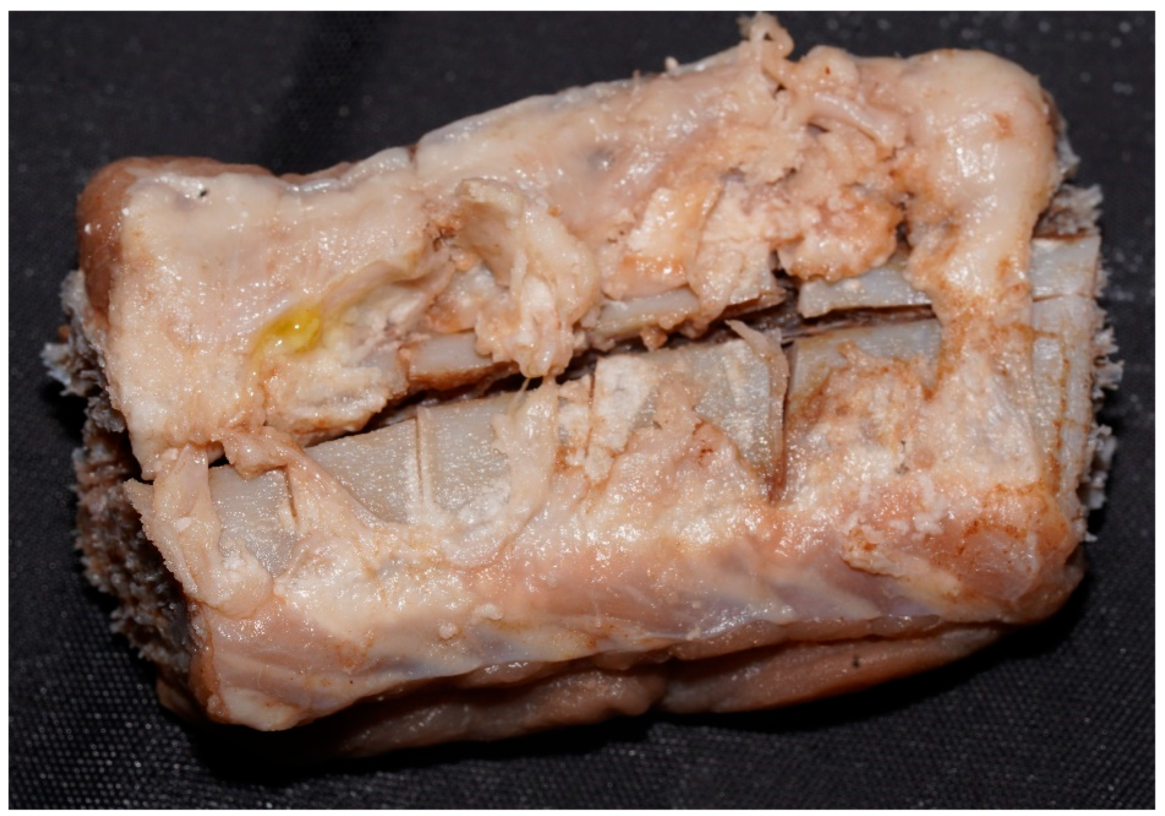
Exposure to air led to dehydration and soft tissue detachment (Figure 11), followed by the formation of fibrous, snowflake-like residues and a white, chalky, friable appearance (Figure 12). Within two weeks, samples became brittle and crumbly. Ultimately, all were fully disintegrated, particularly those removed later—described before as showing a greenish, oxidized tinge.
  • Promax—ACB
Samples exhibited increased firmness upon palpation. In those with deep lesions, wound edges became more prominent, forming raised contours relative to surrounding bone, with associated cortical bone whitening. Notably, bone marrow extrusion was absent.
  • Promax—FL
As drying progressed, samples developed a firmer consistency, losing their initial soaked appearance and acquiring a greasy outer layer. In deeper lesions, whitish discoloration became more evident, resembling immersion-phase deposits. Bone marrow darkened, while soft tissues turned brown, greasy, and cardboard-like. Early-removed samples remained firm, whereas those removed later became increasingly friable and disintegrated under light pressure.
  • Misavan
Samples displayed a globally greasy/oily surface. Initially firm, resembling controls, their consistency progressively declined over time. Fragmentation occurred easily during handling, following a longitudinal pattern, while the exposed surfaces gradually dried.
  • Dizol
Throughout the observation period, samples retained a moist, crumbly or pasty consistency. The tissues did not dry out completly and remained soft and easily disintegrable upon gentle handling.

3.2.2. XRF Measurement Results at Lesional Level

A preliminary XRF examination of all samples (N = 59) was conducted immediately after lesion infliction and prior to immersion, to verify the presence of metallic residues. Subsequent XRF analyses were performed not at fixed intervals, but rather when relevant macroscopic changes were observed during monitoring. (Figure 13).
The results of the examinations performed on the immersed samples, focusing on the inflicted lesions, are as follows:
Samples exposed to caustic soda: Lesions induced by the axe showed the presence of iron (Fe), but only during the early observation period (up to Day 4). For samples with ceramic knife-induced lesions, no metallic traces were detected by XRF measurements after immersion.
Samples exposed to Promax—ACB: Axe-induced lesions revealed the presence of Fe, but only during the initial days (up to Day 4) (see Figure 14). For samples with ceramic knife-induced lesions, no metallic traces were detected by XRF measurements after immersion.
Samples exposed to Promax-FL: Elemental residues were detected throughout the entire duration of the experiment for both instruments (see Figure 15 and Figure 16).
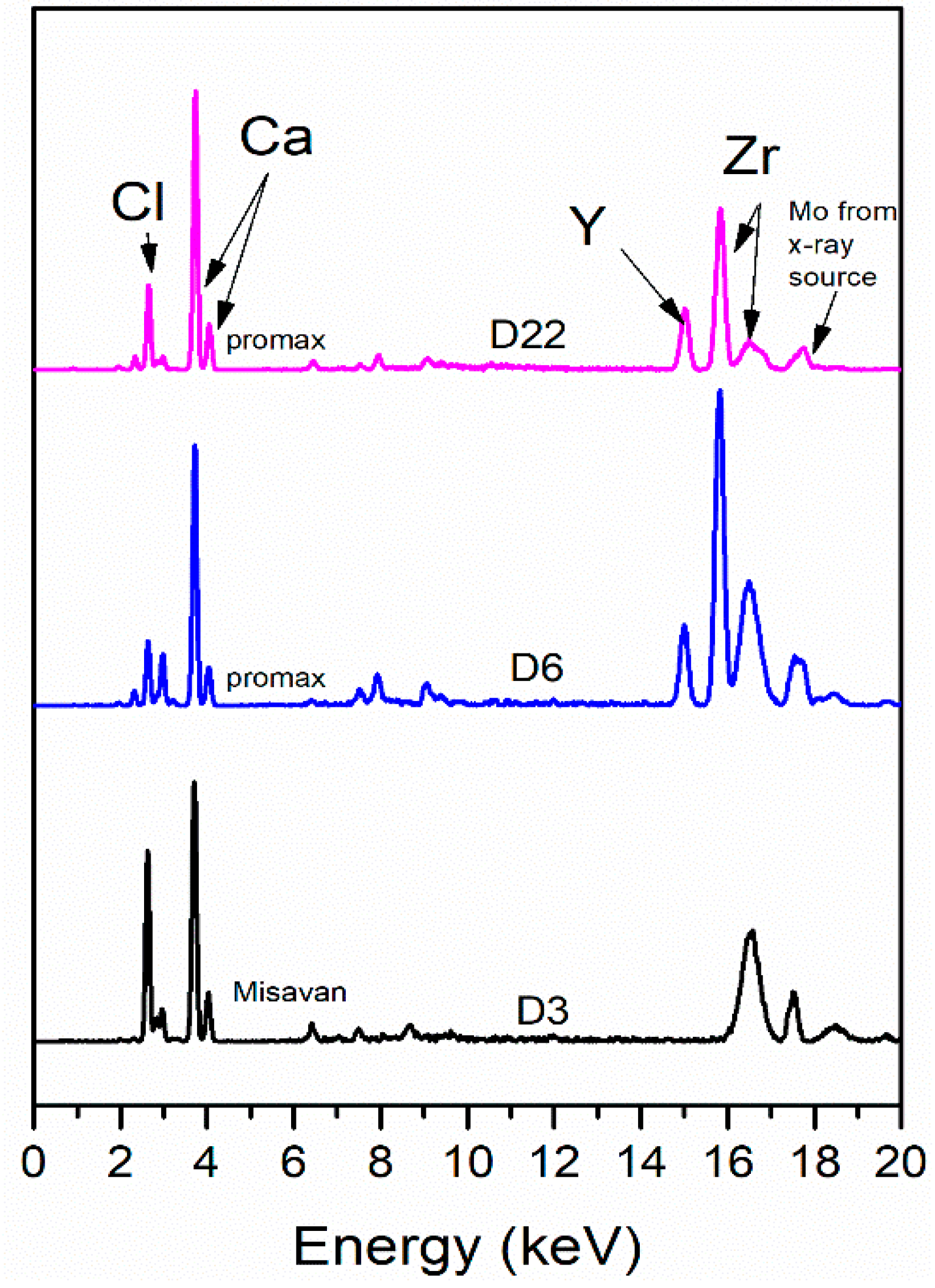
Samples exposed to Misavan: Residual elements from both instruments were detected only during the initial days—up to Day 6 for the ceramic knife and up to Day 4 for the axe (see Figure 16 and Figure 17).
Samples exposed to Dizol: No detectable elemental residues were observed.
A summary of the XRF results presented above is provided in Table 4.

3.2.3. Serological Results (Species Identification)

Samples exposed to Caustic soda and Misavan: Species identification remained positive throughout the entire duration of the experiment (see Figure 18).
Samples exposed to Promax-ACB: Species identification was successful only on Day 1, with no detectable identification afterward.
Samples exposed to Promax-FL and Dizol: All tests yielded negative results (see Figure 19).
A summary of all results of the actual research is provided in Table 5.

4. Discussion

4.1. Methodological Considerations and Forensic Implications

This study aimed to investigate the macroscopic effects of chemical substances on samples containing both bone and soft tissue, in order to better replicate real-life forensic scenarios. Unlike models where soft tissues are mechanically or chemically removed means [21,22], this approach preserves tissue architecture and reflects common disposal methods encountered in forensic practice.
Commercially available substances were used without concentration adjustments (except for the dilution of caustic soda flakes), to simulate real-world conditions where perpetrators typically use household products rather than specialized chemicals noted [23,24,25,26,27]. These substances can mimic traumatic features—such as fractures, soft tissue retraction, or cortical detachment—complicating trauma interpretation.
Two tools were selected for experimental injuries: a ceramic knife and an axe, based on their differing compositions and observed residue patterns. The ceramic knife often left micro-particles. Similar findings regarding ceramic knives have been reported in other studies using SEM-EDX analysis [28]. We explained this result due to its brittle nature of the ceramic blade, which makes it more likely to develop micro-striations and leave behind residual particles while the axe—especially when worn or rusted—produced deeper, more detectable traces. A metal wire was also included in the pilot study to simulate body restraint, not to induce trauma.

4.2. Macroscopic, Serological, and Elemental Findings: Substance-Specific Observations

In the next section, sample evolution is presented by combining macroscopic findings with lesion visibility, XRF detection, and serological outcomes, all framed within their forensic relevance. For clarity, the results are organized by chemical agent, following the same order as in Table 1.

4.2.1. Caustic Soda

For the only caustic substance tested in this study, the results were among the most destructive. The use of this substance in forensic anthropology is not new [29,30]. Caustic soda has long been employed as a method for cleaning skeletal remains to allow for clearer examination, but its destructive potential is also acknowledged.
In our study rapid degradation of soft tissues and progressive cortical bone thinning was observed from the first days of exposure. The substance triggered an exothermic reaction and extensive fat saponification, resulting in the formation of sodium soap and a characteristic pink-reddish discoloration—attributed to its effect on hemoglobin, which turned green upon air exposure due to oxidation. Soft tissue destruction followed a pattern from the exterior inward, while bone degradation occurred primarily via cortical flaking off and longitudinal defragmentation.
Lesion characteristics influenced the degradation pattern: ceramic knife wounds, being more numerous and superficial, facilitated uniform solution penetration and faster bone erosion at section margins—often erasing macroscopic lesion features by day 45. In contrast, axe-induced lesions, though fewer and deeper, remained visible up to day 59, with some traces detectable even in small fragments.
The action of caustic soda appeared to be more rapid and concentrated on soft tissues, in contrast to its effect on bone, which it fragmented mainly through penetration and massive impregnation. This observation is consistent with previous cases, such as that of Leonarda Cianciulli [31].
After removal from the solution and exposure to air, the samples developed a chalky white, friable surface with progressive powder-like disintegration. This appearance—also noted in historical forensic cases [32], supports the potential forensic diagnostic value of such transformations as indicators of caustic exposure.
XRF analysis showed no detectable metallic residues in samples with ceramic knife lesions, due to accelerated material loss. However, iron traces were still present in axe-inflicted wounds up to day 4. Despite the aggressive chemical action, serological species identification using anti-species sera remained positive throughout the 59-day exposure period, indicating preserved protein structures. This is consistent with another author’s study, in which DNA was still recoverable after 28 days of immersion [33]. It has also been shown that stable isotope analysis is not affected after a 20 h treatment with NaOH [34].

4.2.2. Promax-ACB

This hypochlorite solution induced moderate tissue alteration, with a clear predilection for soft tissues. Early macroscopic changes included a salmon-like hue of the muscle and whitish discoloration of adipose tissue. Bone marrow gradually detached and accumulated at the bottom of the container, leaving bones emptied but intact.
Soft tissues detached progressively, while bone lesions remained visible with sharply defined margins. Toward the end of the experiment, bone consistency increased and minimal cortical exfoliation was noted. Overall, the solution exhibited a “cleaning” rather than destructive effect, aiding soft tissue removal while preserving bone structure. The evolution was similar for all samples, regardless of the lesions present.
However, this preservation effect impaired species identification beyond the initial days, likely due to protein denaturation. Similarly, XRF analysis was limited: metallic residues from the axe were detectable only in early stages, while increased calcium signals and chlorine detection pointed to exposed bone surfaces and residual hypochlorite interaction.

4.2.3. Promax-FL

From day 1, macroscopic changes included brownish discoloration and whitish fat deposits, with bone marrow protrusion at sectioned surfaces—similar to observations with Misavan.
Despite the higher hydrochloric acid concentration, bone degradation was limited and structural collapse was not observed. Instead, samples developed an elastic consistency over time. No significant differences were noted between lesions produced by the ceramic knife and those by the axe. However, inter-lesional degradation appeared more pronounced in samples with multiple, superficial injuries.
A longitudinal pattern of cortical degradation, akin to that seen in caustic soda, was observed. Wound edges showed signs of retraction, likely due to a dehydration-like effect. The action of hydrochloric acid appeared more relevant than that of sulfuric acid, although a potential synergistic effect cannot be ruled out.
Species identification failed in all samples, due to molecular-level alterations, despite limited visible degradation. In contrast, XRF analysis yielded strong results, with metallic residues detected even at the experiment’s conclusion—likely preserved at lesion edges beneath surface deposits.

4.2.4. Misavan

This viscous yellow solution containing hydrochloric acid, produced early and progressive changes, particularly affecting the bone marrow, which liquefied and protruded through lesions by day 1. Thin soft tissue layers detached rapidly, while deeper layers persisted but became increasingly heterogeneous. Over time, sample consistency decreased, and the bones underwent gradual decalcification, leading to structural collapse and the transformation of specimens into a pasty mass by day 59.
Macroscopic examination of dried samples revealed the absence of bone beneath a residual shell of soft tissue, confirming the acid’s preferential action on osseous structures, also noted by other authors [27]. The effect of this solution on bone tissue—rather than on soft tissues—was further confirmed through observations of the dried samples, which revealed that, over the course of the experiment, a “shell” of dried soft tissue had formed in the absence of any preserved bony structure. This supports the conclusion that hydrochloric acid acts more aggressively on bone compared to soft tissues. The extraction of calcium from bone was also noted by Sabolová et al. [22].
Lesions remained visible until approximately day 36, after which bone collapse rendered them indistinct. Similarly to caustic soda, a higher number of superficial lesions (samples with ceramic knife-inflicted injuries) facilitated deeper acid penetration and accelerated degradation at the lesion sites.
Despite advanced degradation, species identification via anti-species sera remained possible throughout the experiment, suggesting minimal impact of hydrochloric acid on soft tissue protein structures—even from the paste-like material recovered at the end of the experiment—raising the question of whether a longer exposure would have led to complete tissue dissolution. XRF analysis detected metallic residues from both tools in the first 6 days; however, subsequent detection was hindered by acid-induced decalcification and loss of bone surface, limiting the effectiveness of this surface-sensitive technique.

4.2.5. Dizol

This highly concentrated sulfuric acid solution caused the most rapid and aggressive degradation among all substances tested, although not as immediate as reported in a recent study [35]. Complete loss of soft tissues was observed from day 1, with visible bone alteration or loss in certain areas. Despite prior literature suggesting slower bone degradation [36] in this 9-day experiment, both soft and hard tissues were extensively affected, with no notable difference between ceramic knife and axe lesions. Tissue breakdown advanced rapidly from the exterior inward.
Lesions remained partially visible early on, but margins degraded quickly, and by the end of the experiment, only a single infracentimetric bone fragment remained per sample. Unlike other substances, Dizol caused uniform dissolution without producing loose fragments or particulate residues. The remaining solution increased in viscosity as tissue degradation progressed.
XRF analysis was severely limited by the loss of identifiable bone structure and the dissolution of tissues with any metallic residues. Species identification using anti-species sera yielded poor results, consistent with protein denaturation caused by sulfuric acid [37].
These findings confirm sulfuric acid’s potent destructive capacity, particularly when applied to intact or sectioned tissue, though not necessarily matching the speed often claimed in anecdotal or criminal accounts insiders [3].

4.3. Molecular Considerations on Chemical-Tissue Interactions

Although the present study was not designed to investigate tissue degradation at a molecular level, certain macroscopic and serological findings allow for logical inferences regarding underlying biochemical processes. The observed differences may reflect the distinct chemical pathways of acid hydrolysis, alkaline saponification and oxidative degradation. For instance, the use of caustic soda likely induced alkaline hydrolysis of lipids and proteins, as evidenced by visible saponification (formation of sodium soaps) and the gelatinous, friable appearance of soft tissues. This suggests peptide bond cleavage and secondary structure disruption in proteins [38], which correlates with the preservation of antigenic sites for species identification but loss of structural integrity.
In contrast, Dizol (sulfuric acid) exhibited strong protein denaturation and dehydration capacity [39], consistent with the near-total dissolution of tissues and the inability to detect species-specific proteins. Sulfuric acid’s hygroscopic nature also promotes coagulative necrosis and cross-linking of tissue proteins, impeding antigen–antibody interactions.
Hydrochloric acid-based products (Misavan, Promax-FL) appear to act primarily through acid hydrolysis and decalcification [40], targeting the inorganic matrix of bone via calcium dissolution, while causing less aggressive protein degradation—supported by partially preserved soft tissue architecture and positive serological reactions in Misavan.
Sodium hypochlorite (Promax-ACB), a potent oxidizing agent, likely caused protein oxidation and fragmentation [41], leading to partial structural preservation but early loss of antigenicity, as indicated by negative species identification after Day 1.
These differential molecular mechanisms, though not directly measured, are strongly supported by the observable macroscopical degradation patterns and analytical outcomes, underscoring the forensic value of correlating chemical-tissue interactions across scales—from molecular to anatomical.

4.4. Limitations and Further Directions

This study was conducted exclusively on animal tissues, which, although taphonomically relevant, cannot fully replicate human anatomical and chemical characteristics—so extrapolation to human cadaveric decomposition should be made with caution. Additionally, the use of commercially available products introduces variability due to potential changes in formulation. Advanced biochemical assessments such as proteomics or DNA analysis were not performed. The macroscopic and spectrometric evaluations, while informative, are limited in detecting molecular-level changes. Future research should aim to validate these findings on human tissues and incorporate genetic and biochemical analyses. Studies using micro-CT or infrared spectroscopy may provide deeper insight into subsurface changes and mineral loss. Histological analysis of degraded tissues may also enhance understanding of microscopic alterations and contribute to more robust forensic interpretations. Additionally, evaluating the time-dependent kinetics of chemical interactions could aid in refining postmortem interval estimates in chemically treated remains.

5. Conclusions

To our knowledge, this is the first study to emphasize the importance of employing multidisciplinary methods—macroscopic analysis, XRF measurements and serological testing—for the assessment of traumatic lesions and the interaction of tissues with various chemical substances. The evolution of the samples depending on the solutions used revealed significant differences in the behavior of both soft tissues and bone, confirming that each substance acts in a distinct manner: Caustic soda and Dizol caused extensive destruction, rapidly compromising both bone structure and lesion visibility; Promax-ACB on the other hand, preserved the bone and aided in its cleaning; hydrochloric acid in Misavan led to bone decalcification while relatively preserving the soft tissues; and Promax-FL showed an intermediate effect. Furthermore, the type of instrument used influenced the progression of the samples, with observable differences between lesions produced by the axe and those by the ceramic knife, particularly in the distribution of metallic traces. It was also found that the number of lesions—more than their depth—affected the penetration of substances into the samples. Species identification was possible depending on the type of substance and exposure duration, with the most favorable results obtained in solutions with a lower impact on proteins. These findings support forensic practitioners in identifying chemical body disposal attempts and assessing lesion persistence under corrosive exposure. This experimental model could serve as a reference system for training forensic personnel in recognizing chemical tissue degradation in simulated environments.

Author Contributions

Conceptualization, L.A.U. and G.C.C.; methodology, L.A.U.; investigation, L.A.U. and I.D.; resources, L.A.U.; data curation, L.A.U. and I.D.; writing—original draft preparation, L.A.U. and I.D.; writing—review and editing, L.A.U.; visualization, L.A.U. and I.D.; supervision, G.C.C. All authors have read and agreed to the published version of the manuscript.

Funding

This research received no external funding.

Institutional Review Board Statement

Not applicable.

Informed Consent Statement

Not applicable.

Data Availability Statement

The raw data supporting the conclusions of this article will be made available by the authors on request.

Acknowledgments

We kindly thank the laboratory team for their valuable support in performing the analyses.

Conflicts of Interest

The authors declare no conflicts of interest.

References

  1. Silvestre, C.R. Looking for El Pozolero’s Traces: Identity and Liminal Condition in the War on Drug’s Disappearances. Front. Norte 2014, 26, 5–23. [Google Scholar]
  2. Silva, J.A.; Ferrari, M.M.; Leong, G.B. The Case of Jeffrey Dahmer: Sexual Serial Homicide from a Neuropsychiatric Developmental Perspective. J. Forensic Sci. 2002, 47, 1347–1359. [Google Scholar] [CrossRef] [PubMed]
  3. Grillo, M.; Cascino, F.M.; Murgo, V.; Milone, L. Method of Concealment of Corpses in Mafia Related Homicides: Melting in Strong Acids. In Proceedings of the 63th Annual Scientific Meeting American Academy of Forensic Sciences, Chicago, IL, USA, 21–26 February 2011. [Google Scholar]
  4. Blau, S.; Ranson, D. Body in the Barrel: Complex Body Disposal and Recovery. In Case Studies in Forensic Anthropology: Bonified Skeletons; Garvin, H.M., Langley, N.R., Eds.; CRC Press: Boca Raton, FL, USA, 2020; pp. 91–98. [Google Scholar]
  5. Fulginiti, L.C.; Hartnett-McCann, K.M.; Di Monica, F. Sealed for Your Protection: A Triple Homicide Involving the Use of a Corrosive Agent to Obscure Identity. In Case Studies in Forensic Anthropology: Bonified Skeletons; Garvin, H.M., Langley, N.R., Eds.; CRC Press: Boca Raton, FL, USA, 2020; pp. 99–108. [Google Scholar]
  6. Pokines, J.T.; Springer, K. A Case of Localized Corrosion on Bone Caused by Chemical Contact. J. Forensic Identif. 2016, 66, 173–186. [Google Scholar]
  7. Hinkes, M.J.; Lucas, J. Body Rendering by Drug Cartels in San Diego: Case Study of Caustic Soda. Proc. Am. Acad. Sci. USA 2014, 20, 207. [Google Scholar]
  8. Pomara, C.; Gianpaolo, D.P.; Monica, S.; Maglietta, F.; Sessa, F.; Guglielmi, G.; Turillazzi, E. “Lupara Bianca” a Way to Hide Cadavers after Mafia Homicides. A Cemetery of Italian Mafia. A Case Study. Leg. Med. 2015, 17, 192–197. [Google Scholar] [CrossRef]
  9. Amadasi, A.; Camici, A.; Porta, D.; Cucca, L.; Merli, D.; Milanese, C.; Profumo, A.; Rassifi, N.; Cattaneo, C. Assessment of the Effects Exerted by Acid and Alkaline Solutions on Bone: Is Chemistry the Answer? J. Forensic Sci. 2017, 62, 1297–1303. [Google Scholar] [CrossRef]
  10. Amadasi, A.; Camici, A.; Sironi, L.; Profumo, A.; Merli, D.; Mazzarelli, D.; Porta, D.; Duday, H.; Cattaneo, C. The Effects of Acid and Alkaline Solutions on Cut Marks and on the Structure of Bone: An Experimental Study on Porcine Ribs. Leg. Med. 2015, 17, 503–508. [Google Scholar] [CrossRef]
  11. Udriştioiu, L.A.; Andrei, M.; Perde, F.; Curcă, G.C. Postmortem Tissue Alterations Induced by Corrosive Substances—A Scoping Review. J. Forensic Leg. Med. 2025, 109, 102794. [Google Scholar] [CrossRef]
  12. Matuszewski, S.; Hall, M.J.R.; Moreau, G.; Schoenly, K.G.; Tarone, A.M.; Villet, M.H. Pigs vs People: The Use of Pigs as Analogues for Humans in Forensic Entomology and Taphonomy Research. Int. J. Leg. Med. 2020, 134, 793–810. [Google Scholar] [CrossRef]
  13. Allen, T.; Fox, K.A. Multivariate Dimensions of Age, Gender, and Weapon Use in Spousal Homicides. Vict. Offender 2013, 8, 329–346. [Google Scholar] [CrossRef]
  14. Chan, H.C.O.; Beauregard, E. Choice of Weapon or Weapon of Choice? Examining the Interactions between Victim Characteristics in Single-Victim Male Sexual Homicide Offenders. J. Investig. Psychol. Offender Profiling 2016, 13, 70–88. [Google Scholar] [CrossRef]
  15. Stanley, S.A.; Hainsworth, S.V.; Rutty, G.N. How Taphonomic Alteration Affects the Detection and Imaging of Striations in Stab Wounds. Int. J. Leg. Med. 2018, 132, 463–475. [Google Scholar] [CrossRef]
  16. Komo, L.; Grassberger, M. Experimental Sharp Force Injuries to Ribs: Multimodal Morphological and Geometric Morphometric Analyses Using Micro-CT, Macro Photography and SEM. Forensic Sci. Int. 2018, 288, 189–200. [Google Scholar] [CrossRef]
  17. Komar, A.G.; Motronenko, V.V.; Gorshunov, Y.V. Serological Methods in Modern Biotechnologies and Their Bioanalytical Standardization. Biomed. Eng. Technol. 2019, 2, 108–116. [Google Scholar] [CrossRef]
  18. Dobrea, C.; Tiseanu, I.; Crăciunescu, T.; Grigore, E. Non Destructive Analysis of Large Area Metallic Coatings of Fusion Materials. Rom. J. Phys. 2011, 56, 69–73. [Google Scholar]
  19. Lungu, M.; Dobrea, C.; Tiseanu, I. Enhanced XRF Methods for Investigating the Erosion-Resistant Functional Coatings. Coatings 2019, 9, 847. [Google Scholar] [CrossRef]
  20. High Resolution Nanofocus X-Ray Facility—The Advanced X-Ray. Imagistics Lab. Available online: https://tomography.inflpr.ro/high_resolution_nanofocus_x-ray_facility/ (accessed on 23 July 2025).
  21. Rosa, J.; Batista de Carvalho, L.A.E.; Marques, M.P.M.; Ferreira, M.T.; Gonçalves, D.; Gil, F.P.S.C. XRF Identification of Sharp-Force Trauma in Fresh and Dry Human Bone under Varied Experimental Heat Conditions. Sci. Justice 2024, 64, 305–313. [Google Scholar] [CrossRef]
  22. Sabolová, V.; Brinek, A.; Sládek, V. The Effect of Hydrochloric Acid on Microstructure of Porcine (Sus Scrofa Domesticus) Cortical Bone Tissue. Forensic Sci. Int. 2018, 291, 260–271. [Google Scholar] [CrossRef]
  23. Cope, D.J.; Dupras, T.L. The Effects of Household Corrosive Chemicals on Human Dentition. J. Forensic Sci. 2009, 54, 1238–1246. [Google Scholar] [CrossRef]
  24. Dowell, T. Examining the Effects of Corrosive Household Chemicals on Bone and Tissue. Poster Presented at the Academic Excellence Showcase, Western Oregon University. 2012. Available online: https://core.ac.uk/download/pdf/228830539.pdf (accessed on 23 July 2025).
  25. Baglieri, G. The Effects of Household Corrosive Chemicals on Pig Bones and Human Tissue. In Proceedings of the 68th Annual Meeting of the American Academy of Forensic Sciences, Las Vegas, NV, USA, 22–27 February 2016. [Google Scholar]
  26. Fey, S. Examination of the Effects of Household Corrosive Substances on the Dissolution of Complete Pig (Sus Scrofa) Carcasses. Master’s Thesis, Boston University, Boston, MA, USA, 2022. [Google Scholar]
  27. Maki, A.G. The Effects of Standard Household Chemicals Containing Acids on Bone and Soft Tissue of Complete Pig (Sus Scrofa) Heads. Master’s Thesis, Boston University, Boston, MA, USA, 2017. [Google Scholar]
  28. Porta, D.; Amadasi, A.; Cappella, A.; Mazzarelli, D.; Magli, F.; Gibelli, D.; Rizzi, A.; Picozzi, M.; Gentilomo, A.; Cattaneo, C. Dismemberment and Disarticulation: A Forensic Anthropological Approach. J. Forensic Leg. Med. 2016, 38, 50–57. [Google Scholar] [CrossRef]
  29. Steadman, D.W.; DiAntonio, L.L.; Wilson, J.J.; Sheridan, K.E.; Tammariello, S.P. The Effects of Chemical and Heat Maceration Techniques on the Recovery of Nuclear and Mitochondrial DNA from Bone. J. Forensic Sci. 2006, 51, 11–17. [Google Scholar] [CrossRef]
  30. Cunha, E.; Ubelaker, D.H. Chapter 5: Processing and Examining Human Remains. Forensic Sci. Res. 2020, 5, 89–97. [Google Scholar] [CrossRef]
  31. Di Nunno, N.; Costantinides, F.; Vacca, M.; Di Nunno, C. Dismemberment: A Review of the Literature and Description of 3 Cases. Am. J. Forensic Med. Pathol. 2006, 27, 307–312. [Google Scholar] [CrossRef]
  32. Ubelaker, D.H. Taphonomic Applications in Forensic Anthropology. In Forensic Taphonomy: The Postmortem Fate of Human Remains; Haglund, W.D., Sorg, M.H., Eds.; CRC Press: Boca Raton, FL, USA, 1997; pp. 82–83. ISBN 0-8493-9434-1. [Google Scholar]
  33. Snedeker, J.; Houston, R.; Hughes, S. Twenty-Eight Days Later: The Recovery of DNA from Human Remains Submerged in Aggressive Household Chemicals. J. Forensic Sci. 2025, 70, 460–475. [Google Scholar] [CrossRef]
  34. van der Haas, V.M.; Garvie-Lok, S.; Bazaliiskii, V.I.; Weber, A.W. Evaluating Sodium Hydroxide Usage for Stable Isotope Analysis of Prehistoric Human Tooth Dentine. J. Archaeol. Sci. Rep. 2018, 20, 80–86. [Google Scholar] [CrossRef]
  35. Yadav, P.; Bishariya, N.; Lather, J.; Dhattarwal, S.K.; Sharma, N.; Lohhra, A. Assessing the Impact of Corrosive Acids on Human Bone Integrity in Forensic Context. Forensic Sci. Med. Pathol. 2025, 21, 42–55. [Google Scholar] [CrossRef] [PubMed]
  36. Jose, N.; Varghese, K.A.; Venugopal, S.; Kingsley, D. A study on determining the Effect of Acid on bones. Spec. Ugdym. 2022, 1, 8141–8148. [Google Scholar]
  37. Vermeij, E.; Zoon, P.; van Wijk, M.; Gerretsen, R. Microscopic Residues of Bone from Dissolving Human Remains in Acids. J. Forensic Sci. 2015, 60, 770–776. [Google Scholar] [CrossRef]
  38. Bi, L.; Li, D.C.; Huang, Z.S.; Yuan, Z. Effects of Sodium Hydroxide, Sodium Hypochlorite, and Gaseous Hydrogen Peroxide on the Natural Properties of Cancellous Bone. Artif. Organs 2013, 37, 629–636. [Google Scholar] [CrossRef] [PubMed]
  39. Marković, M.; Kuzmanović, M.; Ranković, D.; Bajuk-Bogdanović, D.; Šajić, A.; Dimić, D. From Structure to Strength: Analyzing the Impact of Sulfuric Acid on Pig Bone Demineralization Through FTIR, LIBS, and AAS. Int. J. Mol. Sci. 2024, 25, 12250. [Google Scholar] [CrossRef]
  40. Guendalina, G.; Stefano, T.; Salvatore, A.; Paolo, B.; Giorgia, B.; Ilaria, G.; Riccardo, Z. Analysis of the Corrosive Effects of Hydrochloric Acid (HCl) on Human Bone: Preliminary Microscopic Study and Observations for Forensic Purposes. Forensic Sci. Int. 2021, 329, 111095. [Google Scholar] [CrossRef] [PubMed]
  41. Hawkins, C.L.; Pattison, D.I.; Davies, M.J. Hypochlorite-Induced Oxidation of Amino Acids, Peptides and Proteins. Amino Acids 2003, 25, 259–274. [Google Scholar] [CrossRef] [PubMed]
Figure 1. Workflow of the experimental study.
Applsci 15 10347 g001
Figure 2. Part of rib fragment after 33 days in NaOH. Note the greenish superficial discoloration (general appearance: 3), progressive cortical detachment (bone modifications: 3), and saponification (soft tissue evolution: 3). Lesions are no longer visible (lesion detectability: 0; final observations: 3).
Applsci 15 10347 g002
Figure 3. Part of rib fragment after 33 days in NaOH. In contrast with Figure 2 (same day), traces of axe-induced lesions were still visible (lesion detectability: 1).
Applsci 15 10347 g003
Figure 4. Rib fragment in contact with Promax–ACB solution, day 23. Soft tissues were nearly completely detached (soft tissue evolution: 3). Vertical lines correspond to the locations of inflicted injuries (lesion detectability: 3; bone modifications: 1–2; general appearance: 2).
Applsci 15 10347 g004
Figure 5. Rib fragment in contact with Promax–ACB solution, day 30. Note the evident bone lesions (lesion detectability: 3; bone modifications: 1–2; soft tissue evolution: 3; general appearance: 2).
Applsci 15 10347 g005
Figure 6. Rib fragment in Promax-FL solution, Day 16. Elastic consistency and fat coagulation observed (soft tissue evolution: 2; general appearance: 2). Vertical lines correspond to the locations of inflicted injuries (lesion detectability: 2; bone modifications: 1).
Applsci 15 10347 g006
Figure 7. Rib fragment exposed to Promax-FL solution, day 48. Vertical lines correspond to the locations of inflicted injuries (lesion detectability: 2). Deep longitudinal cracks (bone modifications: 3; general appearance: 2; final observations: 2).
Applsci 15 10347 g007
Figure 8. Misavan-treated rib fragment, Day 3. Yellow tissue staining and marrow extrusion visible (soft tissue evolution: 2; bone modifications: 1–2; general appearance: 2).
Applsci 15 10347 g008
Figure 9. Rib fragment exposed to Misavan solution, day 45—pasty material appearance (soft tissue evolution: 3; bone modifications: 3; lesion detectability: 0; general appearance: 3; final observations: 3).
Applsci 15 10347 g009
Figure 10. Washed rib fragment after 5 days of exposure to Dizol solution, showing extremely friable tissue. Vertical lines correspond to the locations of inflicted injuries (soft tissue evolution: 3; bone modifications: 3; lesion detectability: 1; general appearance: 2).
Applsci 15 10347 g010
Figure 11. Rib fragment exposed to air after 3 days of contact with caustic soda. Note gelatinous texture, friable cortex, reddish discoloration.
Applsci 15 10347 g011
Figure 12. Rib fragment with a chalky appearance after 5 days of air exposure.
Applsci 15 10347 g012
Figure 13. Radiography shows residues left by the ceramic knife. The radiography was acquired before immersing the sample in any solution. The right-hand image presents a magnified view of the area where contamination from the ceramic knife is used to induce damage.
Applsci 15 10347 g013
Figure 14. XRF spectra acquired from samples damaged with an axe and subsequently immersed in Promax–ACB solution for up to 6 days. The Fe peak intensities in the spectra recorded after 3 and 4 days of exposure were multiplied by a factor of 10 for visualization purposes. D = days of immersion.
Applsci 15 10347 g014
Figure 15. XRF spectra acquired from samples damaged with an axe and subsequently immersed in Promax-FL solution for up to 64 days. The Fe peak intensities in the spectra recorded after 9, 26, and 64 days of exposure were multiplied by a factor of 10 for visualization purposes. D = days of immersion.
Applsci 15 10347 g015
Figure 16. XRF spectra acquired from samples damaged with a ceramic knife and subsequently immersed in Promax solution for 6 and 22 days, or in Misavan solution for 3 days. D = days of immersion.
Applsci 15 10347 g016
Figure 17. XRF spectra acquired from samples damaged with an axe and immersed in Misavan solution for 3, 4, and 6 days. D = days of immersion.
Applsci 15 10347 g017
Figure 18. Positive species identification with Misavan sample. Precipitin ring visible at interface.
Applsci 15 10347 g018
Figure 19. Image showing a negative species identification result for a sample treated with Promax- solution.
Applsci 15 10347 g019
Table 1. Chemical solutions.
No.Product (Commercial Name)Ingredients According to the Packaging/Technical Data SheetpH Indicated on the PackagingpH Measured with pH Strips *
1.Caustic Soda FlakesSodium Hydroxide: min. 98% + water-14
2.PROMAX Active Chlorine Bleach
(PROMAX—ACB)
Sodium Hypochlorite 1–5%
Stabilizers
-5
3.PROMAX Floral Descaler
(PROMAX—FL)
Hydrochloric Acid 15–30%
Sulfuric Acid 1–5%
Non-Ionic Surfactants <1%
Fragrance
2.51
4.Misavan DescalerHydrochloric Acid 5–15%
Fragrance
Preservative
1–21
5.Dizol Quick Clog RemoverSulfuric Acid 75–<100%
Ethoxylated Fat Alcohol 2.5–<10%
-1
* After acquisition, each substance was tested using pH indicator strips, and the pH indicated in the last column was recorded. For products 2–5, the substance itself was tested (without attempting any dilutions or concentrations), while for product number 1 (caustic soda flakes), a solution was prepared by mixing 10 g of caustic soda flakes with 100 mL of tap water, resulting in a pH of 14. For control, tap water was used, with a pH of 7.
Table 2. Tool Composition via XRF analysis.
No.Instrument/ObjectXRF ResultDegree of Use
1Forged axe with wooden handleFeHeavy use
2Orthopedic sawFeLimited use
3Tefal® kitchen knife *Fe + CrNew
4Hacksaw with metal bladeFeHeavy use, rust present
5Metal wireFeMultiple uses
6Universal hand sawFeVery limited use
7Ceramic kitchen knifeHf + Y + ZrNew
* Regarding the Tefal® kitchen knife, it is worth noting that both the packaging and the blade were labeled with the alloy formula “X50Cr15MoV”, indicating the presence of 50% steel, 15% chromium, molybdenum and vanadium. However, the XRF analysis detected only Fe and Cr.
Table 3. Comparative Summary Table—Macroscopic Evolution of Samples by Solution.
SolutionSoft Tissue EvolutionBone ModificationsGeneral AppearanceFinal Observation Including Lesion Evolution
Caustic sodaDetachment, saponification, reddish-gelatinous and later greenish aspectCortical detachment, emaciation, fragmentationPink-reddish, friableComplete fragmentation; only traces of deep lesions remain on some fragments
Promax-ACBWhitening, followed by full detachment and cleaned marginsCortical exfoliation, white boneMatte, firm, with clearly visible bone structureBone preserved, but subject to exfoliation; lesions remain well-defined
Promax-FLWaterlogged, brown, with cauliflower-like fat white depositsAccentuated cracks, marrow exposureElastic, dry, granular; tendency to fragmentationIncreasing fragmentation toward the end;
lesions still visible
MisavanYellowish, viscous transformation, later pastyBone collapse, yellowish appearanceOily, with progressive disintegrationAmorphous paste-advanced degradation; lesions no longer detectable
DizolLiquefaction, complete loss of soft tissuesBlurred lesions, bone dissolutionBrown-violet, friable, pastySample “dissolution”; lesions no longer detectable
Table 4. XRF detection of metallic residues in lesions after sample immersion.
SolutionAxe-Induced LesionsCeramic-Knife Induced LesionsGeneral Observations
Caustic sodaFe detected up to Day 4No traces detectedLimited residues, disappear rapidly
Promax-ACBFe detected up to Day 4No traces detectedSimilar to caustic soda
Promax-FLResidues detected throughout the experimentResidues detected throughout the experimentHigh persistence
MisavanResidues detected up to Day 4Residues detected up to Day 6Transient presence
DizolNo traces detectedNo traces detectedComplete absence of residues
Table 5. Summary of experimental findings (AL = axe-induced lesions, CKL = ceramic-knife-induced lesions).
Chemical AgentPrimary EffectVisibility of LesionsDetectability of Residues (XRF)Species Identification
Caustic sodaFat dissolution; early saponification → progressive cortical bone degradation → complete disintegration AL—partial contours until final stages; CKL—faded faster → absentAL—Fe traces up to Day 4; CKL—no metallic traces after immersionPositive throughout the experiment
Promax—ACBLoss of soft tissues and marrow; later bone whitening and cortical exfoliation → nearly clean, firm white boneAL and CKL remained visible throughoutAL—Fe traces up to Day 4; CKL—no metallic traces after immersionPositive only on Day 1; negative afterward
Promax—FLWaterlogged, cracks, deposits → drying and fissuring → partial fragmentation AL and CKL visible until latest stagesAL and CKL traces detected throughout the experiment Negative for all samples
MisavanSoaked, viscous, greasy appearance → progressive tissue decline → collapse → amorphous pasteAL and CKL visible until later stagesResidues detectable only early: AL—up to Day 4 and CKL—Day 6Positive throughout the experiment
DizolRapid tissue loss → complete disintegrationAL and CKL detectable only in early phaseNo detectable elemental residuesNegative throughout the experiment
Disclaimer/Publisher’s Note: The statements, opinions and data contained in all publications are solely those of the individual author(s) and contributor(s) and not of MDPI and/or the editor(s). MDPI and/or the editor(s) disclaim responsibility for any injury to people or property resulting from any ideas, methods, instructions or products referred to in the content.