Article

Comparative Analysis of the Chelating Capacity of Two Solutions Activated with Sonic and Ultrasonic Systems: HEBP Versus EDTA

by
Chloé Lefevre
1,
Julia Mena-Gómez
2,
Andrea Martin-Vacas
1,
Vicente Vera-Gónzalez
3 and
Jesús Mena-Álvarez
1,3,*
1
Faculty of Dentistry, Alfonso X El Sabio University, 28691 Madrid, Spain
2
Escuela de Enfermería Cruz Roja, Universidad Autónoma, 28049 Madrid, Spain
3
Faculty of Dentistry, Universidad Complutense de Madrid, 28040 Madrid, Spain
*
Author to whom correspondence should be addressed.
Appl. Sci. 2025, 15(18), 9993; https://doi.org/10.3390/app15189993
Submission received: 29 July 2025 / Revised: 9 September 2025 / Accepted: 11 September 2025 / Published: 12 September 2025

Abstract

The success of root canal treatment depends on the proper execution of each phase. However, the instrumentation and irrigation phase is especially important. During this phase, the interior of the root canal system must be removed to facilitate the next phase, obturation, achieving the most airtight seal possible, resulting in the success of the endodontic treatment. This study aimed to compare the chelating capacity and smear layer removal effectiveness of two irrigants—17% ethylenediaminetetraacetic acid (EDTA) and 9% hydroxyethylidene bisphosphonate (HEBP)—when activated using two different irrigant activation systems: sonic and ultrasonic. Additionally, the study assessed the relationship between these variables and the average diameter of dentinal tubules in the coronal, middle, and apical thirds of the root canal. A total of 105 single-rooted human teeth were decoronated and instrumented using a rotary system. Teeth were randomly assigned to four experimental groups based on the irrigant (EDTA or HEBP) and the activation method (sonic or ultrasonic). Final irrigation was performed with the corresponding protocol. Samples were analyzed using scanning electron microscopy (SEM). Smear layer removal was quantified using the Carvalho method, and dentinal tubule diameter was measured with image analysis software. Data were statistically analyzed using Kolmogorov–Smirnov and non-parametric tests, with a significance level set at α = 0.05. EDTA showed superior smear layer removal in the coronal and middle thirds, particularly when activated ultrasonically. In contrast, HEBP was more effective in the apical third, especially when used with sonic activation. There were no statistically significant differences in the overall tubule diameter between the two chelating agents; however, HEBP resulted in significantly larger tubule openings in the apical third. Activation systems played a critical role, with ultrasonic irrigation being more effective for EDTA and sonic irrigation favoring HEBP in specific canal regions. The combination of chelating agent and activation system influences both smear layer removal and dentinal tubule morphology. HEBP demonstrated promising results in the apical third with minimal structural damage, supporting its use as a viable alternative to EDTA in continuous chelation protocols.

1. Introduction

Endodontic treatment is not a consequence of a dentin infection but rather the treatment that addresses the infection of the dental pulp that often results from bacteria entering the tooth through damaged or decayed dentin. Bacterial invasion of the root canal system is the primary cause of pulp infection by a deep carious lesion or, sometimes, fissures, which allow bacteria to enter the pulp, establishing a biofilm in the root canals [1,2,3]. Further, disinfection is complex, both from a microbiological and technical perspective [1,4,5].
Dentin infections are polymicrobial and the organization of microorganisms in multiple layers allows for a gradient of nutrient and oxygen concentrations across their biofilm, forcing the innermost cells to reduce their slow-growing metabolic state, making them less susceptible to antimicrobials [6].
The smear layer is defined as an acid-soluble layer that forms on dentin surfaces during mechanical canal instrumentation [3,7,8,9]. It can be 1–2 μm thick and up to 40 μm deep. It is composed of dentin debris and necrotic pulp tissue, present pathogens and remnants of odontoblastic processes [7]. The presence of this smear layer decreases the diffusion and antimicrobial activity of irrigants, the efficacy of medication and the penetration of filling materials leading to poor sealing and causing microleakage. It may also act as a substrate for bacteria, allowing bacteria to penetrate deeper into the dentin tubules [8,10].
It has been proven that the success rate of root canal treatment increases by 10% to 26% when bacteria are eliminated before filling [6]. Therefore, irrigation is an essential and fundamental part of root canal preparation, as it allows access to areas where instrumentation cannot [1,2]. However, it has been shown that despite the use of these irrigants, bacteria within the root canal system may not be completely eliminated after biomechanical preparation [5].
During endodontic treatment, it seeks to use irrigants that disrupt biofilms by inactivating endotoxins [5]. These are the primary means of cleaning and disinfecting the system [6]. The main one is sodium hypochlorite, which dissolves necrotic tissue and eliminates pathogens in the form of biofilm, also acting on the endotoxins they produce [1,6,8]. Its inability to eliminate the smear layer is its greatest drawback [2]. Currently, no irrigant can act simultaneously on the organic and inorganic components of the system, which requires the use of chelators in all endodontic treatments [2,5]. For this purpose, ethylenediaminetetraacetic acid (EDTA) or citric acid have been described, as well as weak chelators such as etidronic acid (HEBP) [1]. EDTA is considered the most widely used chelating agent currently [7,8]. It is a substance used after instrumentation to remove the smear layer and detach biofilms but it has no antimicrobial action [1,7]. It should be noted that in the absence of sodium hypochlorite, it will not be able to perform its function [11,12].
Hydroxyethylidene bisphosphonate or etidronic acid (HEBP) is an aqueous solution containing A1-hydroxyethylidene-1,1-bisphosphonate. It is a less destructive agent on dentin than citric acid or EDTA and has the great advantage of not interfering with the proteolytic or antimicrobial properties of hypochlorite [7,11]. However, unlike EDTA, it cannot be used as a final irrigant because it is weaker [11]. Therefore, its combination with hypochlorite and its use as the main irrigant is recommended [4]. The combination of HEBP and sodium hypochlorite is made in the form of a single solution, also called continuous chelation [3,10]. The properties of hypochlorite are not enhanced, but the properties of HEBP are added to them by combining the organic and inorganic dissolution of the root canal system [3,13,14].
The effectiveness of irrigation is determined by its distribution in the system, which makes its activation essential [5]. Sonic irrigation operates at a frequency between 1 and 10 kHz [5,6]; however, the frequency is too low and the oscillation amplitude is too large to produce an acoustic flow. This mechanism employs flexible and non-cutting polymer tips to prevent modifications of the root canal’s morphology and avoid unintended dentin removal [6,15]. Ultrasonic irrigation is the most widely used technique, and three techniques have been described in the literature: ultrasonic instrumentation (UI), which combines simultaneous instrumentation and ultrasonic irrigation, passive ultrasonic irrigation (PUI), which operates without simultaneous instrumentation, and continuous ultrasonic irrigation (CUI). Passive ultrasonic irrigation produces an acoustic transmission which is the circular movement of the flow around the tip and a hydrodynamic cavitation which is the formation of cavities in the liquid through pressure gradient [5]. It is this combination that allows the irrigant to move dynamically within the entire system, improving cleaning [4,6,16]. The energy released is transmitted to the root walls which can in turn release the waste found, and, thanks to the microcurrent, get these wastes to be transported coronally to eliminate them from the canal [6].
The presence of apical vapor lock, which is an air or gas bubble trapped in the narrow, closed-ended apical portion of a root canal, obstructs the penetration and action of endodontic irrigants; this blockage prevents effective cleaning and disinfection of the apical region, a critical step in successful endodontic treatment [9]. While syringe irrigation can be limited, sonic and ultrasonic methods are designed to overcome this issue by creating fluid agitation ultimately improving debridement [9,17,18]. Another property of this activation is that part of the kinetic energy created is converted into heat, which accelerates chemical reactions, increasing the collagen-dissolving capacity and the antimicrobial activity of hypochlorite [6].
The study is based on the null hypothesis H0, in which there are no statistically significant differences between the chelating action of EDTA and HEBP, nor between the different irrigation systems used with respect to smear layer removal and the average size of dentin tubules in the three-thirds of the root canal. Statistically significant differences will be found in the alternative hypothesis H1. The aim of this work is to know how the endodontic activation system interferes with EDTA and HEBP used in the different dental thirds to assess the opening and cleaning of the dentin tubules.

2. Materials and Methods

2.1. Study Design

The total sample consisted of 105 single-rooted teeth extracted with the following inclusion criteria: the absence of caries, cervical abfraction or root fracture, a curvature of less than 5° according to Schneider’s technique, a root length of 14 ± 1 mm and rather similar mesiodistal and buccolingual dimensions (±10%). Furthermore, the teeth were submitted to a radiographic exam to analyze the number of root canals, and the absence of previous endodontic treatment, restorations and root resorption; a single-rooted tooth with a single canal verified at the time of decoronation with the DG16 probe. The study was approved by the Bioethics Committee of the Universidad Alfonso X el Sabio (reference number 2023_12/238).
The control groups were not included in the statistical comparison, but their observation with a Scanning Electron Microscope allowed confirmation of the data observed in the literature.
Decoronation of the teeth allowed standardization of the length of each sample. They were cut 14 mm from the apex for subsequent observation with a Scanning Electron Microscope (JEOL 6400 JSM. Jeol Inc., Tokyo, Japan). It also allowed direct access to the root canal system without having to open the teeth. Each tooth was cut with a turbine and diamond burs under maximum cooling. Once the teeth were decapitated, there was direct access to the root canal system, which could be located with the DG 16 probe. This process allowed for the elimination of samples that did not meet the study’s inclusion and exclusion criteria.
After decoronation, teeth were divided into four study groups using the EPIDAT 4.2 program of Epidemiology Service of the General Directorate of Public Health of the Ministry of Health (Galicia/Spain). Therefore, marks were made on each tooth with a graphite mechanical pencil. Once marked, teeth were placed in sterile containers corresponding to each study group.
The working length of each tooth was then determined using a size 10 K-flex file (DentsplySirona Endodontics, Ballaigues, Switzerland). The file was inserted into the root canal, and as soon as the tip of the file was seen through the apical foramen, the exact length was measured, subtracting 1 mm from the measurement obtained to obtain the appropriate working length for each tooth. To begin instrumentation of the samples, a manual glidepath was performed using manual K-files (DentsplySirona Endodontics, Ballaigues, Switzerland) in sizes 10, 15 and 20, irrigating between each file with 5.25% sodium hypochlorite.
Each tooth was then instrumented with rotary instrumentation using Endogal® files (Endogal, Galician Endodontics Company, Lugo, Spain) following the manufacturer’s recommendations (300 r.p.m/2.4 N/cm2. Rotary instrumentation was performed using the Bondent iRoot Pro® (Bondent, Shanghai, China) motor at the speed and torque indicated by the manufacturer of the files.
Throughout the instrumentation process, patency with k-file #10 0.5 mm out of work length was maintained between files. Irrigation was also provided between each file with 5.25% sodium hypochlorite using a 30G Monoject (Monoject, Covidien, Mansfield, MA, USA) syringe passively inserted at the working length minus 2 mm.
Once the sample instrumentation was completed, a closed system was created before final irrigation to maximize the reproducibility of the conditions found in the periodontium. Therefore, a wax seal was applied by inserting gutta-percha into the canal. Once the system was closed, the final irrigation was performed differently for each group. This final irrigation differed for each group, depending on the substances used and the irrigation activation method (Table 1); 17% EDTA (Merck, Darmstadt, Germany) and 9% HEBP (Cublen K8514 GR; Zschimmer & Schwarz, Mohsdorf, Germany) and 5.25% NaOCl (Panreac Química SA, Castellar del Vallés, Spain).
After each tooth in each group was deconvoluted, instrumented and irrigated, the midsagittal plane was cut using a high-speed turbine and cooled with a diamond bur. The cut was made partially so that the bur would not come into contact with the internal walls of the canal, which would cause filings to form inside the dentinal tubules, making later observation under the microscope difficult and potentially obscuring the results of the study. Therefore, a scalpel blade was used to complete the cut.
Once all these processes were completed, the teeth underwent the necessary procedures for subsequent observation under a Scanning Electron Microscope in the ICTS National Center for Electron Microscopy of Faculty of Chemistry of Universidad Complutense de Madrid (Spain) (JEOL 6400 JSM. Jeol Inc., Tokyo, Japan). Images were captured on a monitor connected to the SEM. All photomicrographs were taken at 1500× magnification (×1500).
In this case, we made three observations for each sample: the coronal, middle and apical thirds of each tooth, respectively. Once the desired image was obtained, we captured it photographically and stored it. Once the images were stored, they were evaluated for later statistical analysis.

2.2. Evaluation Using Carvalho’s Quantitative Method

For image evaluation using the quantitative method, we followed the method described by Carvalho et al. [19] but added the contributions of Mayers’ study [20]. In this case, the digital images were analyzed at a size of 29 cm × 22 cm, and a 10 cm × 8 cm box was randomly placed on each image. Image analysis was performed using Carvalho’s method, which involves counting the number of clean dentinal tubules (CDT) as well as the number of dirty dentinal tubules (DDT) and calculating the clean tubules ratio according to the following formula:
% C D T = C D T D D T + C D T × 100
The data obtained were entered into an Excel spreadsheet with a nominal value assigned to each percentage obtained (actual value), as follows:
Percentage between 100 and 81% = Very Clean
Percentage between 80 and 61% = Clean
Percentage between 60 and 41% = Moderate
Percentage between 40 and 21% = Dirty
Percentage between 20 and 0% = Very Dirty
The data were subsequently entered and analyzed statistically.

2.3. Tubule Size Assessment

To assess tubule size, the same box described in the Carvalho method assessment was used. Once the box was placed and counted the tubules, we proceeded to measure the diameter of all the tubules that appeared in that box.
This measurement was performed using Adobe Photoshop software CS6® (Adobe Systems Incorporated, San José, CA, USA). After taking this measurement of all the tubules that appeared in the box, the mean was calculated using Excel. In this way, each image was assigned an average tubular size, which was used for statistical comparison.

2.4. Data Analysis

The data were entered into IBM SPSS version 22.0 (IBM, Chicago, IL, USA) for subsequent statistical analysis. The following statistical methods were used:
Descriptive: Each group was analyzed independently, expressing the usual statistics: mean, median, standard deviation, etc. Graphs were created to visualize the different groups comparatively. Box plots (whisker plots) and bar charts were used, although due to their relevance, we only show bar charts.
Analytical: A significance level of α = 0.05 was used. The following general rules were applied: If p-value < 0.05, the differences between the means obtained are considered significant. If p-value > 0.05, the statistical criteria for significant differences are not met.
The statistical methods applied were as follows: Kolmogorov–Smirnov (with Lilliefors correction) to check the normality of the data. After testing, the data did not follow a normal distribution. Kruskal–Wallis H was used to compare n means of independent samples between groups, when the sample did not follow a normal distribution, and Mann–Whitney U was used to compare n means of independent samples within groups, when the sample did not follow a normal distribution. It was used to perform the analysis by pairs.

3. Results

The relationship between smear layer removal capacity and the use of a chelating agent and irrigants activation method was analyzed. The relationship between the mean diameter size of the dentin tubules and the use of a chelating agent and irrigants activation method was also analyzed. Therefore, two independent statistical analyses were performed: one to determine the remaining smear layer and another for the mean diameter size of the tubule in the canal wall. Regarding smear layer removal efficacy, each image was evaluated using the Carvalho method.
Table 2 shows the results of the percentage of clean tubules grouped according to the irrigation system used, comparing EDTA versus HEBP.
In Figure 1, it is possible to see the results of EDTA in function of the third and irrigant systems, where better cleaning of the coronal third is observed regardless of the activation system used.
Figure 2 shows the results obtained with the use of HEBP based on the irrigation system used in relation to the irrigated thirds. As with EDTA, better cleaning is observed in the coronal third than in the middle or apical third, regardless of the irrigation system used.
In comparative analysis, there are no differences in overall cleaning between HEBP and EDTA regardless of the activation system used (p = 0.299). However, when comparing the degree of cleaning based on the irrigated thirds and the activation system used, there are statistically significant differences. In ultrasound activation of EDTA versus HEBPP, significant differences were found in the coronal and middle thirds, with EDTA being superior (p < 0.005). In contrast, with sonic activation, there were statistically significant differences between HEBP and EDTA in the coronal and apical thirds (p = 0.004/0.00014), with the middle third being close to significance (p = 0.08).
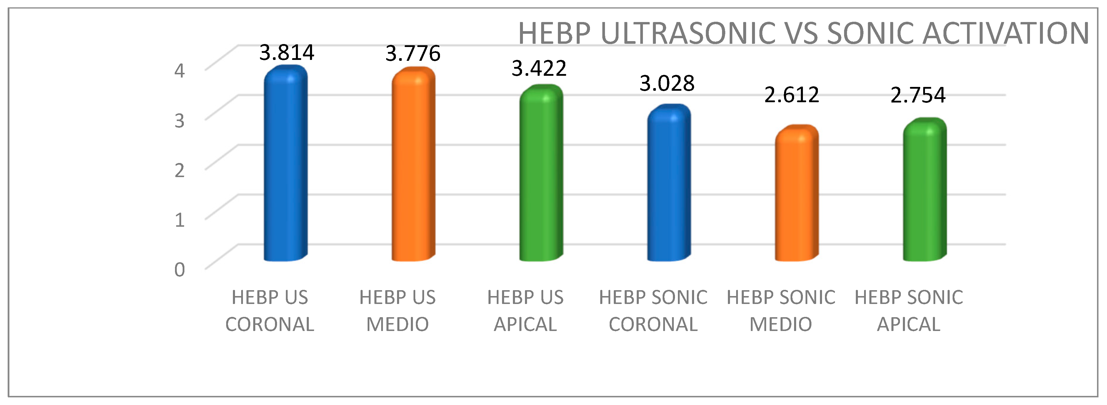
Regarding the diameter of the measured tubules, the results are shown in Table 3 and Figure 3 and Figure 4, where the mean size can be seen based on the thirds studied and the activation systems used.
In comparative analysis, no significant differences were observed between HEBP and EDTA in tubule size. However, when the samples were analyzed by third and according to the activation system, statistically significant differences only appeared in the apical third for both sonic and ultrasonic activation, favoring HEBP over EDTA (p = 0.0002/0.0035). Representative SEM images are shown in Figure 5 and Figure 6.

4. Discussion

The success of root canal treatment depends on thorough instrumentation and irrigation that effectively cleanse the canal system [21,22,23,24,25]. After instrumentation, remnants of inorganic tissue, known as the smear layer, remain within the canal [26,27,28]. This smear layer must be removed using chelating agents, which eliminate inorganic debris and leave the canal clean. For this purpose, we selected two commonly used agents: EDTA [29,30,31,32,33] and 1.1-hydroxyethylidene-1.1-bisphosphonate (HEBP) [34]. The activation time for each solution was standardized at 20 s, to ensure equal treatment duration. This time frame was chosen based on ultrasonic activation (PUI) guidelines, as references indicate that activation beyond 20 s may cause the ultrasonic file to contact dentin walls and produce undesirable effects [35,36]. Although continuous chelation may better reflect clinical reality, in order to standardize our model, we applied HEBP only during the final irrigation, in the same way as EDTA.
Most of the reviewed studies were conducted in vitro, using human-extracted teeth, predominantly single-rooted specimens [29,30,31,32,33]. González-López et al. [37], however, used bovine incisors to investigate the decalcifying effects of citric acid and EDTA. In line with most authors, we chose to carry out an in vitro study with human-extracted single-rooted teeth.
When replicating in vivo endodontic irrigation conditions, it is important to note that the canal functions as a closed system. To reproduce this in vitro, an artificial periodontium must be created. Several authors have recommended sealing the root using wax [26], silicone [38], or glue [35]. Accordingly, we adopted a closed system by coating the root surface with molten wax, following the method described by Prabhu et al. [26].
Finally, regarding incomplete root sectioning, it was essential to ensure that any smear layer observed originated solely from the instrumentation/irrigation process, rather than from the sectioning itself. Therefore, when preparing samples for electron microscopy, we followed methods described in the literature, making an external cut with a cooled low-speed diamond disk [19,32,38] or a cooled high-speed diamond bur [26,39] without entering the root canal, thus preventing the formation of a new smear layer. Separation of the segments was then completed with a chisel [32,38,40], scalpel [26], sharp blade [41] or surgical chisel [39]. In our study, we employed the technique described by Prabhu et al. [26], using a high-speed diamond bur for the external cut, followed by root segmentation with a scalpel. The results obtained in this study confirm that the effectiveness of smear layer removal and dentinal tubules opening depends significantly on both the chelating agent used and the irrigant activation system. This finding is consistent with what has been described in the literature, which has emphasized that no irrigant alone is capable of completely removing both organic and inorganic components from the root canal system without effective activation. In line with previous research, such as that of Mancini et al. [15] and Nogueira et al. [42], these effects are directly related to the chemical interaction between irrigants and the dentin structure.
About the chelating agent, 17% EDTA showed superior performance in cleaning the coronal and middle thirds when combined with ultrasonic activation. These results are consistent with previous studies [43] that have shown greater efficacy of EDTA in surface demineralization and canal debris removal. On the other hand, its erosive action can compromise the mechanical integrity of the tooth, decrease microhardness and increase dentin porosity. However, its performance in the apical third was poor, which may be attributed to the inherent difficulty of the irrigant adequately accessing this area due to the “vapor lock” phenomenon described by Tay et al. [17].
In contrast, HEBP showed better results than EDTA in the apical third, especially when sonically activated. This is interesting because the continuous combination of HEBP and sodium hypochlorite, although the latter is considered a weak chelator, appears to promote cleaning in hard-to-reach areas such as the apical third. Studies such as that of Morago et al. [10] had already pointed out this advantage of the NaOCl + HEBP combination, especially in terms of synergistic interaction between the organic and inorganic solutions. Unlike EDTA, HEBP causes minimal alteration in the morphology and chemical composition of dentin, maintaining a higher calcium and phosphorus content and a more stable Ca/P ratio [3,44]. This suggests a more conservative action that favors the structural integrity of the tooth, an advantage widely supported by previous studies [45].
Regarding the activation method, ultrasonic irrigation proved to be generally superior for EDTA, while sonic activation was more effective for HEBP, especially in the apical third. This finding suggests that the ideal combination may not be uniform for all irrigants and that the physicochemical properties of each solution interact differently with the energy transmitted by the activation system. As described by Mancini et al. [15,16], ultrasonic activation allows the formation of cavitation and microcurrents that promote better irrigant penetration and mechanical removal of debris.
The number and diameter of dentin tubules vary depending on their proximity to the pulp, their anatomical location within the tooth and the patient’s age [46]. In root dentin, tubule density decreases progressively toward the apical region of the canal system [46,47,48]. Reported data on average tubule diameter are highly variable; however, the largest diameters are consistently found in deep dentin (close to the pulp), where the mean diameter is approximately 2.5 µm and macrotubules of up to 5 µm may occur. In contrast, superficial dentin (further from the pulp and closer to enamel or cementum) presents a mean diameter of about 1 µm [49]. To our knowledge, no specific data are available regarding tubule diameters according to the coronal, middle and apical thirds of root dentin, preventing direct comparison with our results.
In our study, HEBP produced significantly greater tubule widening in the apical third compared with EDTA. This effect may be attributed to its slower and less aggressive mechanism of action, which promotes controlled enlargement without collapsing the tubular structure [50]. Conversely, no significant differences between the two chelators were observed in the middle and coronal thirds, suggesting that the impact of chelating agents on tubule diameter is more relevant in areas with limited accessibility. These findings support the hypothesis that the milder action of HEBP enables effective enlargement of dentinal tubules while minimizing structural damage, consistent with previous SEM studies reporting reduced erosion. Figure 6 shows a severe erosion of dentin surface (the tubules are widened due to dissolution of peritubular and interlobular dentin) which leads us to consider the study of such erosion in future research work with a similar methodology.
These results should be interpreted in light of certain methodological limitations. Although the study followed a rigorous and standardized design, including scanning electron microscopy (SEM) analysis, its in vitro nature inherently restricts direct extrapolation to clinical practice. In vitro models cannot fully replicate the biological complexity of the periapical tissues, the dynamics of pulpal and periapical fluids, or the variability of clinical conditions such as anatomical differences, bacterial biofilms and host immune responses. Additionally, the absence of a non-activation irrigation control group limited the ability to isolate and compare the specific effect of activation. Taken together, these factors highlight the need for caution when extrapolating our findings to clinical scenarios and underscore the importance of future in vivo studies to validate these results [51,52].
Finally, the combination of NaOCl with EDTA or HEBP should be carefully analyzed. Studies such as that by Nogueira et al. [42] demonstrated that the use of EDTA followed by NaOCl can cause greater demineralization and exposure of the collagen matrix, resulting in greater erosion and dentin fragility. In contrast, HEBP can be mixed with NaOCl without losing efficacy, supporting its use as the primary irrigant in continuous chelation protocols.

5. Conclusions

The cleaning effectiveness of the root canal system varies significantly depending on the chelating agent and the irrigant activation method used.
17% EDTA showed superior smear layer removal in the coronal and middle thirds, particularly when activated with ultrasonic irrigation. However, its effectiveness in the apical third was limited; 9% HEBP, although a weaker chelator, demonstrated greater efficacy in the apical third, especially when combined with sonic activation, suggesting an advantage in areas with limited irrigant penetration.
No significant overall differences were observed in dentinal tubule diameter between EDTA and HEBP; however, HEBP resulted in significantly larger tubule diameters in the apical third, likely due to its less erosive effect on dentin. Ultrasonic activation was more effective than sonic activation for EDTA, whereas sonic activation yielded better results when used with HEBP, indicating that the choice of activation system should be tailored to the irrigant used.
These findings support the use of HEBP as an alternative to EDTA, particularly in continuous chelation protocols with sodium hypochlorite, offering a conservative approach with favorable cleaning outcomes in the apical third.

Author Contributions

Conceptualization, C.L. and J.M.-Á.; methodology, C.L. and J.M.-Á.; software, C.L.; validation, J.M.-Á., J.M.-G. and A.M.-V.; formal analysis, J.M.-Á.; investigation, C.L.; resources, A.M.-V. and V.V.-G.; data curation, J.M.-Á. and J.M.-G.; writing—original draft preparation, J.M.-Á.; writing—review and editing, J.M.-G.; visualization, A.M.-V. and V.V.-G. All authors have read and agreed to the published version of the manuscript.

Funding

This research received no external funding.

Institutional Review Board Statement

The study was approved by the Bioethics Committee of the Universidad Alfonso X el Sabio (reference number 2023_12/238).

Informed Consent Statement

Informed consent was obtained from all subjects involved in the study.

Data Availability Statement

The datasets used and/or analyzed during the current study are available from the corresponding author on reasonable request.

Acknowledgments

We want to thank the ICTS National Center for Electron Microscopy of Faculty Of Chemistry of Universidad Complutense de Madrid (Spain) for their help with this study.

Conflicts of Interest

The authors declare no conflicts of interest.

Abbreviations

The following abbreviations are used in this manuscript:
CDT:clean dentinal tubules
DDT:dirty dentinal tubules
EDTA:17% ethylenediaminetetraacetic acid
HEBP:9% hydroxyethylidene bisphosphonate
NaOCl:sodium hypochlorite
SEM:scanning electron microscopy

References

  1. Zehnder, M. Root Canal Irrigants. J. Endod. 2006, 32, 389–398. [Google Scholar] [CrossRef] [PubMed]
  2. Arias-Moliz, M.T.; Ordinola-Zapata, R.; Baca, P.; Ruiz-Linares, M.; Ferrer-Luque, C.M. Antimicrobial activity of a sodium hypochlorite/etidronic acid irrigant solution. J. Endod. 2014, 40, 1999–2002. [Google Scholar] [CrossRef] [PubMed]
  3. Morago, A.; Ruiz-Linares, M.; Ferrer-Luque, C.M.; Baca, P.; Rodríguez Archilla, A.; Arias-Moliz, M.T. Dentine tubule disinfection by different irrigation protocols. Microsc. Res. Tech. 2019, 82, 558–563. [Google Scholar] [CrossRef]
  4. Tonini, R.; Salvadori, M.; Audino, E.; Sauro, S.; Garo, M.L.; Salgarello, S. Irrigating Solutions and Activation Methods Used in Clinical Endodontics: A Systematic Review. Front. Oral Health 2022, 3, 838043. [Google Scholar]
  5. Gomes, B.P.F.A.; Aveiro, E.; Kishen, A. Irrigants and irrigation activation systems in Endodontics. Braz. Dent. J. 2023, 34, 1–33. [Google Scholar] [CrossRef]
  6. Boutsioukis, C.; Arias-Moliz, M.T. Present status and future directions–irrigants and irrigation methods. Int. Endod. J. 2022, 55 (Suppl. 3), 588–612. [Google Scholar] [CrossRef]
  7. Emre Erik, C.; Onur Orhan, E.; Maden, M. Qualitative analysis of smear layer treated with different etidronate concentrations: A scanning electron microscopy study. Microsc. Res. Tech. 2019, 82, 1535–1541. [Google Scholar] [CrossRef]
  8. Gómez-Delgado, M.; Camps-Font, O.; Luz, L.; Sanz, D.; Mercade, M. Update on citric acid use in endodontic treatment: A systematic review. Odontology 2023, 111, 1–19. [Google Scholar] [CrossRef]
  9. Ali, A.; Bhosale, A.; Pawar, S.; Kakti, A.; Bichpuriya, A.; Agwan, M.A. Current Trends in Root Canal Irrigation. Cureus 2022, 14, e24833. [Google Scholar] [CrossRef]
  10. Morago, A.; Ordinola-Zapata, R.; Ferrer-Luque, C.M.; Baca, P.; Ruiz-Linares, M.; Arias-Moliz, M.T. Influence of Smear Layer on the Antimicrobial Activity of a Sodium Hypochlorite/Etidronic Acid Irrigating Solution in Infected Dentin. J. Endod. 2016, 42, 1647–1650. [Google Scholar] [CrossRef] [PubMed]
  11. Arslan, S.; Balkaya, H.; Çakir, N. Efficacy of different endodontic irrigation protocols on shear bond strength to coronal dentin. J. Conserv. Dent. 2019, 22, 223–227. [Google Scholar] [CrossRef]
  12. Dioguardi, M.; Di Gioia, G.; Illuzzi, G.; Laneve, E.; Cocco, A.; Troiano, G. Endodontic irrigants: Different methods to improve efficacy and related problems. Eur. J. Dent. 2018, 12, 459–466. [Google Scholar] [CrossRef]
  13. Busanello, F.H.; Petridis, X.; So, M.V.R.; Dijkstra, R.J.B.; Sharma, P.K.; Van Der Sluis, L.W.M. Chemical biofilm removal capacity of endodontic irrigants as a function of biofilm structure: Optical coherence tomography, confocal microscopy and viscoelasticity determination as integrated assessment tools. Int. Endod. J. 2019, 52, 461–474. [Google Scholar] [CrossRef]
  14. Borges, M.M.B.; Dijkstra, R.J.B.; de Andrade, F.B.; Duarte, M.A.H.; Versluis, M.; van der Sluis, L.W.M.; Petridis, X. The response of dual-species bacterial biofilm to 2% and 5% NaOCl mixed with etidronic acid: A laboratory real-time evaluation using optical coherence tomography. Int. Endod. J. 2022, 55, 758–771. [Google Scholar] [CrossRef] [PubMed]
  15. Mancini, M.; Cerroni, L.; Iorio, L.; Armellin, E.; Conte, G.; Cianconi, L. Smear layer removal and canal cleanliness using different irrigation systems (EndoActivator, EndoVac, and passive ultrasonic irrigation): Field emission scanning electron microscopic evaluation in an in vitro study. J. Endod. 2013, 39, 1456–1460. [Google Scholar] [CrossRef] [PubMed]
  16. Mancini, M.; Cerroni, L.; Iorio, L.; Dall’Asta, L.; Cianconi, L. FESEM evaluation of smear layer removal using different irrigant activation methods (EndoActivator, EndoVac, PUI and LAI). An in vitro study. Clin. Oral Investig. 2018, 22, 993–999. [Google Scholar] [CrossRef] [PubMed]
  17. Tay, F.R.; Gu Lsha Schoeffel, G.J.; Wimmer, C.; Susin, L.; Zhang, K.; Arun, S.N.; Kim, J.; Looney, S.W.; Pashley, D.H. Effect of vapor lock on root canal debridement by using a side-vented needle for positive-pressure irrigant delivery. J. Endod. 2010, 36, 745–750. [Google Scholar] [CrossRef]
  18. De Gregorio, C.; Arias, A.; Navarrete, N.; Del Rio, V.; Oltra, E.; Cohenca, N. Effect of apical size and taper on volume of irrigant delivered at working length with apical negative pressure at different root curvatures. J. Endod. 2013, 39, 119–124. [Google Scholar] [CrossRef]
  19. Carvalho, A.S.; Camargo, C.H.R.; Valera, M.C.; Camargo, S.E.A.; Mancini, M.N.G. Smear layer removal by auxiliary chemical substances in biomechanical preparation: A scanning electron microscope study. J. Endod. 2008, 34, 1396–1400. [Google Scholar] [CrossRef]
  20. Mayer, B.E.; Peters, O.; Barbakow, F. Effects of rotary instruments and ultrasonic irrigation on debris and smear layer scores: A scanning electron microscopic study. Int. Endod. J. 2002, 35, 582–589. [Google Scholar] [CrossRef]
  21. Giardino, L.; Ambu, E.; Becce, C.; Rimondini, L.; Morra, M. Surface tension comparison of four common root canal irrigants and two new irrigants containing antibiotic. J. Endod. 2006, 32, 1091–1093. [Google Scholar] [CrossRef] [PubMed]
  22. Zmener, O.; Pameijer, C.H.; Alvarez Serrano, S.; Hernandez, S.R. Cleaning efficacy using two engine-driven systems versus manual instrumentation in curved root canals: A scanning electron microscopic study. J. Endod. 2011, 37, 1279–1282. [Google Scholar] [CrossRef]
  23. Scha, E.; Zapke, K. Investigation of the Efficacy of Manual and Automated Instrumentation of Root Canals. J. Endod. 2000, 26, 660–664. [Google Scholar] [CrossRef]
  24. Hartmann, M.S.M.; Barletta, F.B.; Camargo Fontanella, V.R.; Vanni, J.R. Canal transportation after root canal instrumentation: A comparative study with computed tomography. J. Endod. 2007, 33, 962–965. [Google Scholar] [CrossRef]
  25. Soares Grecca, F.; Brandao García, R.; Monteiro Bramante, C.; Gomez de Moraes, I.; Bernarduneli, N. A quantitative analysis of rotary, ultrasonic and manual techniques to treat proximally. J. Appl. Oral Sci. 2007, 15, 89–93. [Google Scholar] [CrossRef] [PubMed]
  26. Prabhu, S.G.; Rahim, N.; Bath, K.S.; Mathew, J. Comparison of removal of endodontic smear layer using NaOCl, EDTA, and different concentrations of maleic acid—A SEM study. Endodontol. 2003, 15, 20–25. [Google Scholar]
  27. Teixeira, C.S.; Felippe, M.C.S.; Felippe, W.T. The effect of application time of EDTA and NaOCl on intracanal smear layer removal: An SEM analysis. Int. Endod. J. 2005, 38, 285–290. [Google Scholar] [CrossRef]
  28. Violich, D.R.; Chandler, N.P. The smear layer in endodontics-A review. Int. Endod. J. 2010, 43, 2–15. [Google Scholar] [CrossRef]
  29. De Gregorio, C.; Estevez, R.; Cisneros, R.; Heilborn, C.; Cohenca, N. Effect of EDTA, sonic, and ultrasonic activation on the penetration of sodium hypochlorite into simulated lateral canals: An in vitro study. J. Endod. 2009, 35, 891–895. [Google Scholar] [CrossRef] [PubMed]
  30. Uzunoglu, E.; Aktemur, S.; Uyanik, M.O.; Durmaz, V.; Nagas, E. Effect of ethylenediaminetetraacetic acid on root fracture with respect to concentration at different time exposures. J. Endod. 2012, 38, 1110–1113. [Google Scholar] [CrossRef]
  31. Sayin, T.C.; Serper, A.; Cehreli, Z.C.; Kalayci, S. Calcium loss from root canal dentin following EDTA, EGTA, EDTAC, and tetracycline-HCl treatment with or without subsequent NaOCl irrigation. J. Endod. 2007, 33, 581–584. [Google Scholar] [CrossRef]
  32. Khedmat, S.; Shokouhinejad, N. Comparison of the efficacy of three chelating agents in smear layer removal. J. Endod. 2008, 34, 599–602. [Google Scholar] [CrossRef]
  33. Mello, I.; Robazza, C.R.C.; Antoniazzi, J.H.; Coil, J. Influence of different volumes of EDTA for final rinse on smear layer removal. Oral Surg. Oral Med. Oral Pathol. Oral Radiol. Endodontol. 2008, 106, 40–43. [Google Scholar] [CrossRef]
  34. Zehnder, M.; Schmidlin, P.; Sener, B.; Waltimo, T. Chelation in Root Canal Therapy Reconsidered. J. Endod. 2005, 31, 817–820. [Google Scholar] [CrossRef] [PubMed]
  35. Saber, S.E.-D.; Hashem, A.A.R. Efficacy of different final irrigation activation techniques on smear layer removal. J. Endod. 2011, 37, 1272–1275. [Google Scholar] [CrossRef] [PubMed]
  36. De Gregorio, C.; Estevez, R. Efficacy of Different Irrigation and Activation Systems on the Penetration of Sodium Hypochlorite into Simulated Lateral Canals and up to Working Length: An In Vitro Study. J. Endod. 2010, 36, 1216–1221. [Google Scholar] [CrossRef]
  37. González-López, S.; Camejo-Aguilar, D.; Sanchez-Sanchez, P.; Bolaños-Carmona, V. Effect of CHX on the decalcifying effect of 10% citric acid, 20% citric acid, or 17% EDTA. J. Endod. 2006, 32, 781–784. [Google Scholar] [CrossRef]
  38. Blank-Gonçalves, L.M.; Nabeshima, C.K.; Martins, G.H.R.; Machado, M.E.D.L. Qualitative analysis of the removal of the smear layer in the apical third of curved roots: Conventional irrigation versus activation systems. J. Endod. 2011, 37, 1268–1271. [Google Scholar] [CrossRef]
  39. da Silva, L.A.B.; Sanguino, A.C.M.; Rocha, C.T.; Leonardo, M.R.; Silva, R.A.B. Scanning electron microscopic preliminary study of the efficacy of SmearClear and EDTA for smear layer removal after root canal instrumentation in permanent teeth. J. Endod. 2008, 34, 1541–1544. [Google Scholar] [CrossRef] [PubMed]
  40. Da Silva, J.M.; Silveira, A.; Santos, E.; Prado, L.; Pessoa, O.F. Efficacy of sodium hypochlorite, ethylenediaminetetraacetic acid, citric acid and phosphoric acid in calcium hydroxide removal from the root canal: A microscopic cleanliness evaluation. Oral Surg. Oral Med. Oral Pathol. Oral Radiol. Endodontol. 2011, 112, 820–824. [Google Scholar] [CrossRef]
  41. Mello, I.; Kammerer, B.A.; Yoshimoto, D.; Macedo, M.C.S.; Antoniazzi, J.H. Influence of final rinse technique on ability of ethylenediaminetetraacetic acid of removing smear layer. J. Endod. 2010, 36, 512–514. [Google Scholar] [CrossRef]
  42. Lima Nogueira, B.M.; da Costa Pereira, T.I.; Pedrinha, V.F.; de Almeida Rodrigues, P. Effects of Different Irrigation Solutions and Protocols on Mineral Content and Ultrastructure of Root Canal Dentine. Iran. Endod. J. 2018, 13, 209–215. [Google Scholar] [PubMed]
  43. Pedrinha, V.F.; Cardenas Cuellar, M.R.; Velásquez-Espedilla, E.G.; Duarte, M.A.H.; Andrade, F.B.; Rodrigues, P.A. Impact of irrigation protocols with some chelators and mechanical agitation on intratubular decontamination. Braz. Oral. Res. 2021, 35, e127. [Google Scholar] [CrossRef]
  44. Ulusoy, Ö.İ.; Mantı, A.Ş.; Çelik, B. Nanohardness reduction and root dentine erosion after final irrigation with ethylenediaminetetraacetic, etidronic and peracetic acids. Int. Endod. J. 2020, 53, 1549–1558. [Google Scholar] [CrossRef] [PubMed]
  45. Mohammadi, Z.; Shalavi, S.; Yaripour, S.; Kinoshita, J.I.; Manabe, A.; Kobayashi, M.; Giardino, L.; Palazzi, F.; Sharifi, F.; Jafarzadeh, H. Smear Layer Removing Ability of Root Canal Irrigation Solutions: A Review. J. Contemp. Dent. Pract. 2019, 20, 395–402. [Google Scholar] [CrossRef] [PubMed]
  46. Mjör, I.A.; Nordal, I. The density and branching of dentinal tubules in human teeth. Arch. Oral Biol. 1996, 41, 401–412. [Google Scholar] [CrossRef]
  47. Ferrari MMannocci, F.; Vichi, A.; Cagidiaco, M.C.; Mjör, I.A. Bonding of root canal: Structural characteristics of the substrate. Am. J. Dent. 2000, 13, 255–260. [Google Scholar]
  48. Mjör, I.A.; Smith, M.R.; Ferrari, M.; Manocci, F. The structure of dentine in the apical región of human teeth. Int. Endod. J. 2001, 34, 346–353. [Google Scholar] [CrossRef]
  49. Garbergolio, R.; Brännström, M. Scanning electron microscopic investigation of human dentinal tubules. Arch. Oral Biol. 1976, 21, 355–362. [Google Scholar] [CrossRef]
  50. Dineshkumar, M.K.; Vinothkumar, T.S.; Arathi, G.; Shanthisree, P.; Kandaswamy, D. Effect of ethylene diamine tetra-acetic acid, MTAD™, and HEBP as a final rinse on the microhardness of root dentin. J. Conserv. Dent. 2012, 15, 170–173. [Google Scholar]
  51. Kadulkar, N.; Kataki, R.; Deka, A.; Medhi, H.; Chakraborty, S.; Singh, A. Comparative Evaluation of the Effect of Different Chelating Agents on Mineral Content and Erosion of Radicular Dentine: A FESEM-EDS Analysis. Eur. Endod. J. 2024, 9, 73–80. [Google Scholar] [CrossRef] [PubMed]
  52. Tartari, T.; Duarte Junior, A.P.; Silva Júnior, J.O.; Klautau, E.B.; Silva ESouza Junior, M.H.; Silva ESouza Junior, P.d.A. Etidronate from medicine to endodontics: Effects of different irrigation regimes on root dentin roughness. J. Appl. Oral Sci. 2013, 21, 409–415. [Google Scholar] [CrossRef] [PubMed]
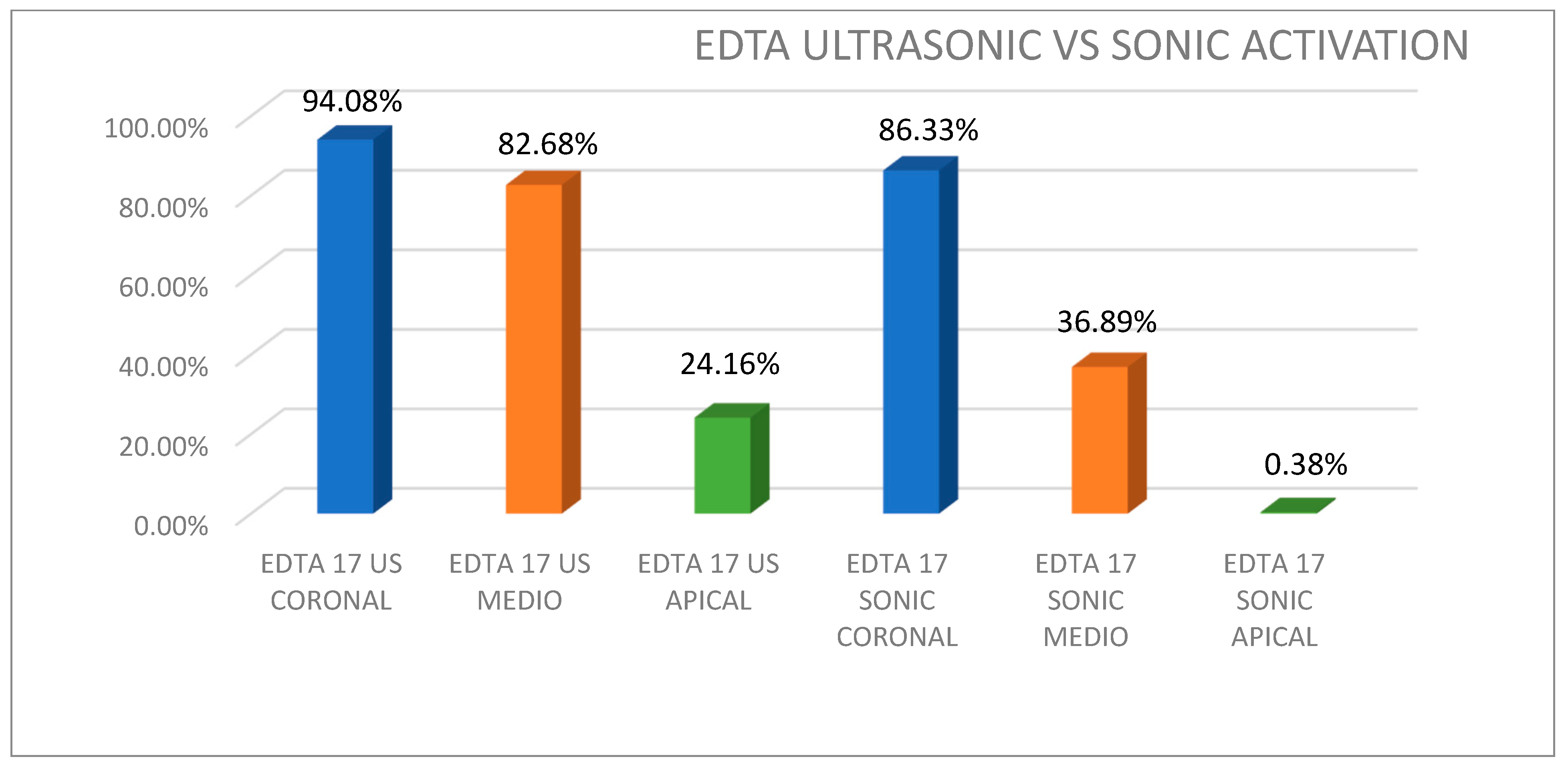
Figure 1. Percentage of clean tubules irrigated with EDTA depending on the thirds studied and the activation system used.
Applsci 15 09993 g001
Figure 2. Percentage of clean tubules irrigated with HEBP depending on the thirds studied and the activation system used.
Applsci 15 09993 g002
Figure 3. Diameter medium of dentin tubules irrigated with EDTA depending on the thirds studied and the activation system used (values expressed in µm).
Applsci 15 09993 g003
Figure 4. Diameter medium of dentin tubules irrigated with HEBP depending on the thirds studied and the activation system used (values expressed in µm).
Applsci 15 09993 g004
Figure 5. Representative SEM photomicrographs at ×1500 after treatment with distilled water (A), 9% HEBP without activation medium (B) and 17% EDTA without activation medium (C).
Applsci 15 09993 g005
Figure 6. Representative SEM photomicrographs at ×1500 after treatment with 17% EDTA Sonic Coronal (A), 9% HEBP Sonic Coronal (B), 17% EDTA Ultrasonic Coronal (C) and 9% HEBP Ultrasonic Coronal (D).
Applsci 15 09993 g006
Table 1. Final irrigation protocol. All final irrigation procedures were the same except for the chelating substance used. EDTA: 17% ethylenediaminetetraacetic acid. HEBP: 9% hydroxyethylidene bisphosphonate. NaOCl: sodium hypochlorite.
Final Irrigation
Control Group (n = 5)Tooth 1 y 2: Without Final Irrigation
Tooth 3: HEBP 9% Irrigation
Tooth 4 y 5: EDTA 17% Irrigation
Activation Time
Group 1:
EDTA 17% + Sonic activation (Smart Lite Pro Endo Activator® [Dentsply Sirona. Ballaigues. Switzerland])
n = 25
2 mLNaOCl 5.25%1 min20 s
2 mLSodium chloride 0.9%1 min-
2 mLEDTA 17%1 min20 s
2 mLSodium chloride 0.9%1 min-
2 mLNaOCl 5.25%1 min20 s
Group 2:
HEBP + Sonic activation (SmartLite Pro EndoActivator® [Denstply Sirona. Ballaigues, Switzerland])
n = 25
2 mLNaOCl 5.25%1 min20 s
2 mLSodium chloride 0.9%1 min-
2 mLHEBP 9%1 min20 s
2 mLSodium chloride 0.9%1 min-
2 mLNaOCl 5.25%1 min20 s
Group 3:
EDTA 17% + Ultrasonic activation (IRRI S 21 mm No 25–VDW, Munchen, Germany])
n = 25
2 mLNaOCl 5.25%1 min20 s
2 mLSodium chloride 0.9%1 min-
2 mLEDTA 17%1 min20 s
2 mLSodium chloride 0.9%1 min-
2 mLNaOCl 5.25%1 min20 s
Group 4:
HEBP 9% + Ultrasonic activation (IRRI S 21 mm No 25–VDW, Munchen, Germany])
2 mLNaOCl 5.25%1 min20 s
2 mLSodium chloride 0.9%1 min-
2 mLHEBP 9%1 min20 s
2 mLSodium chloride 0.9%1 min-
2 mLNaOCl 5.25%1 min20 s
Table 2. Percentage of clean tubules irrigated with EDTA and HEBP depending on the thirds studied and activation system used. Median in parentheses.
Ultrasonic
Coronal
Ultrasonic
Medium
Ultrasonic ApicalSonic CoronalSonic Medium Sonic Apical
EDTA94.08% (94.27)82.68% (84.51)24.16% (10.34)86.33% (80.00)36.89% (35.45)0.38% (0.23)
HEBP56.63% (65.04)15.95% (16.80)7.93% (4.65)97.43% (97.96)68.94% (68.96)39.84% (40.37)
Sig0.0030.0003970.2990.00400.08910.000014
Table 3. Diameter medium of dentin tubules irrigated with EDTA and HEBP depending on the thirds studied and the activation system used (values expressed in µm). Median in parentheses.
Ultrasonic
Coronal
Ultrasonic
Medium
Ultrasonic ApicalSonic CoronalSonic
Medium
Sonic Apical
EDTA3.932 (3.43)3.394 (2.85)1.254 (1.68)3.028 (2.8)2.318 (2.11)0.206 (0.216)
HEBP3.814 (3.5)3.776 (3.9)3.422 (2.68)3.028 (3.14)2.612 (2.71)2.754 (2.61)
Sig0.170.650.000210.6140.0035
Disclaimer/Publisher’s Note: The statements, opinions and data contained in all publications are solely those of the individual author(s) and contributor(s) and not of MDPI and/or the editor(s). MDPI and/or the editor(s) disclaim responsibility for any injury to people or property resulting from any ideas, methods, instructions or products referred to in the content.