Review

From DNA Methylation and Histone Modifications to Non-Coding RNAs: Evaluating Tools for Epigenetic Research

by
Ines Benčik
,
Lara Saftić Martinović
*,
Tea Mladenić
,
Saša Ostojić
and
Sanja Dević Pavlić
*
Department of Medical Biology and Genetics, Faculty of Medicine, University of Rijeka, 51000 Rijeka, Croatia
*
Authors to whom correspondence should be addressed.
Appl. Sci. 2025, 15(18), 9940; https://doi.org/10.3390/app15189940
Submission received: 29 July 2025 / Revised: 3 September 2025 / Accepted: 9 September 2025 / Published: 11 September 2025
(This article belongs to the Section Applied Biosciences and Bioengineering)

Abstract

Epigenetic biomarkers, such as DNA methylation, histone alterations, and non-coding RNAs, are increasingly recognized as essential instruments in disease diagnoses, prognostics, and customized therapy. As their clinical significance increases, so does the necessity for robust, interpretable, and scalable techniques that can accurately detect these molecular alterations. This review provides a critical and organized overview of contemporary technologies employed to characterize the principal categories of epigenetic modifications, encompassing PCR- and sequencing-based methods, high-resolution immunoprecipitation techniques, and CRISPR-enhanced detection systems. Although numerous procedures are technically well-established, their implementation outside research laboratories frequently faces substantial challenges: elevated prices, data intricacy, absence of standardization, and restricted translational frameworks. Furthermore, the interpretation of epigenetic data continues to pose a significant difficulty, especially for heterogeneous clinical samples and the swiftly advancing computational techniques. We examine the advantages and drawbacks of existing approaches, focusing on their incorporation into biomedical engineering platforms, including biosensors, lab-on-a-chip devices, and AI-assisted diagnostics. This review seeks to assist researchers, physicians, and engineers in choosing suitable technologies, comprehending their limitations, and pinpointing areas requiring urgent innovation by merging analytical rigor with a pragmatic viewpoint.

1. Introduction

Epigenetics is the term used to describe heritable changes in gene function that occur without alterations to the deoxyribonucleic acid (DNA) sequence. These modifications, which encompass DNA methylation, histone modifications, and non-coding ribonucleic acids (ncRNAs), dynamically regulate gene expression in various cellular contexts [1]. The pathogenesis of complex diseases, including allergic rhinitis [2], cancer [3], cardiovascular diseases [4], and epilepsy [5], as well as metabolic [6], autoimmune [7], and neuropsychiatric disorders [8], is significantly influenced by epigenetic processes, which also affect normal physiological development [9].
In contrast to irreversible genetic mutations, epigenetic alterations are dynamic and reversible. This plasticity makes them attractive therapeutic targets because pharmacological agents can modulate or restore normal epigenetic states. Their reversibility also underpins their value as biomarkers for early diagnosis and risk evaluation, as changes can be detected at early stages and may be reverted through intervention [1]. Recent data endorse the use of epigenetic markers in precision medicine, especially for patient stratification and forecasting therapeutic response, as demonstrated in immunotherapy [10], obesity-related metabolic risk [6], and temporal lobe epilepsy [5].
Epigenetic regulation is presently being investigated at many levels of precision, ranging from specific single-locus DNA methylation assays to extensive genome-wide mapping facilitated by third-generation sequencing technology [11]. These analyses are simplified by advanced bioinformatics pipelines that can integrate and analyze multi-omics datasets [12]. The advancement of analytical tools for detecting epigenetic changes is critical not only for understanding disease causes but also for enabling downstream functional applications. High-resolution epigenetic mapping lays the groundwork for more advanced therapies, such as gene expression modification at specific loci. In this context, CRISPR/dCas9-based epigenome editing tools offer a promising translational extension by allowing precise alteration of identified epigenetic targets for both research and potential therapeutic applications [13].
The decision between untargeted and targeted epigenetic techniques is based on the study environment and the amount of resolution required. Untargeted approaches, such as whole-genome bisulfite sequencing (WGBS), chromatin Immunoprecipitation (ChIP) sequencing (ChIP-seq), and RNA sequencing (RNA-seq), are critical during the exploratory stages of research. They enable the discovery of novel regulatory areas, previously unknown epigenetic biomarkers, and global patterns of chromatin remodeling. Targeted approaches, such as locus-specific bisulfite polymerase chain reaction (PCR), methylation-sensitive qPCR, targeted ChIP, or microRNA (miRNA) panels, are better suited for hypothesis-driven research, marker validation, and clinical applications that require precision and reproducibility.
Epigenetic research is typically categorized into three methodological frameworks: (1) analysis of DNA methylation, (2) profiling of histone modifications, and (3) expression and interaction mapping of non-coding RNAs. Each category encompasses both untargeted and targeted methodologies.
In this review, we present a critical assessment of existing techniques for all three epigenetic levels (DNA methylation, histone modifications, and non-coding RNAs). We thoroughly assess each technique’s strengths and weaknesses in terms of resolution, scalability, reproducibility, and translational potential. Finally, we demonstrate how combining artificial intelligence (AI) and machine learning can improve the analysis, interpretation, and forecasting potential of epigenetic data. These technologies open up new possibilities for biomarker identification, patient stratification, and the creation of adaptive, data-driven diagnostic systems.

2. DNA Methylation

DNA methylation is a covalent chemical alteration in which a methyl group is added to cytosine’s 5-carbon position, most typically in the context of CpG dinucleotides, but also in CHG and CHH (H = A, T, or C) sequences in plants [14]. This epigenetic mark alters chromatin conformation, which regulates gene expression and contributes to processes like as transcriptional silence, genomic imprinting, X-chromosome inactivation, transposable element suppression, and genome stability. Methylation patterns are dynamic and context-dependent, therefore proper characterization is critical for understanding how environmental and developmental factors influence gene regulation [14].
Depending on the biological topic, multiple methodological approaches can be used, such as genome-wide resolution, analysis of specific genomic areas, or assessment of methylation at designated loci. These include bisulfite-based sequencing, tailored assays, and new long-read technologies. The key strategies are described in the next section and schematically illustrated in Figure 1.

2.1. Bisulfite Sequencing: A Gold Standard for Base-Resolution DNA Methylation Profiling

Bisulfite sequencing (BS-seq) remains the foundational technique for mapping 5-methylcytosine (5mC) at single-nucleotide resolution and continues to serve as a benchmark for epigenomic studies. First introduced by Frommer et al. in 1992, the method is based on the selective deamination of unmethylated cytosine to uracil by sodium bisulfite, while 5mC residues remain unconverted [15]. After PCR amplification and sequencing, uracils are read as thymine, while methylated cytosines remain as cytosine, allowing direct inference of methylation status [16]. This simple chemical distinction has enabled wide-ranging applications from targeted gene studies to genome-scale epigenome mapping. In targeted contexts, BS-seq has been used to interrogate immune-related loci in asthma [17], candidate stress and behavior genes in psychiatric research [18], and cancer-associated genes in colorectal tumors [19], underscoring its value for high-resolution methylation profiling in disease. Examples of genome-scale BS-seq applications are generation of high-resolution methylomes for clinical and FFPE samples [20], assessment of technical feasibility in livestock [21], and mapping of drought-responsive methylation changes in plants [22].
Despite its great power, bisulfite conversion requires a harsh chemical procedure that can lead to DNA fragmentation and reduced sequence complexity, complicating downstream sequencing and alignment [23]. Moreover, the technique cannot differentiate between 5mC and 5-hydroxymethylcytosine (5hmC), limiting its utility in studies where oxidative cytosine derivatives are functionally important [24,25]. However, given its single-base precision, robustness, and broad availability, BS-seq is still a key method in epigenetics despite these limitations.

2.1.1. Whole-Genome Bisulfite Sequencing (WGBS)

WGBS, also referred to as BS-seq or MethylC-seq, provides the most comprehensive view of cytosine methylation, covering nearly all CpG sites in the genome [25,26]. WGBS has been extensively employed in large-scale epigenome mapping projects such as ENCODE, NIH Roadmap Epigenomics, and IHEC [26]. With absolute quantification at each cytosine, it can simultaneously capture methylation in regions with high and low CpG densities, such as gene bodies, enhancers, and intergenic domains [27,28]. In applications such as cancer epigenetics, WGBS has been used to identify hypomethylated domains associated with cell transformation, as well as hypermethylation at tumor suppressor loci [28]. It is also highly valuable for studies of development, imprinting, and allele-specific methylation [29,30].
However, WGBS is very resource-intensive. High sequencing depth is required to obtain adequate coverage (>30× for diploid methylation calling), and the C-to-T conversion reduces sequence complexity, complicating alignment and increasing mapping bias [25,31]. GC-rich regions, including CpG islands, may be underrepresented due to bisulfite-induced DNA damage [32]. Moreover, the method cannot resolve 5mC from 5hmC, which may result in overestimation of canonical methylation [24]. Recent refinements in protocol design and library preparation have enabled WGBS on low-input samples, including nanogram quantities of cell-free DNA (cfDNA), extending its applicability to liquid biopsies and prenatal diagnostics [32]. However, even with such improvements, the trade-off between genome-wide coverage and sequencing depth remains a limiting factor for large-cohort studies [31].

2.1.2. Reduced Representation Bisulfite Sequencing (RRBS)

Reduced representation bisulfite sequencing (RRBS) offers a cost-effective alternative to WGBS by focusing sequencing efforts on CpG-rich regions of the genome. RRBS combines methylation-insensitive restriction enzyme digestion (typically MspI) with size selection to enrich for promoter regions, CpG islands, and gene regulatory elements [33]. After library preparation and bisulfite conversion, sequencing is performed on a reduced but functionally relevant subset of the genome. The method enables efficient profiling of ~4 million CpG sites in the human genome, making it well-suited for large-cohort studies, clinical diagnostics, and non-model organisms [32,34]. RRBS has been successfully applied to assess differential methylation associated with disease [35,36], development, and environmental exposure [37]. The epigenotyping-by-sequencing (epiGBS) method, an RRBS-based approach, enables methylation analysis without relying on a reference genome, which makes it highly applicable in ecological studies of non-model plant species [34].
Despite its efficiency, RRBS has limitations in genome coverage. It excludes distal enhancers, low-CpG-density intergenic regions, and repetitive elements, which may harbor functionally relevant methylation changes [28,38]. The method’s dependence on restriction sites also introduces sequence bias and limits comparability with WGBS or array-based platforms [33,38]. Mapping efficiency is often reduced due to sequence divergence after bisulfite treatment, particularly in non-model species or poorly annotated genomes [38]. Nonetheless, RRBS remains a practical compromise for applications that prioritize precision over breadth. It is advantageous in contexts where cost and throughput outweigh the need for complete methylome coverage [39].

2.1.3. Ultrafast and RNA Bisulfite Sequencing (UBS-Seq)

Ultrafast bisulfite sequencing (UBS-seq) was developed to overcome the limitations of conventional BS-seq protocols, particularly DNA fragmentation and incomplete conversion. By incorporating optimized bisulfite chemistry, UBS-seq enables efficient conversion in approximately 10 min, significantly reducing DNA degradation and improving methylation detection in GC-rich or structured regions such as mitochondrial DNA [24]. UBS-seq has demonstrated broader genome coverage, reduced background noise, and high sensitivity even with low-input material, making it suitable for low-yield samples such as cfDNA and single cells. Additionally, UBS-seq has been adapted for transcriptomic applications, enabling the detection of 5mC in RNA molecules, including messenger RNA (mRNA), ribosomal RNA (rRNA), and transport RNA (tRNA). Thousands of m5C sites regulated by 2 RNA methyltransferases NSUN2 and NSUN6 have been identified, revealing patterns enriched in 5′-untranslated regions (UTRs), suggesting potential roles in translational regulation [24]. Compared to antibody-based enrichment strategies, RNA bisulfite sequencing provides higher resolution and greater quantitative accuracy, although challenges remain in profiling low-abundance or highly structured RNAs.

2.2. Methylation-Specific PCR (MSP): Gene-Specific Epigenetic Analysis

Methylation-specific PCR (MSP) is a highly sensitive and gene-targeted method for detecting CpG methylation, particularly in promoter regions. First introduced by Herman et al. in 1996, the technique uses bisulfite-treated DNA to distinguish between methylated and unmethylated cytosines through the use of primer pairs designed to bind sequence variants resulting from differential conversion [40]. MSP marked a major advancement in early epigenetic research by enabling the reliable detection of methylation in small or degraded DNA samples, such as those from formalin-fixed, paraffin-embedded (FFPE) tissues, where traditional methods, like Southern blotting, proved ineffective [40].
A typical MSP workflow consists of DNA extraction, bisulfite conversion, primer design, PCR amplification, and post-PCR analysis. Bisulfite conversion is central to the assay and must be optimized to ensure complete deamination of unmethylated cytosines without excessive DNA degradation. Primer design is critical; primers must target CpG-rich promoter regions, ideally near transcription start sites, and avoid CpG boundaries, which may reduce amplification efficiency. Notably, methylated primers are generally shorter and richer in G/C content, while unmethylated primers are longer and A/T-rich to accommodate converted uracil [16,41].
Primer pairs should cover equivalent regions, contain a comparable number of CpG sites, and generate amplicons less than 300 bp with melting temperatures within 5 °C of each other. Optimization ensures differential binding and amplification, allowing accurate discrimination between methylation states. The assay typically requires 100 ng–2 μg of input genomic DNA, making it feasible for small-volume clinical samples [42].
PCR amplification with these primers allows qualitative or semi-quantitative determination of methylation status based on gel electrophoresis banding patterns or quantitative PCR signal [40,43]. This design circumvents false positives associated with partial restriction digestion, a common limitation in earlier methylation detection methods [16].
While classical MSP is inherently qualitative, advancements have extended the method into real-time quantitative applications (qMSP), incorporating intercalating dyes or TaqMan probes to enable relative or absolute quantification of methylated alleles. These innovations are increasingly used in cancer diagnostics, correlating methylation levels with disease progression, severity, and treatment response [43,44]. MSP and its derivatives are widely used in clinical and research contexts to assess the methylation status of disease-associated genes. For example, methylation changes in the ADAM33 gene have been implicated in asthma, COPD, and COVID-19. A recent study using MSP-dPCR showed differential methylation of ADAM33 in saliva and lung tissues, with implications for prognosis and pathogenesis in respiratory diseases [43].
Several derivative methods have been developed to address the limitations of conventional MSP:
Nested MSP increases sensitivity by using two rounds of amplification—an initial outer primer set followed by methylation-specific primers, which is useful for detecting low-abundance methylation events [42].
Multiplex MSP (MMSP) allows the simultaneous analysis of multiple methylation targets in a single reaction. This approach increases throughput but requires rigorous primer design to prevent non-specific amplification and PCR competition [39,42].
Quantitative MSP (qMSP) provides methylation quantification using real-time PCR. Dye-based (e.g., SYBR Green) or probe-based (e.g., TaqMan) detection enables relative quantification against reference standards and has been adopted in clinical diagnostics for cancer and other epigenetic disorders [44].
Methylation-Specific High-Resolution Melting (MS-HRM) avoids post-PCR processing by distinguishing methylation states based on differential melting profiles of PCR amplicons, offering a rapid and high-throughput alternative [16,42].
Combined Bisulfite Restriction Analysis (COBRA) uses restriction enzymes to digest PCR amplicons post-bisulfite treatment. It enables qualitative assessment of methylation at specific restriction sites, but is limited to regions containing suitable enzyme recognition motifs [16].
Hairpin-Bisulfite PCR, developed by Laird et al., facilitates the study of methylation symmetry by preserving complementary strands with a covalently linked hairpin structure, allowing simultaneous analysis of both DNA strands [16].
A recent advancement in MSP technology is the development of digital methylation-specific PCR (dMSP), which partitions DNA into thousands of nanochambers to allow absolute quantification of methylated alleles. Zhao et al. (2023) introduced a microfluidic-based digital multiplex methylation-specific PCR (mdMSP) platform for the simultaneous analysis of four hypermethylated biomarkers (SOX17, TAC1, CDO1, and HOXA7) in the early detection of non-small cell lung cancer [39]. This was achieved from just 100 μL of plasma-derived cfDNA, obtaining a sensitivity of 90% and a specificity of 82%, representing a significant improvement over earlier singleplex assays. The mdMSP system did not require proprietary droplet-generation hardware since it used polydimethylsiloxane devices made by soft lithography in conjunction with fluorescently labeled TaqMan probes. The resulting platform was low-cost, compatible with standard laboratory imaging systems, and provided a practical alternative to commercial digital PCR systems [39].
Despite the advantages of mdMSP, challenges remaining include limited throughput, potential “rain” artifacts in fluorescence readouts due to short amplicon sizes, and DNA fragmentation from bisulfite treatment that may compromise partition accuracy [43]. Nevertheless, the system exemplifies the potential of integrating MSP with digital PCR for high-precision, minimally invasive cancer diagnostics.

2.3. Quantitative Methylation Detection Using Pyrosequencing Technology

Pyrosequencing is a real-time, sequencing-by-synthesis technology that enables both genetic and epigenetic analysis with high sensitivity, precision, and quantitative capacity. It is based on enzymatic cascade reactions that detect nucleotide incorporation through light emission proportional to the number of added bases [45]. Originally conceptualized by Pål Nyrén in 1987 and commercialized in the late 1990s, pyrosequencing measures light emission generated from pyrophosphate (PPi) release during DNA polymerization to determine nucleotide incorporation, base by base [46,47]. In contrast to electrophoretic or traditional sequencing methods, it offers a rapid and non-radioactive alternative, particularly suited for targeted applications, including mutation detection, single-nucleotide polymorphism (SNP) analysis, and DNA methylation quantification [47].
One of the most impactful applications of pyrosequencing is in the analysis of DNA methylation. Following sodium bisulfite treatment, non-methylated cytosines are converted to uracils (read as thymine), while methylated cytosines remain unchanged. Pyrosequencing quantifies methylation levels by comparing the peak heights of cytosine- and thymine-derived signals at CpG sites, enabling precise assessment of methylation percentage at individual loci [47,48]. This technique is widely utilized in clinical diagnostics, particularly in cancer, where it enables quantification of methylation in tumor suppressor genes, oncogenes, and biomarkers relevant for prognosis or therapeutic response. For example, Park et al. demonstrated the method’s reliability in quantifying SNPs within the FMO3 gene for pharmacogenomic analysis in Korean populations, noting its simplicity, cost-efficiency, and specificity compared to conventional methods [49]. Pyrosequencing has also been applied to genetic identification in biodiversity studies [50].
Modern pyrosequencing is primarily performed on benchtop platforms that operate in a locus-specific, non-parallel manner, better suited for precision testing rather than genome-wide surveys [47]. Multiplexing in pyrosequencing is achievable by either amplifying several targets in one PCR, followed by sequencing in separate wells with individual sequencing primers, or by optimizing primer binding stringency using additives such as formamide. Primer design is crucial in avoiding dimer formation, nonspecific amplification, and ensuring adequate signal across CpG-rich regions, particularly in bisulfite-treated templates with reduced complexity [47].
The major advantages of pyrosequencing include its high analytical sensitivity (detecting as low as 5% mutant allele fraction), real-time data generation, and ease of use with minimal post-PCR processing [51]. It excels in resolving closely spaced mutations, ambiguous sequencing reads, and distinguishing cis- from trans-mutations in tumor DNA samples with low purity [46,47]. Pyrosequencing also allows for rapid assay development using tools such as Pyromaker to predict and interpret pyrograms [46].
However, several limitations constrain its broader utility. Sequencing read lengths are typically limited to <400 base pairs due to diminishing enzyme activity over cycles and dilution of reagents [46]. PCR bias, especially in bisulfite-treated DNA, can distort quantitative accuracy by favoring amplification of either methylated or unmethylated templates, potentially affecting dynamic range and measurement fidelity [48]. Furthermore, heterogeneous methylation patterns may be masked unless individual clones are sequenced, limiting analysis of complex epigenetic regions. Technical factors can also impact assay reliability. Konrad et al. reported that vibrations substantially degraded pyrosequencing performance, causing baseline drifts and increased failure rates [52]. The use of anti-vibration tables significantly improved reproducibility and reduced error signals, emphasizing the sensitivity of the optical detection system to environmental disturbance [52].
Recent studies have broadened the scope of pyrosequencing applications. In cervical cancer screening, long interspersed nucleotide element 1 L1 (LINE1) pyrosequencing has been used to detect global DNA hypomethylation, suggesting its potential role as a triage tool for large-scale screening efforts [53]. In forensic contexts, the technique has been applied to develop assays for tissue-specific DNA methylation markers (particularly in semen, saliva, and blood) offering a robust method for body fluid identification [54]. In lifestyle-related research, a four-CpG pyrosequencing assay has demonstrated high accuracy in predicting smoking status from blood and saliva samples, indicating promise for biomarker-based exposure assessment [55]. Furthermore, a multiplex pyrosequencing approach has been introduced for age estimation using limited forensic DNA, improving throughput while maintaining measurement reliability [56]. These applications reinforce the method’s growing relevance across clinical, forensic, and environmental epigenetics.
Although its read length and multiplexing capabilities are limited compared to next-generation sequencing, its quantitative precision, user-friendly interface, and adaptability make it a preferred choice for targeted assays in clinical and research settings. Future developments may focus on reducing PCR bias in methylation studies, improving enzyme stability for longer read lengths, and expanding automated analysis pipelines. The continued relevance of pyrosequencing in precision medicine is ensured by its versatility in handling multiplex formats and complex assays.

2.4. Nanopore Technology for Simultaneous Basecalling and Methylation Detection

Nanopore sequencing provides a single-molecule, real-time platform for analyzing native DNA and RNA, including epigenetic modifications, such as 5mC, without the need for amplification or chemical conversion. Developed by Oxford Nanopore Technologies, this approach relies on the translocation of nucleic acid strands through a nanoscale biological pore embedded in a synthetic membrane [57,58,59]. As nucleotides pass through the pore, they cause characteristic disruptions in an ionic current; these signal patterns are recorded and subsequently interpreted into sequence information using trained basecalling algorithms [57,58,59]. In contrast to sequencing-by-synthesis approaches, nanopore sequencing preserves the native structure of DNA, enabling direct detection of modified bases alongside canonical nucleotides. The system employs a motor protein, typically a DNA helicase or polymerase, to regulate the translocation rate, enhancing signal resolution and facilitating accurate base identification. Modified bases such as 5mC, 5hmC, and N6-methyladenine (6mA) can be distinguished by characteristic alterations in current signal, offering an alternative to bisulfite- or antibody-based methylation detection [59,60].
A range of computational tools has been developed to enable methylation calling from nanopore data. Nanopolish applies hidden Markov models to detect methylation at CpG and GpC sites, while DeepSignal employs neural network models for improved detection accuracy, particularly in plants and CpG-rich regions [61,62]. Guppy and Megalodon, maintained by ONT, integrate basecalling and methylation calling within a unified framework and are widely used for both research and diagnostic purposes. Additional software packages (such as Tombo, DeepMod, and SignalAlign) offer alternative models optimized for specific modifications or chemistries [58,59,63]. Recent hardware and software advances have enabled basecalling accuracies exceeding 99.7%, while adaptive sampling allows for real-time selection of target regions, improving data efficiency and reducing off-target sequencing [64].
Nanopore sequencing has been applied in diverse research contexts, including genome-wide methylation profiling, allelic methylation detection, and single-molecule haplotype resolution [65,66,67]. The technique has been used to study regulatory methylation patterns in human tissue, detect tumor-associated methylation signatures in circulating cfDNA, and characterize epigenetic landscapes in non-model organisms lacking reference genomes [68,69,70]. Its compatibility with long-read sequencing enables the interrogation of repeat-rich and structurally complex genomic regions that are typically inaccessible to short-read approaches. In oncology, methylation profiling using nanopore sequencing has been reported in the context of glioma classification, metastatic disease monitoring, and cfDNA-based biomarker discovery [68,71]. In forensic applications, methylation-based tissue identification and age estimation have been implemented using low-input or degraded DNA, demonstrating utility in applied settings [64]. The technology also facilitates the phasing of epigenetic modifications with nearby genetic variants, supporting studies of allele-specific methylation and cis-regulatory effects [58].
Despite its broad utility, nanopore sequencing has several technical limitations. While the latest chemistries have substantially improved basecalling accuracy, insertion–deletion errors remain more frequent than in short-read platforms [59]. Methylation callers vary in computational requirements and performance, with some tools requiring high-memory environments or GPU acceleration. Many algorithms show reduced sensitivity for non-singleton CpG sites, where closely spaced cytosines exhibit heterogeneous methylation states [59]. The method is also sensitive to input DNA quality, often requiring high molecular weight fragments to achieve optimal results [64]. Benchmarking studies have compared the accuracy and coverage of various methylation callers, with Nanopolish, DeepSignal, Guppy, and Megalodon consistently ranking among the most robust. Consensus approaches such as METEORE have been developed to improve reliability by integrating predictions from multiple tools [63]. Recent work has also explored the use of PCR-free classifiers for detecting tumor-derived cfDNA, and machine learning–based models for real-time methylation classification and diagnostic decision-making [68]. Together, these methodological developments position nanopore sequencing as a viable platform for genome-wide methylation analysis, offering flexibility in study design, minimal pre-processing, and the potential for simultaneous detection of genetic and epigenetic variation. Continued improvements in pore chemistry, signal modeling, and basecalling accuracy are expected to enhance its applicability in clinical and research settings, particularly in contexts where real-time data acquisition, portability, and long-range epigenetic information are advantageous.

2.5. Single-Molecule Real-Time Sequencing (SMRT): Direct Epigenetic Profiling via Polymerase Kinetics

Single-Molecule Real-Time (SMRT) sequencing, developed by Pacific Biosciences (PacBio), represents one of the leading third-generation sequencing technologies, enabling the simultaneous detection of DNA sequence and epigenetic modifications in native nucleic acid molecules. Unlike short-read methods and bisulfite-based approaches that infer methylation through conversion or indirect chemical signals, SMRT sequencing allows direct measurement of base modifications based on real-time observation of DNA synthesis, leveraging polymerase kinetics as a molecular signature of epigenetic state [72,73]. SMRT sequencing was first commercialized in 2011, enabling nanophotonic confinement of fluorescent signals from individual nucleotides incorporated by DNA polymerase [74]. Sequencing is performed on SMRTbell libraries (circularized DNA fragments flanked by hairpin adapters), which are read multiple times in continuous cycles. This repeated pass-through allows generation of circular consensus sequencing (CCS or HiFi) reads with high base-call accuracy (>99.9%) [73,75]. Pulse characteristics such as interpulse duration (IPD) and pulse width (PW) are recorded for each base incorporation event, and these kinetic parameters form the basis for detecting modified bases, including 5mC, 6mA, and 4-methylcytosine (4mC), without requiring chemical conversion or amplification [72,75].
The capacity of SMRT sequencing to simultaneously resolve genetic and epigenetic information has enabled diverse applications across microbial, viral, plant, and mammalian genomes. For example, in viral epigenomics, SMRT sequencing was used to characterize m6A methylation in the chlorovirus PBCV-1 genome [76]. In microbial genomics, it enabled the resolution of previously intractable prophage structures and insertion sequence elements in pathogenic Escherichia coli [77], while in plant and fungal systems, the method facilitated complete mitochondrial genome assembly and detection of repeat-associated modifications, contributing to comparative evolutionary studies [78]. Beyond its utility in structural genomics, recent advancements have expanded the use of SMRT sequencing for genome-wide methylome profiling. While the direct detection of 5mC traditionally required ultra-deep coverage due to signal variability, recent algorithmic innovations now achieve over 90% accuracy at single-molecule resolution using high-fidelity circular consensus sequencing (CCS) reads [75]. These approaches leverage convolutional neural networks (CNNs) or recurrent architectures to integrate interpulse duration (IPD), pulse width (PW), and sequence context, enabling both site-specific and allele-specific methylation detection. Notably, ccsmethphase was introduced to phase allele-specific methylation across complex loci using PacBio CCS data, offering a powerful tool for dissecting epigenetic heterogeneity in imprinting and disease-relevant regions [75]. Innovations in library preparation have further reduced input requirements and broadened accessibility. SMRT-Tag and SAMOSA-Tag, two tagmentation-based protocols using Tn5 transposase, allow construction of PacBio-compatible libraries from as little as 40 ng of input DNA or 30,000 nuclei, enabling genetic, epigenetic, and chromatin accessibility mapping from limited or heterogeneous samples [79]. These approaches integrate seamlessly with multimodal epigenomic assays, representing an important step toward routine single-cell and spatial genomics using third-generation sequencing platforms. Additional modifications to SMRT sequencing have aimed at enhancing specificity for particular base modifications. EMox-seq, for example, oxidizes 5mC to 5-carboxycytidine (cadC) using a novel truncated Tet3 enzyme, producing kinetic signatures during sequencing that are readily distinguishable by deep learning algorithms. This enables accurate quantification of 5mC at single-base resolution and holds potential for future analysis of 5hmC or other cytosine derivatives [80]. A number of targeted approaches have also been developed. SMRT-BS, a bisulfite-based SMRT sequencing method, allows methylation quantification across longer amplicons (~1.5–2 kb) than typical bisulfite PCR, enhancing resolution across CpG islands and enabling multiplexed panels for diagnostic and epigenome-wide association studies [81]. Meanwhile, whole-genome long-read TAPS (wglrTAPS) combines enzymatic cytosine conversion with PacBio sequencing to resolve long-range methylation patterns, including those in repetitive and structurally complex regions, with reduced DNA degradation relative to bisulfite treatment [82].
However, SMRT sequencing remains limited by its relatively high input DNA requirements and lower throughput per cost unit, especially when long-read coverage is required for de novo methylation detection [75,82]. Additionally, short DNA fragments are often excluded during size selection, which can bias results or exclude small plasmids and highly fragmented clinical DNA [77]. Despite these constraints, SMRT sequencing remains an essential tool for epigenomic research, particularly when accurate methylation detection across long-range repetitive elements, structural variants, or phased haplotypes is required. Its ability to resolve primary sequence and base modifications concurrently, without chemical conversion, establishes it as a unique complement to bisulfite- and nanopore-based approaches, particularly for high-resolution studies of genome architecture, gene regulation, and methylation-driven disease mechanisms.

2.6. Comparative Evaluation of DNA Methylation Analysis Methods

Advances in both experimental techniques and computational approaches have significantly improved our ability to analyze DNA methylation, allowing for more precise insights into how epigenetic mechanisms control gene expression in different biological settings. Yet, no single method provides a complete view of the methylome across all genomic features and biological conditions. Each technique offers distinct strengths and limitations depending on the research objective, sample type, genomic coverage, and quantitative precision required. A comparative summary of key DNA methylation profiling strategies is presented in Table 1.

2.7. Experimental Considerations and Pipeline Optimization

In addition to methodological differences, DNA methylation profiling performance depends on study design and data processing pipelines. Sample preparation is a decisive factor: DNA integrity, purity, and extraction protocols directly influence bisulfite conversion efficiency and sequencing performance. Intact, high-quality DNA is essential for bisulfite-based methylation studies, as the bisulfite conversion process is rigorous and may result in DNA fragmentation and degradation, hence diminishing sequencing accuracy and yield [83]. Also, low-input protocols (e.g., cfDNA, FFPE) require optimum conversion and library preparation to reduce fragmentation and bias. Indeed, for FFPE-derived DNA in particular, formalin-induced cross-linking and fragmentation pose significant obstacles for downstream methylation profiling, necessitating optimized library preparation, DNA repair treatments, and bioinformatic correction strategies. Recent systematic evaluations highlight that, even from highly degraded archival FFPE samples, reliable sequencing results can be achieved if standardized quality control and processing pipelines are implemented [84]. Furthermore, to detect and evaluate DNA methylation at single-base resolution across large eukaryotic genomes like the human genome, whole-genome bisulfite sequencing frequently requires >30× sequencing depth. It takes great coverage to capture even low levels of methylation, overcome technical obstacles, and account for the reality that some sections of the genome will always have lower coverage than others [85].
Once sequencing libraries are generated, the main challenge shifts from the wet-lab to the computational domain. In bisulfite-based approaches, the artificial C-to-T conversion combined with reduced sequence complexity complicates read alignment and downstream analysis. Bismark, BS-Seeker2, and MethPipe [86] are used for accurate alignment and methylation calling. Nanopolish, DeepSignal, and PacBio’s SMRT Analysis package are increasingly utilized to directly call methylation from raw data for long-read platforms [87,88,89]. Downstream interpretation requires stringent quality control after alignment, including duplication rates, conversion efficiency, and coverage consistency.
In large-scale or multicentric investigations, where reproducibility and cross-platform comparability are persistent issues, it is essential to standardize protocols. The adoption of community-driven minimum reporting criteria will enhance cross-study comparability and clinical translation, while multi-omics data integration (e.g., methylation with transcriptomics) and containerized processes can further enhance transparency, scalability, and repeatability. In our opinion, these measures should not be regarded as optional; rather, they are essential prerequisites for any methylation study that seeks clinical or translational relevance. A comprehensive comprehension of the sample type—whether it be cfDNA, FFPE, or fresh tissue, is equally critical, as it influences the experimental setup, achievable resolution, and biological interpretability. Epigenetic studies are at risk of producing data that is technically accurate but biologically misleading in the absence of this dual awareness of methodological standardization and biological context.
Each methylation detection method offers distinct advantages depending on the research or clinical objective. Bisulfite sequencing, particularly WGBS, remains the most comprehensive approach for genome-wide, base-resolution methylation mapping, but is limited by cost, DNA degradation, and its inability to distinguish 5mC from 5hmC. RRBS provides a more economical alternative with targeted CpG coverage, well-suited for large-scale or non-model organism studies.
MSP is a low-cost, high-sensitivity technique optimized for targeted loci and clinical diagnostics, though its qualitative nature and susceptibility to primer design bias limit its use in high-resolution applications. Pyrosequencing bridges the gap with robust, quantitative CpG methylation analysis at specific loci and is widely used in forensics, exposure assessment, and aging research. However, it is limited by read length and multiplexing.
Third-generation sequencing technologies (nanopore and SMRT) provide new capabilities for direct methylation detection. Nanopore sequencing enables methylation analysis in native DNA without chemical conversion, offering portability, long reads, and multiplex detection of modifications. Its lower base accuracy and complex signal interpretation remain challenges. SMRT sequencing achieves high accuracy and kinetic-based detection of multiple DNA modifications, with increasing applications in allele-specific methylation, phased methylomes, and structural variant analysis. Its main limitations are high input requirements and sequencing cost.
Overall, method choice should reflect study goals: genome-wide coverage and discovery call for WGBS or long-read platforms, whereas focused questions may be better addressed by MSP or pyrosequencing. Emerging methods like nanopore and SMRT sequencing offer promising integrative capabilities but currently complement rather than replace bisulfite-based approaches in many contexts.

3. Histone Modifications

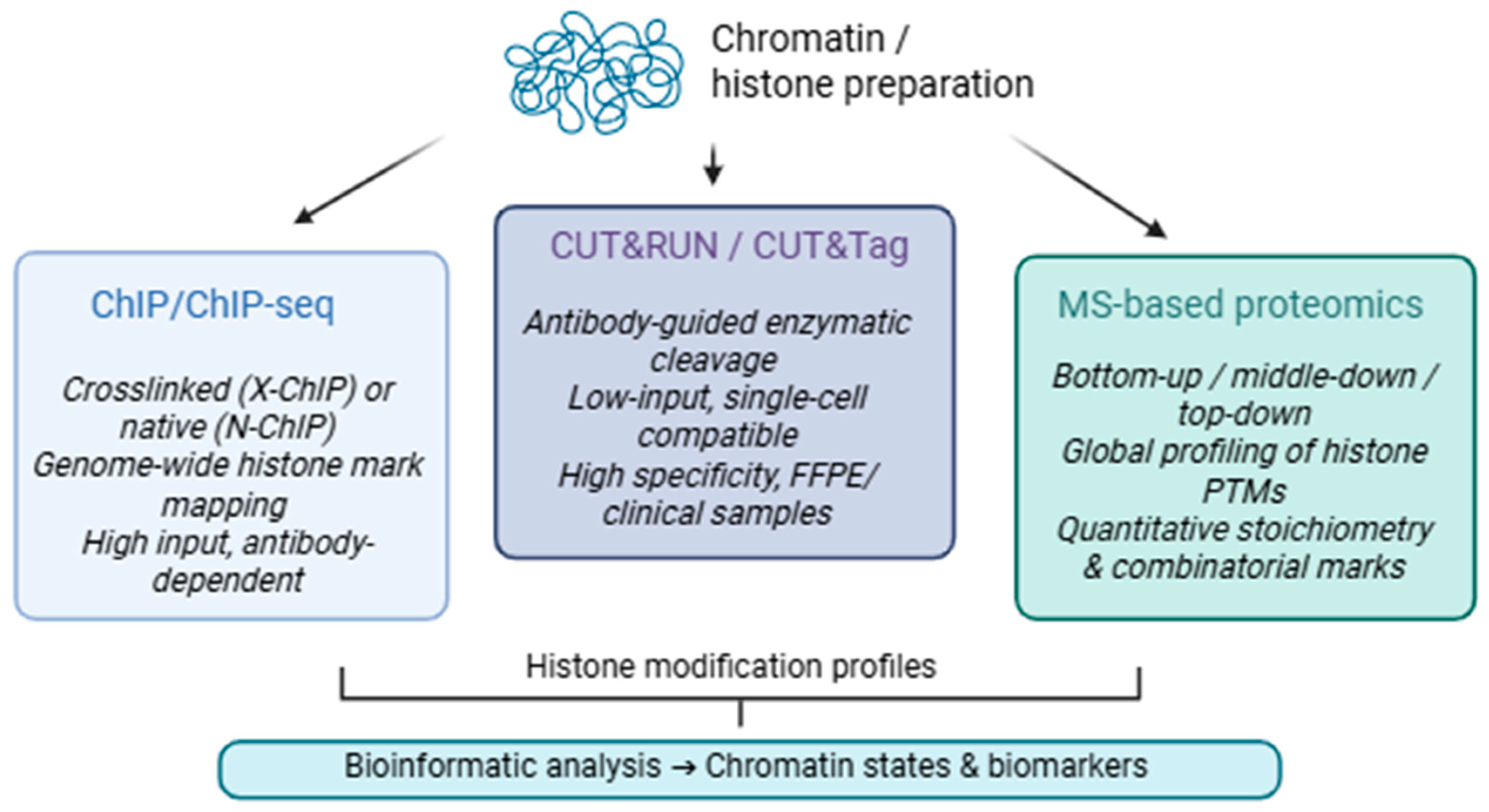
Histone modifications serve as essential epigenetic regulators that alter chromatin structure and hence affect gene expression [90]. Histone proteins, especially the N-terminal tails of H3 and H4, experience numerous post-translational modifications (PTMs), such as methylation, acetylation, phosphorylation and ubiquitination. These alterations are facilitated by specialized enzymes known as “writers” (e.g., histone acetyltransferases, methyltransferases), interpreted by “readers” (e.g., bromodomain- or chromodomain-containing proteins), and eliminated by “erasers” (e.g., histone deacetylases, demethylases) [91]. Certain histone modifications are functionally associated with transcriptional activation, including H3K4me3 and H3K27ac at active promoters and enhancers [92], whereas others, such as H3K27me3 and H3K9me3, are connected to transcriptional repression and the creation of heterochromatin [93]. The interplay of writer, reader, and eraser activity establishes chromatin states and supports processes including development, differentiation, and disease progression. In the subsequent subsections, we examine the primary methodological approaches (Figure 2) for profiling histone changes and evaluate their merits, limits, and recent advancements.

3.1. Chromatin Immunoprecipitation

ChIP is a crucial technique in epigenetics that facilitates the examination of protein–DNA interactions and histone changes within their native chromatin environment. This method, when combined with next-generation sequencing (ChIP-seq), produces genome-wide enrichment profiles with positional resolution determined by fragment size/peak width (typically tens to a few hundred bases). In specialized footprinting variants (e.g., ChIP-exo), it provides single-base resolution [94,95].
There are two primary categories of ChIP: crosslinked ChIP (X-ChIP) and native ChIP (N-ChIP). X-ChIP employs formaldehyde to stabilize protein–DNA interactions, making it especially beneficial for transcription factors and transient binding events, whereas N-ChIP circumvents crosslinking and is more appropriate for investigating histone modifications, owing to its diminished background and maintenance of native chromatin architecture [75,77]. Both procedures require meticulous tuning of crosslinking efficiency, chromatin shearing, antibody specificity, and control sample design to guarantee biological validity [94,96].
ChIP-seq utilizes high-throughput sequencing to significantly enhance resolution, diminish background noise, and broaden genome coverage. This facilitates the detection of distinct histone modifications linked to active transcription (e.g., H3K4me3, H3K27ac), repression (e.g., H3K27me3), enhancer activity (e.g., H3K4me1), or constitutive heterochromatin (e.g., H3K9me3) [97]. These markings delineate “chromatin states” throughout the genome, which are fundamental to essential regulatory processes such as development, differentiation, and tumor progression [97].
Nonetheless, ChIP-seq poses significant technological difficulties. The ENCODE and modENCODE consortia have instituted rigorous protocols, including antibody validation, sequencing depth, biological replication, and data quality control, to enhance experimental rigor and inter-study comparability [98]. Despite uniformity, experimental artifacts may emerge from inconsistent fixation durations, ineffective fragmentation, or nonspecific antibody enrichment [99]. The analytical aspect of ChIP-seq necessitates comprehensive bioinformatic processing, and specialized techniques are necessary to manage diffuse enrichment domains and super-enhancer clusters for broad histone marks (e.g., H3K27me3) [97].
ChIP has been effectively modified for use in species and tissues typically regarded as challenging for chromatin-based tests. In plant systems like Prunus persica, refined ChIP methods have facilitated the examination of histone modifications (H3K4me3, H3K27me3) in floral buds and fruit mesocarp, elucidating the epigenetic regulation of dormancy and development [100]. Correspondingly, innovative techniques have been established for intricate tissues like Nicotiana benthamiana leaves, which are abundant in starch and present particular biochemical difficulties [101].
An especially transformational application of ChIP-seq is its adaptation for formalin-fixed paraffin-embedded (FFPE) human tissue samples. Techniques like FiT-seq have facilitated chromatin profiling from archival tumor tissues [102], enabling the identification of disease-specific regulatory elements and epigenomic markers directly from clinical samples. This advancement connects epigenetic research with translational diagnostics, facilitating retrospective biomarker identification and mechanistic disease modeling [102,103].
ChIP-seq, as a high-throughput integrative platform, is continuously advancing with the incorporation of single-cell and multi-omics approaches [97]. When integrated with computer modeling and artificial intelligence, it offers potential benefits for both fundamental regulation of biology and predictive, tailored therapy [104,105].

3.2. Cleavage Under Targets (CUT&RUN) and Release Using Nuclease) and Cleavage Under Targets and Tagmentation (CUT&Tag)

The emergence of Cleavage Under Targets (CUT&RUN) and its sequel, Cleavage Under Targets and Tagmentation (CUT&Tag), has significantly transformed chromatin profiling by providing reliable, low-input alternatives to traditional ChIP-seq. These antibody-mediated enzymatic methods have demonstrated notable efficacy in delineating histone changes, attributable to their elevated signal-to-noise ratios, less background interference, and suitability for small or primary samples [106].
In the CUT&RUN technique permeabilized cells or isolated nuclei are fixed onto Concanavalin A-coated beads and treated with a primary antibody specific to a target histone alteration. A conjugate of protein A (or protein A/G) with micrococcal nuclease (pA-MNase) is subsequently injected, which associates with the antibody and selectively cleaves DNA next to the targeted histones. The resultant DNA fragments disperse from the nucleus and are harvested for sequencing. This technique produces highly localized cleavage patterns with significantly reduced background compared to ChIP-seq and can be executed with as few as 100,000 nuclei or fewer [107].
CUT&Tag enhances this framework by linking Tn5 transposase, instead of MNase, to the antibody-bound complex. This enzyme cleaves DNA while concurrently inserting sequencing adapters (tagmentation), optimizing library creation, and minimizing enzymatic bias. It is especially beneficial for profiling unusual cell types or single-cell scenarios. Moreover, the application of biotinylated Tn5 (as utilized in B-CUT&Tag) has facilitated the more efficient purification of specific chromatin fragments in plants, necessitating nuclei isolation due to the existence of stiff cell walls [108].
Fundamentally, both CUT&RUN and CUT&Tag share the same principle: a target-specific antibody guides a tethered enzyme to cleave chromatin near the epitope of interest. In CUT&RUN, micrococcal nuclease is used to digest DNA around the binding site, and the cleaved fragments are recovered for sequencing Via standard library preparation protocols [107]. In contrast, CUT&Tag uses Tn5 transposase, which performs simultaneous cleavage and adapter insertion, greatly simplifying workflow and enabling low-input and high-throughput applications [108]. Compared to classical ChIP-seq, these techniques require fewer cells, avoid crosslinking and sonication, and yield higher signal specificity, making them ideal for fragile, rare, or clinical samples [107,108].
Recent research has illustrated the efficacy of CUT&RUN and CUT&Tag across several systems, including mammalian primary B lymphocytes [107] and fungal pathogens [109]. These techniques have been refined for transcription factors and histone modifications, including H3K4me3, H3K27me3, and H3K27ac, enabling accurate epigenetic mapping with minimum input. CUT&RUN has been effectively modified for Candida albicans, facilitating the genome-wide mapping of histone changes under biofilm-inducing conditions with merely 10% of the sequencing depth necessary for ChIP-seq [109]. CUT&Tag has been utilized to investigate non-canonical DNA structures, including G-quadruplexes (G4s), with particular antibodies such as BG4 [110]. These results demonstrated that BG4 CUT&Tag signals correspond with G4 loci and considerably overlap with accessible chromatin, underscoring the method’s sensitivity and the necessity for suitable controls to identify genuine target specificity [110].
CUT&RUN and CUT&Tag facilitate accurate, reproducible, and scalable profiling of histone changes in scenarios where conventional ChIP is constrained by technical noise, low resolution, or substantial input demands.

3.3. Mass Spectrometry-Based Proteomics

Mass spectrometry (MS) has become the established gold standard for the unbiased and multiplexed analysis of histone post-translational modifications (hPTMs). It provides a combinatorial insight and depth of coverage that antibody-based methods inherently lack. While techniques such as ChIP-seq or CUT&Tag offer information on the genomic localization of individual histone marks, MS is particularly adept at generating a global and quantitative profile of all modifications that are present on histones, including rare, novel, or combinatorial acting PTMs.
Histones are basic proteins abundant in lysine and arginine residues, especially in their N-terminal tails, which undergo several chemical changes, including methylation, acetylation, phosphorylation, ubiquitination, and crotonylation. These alterations affect chromatin accessibility and architecture and are crucial facilitators of epigenetic control. To encapsulate this complexity, MS-based proteomics utilizes three principal methodologies: bottom-up, middle-down, and top-down techniques [111,112].
  • Bottom-up proteomics, the predominant methodology, entails trypsin digestion of chemically modified histones to produce short peptides for liquid chromatography-tandem mass spectrometry (LC-MS/MS) analysis. This technology is resilient and ideally suited for high-throughput operations, including the analysis of clinical samples, as evidenced in patient stratification studies for cancer and other disorders [112].
  • Middle–down proteomics facilitates the examination of extended polypeptides, preserving greater information regarding concurrent post-translational modifications (PTMs) and permitting the identification of combinatorial patterns, such as bivalent marks like H3K4me3/H3K27me3. This method integrates the sensitivity and throughput of bottom-up techniques with the superior resolution of top-down procedures, albeit it necessitates sophisticated chromatographic and computational tools [111].
  • Top-down proteomics directly examines intact histones without preliminary digestion, providing exceptional insight into individual proteoforms, particularly those with multiple, distant alterations. Although technically challenging and currently constrained in large-scale use, advancements in ion mobility and computational deconvolution are progressively enhancing the accessibility of this method [111].
Recent research has illustrated the efficacy of mass spectrometry-based methodologies in toxicoepigenetics, particularly by examining the impact of developmental toxins on histone post-translational modification landscapes in human embryonic stem cells (hESCs) [113,114]. Using valproic acid (VPA) and other chemicals, mass spectrometry allowed for the simultaneous identification of global acetylation elevations and intricate combinatorial post-translational modification alterations in a dose-dependent manner, providing a robust in vitro model for developmental toxicity [113,114].
Moreover, the clinical relevance of MS is continually broadening. A recent transition to directly assessing histones in patient-derived biopsies and circulating nucleosomes, as non-invasive biomarkers, has highlighted the translational potential of this methodology [97]. Altered levels of H4K16ac and H4K20me3 have been consistently associated with various malignancies and were effectively identified in both tumor tissue and serum by quantitative bottom-up mass spectrometry [112]. Ultimately, MS-based proteomics enhances genomic approaches by providing stoichiometric, combinatorial, and dynamic insights into the histone code, essential for comprehending chromatin regulation in development, disease, and therapy. The incorporation of these proteomic data into multi-omics frameworks signifies a viable avenue for forthcoming epigenetic research.

3.4. Comparative Evaluation of Histone Modification Profiling Techniques

The growing variety of experimental and computational techniques for analyzing histone modifications has allowed researchers to examine chromatin states from complementary viewpoints. Nonetheless, no singular approach offers a comprehensive understanding of hPTMs. Each technique presents distinct advantages and limits based on the biological inquiry, sample accessibility, resolution needs, and required data output. ChIP-seq, CUT&RUN/CUT&Tag, and MS-based proteomics represent complementary approaches to histone modification analysis, each offering distinct advantages in terms of resolution, throughput, and target specificity (Table 2).
ChIP-seq continues to serve as a standard for genome-wide mapping of histone modifications and transcription factor interactions. It offers positional data with adequate resolution (~150–300 bp), rendering it suitable for delineating chromatin states throughout the genome. Nonetheless, it is technically challenging, necessitates considerable input material, and is affected by variability stemming from crosslinking and antibody specificity.
CUT&RUN and CUT&Tag are advanced profiling systems that enhance sensitivity and diminish background noise. CUT&RUN employs micrococcal nuclease for specific DNA cleavage, whereas CUT&Tag utilizes Tn5 transposase to incorporate sequencing adapters at the binding site. Both techniques can be executed with restricted cell quantities and are suitable for low-input or single-cell applications. Their efficient methods, minimal sample processing, and compatibility with clinical materials render them formidable alternatives to ChIP-seq, especially in scenarios necessitating high sensitivity and diminished technical variability.
As a contrast to other methods, MS-based proteomics lacks positional information but is proficient at identifying combinatorial post-translational modifications, assessing modification stoichiometry, and uncovering new markers. It facilitates the characterization of global histone modification landscapes, encompassing intricate proteoform variety that remains unattainable by antibody-based methods. Recent applications have illustrated its efficacy in developmental biology, toxicity, and clinical epigenomics.
These strategies are progressively utilized in conjunction, capitalizing on their advantages. The amalgamation of genomic localization Via sequencing techniques with proteomic quantification by mass spectrometry offers a robust framework for elucidating the chromatin code in both health and sickness. A comparative summary of the main technical and functional features of ChIP-seq, CUT&RUN/CUT&Tag, and MS-based proteomics is provided in Table 2, which outlines key aspects such as resolution, input material requirements, compatibility with clinical samples, single-cell adaptability, and ability to detect stoichiometric and combinatorial histone marks.

3.5. Experimental Considerations and Pipeline Optimization

Histone modification profiling methods must match biological questions, sample types, and resolution. Data quality depends on sample preparation. Fresh or rapidly frozen tissues preserve chromatin structure and histone modifications better than FFPE tissues, which fragment DNA and cross-link proteins, damaging chromatin integrity and hiding histone modifications. Nevertheless, FFPE remains the gold standard for tissue preservation in histopathology and thus represents the most frequently available material in clinical research [115]. For this reason, novel extraction protocols and crosslink-reversal strategies, such as FiT-seq and the recently introduced Chrom-EX PE method, are being actively developed to overcome the limitations of FFPE-derived chromatin and to enable its broader application in chromatin-based epigenetic assays [115].
Cell number and viability considerably affect cell-based assay repeatability, especially low-input techniques (CUT&RUN, CUT&Tag) that are sensitive to sample handling and buffer conditions. These approaches can provide robust data from a few hundred to a thousand cells if strict quality controls are followed throughout cell lysis and chromatin accessibility phases. Cell loss or reduced viability can reduce signal-to-noise ratios and skew histone mark detection [116]. Histone extraction for MS-based proteomics must be carefully managed to avoid artificial alteration, using acid extraction or salt gradient methods to preserve endogenous post-translational markers [117,118]. Histone tails are chemically unstable, which makes them susceptible to acetylation, deamidation, or oxidation during sample processing [119]. In addition, incomplete extraction or contamination with non-histone proteins might complicate downstream analysis. Moreover, highly basic histones tend to aggregate [120], making solubilization and reproducibility difficult across laboratories. These factors underscore the need for standardized extraction protocols that minimize technical variability and maintain the native modification landscape.
ChIP-based techniques require careful adjustment of crosslinking duration, antibody specificity, and chromatin fragmentation protocols [121], while CUT&RUN and CUT&Tag minimize such artifacts but depend on enzyme accessibility and sequencing depth [122]. Recent benchmarking against ENCODE ChIP-seq datasets demonstrated that CUT&Tag recovers over half of known histone acetylation peaks with comparable functional enrichment, highlighting both its potential for sensitive profiling and its current limitations in peak recall and coverage [122]. MS-based proteomics offers unique insights into combinatorial PTMs but requires superior chromatographic separation and computational deconvolution for reliable interpretation [123].
Once challenges related to sample preparation and the experimental workflow are addressed, the critical next step lies in data interpretation and computational processing. Peak calling is usually performed utilizing MACS2 [124] and SICER [125] to identify genome-wide enrichment regions for sequencing-based assays like ChIP-seq, CUT&RUN, and CUT&Tag. Downstream integrative systems like deepTools and ChromHMM annotate chromatin states, show enrichment patterns, and characterize regulatory elements [126]. MS-based proteomics creates complicated spectra from histone peptides or complete proteoforms, necessitating specific algorithms for proteoform identification and quantification. Spectral deconvolution, normalization, and FDR control are often required to accurately identify real histone alterations [127].
Ultimately, the decision between sequencing-based assays and MS-based proteomics must be made at the outset of experimental design, as it determines the entire strategy of sample preparation, data processing, and biological interpretation, in addition to the type of data achievable. It is imperative to maintain a critical awareness of these trade-offs, as histone modification studies are at risk of producing technically competent but biologically uninformative results if the methodological approach is not aligned with the research topic.

4. Non-Coding RNAs

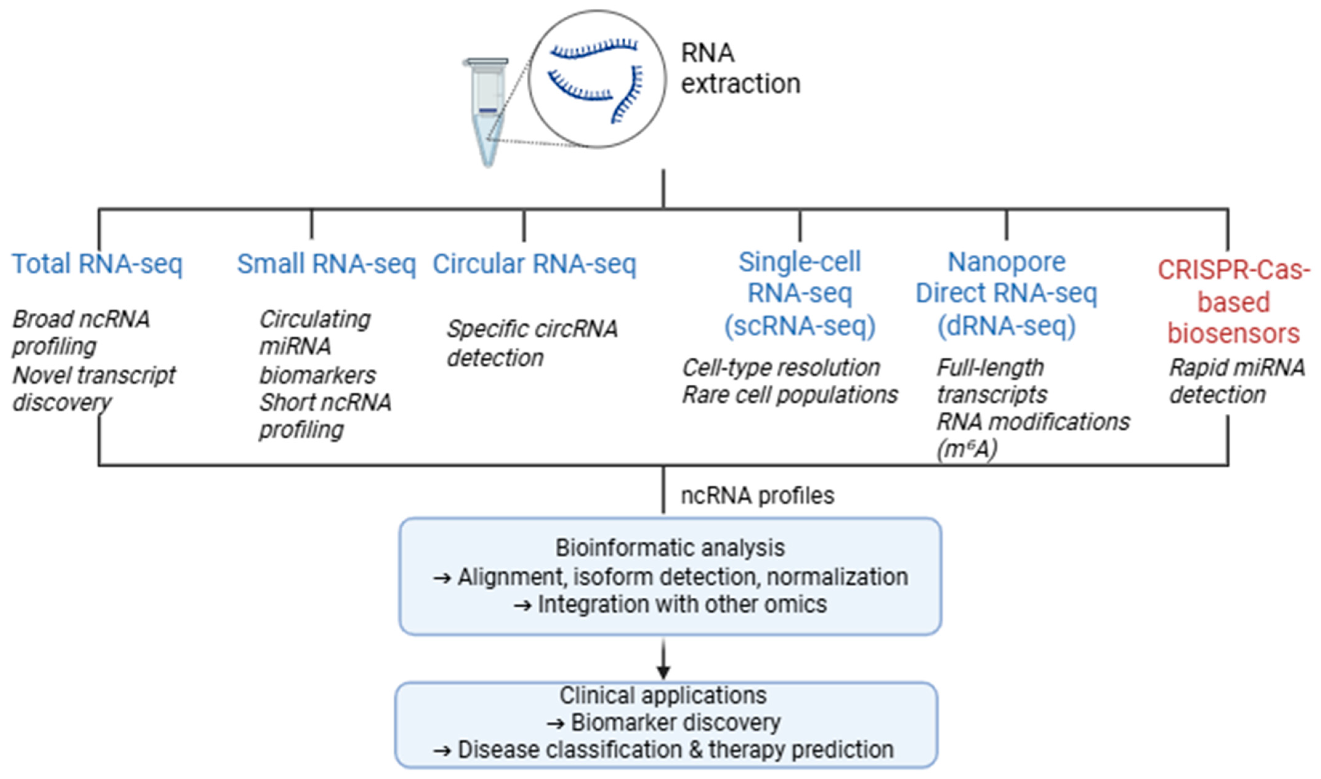
ncRNAs are transcripts that do not encode proteins but exert diverse regulatory roles in gene expression. They influence transcriptional and post-transcriptional processes and modulate chromatin architecture [128]. There are three main types of ncRNAs: small RNAs (like miRNAs, which are about 18–25 nt long and mostly work by breaking down mRNA or stopping translation), long non-coding RNAs (lncRNAs, which are more than 200 nt long and often act as scaffolds, decoys, or regulators of chromatin state), and circular RNAs (circRNAs, which are molecules that are covalently closed and can act as microRNA sponges or modulate transcription and translation) [128]. Because of their relative stability, abundance, and direct involvement in regulatory networks, ncRNAs have attracted considerable attention as epigenetic biomarkers. They can be detected in tissues as well as in body fluids, such as serum or plasma, making them suitable for non-invasive biomarker discovery in development, immunity, and tumorigenesis [129,130]. Given the large number of ncRNA species with diverse sizes, functions, and expression patterns, as well as the wide range of biological samples from which they can be studied, multiple analytical strategies have been developed for their detection and characterization (Figure 3). In the following sections, we outline the principles of these methodologies and, subsequently, highlight their key challenges and limitations.

4.1. RNA-Seq Approaches

4.1.1. Total RNA Sequencing

Total RNA-seq captures a wide range of RNA species by removing rRNA, which otherwise constitutes more than 80% of total RNA, thereby enriching for other transcript types and preserving both polyadenylated and non-polyadenylated transcripts [131]. This is particularly important for profiling lncRNAs and other non-coding species that lack polyA tails [130]. In contrast to earlier transcriptomic platforms like microarrays, RNA-seq does not rely on prior genome annotation for probe design, offering greater flexibility and reusability of datasets as reference annotations evolve [131].
Library preparation for total RNA-seq typically involves fragmentation, reverse transcription, and adapter ligation. Sequencing is then followed by computational alignment and quantification steps, often using genome-wide references [132]. However, the resulting data are affected by both technical and biological noise—variability stemming from sample handling, sequencing, and inter-individual heterogeneity [131,133]. Consequently, the reproducibility of RNA-seq experiments hinges on the precise documentation of computational steps, reference versions, and software parameters [131].
Total RNA-seq has been instrumental in characterizing novel lncRNAs and detecting low-abundance regulatory transcripts in tissues, tumors, and extracellular vesicles [129,130]. Despite its power, the method demands significant depth and processing, with computational “design noise” remaining a key limitation for reproducibility [131].

4.1.2. Small RNA Sequencing

Small RNA-seq is optimized for RNAs under 200 nucleotides, including miRNAs, small interfering RNAs (siRNAs), and piwi-interacting RNAs (piRNAs). It typically employs size-selection and sequential adapter ligation, followed by reverse transcription and amplification [133]. This methodology is central to the discovery of circulating miRNA biomarkers, given the stability and abundance of these molecules in plasma, serum, etc. [129,134].
One of the key advantages of small RNA-seq is its ability to resolve sequence isoforms and novel small RNAs across different tissues and conditions [132]. However, biases in adapter ligation efficiency and variability in reverse transcription can impact quantification, necessitating rigorous normalization strategies [131,133]. Additionally, the method is not immune to sequencing depth limitations, particularly for low-expression transcripts such as tissue-specific miRNAs [130].
Recent efforts have integrated small RNA-seq data into machine learning models to classify disease states, including cancer subtypes, with promising accuracy [129,135]. Moreover, comparative studies have emphasized the need for standardization in library preparation protocols to ensure cross-study comparability [136].

4.1.3. Circular RNA Sequencing

circRNAs are covalently closed transcripts that arise through back-splicing and often function as miRNA sponges or regulators of protein translation [129,130]. Owing to their exonuclease resistance and high stability, they are considered attractive biomarkers, especially in liquid biopsy contexts [134]. Standard RNA-seq protocols are not tailored for their detection, necessitating specialized methods such as RNase R treatment to remove linear RNAs, or computational pipelines that identify back-splice junctions (e.g., CIRCexplorer, CIRI) [137,138]. These approaches require high read depth and precise alignment algorithms to distinguish circRNAs from linear splicing events. Misannotation and the presence of repetitive elements remain challenges in circRNA profiling [138]. Nonetheless, growing evidence supports their diagnostic utility in oncology, neurodegeneration, and cardiovascular disease [129,130].

4.1.4. Single-Cell RNA Sequencing

While bulk RNA-seq captures the average expression across a cell population, single-cell RNA sequencing (scRNA-seq) enables the profiling of individual cells, revealing stochastic gene expression, rare cell types, developmental trajectories, and the tissue microenvironment [129,130].
The general workflow for scRNA-seq includes single-cell isolation, cell lysis, reverse transcription, cDNA amplification, library preparation, sequencing, and downstream computational analysis [130]. A variety of scRNA-seq protocols have been developed with trade-offs in sensitivity, throughput, cost, and transcript coverage. Plate-based methods like Smart-seq2 and Smart-seq3 offer high sensitivity and full-length transcript detection, making them well suited for isoform analysis and low-abundance non-coding RNAs [136,139]. In contrast, droplet-based platforms (e.g., 10× Genomics Chromium, Drop-seq, inDrop) provide higher throughput but often restrict sequencing to the 3′ or 5′ end of transcripts, limiting their capacity for isoform-level resolution [135].
While scRNA-seq is a powerful tool for dissecting gene expression regulation, its effective application depends heavily on sample quality, cell viability, RNA integrity, and precise computational processing [133,135]. Although scRNA-seq was initially designed for polyadenylated mRNA, it has increasingly been used to explore the landscape of ncRNAs, including lncRNAs, antisense transcripts, and certain circular RNAs [129,134]. However, detecting miRNAs and other small RNAs remains a significant challenge in scRNA-seq, primarily due to their short length, lack of polyA tails, and incompatibility with standard reverse transcription strategies [140]. Emerging adaptations include small RNA-focused single-cell protocols and integrated workflows that combine miRNA-seq with transcriptomic profiling in dual-omics approaches [141]. CircRNAs pose another methodological challenge due to their back-spliced junctions and absence of polyadenylation. Although not routinely captured by conventional scRNA-seq protocols, specialized bioinformatic tools and enrichment strategies are beginning to enable circRNA detection at the single-cell level [137,138].
The resolution provided by scRNA-seq has made it a cornerstone in epigenetic biomarker research, particularly in fields such as oncology, neurodevelopment, and immunology [129,135]. For instance, single-cell transcriptomics has been used to track lineage-specific lncRNA expression in developing tissues and to identify tumor subpopulations expressing regulatory ncRNAs linked to prognosis and therapy resistance [129,130]. By revealing transcriptional heterogeneity and lineage trajectories, scRNA-seq provides critical insights into how epigenetic states, including those mediated by ncRNAs, evolve across development and disease. As spatial transcriptomics and multi-modal platforms mature, their integration with scRNA-seq will enable simultaneous analysis of gene expression, chromatin accessibility, and epigenetic regulation at single-cell resolution [139,141].
While the detection of non-coding RNAs remains challenging, especially for small and circular species, emerging methods and computational advancements are steadily enhancing the resolution and reliability of single-cell transcriptomic data. As technologies mature and integrate, scRNA-seq will play a pivotal role in the clinical application of epigenetic biomarker discovery.

4.1.5. Nanopore Direct RNA Sequencing (dRNA-Seq)

Nanopore-based direct RNA sequencing (dRNA-seq) has emerged as a powerful technology for profiling the transcriptome at single-molecule resolution without requiring reverse transcription or amplification. Developed by Oxford Nanopore Technologies, this approach enables the direct sequencing of full-length native RNA strands through a biological nanopore, preserving nucleotide modifications and revealing isoform complexity in a manner not achievable by short-read or cDNA-based methods [142]. The dRNA-seq protocol involves ligating an adapter–motor complex to the poly(A) tail of intact RNA molecules, guiding them through a nanopore under an applied voltage. As the RNA translocates through the pore, changes in ionic current are measured and interpreted to infer nucleotide sequence. Crucially, this technique retains base modifications (such as m6A) as they influence the current signal, offering a window into the epitranscriptome [143,144].
One of the central advantages of dRNA-seq is its capacity to sequence full-length transcripts, including UTRs, alternative splicing events, and transcript isoforms, without the biases introduced by reverse transcription. This has made it particularly valuable in identifying transcriptome complexity in both human and non-model organisms [145,146]. Moreover, the method enables direct detection of polyadenylation sites and estimates of poly(A) tail lengths, which are essential for understanding RNA stability and translational control. Applications of dRNA-seq span from transcript discovery to isoform quantification and RNA modification profiling. Recent studies have shown that it enables more detailed analysis of transcript diversity, RNA processing, and regulatory dynamics, offering insights that are often missed by conventional short-read sequencing approaches [147,148]. For instance, Leger et al. (2021) demonstrated how this approach can identify modification signatures with improved resolution when combined with comparative models and controlled synthetic standards [147]. Similarly, Burdick et al. (2023) employed dRNA-seq to resolve dynamic RNA processing events and transcriptional regulation in viral infection models [149]. In the context of neurodegeneration, Naarmann-de Vries et al. (2022) applied dRNA-seq to study transcript diversity and isoform-specific expression shifts in disease-relevant neuronal populations, revealing insights not captured by conventional short-read methods [148].
Despite these strengths, dRNA-seq also presents challenges. Its relatively high input requirements (typically 500 ng or more of polyadenylated RNA) limit its use in low-input or single-cell applications. Furthermore, the raw read accuracy remains lower, although improvements in basecalling models and training data continue to enhance reliability [150]. Importantly, because sequencing is anchored to the poly(A) tail, non-polyadenylated RNAs (such as histone mRNAs and many long non-coding RNAs) are generally not captured unless specialized protocols are applied [151].
Efforts to refine the technology include algorithmic developments for RNA modification detection and transcript phasing. For example, Hong et al. (2022) advanced the modeling of signal shifts induced by m6A modifications using enhanced statistical frameworks trained on synthetic RNA standards [146]. These approaches have begun to disentangle modified from unmodified bases in native RNA, although the accuracy of modification calling remains an active area of development. Although current limitations in throughput, cost, and modification sensitivity remain, dRNA-seq uniquely enables transcriptome-wide interrogation of RNA biology with molecular fidelity.

4.1.6. Computational Pipelines and Reproducibility

A major issue in RNA-seq analysis is the lack of standardized computational pipelines. Even with identical raw data, different tool combinations and parameters can yield divergent transcript quantification results, a phenomenon referred to as “in silico design noise” [131]. Despite existing guidelines such as MINSEQE, reporting practices remain inconsistent; only a minority of studies disclose complete pipeline details or software versions [131,132]. This problem is compounded in ncRNA analysis, where annotations are still evolving and tools often lack consensus on transcript definitions [129]. Open-access workflows, version control systems like Git, and containerized environments (e.g., Docker, Nextflow) have been proposed as solutions to enhance reproducibility and transparency [131,141].

4.2. CRISPR-Cas-Based Sensors for miRNA

CRISPR-Cas-based biosensors have arisen as potent tools for the precise and sensitive detection of miRNAs, with extensive implications in diagnostics and targeted therapies, especially in oncology. These systems leverage the programmability of CRISPR-associated proteins and the distinctive sequences of miRNAs to attain accurate molecular recognition and signal amplification.
The technology of CRISPR-Cas-based miRNA applications relies on programmable nucleases, mainly Cas9, Cas12a, and Cas13a, guided by sequence-specific RNAs for targeted detection or modification of miRNAs. Cas9, guided by a single-guide RNA (sgRNA), creates double-strand breaks in DNA to modify or destroy miRNA genes or their target binding sites, hence affecting post-transcriptional regulation [152,153]. Cas12a and Cas13a are utilized in biosensing applications due to their trans-cleavage activity; upon recognizing a specific miRNA, the activated enzyme cleaves reporter molecules, producing observable signals such as fluorescence [154,155].
In addition to detection, CRISPR can modify miRNA binding sites on mRNAs, thereby inhibiting repression and augmenting gene expression. This has been evidenced in plant models and has potential for translational applications [156]. In oncology, CRISPR systems have been utilized to regulate miRNA genes associated with tumor growth, facilitating both diagnostic evaluation and therapeutic intervention [153].
Methodologically, the CRISPR system and biosensor design depend on numerous factors: (1) Cas protein type (e.g., Cas9 vs. Cas12a), (2) target miRNA sequence and cellular context, (3) detection or functional modification, and (4) matrix complexity (e.g., serum vs. pure RNA) [155]. Due to its collateral cleavage activity, biosensors use Cas12a for rapid signal amplification, while Cas9 is better for site-specific genome editing. Understanding miRNA biosynthesis, location, and expression levels in the sample type helps choose the best miRNA detection or editing approach. Circulating miRNAs in serum or exosomes may require different lysis and purification methods than intracellular ones [136,138]. Therapeutic applications must also address miRNA redundancy and off-target effects, requiring careful guide RNA and delivery vector design to prevent cytotoxicity and immune responses [152,154].

4.3. Comparative Evaluation of RNA-Based Methods for Non-Coding RNA Profiling

Numerous transcriptomic methodologies have been established to analyze non-coding RNAs, owing to their significant functions in gene regulation and disease. A summary of the five principal RNA-based techniques is provided in Table 3, emphasizing their respective benefits, drawbacks, and significance for ncRNA biomarker identification.
Together, these technologies provide a powerful and evolving toolkit for interrogating the epigenetic functions of ncRNAs in health and disease, with future progress likely to depend on improved multi-modal integration, standardization of computational workflows, and methodological innovations tailored to specific RNA biotypes.

4.4. Experimental Considerations and Pipeline Optimization

The diverse biotypes and biochemical properties of ncRNAs [128] present unique challenges during sample preparation. miRNAs are highly structured, short molecules (approximately 22 nt), and are susceptible to ligation biases during library preparation. lncRNAs are low-abundance and frequently lack poly(A) tails, necessitating rRNA depletion or capture-based enrichment. In contrast, circRNAs are covalently closed and resistant to exonuclease digestion, necessitating RNase R treatment to selectively degrade linear transcripts. RNA integrity is a critical pre-analytical concern. Longer lncRNAs are disproportionately affected by degradation during extraction or protracted storage, while extracellular miRNAs in plasma or exosomes necessitate optimized isolation to prevent the loss of low-yield species [128]. For instance, plasma miRNA biomarker studies have demonstrated that variations in extraction tools can result in variability in yield and bias downstream quantification, emphasizing the direct influence of early technical decisions on biological interpretation [157]. RNA isolation is a critical stage in the study of ncRNAs [158]. Commercial kits that are optimized for either total RNA or short RNA fractions are utilized by the majority of laboratories. These kits are frequently based on magnetic bead technology or silica-membrane spin columns. Although these standardized protocols enhance reproducibility, they also introduce specific biases [158]. For instance, some total RNA kits under-represent small RNAs, while small RNA-enrichment kits may lose longer lncRNAs. Therefore, it is crucial to select an optimal RNA isolation kit, particularly when working with low-yield samples such as plasma, exosomes, or FFPE tissues. To preserve RNA integrity, freeze–thaw cycles and delays in processing should be minimized, since long and low-abundance ncRNAs are especially sensitive to degradation [159]. Before library preparation, quality should also be checked to confirm that differences in transcript levels reflect biology and not technical artifacts [160].
The choice of methodology for ncRNA profiling cannot be made arbitrarily; it must reflect the RNA biotype and practical factors such as input amount, RNA integrity, and required resolution. After sample preparation and the chosen analysis, the analytical focus transitions to bioinformatic processing. The specific characteristics of ncRNAs must be taken into consideration by pipelines. For highly structured miRNAs, read trimming and alignment are more complex, circRNA identification necessitates algorithms such as CIRCexplorer or CIRI [161], and scRNA-seq necessitates correction for dropout effects and amplification artifacts [162]. Normalization for sequencing depth, bulk effects, and compositional biases is indispensable on all platforms. However, the evolving and occasionally inconsistent annotation of ncRNAs remains a persistent challenge, as it incorporates variability into transcript definitions and complicates cross-study comparison. Using containerized workflows such as Docker [163], version control tools like Git [164], and clear reporting standards makes analyses easier to repeat and compare across studies. These practices ensure that the same software versions and settings are applied, so results are consistent and more likely to reflect true biological differences.
Ultimately, choices taken early on, like sample preparation, RNA integrity, and methodological alignment with the target biotype, have a greater impact on the outcome of ncRNA profiling than the sequencing technology itself. Even while single-cell platforms, nanopore technologies, and RNA-seq have increased analytical options, their results are still quite susceptible to computational variability and pre-analytical bias. Without careful matching of protocol to the biological question and transparent, standardized bioinformatic workflows, there is a risk of producing technically valid but biologically misleading results.

5. Artificial Intelligence Models Integrated for Epigenetics Research

In contrast to static genomic information, epigenetic modifications are dynamic, context-dependent, and reactive to environmental stimuli. These characteristics render epigenetic data exceptionally intricate but computationally intensive, requiring sophisticated analytical approaches to discern patterns within high-dimensional, heterogeneous datasets. AI, especially Via machine learning and deep learning methodologies, offers a scalable framework to challenge these difficulties and derive therapeutically significant findings [165]. AI models, particularly those employing deep neural networks, are exceptionally skilled at tasks related to pattern recognition, feature extraction, and result prediction from extensive, intricate datasets. Traditional machine learning approaches depend on manually crafted features, but deep learning architectures can independently acquire latent data representations through hierarchical layers [165]. These layers include nonlinear relationships between epigenetic characteristics and phenotypic results, facilitating strong performance despite the presence of incomplete or noisy data [165].
Building on these capabilities, a diverse set of AI algorithms has been applied to epigenetic data, particularly DNA methylation profiles [166], to address clinical classification and biomarker discovery.
Traditional models, such as support vector machines (SVMs) and random forests (RFs), are still commonly utilized due to their ability to manage large feature sets and produce interpretable results [167,168]. However, the area has experienced a significant movement toward deep learning architectures that can capture complicated, nonlinear connections without requiring extensive preprocessing. Table 4 lists representative AI models integrating epigenetic data. These models are merely a few examples of effective AI applications in epigenetic data processing. Their structure and implementation are tailored to the type of input data (e.g., DNA methylation, histone modifications), the clinical task (e.g., classification, prognosis, or biomarker discovery), and the complementary data (e.g., gene expression, chromatin accessibility, 3D genome structure). Models are often chosen depending on the study purpose, data complexity, and the balance of interpretability and forecast accuracy.
MethylNet, for example, employs variational autoencoders to compress high-dimensional methylation data and predict clinical outcomes, including age, tumor subtype, and immune infiltration [169]. DeepChrome [170] and DeepHistone [171] use convolutional neural networks to capture regulatory patterns in histone mark data, with DeepHistone improving accuracy by including chromatin accessibility. INTERACT [28], a hybrid CNN-transformer model, accurately predicts CpG methylation levels by modeling long-range dependencies in genomic sequence, which is particularly useful for deciphering regulatory variations. ChromeGCN [172] uses graph convolutional networks and Hi-C data to predict chromatin states and enhancer-promoter interactions in geographical contexts. Finally, DDAE + MLP [173] incorporates methylation and copy number variation data to estimate gene expression, providing a multi-omics method that is very effective in cancer transcriptomics.
Numerous instances exist of AI being utilized on epigenetic data for diagnostic and prognostic applications across various diseases. Three systematic review studies have been conducted up until now that illustrate the development of this topic. Rauschert et al. (2020) reviewed early applications of machine learning to DNA methylation data, showing promising results in cancer, fetal growth restriction, and autism, but emphasized challenges such as small sample sizes, overfitting, and lack of external validation [174]. In 2023, Yassi et al. performed a thorough evaluation of 35 deep learning applications in cancer epigenetics, discovering several architectures, including CNNs, autoencoders, and transformer variations, utilized for both array-based and sequencing-derived methylation data [175]. These models were employed for tasks including the identification of differentially methylated regions (DMRs), the categorization of tumor vs. normal tissue, subtype stratification, and survival prediction. A consistent observation was the enhanced efficacy of multimodal deep learning frameworks in forecasting disease outcomes relative to unimodal baselines. This perspective was further developed by Castilho et al. (2024), who conducted a systematic analysis of 54 machine learning studies that employed methylation data for cancer detection [176]. Their review verified that oncology is the most extensively researched field, with the majority of models achieving over 90% accuracy, notably in breast, lung, and colorectal cancers. The authors emphasize the increasing prevalence of deep learning and the potential of explainable AI methods to enhance the interpretability of models and their adoption in the clinical setting [176]. These improvements collectively illustrate that AI-based approaches may efficiently manage the complexity of epigenetic data while facilitating their translation into clinically relevant, robust, and scalable diagnostic tools.
Despite the potential of AI in epigenetic data analysis, there are still numerous critical challenges that exceed the scope of computational modeling. The lack of laboratory-wide sample processing, data standardization, and biomarker validation standards hinders repeatability and clinical adoption. Epigenetic markings are tissue-specific, especially in diverse or inaccessible regions like the brain and placenta, making their identification difficult. To move toward clinical application, epigenetic biomarkers will require multi-center validation, regulatory approval, and the development of affordable and accessible assays. AI can accelerate this process, but it cannot replace rigorous experimental design and validation; only their integration will make AI-assisted epigenetic diagnostics clinically reliable.

6. Conclusions

Over the past two decades, the advancement and implementation of approaches for identifying epigenetic biomarkers have significantly progressed, allowing researchers to investigate the epigenetic landscape with unparalleled depth and accuracy. Among the methodologies examined, bisulfite sequencing is regarded as the gold standard for DNA methylation studies owing to its single-base resolution and exceptional specificity. Chromatin immunoprecipitation followed by sequencing (ChIP-seq) is considered the standard method for histone modification profiling, providing specificity and comprehensive genome-wide mapping. In the field of non-coding RNA identification, RT-qPCR and next-generation RNA sequencing (RNA-seq) deliver great sensitivity and quantitative precision, with RNA-seq furthermore providing comprehensive transcriptome coverage.
Future prospects involve the enhancement of single-cell epigenomic technologies, enabling the analysis of cellular heterogeneity within intricate tissues. The integration of machine learning algorithms with multi-omics data shows potential for creating prediction models in personalized medicine. The ongoing decrease in sequencing expenses and enhancement of data processing workflows will render these instruments more attainable for clinical laboratories.

Author Contributions

Conceptualization, S.D.P. and L.S.M.; investigation, I.B.; writing—original draft preparation, I.B. and L.S.M.; writing—review and editing, I.B., T.M., S.O., S.D.P. and L.S.M.; visualization, I.B.; supervision, S.D.P. and L.S.M. All authors have read and agreed to the published version of the manuscript.

Funding

This research received no external funding.

Institutional Review Board Statement

Not applicable.

Informed Consent Statement

Not applicable.

Data Availability Statement

No new data were created or analyzed in this study. Data sharing is not applicable to this article.

Conflicts of Interest

The authors declare no conflicts of interest.

References

  1. Cavalli, G.; Heard, E. Advances in Epigenetics Link Genetics to the Environment and Disease. Nature 2019, 571, 489–499. [Google Scholar] [CrossRef]
  2. Choi, B.Y.; Han, M.; Kwak, J.W.; Kim, T.H. Genetics and Epigenetics in Allergic Rhinitis. Genes 2021, 12, 2004. [Google Scholar] [CrossRef]
  3. Recillas-Targa, F. Cancer Epigenetics: An Overview. Arch. Med. Res. 2022, 53, 732–740. [Google Scholar] [CrossRef]
  4. Prasher, D.; Greenway, S.C.; Singh, R.B. The Impact of Epigenetics on Cardiovascular Disease. Biochem. Cell Biol. 2020, 98, 12–22. [Google Scholar] [CrossRef]
  5. Kumar, S.; Shanker, O.R.; Banerjee, J.; Tripathi, M.; Chandra, P.S.; Dixit, A.B. Epigenetics in Epilepsy. Prog. Mol. Biol. Transl. Sci. 2023, 198, 249–269. [Google Scholar] [CrossRef] [PubMed]
  6. Ling, C.; Rönn, T. Epigenetics in Human Obesity and Type 2 Diabetes. Cell Metab. 2019, 29, 1028–1044. [Google Scholar] [CrossRef]
  7. Surace, A.E.A.; Hedrich, C.M. The Role of Epigenetics in Autoimmune/Inflammatory Disease. Front. Immunol. 2019, 10, 1525. [Google Scholar] [CrossRef] [PubMed]
  8. Ghosh, P.; Saadat, A. Neurodegeneration and Epigenetics: A Review. Neurologia 2023, 38, e62–e68. [Google Scholar] [CrossRef]
  9. Ho, D.H.; Burggren, W.W. Epigenetics and Transgenerational Transfer: A Physiological Perspective. J. Exp. Biol. 2010, 213, 3–16. [Google Scholar] [CrossRef] [PubMed]
  10. Villanueva, L.; Álvarez-Errico, D.; Esteller, M. The Contribution of Epigenetics to Cancer Immunotherapy. Trends Immunol. 2020, 41, 676–691. [Google Scholar] [CrossRef]
  11. Li, Y. Modern Epigenetics Methods in Biological Research. Methods 2020, 187, 104. [Google Scholar] [CrossRef]
  12. Comendul, A.; Ruf-Zamojski, F.; Ford, C.T.; Agarwal, P.; Zaslavsky, E.; Nudelman, G.; Hariharan, M.; Rubenstein, A.; Pincas, H.; Nair, V.D.; et al. Comprehensive Guide for Epigenetics and Transcriptomics Data Quality Control. STAR Protoc. 2025, 6, 103607. [Google Scholar] [CrossRef] [PubMed]
  13. Cai, R.; Lv, R.; Shi, X.; Yang, G.; Jin, J. CRISPR/DCas9 Tools: Epigenetic Mechanism and Application in Gene Transcriptional Regulation. Int. J. Mol. Sci. 2023, 24, 14865. [Google Scholar] [CrossRef]
  14. Moore, L.D.; Le, T.; Fan, G. DNA Methylation and Its Basic Function. Neuropsychopharmacology 2013, 38, 23–38. [Google Scholar] [CrossRef]
  15. Frommer, M.; McDonald, L.E.; Millar, D.S.; Collis, C.M.; Watt, F.; Grigg, G.W.; Molloy, P.L.; Paul, C.L. A Genomic Sequencing Protocol That Yields a Positive Display of 5-Methylcytosine Residues in Individual DNA Strands. Proc. Natl. Acad. Sci. USA 1992, 89, 1827–1831. [Google Scholar] [CrossRef]
  16. Harrison, A.; Parle-McDermott, A. DNA Methylation: A Timeline of Methods and Applications. Front. Genet. 2011, 2, 74. [Google Scholar] [CrossRef]
  17. Söderhäll, C.; Reinius, L.E.; Salmenperä, P.; Gentile, M.; Acevedo, N.; Konradsen, J.R.; Nordlund, B.; Hedlin, G.; Scheynius, A.; Myllykangas, S.; et al. High-Resolution Targeted Bisulfite Sequencing Reveals Blood Cell Type-Specific DNA Methylation Patterns in IL13 and ORMDL3. Clin. Epigenetics 2021, 13, 106. [Google Scholar] [CrossRef] [PubMed]
  18. Moser, D.A.; Müller, S.; Hummel, E.M.; Limberg, A.S.; Dieckmann, L.; Frach, L.; Pakusch, J.; Flasbeck, V.; Brüne, M.; Beygo, J.; et al. Targeted Bisulfite Sequencing: A Novel Tool for the Assessment of DNA Methylation with High Sensitivity and Increased Coverage. Psychoneuroendocrinology 2020, 120, 104784. [Google Scholar] [CrossRef]
  19. Pu, W.; Qian, F.; Liu, J.; Shao, K.; Xiao, F.; Jin, Q.; Liu, Q.; Jiang, S.; Zhang, R.; Zhang, J.; et al. Targeted Bisulfite Sequencing Reveals DNA Methylation Changes in Zinc Finger Family Genes Associated with KRAS Mutated Colorectal Cancer. Front. Cell Dev. Biol. 2021, 9, 759813. [Google Scholar] [CrossRef] [PubMed]
  20. Gu, H.; Bock, C.; Mikkelsen, T.S.; Jäger, N.; Smith, Z.D.; Tomazou, E.; Gnirke, A.; Lander, E.S.; Meissner, A. Genome-Scale DNA Methylation Mapping of Clinical Samples at Single-Nucleotide Resolution. Nat. Methods 2010, 7, 133. [Google Scholar] [CrossRef]
  21. Doherty, R.; Couldrey, C. Exploring Genome Wide Bisulfite Sequencing for DNA Methylation Analysis in Livestock: A Technical Assessment. Front. Genet. 2014, 5, 126. [Google Scholar] [CrossRef] [PubMed]
  22. Li, R.; Hu, F.; Li, B.; Zhang, Y.; Chen, M.; Fan, T.; Wang, T. Whole Genome Bisulfite Sequencing Methylome Analysis of Mulberry (Morus alba) Reveals Epigenome Modifications in Response to Drought Stress. Sci. Rep. 2020, 10, 8013. [Google Scholar] [CrossRef] [PubMed]
  23. Simons, R.B.; Karkala, F.; Kukk, M.M.; Adams, H.H.H.; Kayser, M.; Vidaki, A. Comparative Performance Evaluation of Bisulfite- and Enzyme-Based DNA Conversion Methods. Clin. Epigenetics 2025, 17, 56. [Google Scholar] [CrossRef] [PubMed]
  24. Dai, Q.; Ye, C.; Irkliyenko, I.; Wang, Y.; Sun, H.L.; Gao, Y.; Liu, Y.; Beadell, A.; Perea, J.; Goel, A.; et al. Ultrafast Bisulfite Sequencing Detection of 5-Methylcytosine in DNA and RNA. Nat. Biotechnol. 2024, 42, 1559–1570. [Google Scholar] [CrossRef] [PubMed]
  25. Kurdyukov, S.; Bullock, M. DNA Methylation Analysis: Choosing the Right Method. Biology 2016, 5, 3. [Google Scholar] [CrossRef]
  26. Rauluseviciute, I.; Drabløs, F.; Rye, M.B. DNA Methylation Data by Sequencing: Experimental Approaches and Recommendations for Tools and Pipelines for Data Analysis. Clin. Epigenetics 2019, 11, 193. [Google Scholar] [CrossRef]
  27. Khodadadi, E.; Fahmideh, L.; Khodadadi, E.; Dao, S.; Yousefi, M.; Taghizadeh, S.; Asgharzadeh, M.; Yousefi, B.; Kafil, H.S. Current Advances in DNA Methylation Analysis Methods. BioMed Res. Int. 2021, 2021, 8827516. [Google Scholar] [CrossRef]
  28. Zhou, J.; Chen, Q.; Braun, P.R.; Perzel Mandell, K.A.; Jaffe, A.E.; Tan, H.Y.; Hyde, T.M.; Kleinman, J.E.; Potash, J.B.; Shinozaki, G.; et al. Deep Learning Predicts DNA Methylation Regulatory Variants in the Human Brain and Elucidates the Genetics of Psychiatric Disorders. Proc. Natl. Acad. Sci. USA 2022, 119, e2206069119. [Google Scholar] [CrossRef]
  29. Dixon, G.; Matz, M. Benchmarking DNA Methylation Assays in a Reef-Building Coral. Mol. Ecol. Resour. 2021, 21, 464–477. [Google Scholar] [CrossRef]
  30. Luo, X.; Wang, Y.; Zou, Q.; Xu, L. Recall DNA Methylation Levels at Low Coverage Sites Using a CNN Model in WGBS. PLoS Comput. Biol. 2023, 19, e1011205. [Google Scholar] [CrossRef]
  31. Simpson, D.J.; Zhao, Q.; Olova, N.N.; Dabrowski, J.; Xie, X.; Latorre-Crespo, E.; Chandra, T. Region-Based Epigenetic Clock Design Improves RRBS-Based Age Prediction. Aging Cell 2023, 22, e13866. [Google Scholar] [CrossRef] [PubMed]
  32. Guanzon, D.; Ross, J.P.; Ma, C.; Berry, O.; Liew, Y.J. Comparing Methylation Levels Assayed in GC-Rich Regions with Current and Emerging Methods. BMC Genom. 2024, 25, 741. [Google Scholar] [CrossRef]
  33. Liu, X.; Pang, Y.; Shan, J.; Wang, Y.; Zheng, Y.; Xue, Y.; Zhou, X.; Wang, W.; Sun, Y.; Yan, X.; et al. Beyond the Base Pairs: Comparative Genome-Wide DNA Methylation Profiling across Sequencing Technologies. Brief. Bioinform. 2024, 25, bbae440. [Google Scholar] [CrossRef]
  34. Troyee, A.N.; Peña-Ponton, C.; Medrano, M.; Verhoeven, K.J.F.; Alonso, C. Herbivory Induced Methylation Changes in the Lombardy Poplar: A Comparison of Results Obtained by EpiGBS and WGBS. PLoS ONE 2023, 18, e0291202. [Google Scholar] [CrossRef]
  35. Attree, E.; Griffiths, B.; Panchal, K.; Xia, D.; Werling, D.; Banos, G.; Oikonomou, G.; Psifidi, A. Identification of DNA Methylation Markers for Age and Bovine Respiratory Disease in Dairy Cattle: A Pilot Study Based on Reduced Representation Bisulfite Sequencing. Commun. Biol. 2024, 7, 1251. [Google Scholar] [CrossRef] [PubMed]
  36. Aroke, E.N.; Overstreet, D.S.; Penn, T.M.; Crossman, D.K.; Jackson, P.; Tollefsbol, T.O.; Quinn, T.L.; Yi, N.; Goodin, B.R. Identification of DNA Methylation Associated Enrichment Pathways in Adults with Non-Specific Chronic Low Back Pain. Mol. Pain. 2020, 16, 1744806920972889. [Google Scholar] [CrossRef] [PubMed]
  37. Nkongolo, K.; Michael, P. Reduced Representation Bisulfite Sequencing (RRBS) Analysis Reveals Variation in Distribution and Levels of DNA Methylation in White Birch (Betula papyrifera) Exposed to Nickel. Genome 2024, 67, 351–367. [Google Scholar] [CrossRef]
  38. Beck, D.; Ben Maamar, M.; Skinner, M.K. Genome-Wide CpG Density and DNA Methylation Analysis Method (MeDIP, RRBS, and WGBS) Comparisons. Epigenetics 2022, 17, 518–530. [Google Scholar] [CrossRef]
  39. Zhao, Y.; O’Keefe, C.M.; Hsieh, K.; Cope, L.; Joyce, S.C.; Pisanic, T.R.; Herman, J.G.; Wang, T.H. Multiplex Digital Methylation-Specific PCR for Noninvasive Screening of Lung Cancer. Adv. Sci. 2023, 10, 2206518. [Google Scholar] [CrossRef]
  40. Herman, J.G.; Graff, J.R.; Myohanen, S.; Nelkin, B.D.; Baylin, S.B. Methylation-Specific PCR: A Novel PCR Assay for Methylation Status of CpG Islands (DNA Methylation/Tumor Suppressor Genes/Pl6/P15). Proc. Natl. Acad. Sci. USA 1996, 93, 9821–9826. [Google Scholar] [CrossRef]
  41. Yoshioka, M.; Matsutani, T.; Hara, A.; Hirono, S.; Hiwasa, T.; Takiguchi, M.; Iwadate, Y.; Yoshioka, M.; Matsutani, T.; Hara, A.; et al. Real-Time Methylation-Specific PCR for the Evaluation of Methylation Status of MGMT Gene in Glioblastoma. Oncotarget 2018, 9, 27728–27735. [Google Scholar] [CrossRef]
  42. Wani, K.; Aldape, K.D. PCR Techniques in Characterizing DNA Methylation. Methods Mol. Biol. 2016, 1392, 177–186. [Google Scholar] [CrossRef] [PubMed]
  43. Cardoso, G.C.; de Oliveira Ganzella, F.A.; Miniskiskosky, G.; da Cunha, R.S.; de Souza Ramos, E.A. Digital Methylation-Specific PCR: New Applications for Liquid Biopsy. Biomol. Concepts 2024, 15, 20220041. [Google Scholar] [CrossRef] [PubMed]
  44. Santourlidis, S.; Ghanjati, F.; Beermann, A.; Hermanns, T.; Poyet, C. IDLN-MSP: Idiolocal Normalization of Real-Time Methylation-Specific PCR for Genetic Imbalanced DNA Specimens. Biotechniques 2016, 60, 84–87. [Google Scholar] [CrossRef]
  45. Tost, J.; Gut, I.G. Analysis of Gene-Specific DNA Methylation Patterns by Pyrosequencing® Technology. Methods Mol. Biol. 2007, 373, 89–102. [Google Scholar] [CrossRef]
  46. Harrington, C.T.; Lin, E.I.; Olson, M.T.; Eshleman, J.R. Fundamentals of Pyrosequencing. Arch. Pathol. Lab. Med. 2013, 137, 1296–1303. [Google Scholar] [CrossRef] [PubMed]
  47. Ghemrawi, M.; Tejero, N.F.; Duncan, G.; McCord, B. Pyrosequencing: Current Forensic Methodology and Future Applications—A Review. Electrophoresis 2023, 44, 298–312. [Google Scholar] [CrossRef]
  48. Taryma-Lesniak, O.; Kjeldsen, T.E.; Hansen, L.L.; Wojdacz, T.K. Influence of Unequal Amplification of Methylated and Non-Methylated Template on Performance of Pyrosequencing. Genes 2022, 13, 1418. [Google Scholar] [CrossRef]
  49. Park, J.W.; Park, I.H.; Kim, J.M.; Noh, J.H.; Kim, K.A.; Park, J.Y. Rapid Detection of FMO3 Single Nucleotide Polymorphisms Using a Pyrosequencing Method. Mol. Med. Rep. 2022, 25, 48. [Google Scholar] [CrossRef]
  50. De Battisti, C.; Marciano, S.; Magnabosco, C.; Busato, S.; Arcangeli, G.; Cattoli, G. Pyrosequencing as a Tool for Rapid Fish Species Identification and Commercial Fraud Detection. J. Agric. Food Chem. 2014, 62, 198–205. [Google Scholar] [CrossRef]
  51. Ogino, S.; Kawasaki, T.; Brahmandam, M.; Yan, L.; Cantor, M.; Nangyal, C.; Mino-Kenudson, M.; Lauwers, G.Y.; Loda, M.; Fuchs, C.S. Sensitive Sequencing Method for KRAS Mutation Detection by Pyrosequencing. J. Mol. Diagn. 2005, 7, 413. [Google Scholar] [CrossRef]
  52. Konrad, H.; Schäfer, L.; Sturm, H.; Hördt, L.; Bajanowski, T.; Poetsch, M. Vibration as a Pitfall in Pyrosequencing Analyses. Int. J. Legal Med. 2022, 136, 103–105. [Google Scholar] [CrossRef] [PubMed]
  53. Thumbovorn, R.; Bhattarakosol, P.; Chaiwongkot, A. Detection of Global DNA Methylation in Cervical Intraepithelial Neoplasia and Cancerous Lesions by Pyrosequencing and Enzyme-Linked Immunosorbent Assays. Asian Pac. J. Cancer Prev. 2022, 23, 143–149. [Google Scholar] [CrossRef]
  54. Alghanim, H.; Balamurugan, K.; McCord, B. Development of DNA Methylation Markers for Sperm, Saliva and Blood Identification Using Pyrosequencing and QPCR/HRM. Anal. Biochem. 2020, 611, 113933. [Google Scholar] [CrossRef]
  55. Alghanim, H.; Wu, W.; McCord, B. DNA Methylation Assay Based on Pyrosequencing for Determination of Smoking Status. Electrophoresis 2018, 39, 2806–2814. [Google Scholar] [CrossRef]
  56. Fleckhaus, J.; Schneider, P.M. Novel Multiplex Strategy for DNA Methylation-Based Age Prediction from Small Amounts of DNA via Pyrosequencing. Forensic Sci. Int. Genet. 2020, 44, 102189. [Google Scholar] [CrossRef] [PubMed]
  57. Dorey, A.; Howorka, S. Nanopore DNA Sequencing Technologies and Their Applications towards Single-Molecule Proteomics. Nat. Chem. 2024, 16, 314–334. [Google Scholar] [CrossRef] [PubMed]
  58. Chera, A.; Stancu-Cretu, M.; Zabet, N.R.; Bucur, O. Shedding Light on DNA Methylation and Its Clinical Implications: The Impact of Long-Read-Based Nanopore Technology. Epigenetics Chromatin 2024, 17, 39. [Google Scholar] [CrossRef]
  59. Liu, Y.; Rosikiewicz, W.; Pan, Z.; Jillette, N.; Wang, P.; Taghbalout, A.; Foox, J.; Mason, C.; Carroll, M.; Cheng, A.; et al. DNA Methylation-Calling Tools for Oxford Nanopore Sequencing: A Survey and Human Epigenome-Wide Evaluation. Genome Biol. 2021, 22, 295. [Google Scholar] [CrossRef]
  60. Tourancheau, A.; Mead, E.A.; Zhang, X.S.; Fang, G. Discovering Multiple Types of DNA Methylation from Bacteria and Microbiome Using Nanopore Sequencing. Nat. Methods 2021, 18, 491–498. [Google Scholar] [CrossRef]
  61. Yue, X.; Xie, Z.; Li, M.; Wang, K.; Li, X.; Zhang, X.; Yan, J.; Yin, Y. Simultaneous Profiling of Histone Modifications and DNA Methylation via Nanopore Sequencing. Nat. Commun. 2022, 13, 7939. [Google Scholar] [CrossRef] [PubMed]
  62. Ni, P.; Huang, N.; Nie, F.; Zhang, J.; Zhang, Z.; Wu, B.; Bai, L.; Liu, W.; Xiao, C.L.; Luo, F.; et al. Genome-Wide Detection of Cytosine Methylations in Plant from Nanopore Data Using Deep Learning. Nat. Commun. 2021, 12, 5976. [Google Scholar] [CrossRef]
  63. Yuen, Z.W.S.; Srivastava, A.; Daniel, R.; McNevin, D.; Jack, C.; Eyras, E. Systematic Benchmarking of Tools for CpG Methylation Detection from Nanopore Sequencing. Nat. Commun. 2021, 12, 3438. [Google Scholar] [CrossRef]
  64. Yuen, Z.W.S.; Shanmuganandam, S.; Stanley, M.; Jiang, S.; Hein, N.; Daniel, R.; McNevin, D.; Jack, C.; Eyras, E. Profiling Age and Body Fluid DNA Methylation Markers Using Nanopore Adaptive Sampling. Forensic Sci. Int. Genet. 2024, 71, 103048. [Google Scholar] [CrossRef]
  65. Jain, M.; Koren, S.; Miga, K.H.; Quick, J.; Rand, A.C.; Sasani, T.A.; Tyson, J.R.; Beggs, A.D.; Dilthey, A.T.; Fiddes, I.T.; et al. Nanopore Sequencing and Assembly of a Human Genome with Ultra-Long Reads. Nat. Biotechnol. 2018, 36, 338–345. [Google Scholar] [CrossRef]
  66. Akbari, V.; Garant, J.M.; O’Neill, K.; Pandoh, P.; Moore, R.; Marra, M.A.; Hirst, M.; Jones, S.J.M. Genome-Wide Detection of Imprinted Differentially Methylated Regions Using Nanopore Sequencing. eLife 2022, 11, e77898. [Google Scholar] [CrossRef]
  67. Xia, Q.; Chang, T.; Ding, T.; Liu, Z.; Liu, J.; Li, Y.; Yao, Z. Clinical Application of Nanopore Sequencing for Haplotype Linkage Analysis in Preimplantation Genetic Testing for Duchenne Muscular Dystrophy. Sci. Rep. 2025, 15, 30498. [Google Scholar] [CrossRef]
  68. Lau, B.T.; Almeda, A.; Schauer, M.; McNamara, M.; Bai, X.; Meng, Q.; Partha, M.; Grimes, S.M.; Lee, H.J.; Heestand, G.M.; et al. Single-Molecule Methylation Profiles of Cell-Free DNA in Cancer with Nanopore Sequencing. Genome Med. 2023, 15, 33. [Google Scholar] [CrossRef] [PubMed]
  69. Katsman, E.; Orlanski, S.; Martignano, F.; Fox-Fisher, I.; Shemer, R.; Dor, Y.; Zick, A.; Eden, A.; Petrini, I.; Conticello, S.G.; et al. Detecting Cell-of-Origin and Cancer-Specific Methylation Features of Cell-Free DNA from Nanopore Sequencing. Genome Biol. 2022, 23, 158. [Google Scholar] [CrossRef] [PubMed]
  70. Dimond, J.L.; Nguyen, N.; Roberts, S.B. DNA Methylation Profiling of a Cnidarian-Algal Symbiosis Using Nanopore Sequencing. G3 Genes Genomes Genet. 2021, 11, jkab148. [Google Scholar] [CrossRef] [PubMed]
  71. Drag, M.H.; Debes, K.P.; Franck, C.S.; Flethøj, M.; Lyhne, M.K.; Møller, J.E.; Ludvigsen, T.P.; Jespersen, T.; Olsen, L.H.; Kilpeläinen, T.O. Nanopore Sequencing Reveals Methylation Changes Associated with Obesity in Circulating Cell-Free DNA from Göttingen Minipigs. Epigenetics 2023, 18, 2199374. [Google Scholar] [CrossRef]
  72. Flusberg, B.A.; Webster, D.R.; Lee, J.H.; Travers, K.J.; Olivares, E.C.; Clark, T.A.; Korlach, J.; Turner, S.W. Direct Detection of DNA Methylation during Single-Molecule, Real-Time Sequencing. Nat. Methods 2010, 7, 461–465. [Google Scholar] [CrossRef]
  73. Nakano, K.; Shiroma, A.; Shimoji, M.; Tamotsu, H.; Ashimine, N.; Ohki, S.; Shinzato, M.; Minami, M.; Nakanishi, T.; Teruya, K.; et al. Advantages of Genome Sequencing by Long-Read Sequencer Using SMRT Technology in Medical Area. Hum. Cell 2017, 30, 149–161. [Google Scholar] [CrossRef] [PubMed]
  74. Puchtler, T.J.; Johnson, K.; Palmer, R.N.; Talbot, E.L.; Ibbotson, L.A.; Powalowska, P.K.; Knox, R.; Shibahara, A.; Cunha, P.M.S.; Newell, O.J.; et al. Single-Molecule DNA Sequencing of Widely Varying GC-Content Using Nucleotide Release, Capture and Detection in Microdroplets. Nucleic Acids Res. 2020, 48, e132. [Google Scholar] [CrossRef]
  75. Ni, P.; Nie, F.; Zhong, Z.; Xu, J.; Huang, N.; Zhang, J.; Zhao, H.; Zou, Y.; Huang, Y.; Li, J.; et al. DNA 5-Methylcytosine Detection and Methylation Phasing Using PacBio Circular Consensus Sequencing. Nat. Commun. 2023, 14, 4054. [Google Scholar] [CrossRef] [PubMed]
  76. Coy, S.R.; Gann, E.R.; Papoulis, S.E.; Holder, M.E.; Ajami, N.J.; Petrosino, J.F.; Zinser, E.R.; Van Etten, J.L.; Wilhelm, S.W. SMRT Sequencing of Paramecium Bursaria Chlorella Virus-1 Reveals Diverse Methylation Stability in Adenines Targeted by Restriction Modification Systems. Front. Microbiol. 2020, 11, 887. [Google Scholar] [CrossRef]
  77. Forde, B.M.; McAllister, L.J.; Paton, J.C.; Paton, A.W.; Beatson, S.A. SMRT Sequencing Reveals Differential Patterns of Methylation in Two O111:H- STEC Isolates from a Hemolytic Uremic Syndrome Outbreak in Australia. Sci. Rep. 2019, 9, 9436. [Google Scholar] [CrossRef]
  78. Wang, C.; Feng, J.; Chen, Y.; Li, D.; Liu, L.; Wu, Y.; Zhang, S.; Du, S.; Zhang, Y. Revealing Mitogenome-Wide DNA Methylation and RNA Editing of Three Ascomycotina Fungi Using SMRT Sequencing. Mitochondrion 2020, 51, 88–96. [Google Scholar] [CrossRef]
  79. Nanda, A.S.; Wu, K.; Irkliyenko, I.; Woo, B.; Ostrowski, M.S.; Clugston, A.S.; Sayles, L.C.; Xu, L.; Satpathy, A.T.; Nguyen, H.G.; et al. Direct Transposition of Native DNA for Sensitive Multimodal Single-Molecule Sequencing. Nat. Genet. 2024, 56, 1300–1309. [Google Scholar] [CrossRef]
  80. Sahin, H.; Salehi, R.; Islam, S.; Müller, M.; Giehr, P.; Carell, T. Robust Bisulfite-Free Single-Molecule Real-Time Sequencing of Methyldeoxycytidine Based on a Novel HpTet3 Enzyme. Angew. Chem.—Int. Ed. 2024, 63, e202418500. [Google Scholar] [CrossRef] [PubMed]
  81. Yang, Y.; Sebra, R.; Pullman, B.S.; Qiao, W.; Peter, I.; Desnick, R.J.; Geyer, C.R.; DeCoteau, J.F.; Scott, S.A. Quantitative and Multiplexed DNA Methylation Analysis Using Long-Read Single-Molecule Real-Time Bisulfite Sequencing (SMRT-BS). BMC Genom. 2015, 16, 350. [Google Scholar] [CrossRef]
  82. Chen, J.; Cheng, J.; Chen, X.; Inoue, M.; Liu, Y.; Song, C.X. Whole-Genome Long-Read TAPS Deciphers DNA Methylation Patterns at Base Resolution Using PacBio SMRT Sequencing Technology. Nucleic Acids Res. 2022, 50, E104. [Google Scholar] [CrossRef] [PubMed]
  83. Tierling, S.; Schmitt, B.; Walter, J. Comprehensive Evaluation of Commercial Bisulfite-Based DNA Methylation Kits and Development of an Alternative Protocol with Improved Conversion Performance. Genet. Epigenetics 2018, 10. [Google Scholar] [CrossRef]
  84. Steiert, T.A.; Parra, G.; Gut, M.; Arnold, N.; Trotta, J.R.; Tonda, R.; Moussy, A.; Gerber, Z.; Abuja, P.M.; Zatloukal, K.; et al. A Critical Spotlight on the Paradigms of FFPE-DNA Sequencing. Nucleic Acids Res. 2023, 51, 7143. [Google Scholar] [CrossRef]
  85. Hansen, K.D.; Langmead, B.; Irizarry, R.A. BSmooth: From Whole Genome Bisulfite Sequencing Reads to Differentially Methylated Regions. Genome Biol. 2012, 13, R83. [Google Scholar] [CrossRef] [PubMed]
  86. Gong, W.; Pan, X.; Xu, D.; Ji, G.; Wang, Y.; Tian, Y.; Cai, J.; Li, J.; Zhang, Z.; Yuan, X. Benchmarking DNA Methylation Analysis of 14 Alignment Algorithms for Whole Genome Bisulfite Sequencing in Mammals. Comput. Struct. Biotechnol. J. 2022, 20, 4704–4716. [Google Scholar] [CrossRef] [PubMed]
  87. McEvoy, S.L.; Grady, P.G.S.; Pauloski, N.; O’Neill, R.J.; Wegrzyn, J.L. Profiling Genome-wide Methylation in Two Maples: Fine-scale Approaches to Detection with Nanopore Technology. Evol. Appl. 2024, 17, e13669. [Google Scholar] [CrossRef]
  88. Amarasinghe, S.L.; Su, S.; Dong, X.; Zappia, L.; Ritchie, M.E.; Gouil, Q. Opportunities and Challenges in Long-Read Sequencing Data Analysis. Genome Biol. 2020, 21, 30. [Google Scholar] [CrossRef]
  89. Halliwell, D.O.; Honig, F.; Bagby, S.; Roy, S.; Murrell, A. Double and Single Stranded Detection of 5-Methylcytosine and 5-Hydroxymethylcytosine with Nanopore Sequencing. Commun. Biol. 2025, 8, 243. [Google Scholar] [CrossRef]
  90. Bannister, A.J.; Kouzarides, T. Regulation of Chromatin by Histone Modifications. Cell Res. 2011, 21, 381–395. [Google Scholar] [CrossRef]
  91. Gillette, T.G.; Hill, J.A. Readers, Writers and Erasers: Chromatin as the Whiteboard of Heart Disease. Circ. Res. 2015, 116, 1245. [Google Scholar] [CrossRef]
  92. Zhao, W.; Xu, Y.; Wang, Y.; Gao, D.; King, J.; Xu, Y.; Liang, F. Sen Investigating Crosstalk between H3K27 Acetylation and H3K4 Trimethylation in CRISPR/DCas-Based Epigenome Editing and Gene Activation. Sci. Rep. 2021, 11, 15912. [Google Scholar] [CrossRef]
  93. Kim, J.; Kim, H. Recruitment and Biological Consequences of Histone Modification of H3K27me3 and H3K9me3. ILAR J. 2012, 53, 232. [Google Scholar] [CrossRef]
  94. Gade, P.; Kalvakolanu, D.V. Chromatin Immunoprecipitation Assay as a Tool for Analyzing Transcription Factor Activity. Methods Mol. Biol. 2012, 809, 85–104. [Google Scholar] [CrossRef]
  95. Park, P.J. ChIP-Seq: Advantages and Challenges of a Maturing Technology. Nat. Rev. Genet. 2009, 10, 669–680. [Google Scholar] [CrossRef] [PubMed]
  96. Wiehle, L.; Breiling, A. Chromatin Immunoprecipitation. Methods Mol. Biol. 2016, 1480, 7–21. [Google Scholar] [CrossRef]
  97. Nakato, R.; Sakata, T. Methods for ChIP-Seq Analysis: A Practical Workflow and Advanced Applications. Methods 2021, 187, 44–53. [Google Scholar] [CrossRef] [PubMed]
  98. Landt, S.G.; Marinov, G.K.; Kundaje, A.; Kheradpour, P.; Pauli, F.; Batzoglou, S.; Bernstein, B.E.; Bickel, P.; Brown, J.B.; Cayting, P.; et al. ChIP-Seq Guidelines and Practices of the ENCODE and ModENCODE Consortia. Genome Res. 2012, 22, 1813–1831. [Google Scholar] [CrossRef]
  99. Carroll, T.S.; Liang, Z.; Salama, R.; Stark, R.; de Santiago, I. Impact of Artifact Removal on ChIP Quality Metrics in ChIP-Seq and ChIP-Exo Data. Front. Genet. 2014, 5, 75. [Google Scholar] [CrossRef] [PubMed]
  100. Canton, M.; Farinati, S.; Forestan, C.; Joseph, J.; Bonghi, C.; Varotto, S. An Efficient Chromatin Immunoprecipitation (ChIP) Protocol for Studying Histone Modifications in Peach Reproductive Tissues. Plant Methods 2022, 18, 43. [Google Scholar] [CrossRef]
  101. Ranawaka, B.; Tanurdzic, M.; Waterhouse, P.; Naim, F. An Optimised Chromatin Immunoprecipitation (ChIP) Method for Starchy Leaves of Nicotiana Benthamiana to Study Histone Modifications of an Allotetraploid Plant. Mol. Biol. Rep. 2020, 47, 9499–9509. [Google Scholar] [CrossRef]
  102. Cejas, P.; Li, L.; O’Neill, N.K.; Duarte, M.; Rao, P.; Bowden, M.; Zhou, C.W.; Mendiola, M.; Burgos, E.; Feliu, J.; et al. Chromatin Immunoprecipitation from Fixed Clinical Tissues Reveals Tumor-Specific Enhancer Profiles. Nat. Med. 2016, 22, 685–691. [Google Scholar] [CrossRef]
  103. Amatori, S.; Fanelli, M. The Current State of Chromatin Immunoprecipitation (ChIP) from FFPE Tissues. Int. J. Mol. Sci. 2022, 23, 1103. [Google Scholar] [CrossRef]
  104. Gorkin, D.U.; Lee, D.; Reed, X.; Fletez-Brant, C.; Bessling, S.L.; Loftus, S.K.; Beer, M.A.; Pavan, W.J.; McCallion, A.S. Integration of ChIP-Seq and Machine Learning Reveals Enhancers and a Predictive Regulatory Sequence Vocabulary in Melanocytes. Genome Res. 2012, 22, 2290–2301. [Google Scholar] [CrossRef] [PubMed]
  105. Oh, D.; Strattan, J.S.; Hur, J.K.; Bento, J.; Urban, A.E.; Song, G.; Cherry, J.M. CNN-Peaks: ChIP-Seq Peak Detection Pipeline Using Convolutional Neural Networks That Imitate Human Visual Inspection. Sci. Rep. 2020, 10, 7933. [Google Scholar] [CrossRef]
  106. de Mello, F.N.; Tahira, A.C.; Berzoti-Coelho, M.G.; Verjovski-Almeida, S. The CUT&RUN Greenlist: Genomic Regions of Consistent Noise Are Effective Normalizing Factors for Quantitative Epigenome Mapping. Brief. Bioinform. 2024, 25, bbad538. [Google Scholar] [CrossRef]
  107. Ruiz, S.E.; Maul, R.W.; Gearhart, P.J. Optimized CUT&RUN Protocol for Activated Primary Mouse B Cells. PLoS ONE 2025, 20, e0322139. [Google Scholar] [CrossRef]
  108. Tao, X.Y.; Guan, X.Y.; Hong, G.J.; He, Y.Q.; Li, S.J.; Feng, S.L.; Wang, J.; Chen, G.; Xu, F.; Wang, J.W.; et al. Biotinylated Tn5 Transposase-Mediated CUT&Tag Efficiently Profiles Transcription Factor-DNA Interactions in Plants. Plant Biotechnol. J. 2023, 21, 1191–1205. [Google Scholar] [CrossRef]
  109. Qasim, M.N.; Valle Arevalo, A.; Paropkari, A.D.; Ennis, C.L.; Sindi, S.S.; Nobile, C.J.; Hernday, A.D. Genome-Wide Profiling of Transcription Factor-DNA Binding Interactions in Candida Albicans: A Comprehensive CUT&RUN Method and Data Analysis Workflow. J. Vis. Exp. 2022, 2022, e63655. [Google Scholar] [CrossRef]
  110. Thompson, M.; Byrd, A. Untargeted CUT&Tag and BG4 CUT&Tag Are Both Enriched at G-Quadruplexes and Accessible Chromatin. bioRxiv 2024. [Google Scholar] [CrossRef]
  111. Lu, C.; Coradin, M.; Porter, E.G.; Garcia, B.A. Accelerating the Field of Epigenetic Histone Modification through Mass Spectrometry–Based Approaches. Mol. Cell. Proteom. 2021, 20, 100006. [Google Scholar] [CrossRef] [PubMed]
  112. Noberini, R.; Robusti, G.; Bonaldi, T. Mass Spectrometry-Based Characterization of Histones in Clinical Samples: Applications, Progress, and Challenges. FEBS J. 2022, 289, 1191–1213. [Google Scholar] [CrossRef] [PubMed]
  113. Verhelst, S.; De Clerck, L.; Willems, S.; Van Puyvelde, B.; Daled, S.; Deforce, D.; Dhaenens, M. Comprehensive Histone Epigenetics: A Mass Spectrometry Based Screening Assay to Measure Epigenetic Toxicity. MethodsX 2020, 7, 101055. [Google Scholar] [CrossRef]
  114. Verhelst, S.; Van Puyvelde, B.; Willems, S.; Daled, S.; Cornelis, S.; Corveleyn, L.; Willems, E.; Deforce, D.; De Clerck, L.; Dhaenens, M. A Large Scale Mass Spectrometry-Based Histone Screening for Assessing Epigenetic Developmental Toxicity. Sci. Rep. 2022, 12, 1256. [Google Scholar] [CrossRef]
  115. Zhong, J.; Ye, Z.; Clark, C.R.; Lenz, S.W.; Nguyen, J.H.; Yan, H.; Robertson, K.D.; Farrugia, G.; Zhang, Z.; Ordog, T.; et al. Enhanced and Controlled Chromatin Extraction from FFPE Tissues and the Application to ChIP-Seq. BMC Genom. 2019, 20, 249. [Google Scholar] [CrossRef] [PubMed]
  116. Skene, P.J.; Henikoff, J.G.; Henikoff, S. Targeted in Situ Genome-Wide Profiling with High Efficiency for Low Cell Numbers. Nat. Protoc. 2018, 13, 1006–1019. [Google Scholar] [CrossRef]
  117. Kulej, K.; Avgousti, D.C.; Weitzman, M.D.; Garcia, B.A. Characterization of Histone Post-Translational Modifications during Virus Infection Using Mass Spectrometry-Based Proteomics. Methods 2015, 90, 8. [Google Scholar] [CrossRef]
  118. Scheid, R.; Dowell, J.A.; Sanders, D.; Jiang, J.; Denu, J.M.; Zhong, X. Histone Acid Extraction and High Throughput Mass Spectrometry to Profile Histone Modifications in Arabidopsis Thaliana. Curr. Protoc. 2022, 2, e527. [Google Scholar] [CrossRef]
  119. Maksimovic, I.; David, Y. Non-Enzymatic Covalent Modifications as a New Chapter in the Histone Code. Trends Biochem. Sci. 2021, 46, 718. [Google Scholar] [CrossRef]
  120. Hansen, J.C.; Maeshima, K.; Hendzel, M.J. The Solid and Liquid States of Chromatin. Epigenetics Chromatin 2021, 14, 50. [Google Scholar] [CrossRef]
  121. Khanduja, J.S.; Motamedi, M. Protocol for Chromatin Immunoprecipitation of Chromatin-Binding Proteins in Schizosaccharomyces Pombe Using a Dual-Crosslinking Approach. STAR Protoc. 2025, 6, 103695. [Google Scholar] [CrossRef]
  122. Abbasova, L.; Urbanaviciute, P.; Hu, D.; Ismail, J.N.; Schilder, B.M.; Nott, A.; Skene, N.G.; Marzi, S.J. CUT&Tag Recovers up to Half of ENCODE ChIP-Seq Histone Acetylation Peaks. Nat. Commun. 2025, 16, 2993. [Google Scholar] [CrossRef]
  123. Angel, T.E.; Aryal, U.K.; Hengel, S.M.; Baker, E.S.; Kelly, R.T.; Robinson, E.W.; Smith, R.D. Mass Spectrometry Based Proteomics: Existing Capabilities and Future Directions. Chem. Soc. Rev. 2012, 41, 3912. [Google Scholar] [CrossRef]
  124. Liu, T. Use Model-Based Analysis of ChIP-Seq (MACS) to Analyze Short Reads Generated by Sequencing Protein–DNA Interactions in Embryonic Stem Cells. Methods Mol. Biol. 2014, 1150, 81–95. [Google Scholar] [CrossRef]
  125. Zang, C.; Schones, D.E.; Zeng, C.; Cui, K.; Zhao, K.; Peng, W. A Clustering Approach for Identification of Enriched Domains from Histone Modification ChIP-Seq Data. Bioinformatics 2009, 25, 1952–1958. [Google Scholar] [CrossRef] [PubMed]
  126. Orouji, E.; Raman, A.T. Computational Methods to Explore Chromatin State Dynamics. Brief. Bioinform. 2022, 23, bbac439. [Google Scholar] [CrossRef] [PubMed]
  127. Yuan, Z.F.; Arnaudo, A.M.; Garcia, B.A. Mass Spectrometric Analysis of Histone Proteoforms. Annu. Rev. Anal. Chem. 2014, 7, 113. [Google Scholar] [CrossRef] [PubMed]
  128. Diermeier, S.D.; Leask, M.P. History and Definitions of NcRNAs. In Navigating Non-Coding RNA: From Biogenesis to Therapeutic Application; Academic Press: Cambridge, MA, USA, 2023; pp. 1–46. [Google Scholar] [CrossRef]
  129. Jovic, D.; Liang, X.; Zeng, H.; Lin, L.; Xu, F.; Luo, Y. Single-cell RNA Sequencing Technologies and Applications: A Brief Overview. Clin. Transl. Med. 2022, 12, e694. [Google Scholar] [CrossRef]
  130. Wang, S.; Sun, S.T.; Zhang, X.Y.; Ding, H.R.; Yuan, Y.; He, J.J.; Wang, M.S.; Yang, B.; Li, Y.B. The Evolution of Single-Cell RNA Sequencing Technology and Application: Progress and Perspectives. Int. J. Mol. Sci. 2023, 24, 2943. [Google Scholar] [CrossRef]
  131. Simoneau, J.; Dumontier, S.; Gosselin, R.; Scott, M.S. Current RNA-Seq Methodology Reporting Limits Reproducibility. Brief. Bioinform. 2021, 22, 140–145. [Google Scholar] [CrossRef]
  132. Pisu, D.; Huang, L.; Grenier, J.K.; Russell, D.G. Dual RNA-Seq of Mtb-Infected Macrophages In Vivo Reveals Ontologically Distinct Host-Pathogen Interactions. Cell Rep. 2020, 30, 335–350.e4. [Google Scholar] [CrossRef]
  133. Westermann, A.J.; Vogel, J. Cross-Species RNA-Seq for Deciphering Host–Microbe Interactions. Nat. Rev. Genet. 2021, 22, 361–378. [Google Scholar] [CrossRef]
  134. Smail, C.; Montgomery, S.B. RNA Sequencing in Disease Diagnosis. Annu. Rev. Genom. Hum. Genet. 2025, 25, 353–367. [Google Scholar] [CrossRef]
  135. Sant, P.; Rippe, K.; Mallm, J.P. Approaches for Single-Cell RNA Sequencing across Tissues and Cell Types. Transcription 2023, 14, 127–145. [Google Scholar] [CrossRef]
  136. Ziegenhain, C.; Vieth, B.; Parekh, S.; Reinius, B.; Guillaumet-Adkins, A.; Smets, M.; Leonhardt, H.; Heyn, H.; Hellmann, I.; Enard, W. Comparative Analysis of Single-Cell RNA Sequencing Methods. Mol. Cell 2017, 65, 631–643.e4. [Google Scholar] [CrossRef] [PubMed]
  137. Jiang, H.; Yu, D.; Yang, P.; Guo, R.; Kong, M.; Gao, Y.; Yu, X.; Lu, X.; Fan, X. Revealing the Transcriptional Heterogeneity of Organ-specific Metastasis in Human Gastric Cancer Using Single-cell RNA Sequencing. Clin. Transl. Med. 2022, 12, e730. [Google Scholar] [CrossRef] [PubMed]
  138. Zhu, X.T.; Sanz-Jimenez, P.; Ning, X.T.; Tahir ul Qamar, M.; Chen, L.L. Direct RNA Sequencing in Plants: Practical Applications and Future Perspectives. Plant Commun. 2024, 5, 101064. [Google Scholar] [CrossRef]
  139. Walter, T.J.; Suter, R.K.; Ayad, N.G. An Overview of Human Single-Cell RNA Sequencing Studies in Neurobiological Disease. Neurobiol. Dis. 2023, 184, 106201. [Google Scholar] [CrossRef]
  140. Bawa, G.; Liu, Z.; Yu, X.; Qin, A.; Sun, X. Single-Cell RNA Sequencing for Plant Research: Insights and Possible Benefits. Int. J. Mol. Sci. 2022, 23, 4497. [Google Scholar] [CrossRef] [PubMed]
  141. Tirosh, I.; Suva, M.L. Cancer Cell States: Lessons from Ten Years of Single-Cell RNA-Sequencing of Human Tumors. Cancer Cell 2024, 42, 1497–1506. [Google Scholar] [CrossRef]
  142. Jain, M.; Abu-Shumays, R.; Olsen, H.E.; Akeson, M. Advances in Nanopore Direct RNA Sequencing. Nat. Methods 2022, 19, 1160–1164. [Google Scholar] [CrossRef]
  143. Kim, Y.; Saville, L.; O’Neill, K.; Garant, J.M.; Liu, Y.; Haile-Merhu, S.; Ghashghaei, M.; Hoang, Q.A.; Louwagie, A.; Park, Y.P.; et al. Nanopore Direct RNA Sequencing of Human Transcriptomes Reveals the Complexity of MRNA Modifications and Crosstalk between Regulatory Features. Cell Genom. 2025, 5, 100872. [Google Scholar] [CrossRef] [PubMed]
  144. Pratanwanich, P.N.; Yao, F.; Chen, Y.; Koh, C.W.Q.; Wan, Y.K.; Hendra, C.; Poon, P.; Goh, Y.T.; Yap, P.M.L.; Chooi, J.Y.; et al. Identification of Differential RNA Modifications from Nanopore Direct RNA Sequencing with XPore. Nat. Biotechnol. 2021, 39, 1394–1402. [Google Scholar] [CrossRef] [PubMed]
  145. Liang, Q.; Zhang, J.; Lam, H.M.; Chan, T.F. Nanopore Direct RNA Sequencing Reveals N6-Methyladenosine and Polyadenylation Landscapes on Long Non-Coding RNAs in Arabidopsis Thaliana. BMC Plant Biol. 2024, 24, 1126. [Google Scholar] [CrossRef]
  146. Hong, A.; Kim, D.; Kim, V.N.; Chang, H. Analyzing Viral Epitranscriptomes Using Nanopore Direct RNA Sequencing. J. Microbiol. 2022, 60, 867–876. [Google Scholar] [CrossRef]
  147. Leger, A.; Amaral, P.P.; Pandolfini, L.; Capitanchik, C.; Capraro, F.; Miano, V.; Migliori, V.; Toolan-Kerr, P.; Sideri, T.; Enright, A.J.; et al. RNA Modifications Detection by Comparative Nanopore Direct RNA Sequencing. Nat. Commun. 2021, 12, 7198. [Google Scholar] [CrossRef]
  148. Naarmann-de Vries, I.S.; Eschenbach, J.; Dieterich, C. Improved Nanopore Direct RNA Sequencing of Cardiac Myocyte Samples by Selective Mt-RNA Depletion. J. Mol. Cell Cardiol. 2022, 163, 175–186. [Google Scholar] [CrossRef] [PubMed]
  149. Burdick, J.T.; Comai, A.; Bruzel, A.; Sun, G.; Dedon, P.C.; Cheung, V.G. Nanopore-Based Direct Sequencing of RNA Transcripts with 10 Different Modified Nucleotides Reveals Gaps in Existing Technology. G3 Genes Genomes Genet. 2023, 13, jkad200. [Google Scholar] [CrossRef]
  150. Prawer, Y.D.J.; Gleeson, J.; De Paoli-Iseppi, R.; Clark, M.B. Pervasive Effects of RNA Degradation on Nanopore Direct RNA Sequencing. NAR Genom. Bioinform. 2023, 5, lqad060. [Google Scholar] [CrossRef]
  151. Pust, M.-M.; Davenport, C.F.; Wiehlmann, L.; Tümmler, B. Direct RNA Nanopore Sequencing of Pseudomonas Aeruginosa Clone C Transcriptomes. J. Bacteriol. 2022, 204, e0041821. [Google Scholar] [CrossRef]
  152. Hussen, B.M.; Rasul, M.F.; Abdullah, S.R.; Hidayat, H.J.; Faraj, G.S.H.; Ali, F.A.; Salihi, A.; Baniahmad, A.; Ghafouri-Fard, S.; Rahman, M.; et al. Targeting MiRNA by CRISPR/Cas in Cancer: Advantages and Challenges. Mil. Med. Res. 2023, 10, 32. [Google Scholar] [CrossRef]
  153. Alinejad, T.; Modarressi, S.; Sadri, Z.; Hao, Z.; Chen, C.S. Diagnostic Applications and Therapeutic Option of Cascade CRISPR/Cas in the Modulation of MiRNA in Diverse Cancers: Promises and Obstacles. J. Cancer Res. Clin. Oncol. 2023, 149, 9557–9575. [Google Scholar] [CrossRef]
  154. Yun, D.; Jung, C. MiRNA-Responsive CRISPR-Cas System via a DNA Regulator. Biosensors 2023, 13, 975. [Google Scholar] [CrossRef]
  155. Zhang, Q.; Zhang, X.; Zou, X.; Ma, F.; Zhang, C.Y. CRISPR/Cas-Based MicroRNA Biosensors. Chem.-Eur. J. 2023, 29, e202203412. [Google Scholar] [CrossRef]
  156. Ferreira, S.S.; Reis, R.S. Using CRISPR/Cas to Enhance Gene Expression for Crop Trait Improvement by Editing MiRNA Targets. J. Exp. Bot. 2023, 74, 2208–2212. [Google Scholar] [CrossRef] [PubMed]
  157. McAlexander, M.A.; Phillips, M.J.; Witwer, K.W. Comparison of Methods for MiRNA Extraction from Plasma and Quantitative Recovery of RNA from Cerebrospinal Fluid. Front. Genet. 2013, 4, 83. [Google Scholar] [CrossRef] [PubMed]
  158. Kukurba, K.R.; Montgomery, S.B. RNA Sequencing and Analysis. Cold Spring Harb. Protoc. 2015, 2015, 951. [Google Scholar] [CrossRef]
  159. Kellman, B.P.; Baghdassarian, H.M.; Pramparo, T.; Shamie, I.; Gazestani, V.; Begzati, A.; Li, S.; Nalabolu, S.; Murray, S.; Lopez, L.; et al. Multiple Freeze-Thaw Cycles Lead to a Loss of Consistency in Poly(A)-Enriched RNA Sequencing. BMC Genom. 2021, 22, 69. [Google Scholar] [CrossRef]
  160. Conesa, A.; Madrigal, P.; Tarazona, S.; Gomez-Cabrero, D.; Cervera, A.; McPherson, A.; Szcześniak, M.W.; Gaffney, D.J.; Elo, L.L.; Zhang, X.; et al. A Survey of Best Practices for RNA-Seq Data Analysis. Genome Biol. 2016, 17, 13. [Google Scholar] [CrossRef] [PubMed]
  161. Hansen, T.B.; Venø, M.T.; Damgaard, C.K.; Kjems, J. Comparison of Circular RNA Prediction Tools. Nucleic Acids Res. 2015, 44, e58. [Google Scholar] [CrossRef]
  162. Ran, D.; Zhang, S.; Lytal, N.; An, L. ScDoc: Correcting Drop-out Events in Single-Cell RNA-Seq Data. Bioinformatics 2020, 36, 4233–4239. [Google Scholar] [CrossRef]
  163. GitHub—FaryabiLab/Dockerize-Workflows: Genomic Data Processing Pipelines Written in WDL Making Use of Docker Containers to Ensure Stability. Available online: https://github.com/faryabiLab/dockerize-workflows (accessed on 29 August 2025).
  164. Blischak, J.D.; Davenport, E.R.; Wilson, G. A Quick Introduction to Version Control with Git and GitHub. PLoS Comput. Biol. 2016, 12, e1004668. [Google Scholar] [CrossRef]
  165. Tahir, M.; Norouzi, M.; Khan, S.S.; Davie, J.R.; Yamanaka, S.; Ashraf, A. Artificial Intelligence and Deep Learning Algorithms for Epigenetic Sequence Analysis: A Review for Epigeneticists and AI Experts. Comput. Biol. Med. 2024, 183, 109302. [Google Scholar] [CrossRef] [PubMed]
  166. Benfatto, S.; Sill, M.; Jones, D.T.W.; Pfister, S.M.; Sahm, F.; von Deimling, A.; Capper, D.; Hovestadt, V. Explainable Artificial Intelligence of DNA Methylation-Based Brain Tumor Diagnostics. Nat. Commun. 2025, 16, 1787. [Google Scholar] [CrossRef] [PubMed]
  167. Guido, R.; Ferrisi, S.; Lofaro, D.; Conforti, D. An Overview on the Advancements of Support Vector Machine Models in Healthcare Applications: A Review. Information 2024, 15, 235. [Google Scholar] [CrossRef]
  168. Kausik, A.K.; Rashid, A.B.; Baki, R.F.; Jannat Maktum, M.M. Machine Learning Algorithms for Manufacturing Quality Assurance: A Systematic Review of Performance Metrics and Applications. Array 2025, 26, 100393. [Google Scholar] [CrossRef]
  169. Levy, J.J.; Titus, A.J.; Petersen, C.L.; Chen, Y.; Salas, L.A.; Christensen, B.C. MethylNet: An Automated and Modular Deep Learning Approach for DNA Methylation Analysis. BMC Bioinform. 2020, 21, 108. [Google Scholar] [CrossRef]
  170. Singh, R.; Lanchantin, J.; Robins, G.; Qi, Y. DeepChrome: Deep-Learning for Predicting Gene Expression from Histone Modifications. Bioinformatics 2016, 32, i639–i648. [Google Scholar] [CrossRef]
  171. Yin, Q.; Wu, M.; Liu, Q.; Lv, H.; Jiang, R. DeepHistone: A Deep Learning Approach to Predicting Histone Modifications. BMC Genom. 2019, 20, 11–23. [Google Scholar] [CrossRef]
  172. Lanchantin, J.; Qi, Y. Graph Convolutional Networks for Epigenetic State Prediction Using Both Sequence and 3D Genome Data. Bioinformatics 2020, 36, i659–i667. [Google Scholar] [CrossRef]
  173. Seal, D.B.; Das, V.; Goswami, S.; De, R.K. Estimating Gene Expression from DNA Methylation and Copy Number Variation: A Deep Learning Regression Model for Multi-Omics Integration. Genomics 2020, 112, 2833–2841. [Google Scholar] [CrossRef]
  174. Rauschert, S.; Raubenheimer, K.; Melton, P.E.; Huang, R.C. Machine Learning and Clinical Epigenetics: A Review of Challenges for Diagnosis and Classification. Clin. Epigenetics 2020, 12, 51. [Google Scholar] [CrossRef]
  175. Yassi, M.; Chatterjee, A.; Parry, M. Application of Deep Learning in Cancer Epigenetics through DNA Methylation Analysis. Brief. Bioinform. 2023, 24, bbad411. [Google Scholar] [CrossRef] [PubMed]
  176. Castilho, R.M.; Castilho, L.S.; Palomares, B.H.; Squarize, C.H. Determinants of Chromatin Organization in Aging and Cancer—Emerging Opportunities for Epigenetic Therapies and AI Technology. Genes 2024, 15, 710. [Google Scholar] [CrossRef]
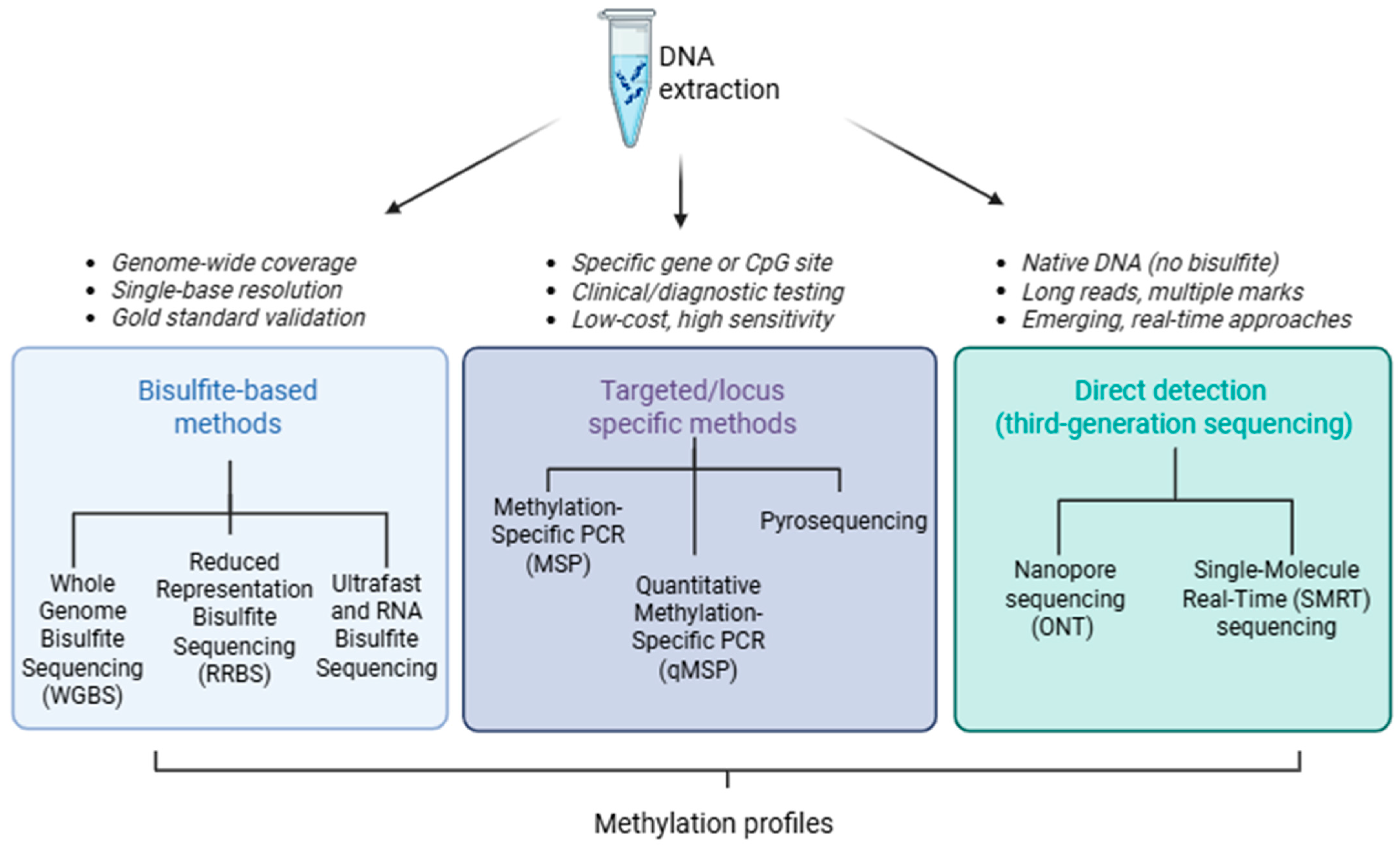
Figure 1. Schematic overview of DNA methylation detection workflows. Bisulfite-based methods (left) enable genome-wide and base-resolution analysis (e.g., WGBS, RRBS, UBS-Seq), targeted/locus-specific methods (middle) are used in clinical and diagnostic applications, and direct detection by third-generation sequencing (right) using nanopore or SMRT platforms enables native DNA analysis. Each approach differs in coverage, resolution, and clinical applicability. Created with BioRender.com.
Applsci 15 09940 g001
Figure 2. Schematic overview of histone modification profiling workflows. Key methods include ChIP/ChIP-seq (left) for genome-wide mapping of histone marks, CUT&RUN and CUT&Tag (middle) as antibody-guided, low-input alternatives with high specificity, and mass spectrometry-based proteomics (right) for quantitative and combinatorial profiling of histone post-translational modifications. Bioinformatic analyses integrate these datasets to define chromatin states and identify biomarkers. Created with BioRender.com.
Applsci 15 09940 g002
Figure 3. Schematic overview of non-coding RNA (ncRNA) detection workflows. Following RNA extraction, multiple strategies can be implemented, depending on target size, epigenetic role or location. Total RNA-seq for broad ncRNA profiling, small RNA-seq for miRNA and short ncRNA discovery, circular RNA-seq for cirRNA detection, single-cell RNA-seq for rare cell populations, nanopore direct RNA-seq for full-length transcript and modification profiling, and CRISPR-Cas-based biosensors for rapid, sequence-specific miRNA detection. Bioinformatic analysis enables ncRNA profiling, integration with multi-omics, and translation into clinical applications such as biomarker discovery and therapy prediction. Created with BioRender.com.
Applsci 15 09940 g003
Table 1. Comparison of DNA methylation detection methods. Summary of commonly used techniques for DNA methylation analysis, including genome-wide bisulfite sequencing approaches, locus-specific assays, and long-read sequencing. Key features, such as resolution, cost, input requirements, and ability to distinguish 5mC from 5hmC, are compared.
FeatureBisulfite SequencingMethylation-Specific PCR (MSP)PyrosequencingNanopore SequencingSMRT Sequencing
Detection PrincipleChemical conversion of unmethylated cytosinesPrimer specificity to methylated/unmethylated DNASequencing-by-synthesis + bisulfite conversionElectrical current changes in native DNAPolymerase kinetics in real-time synthesis
Methylation ResolutionSingle-nucleotideCpG site (qualitative/semi-quantitative)Single CpG (quantitative)Single baseSingle base
5mC vs. 5hmC DistinctionNot distinguishedNot distinguishedNot distinguishedPartially possibleDetectable Via kinetic signatures
Input DNA RequirementModerate–high (low-input possible)LowLowModerate–highHigh (some low-input adaptations exist)
Genome CoverageGenome-wide (WGBS); CpG-rich regions (RRBS)Locus-specificLocus-specificGenome-wide (long-read)Genome-wide or targeted (HiFi/SMRT-BS)
Quantitative OutputAbsolute methylation %Qualitative/semi-quantitativeQuantitativeQuantitativeQuantitative
Cost and ScalabilityHigh (WGBS); moderate (RRBS)LowModerateModerateHigh
Equipment RequirementsNGS platformStandard PCR + gel/qPCRPyrosequencerNanopore devicePacBio sequencer
ThroughputHighLow–moderateLowHighHigh
Special ApplicationsMethylome maps, cancer, developmentClinical panels, diagnosticsForensics, exposure, agingLong-range phasing, cfDNA, forensic IDMethylation + genome architecture, allele phasing
Major LimitationsDNA degradation, no 5hmC resolutionFalse positives, low throughputShort read length, low multiplexingLower raw accuracy, high computationHigh input, expensive, size selection bias
5mC—5-Methylcytosine; 5hmC—5-Hydroxymethylcytosine; WGBS—Whole-Genome Bisulfite Sequencing; RRBS—Reduced Representation Bisulfite Sequencing; MSP—Methylation-Specific PCR; cfDNA—Cell-Free DNA; SMRT—Single-Molecule Real-Time Sequencing; PCR—Polymerase Chain Reaction; NGS—Next-Generation Sequencing.
Table 2. Comparison of histone modification profiling methods. Overview of sequencing- and mass-spectrometry–based techniques for histone PTM mapping. Trade-offs in resolution, cost, and input material are highlighted.
FeatureChIP-SeqCUT&RUN/CUT&TagMS-Based Proteomics
Genomic resolution150–300 bp20–50 bp (narrower peaks, higher specificity)Not applicable (no genomic localization)
Input requirement1–10 million cells10,000–100,000 cells (or fewer in optimized protocols)Micrograms of protein or acid-extracted histones
Crosslinking requiredYesNoNo
Detection of PTM stoichiometryLimited, semi-quantitativeLimited, semi-quantitativeHigh, quantitative (esp. middle-/top-down MS)
Detection of combinatorial PTMsNoPartial (one per antibody)Yes—multiple PTMs on same proteoform
Single-cell compatibilityEmergingHigh (e.g., scCUT&Tag, scCUT&RUN)Currently limited, under development
Sample versatility (e.g., FFPE)Limited (requires optimization)High (esp. CUT&Tag in clinical samples)High (Via histone extraction from FFPE or serum nucleosomes)
hPTMs—histone post-translational modifications; ChIP-seq—chromatin immunoprecipitation followed by sequencing; CUT&RUN—Cleavage Under Targets and Release Using Nuclease; CUT&Tag—Cleavage Under Targets and Tagmentation; MS—mass spectrometry; PTMs—post-translational modifications; FFPE—formalin-fixed paraffin-embedded (clinical samples).
Table 3. Comparison of non-coding RNA detection methods. Summary of approaches for detecting and quantifying ncRNAs, including total RNA-seq, small RNA-seq, circRNA detection, targeted PCR-based assays, and direct RNA nanopore sequencing. Methods are compared in terms of size range, input material, resolution, and common limitations such as ligation bias, RNase R enrichment efficiency, or sequencing error profiles.
MethodTotal RNA-SeqSmall RNA-SeqcircRNA-SeqscRNA-SeqdRNA-Seq (Nanopore)
Target RNAslncRNAs, polyA−/polyA + RNAs, othersmiRNAs, piRNAs, siRNAsCircular RNAsmRNAs, lncRNAs, some circRNAsFull-length RNAs, isoforms, modifications
Input TypeRibosomal RNA-depleted total RNASize-selected RNA (~18–30 nt (protocol dependent))RNase R-treated RNASingle cellsPolyadenylated RNA (native)
StrengthsBroad RNA profiling, captures both coding and non-coding transcriptsSensitive to short RNAs, useful for isoforms, circulating miRNAsHigh specificity for circRNAs, back-splice detection, exonuclease resistanceHigh-resolution profiling, cell-type-specific expression, heterogeneity captureDirect sequencing, detects modifications (e.g., m6A), isoform-level resolution
LimitationsHigh computational noise, requires deep sequencing and standardizationBias from adapter ligation, low reproducibility across protocolsRequires special bioinformatics (e.g., CIRCexplorer, CIRI), prone to misannotationPoor detection of small RNAs, polyA bias, dropout and amplification artifactsHigh input requirement, lower accuracy, polyA bias excludes some ncRNAs
Key ApplicationsDiscovery of novel lncRNAs, low-abundance ncRNAs in tissue, tumor, extracellular vesiclesPlasma/serum biomarker discovery, small RNA profiling, cancer diagnosticsLiquid biopsy, neurodegeneration, cardiovascular disease markersTumor heterogeneity, rare cell types, lncRNA biomarker discoveryIsoform diversity, epitranscriptomic biomarker mapping, neurodegenerative disease
lncRNA—long non-coding RNA; miRNA—micro RNA; piRNA—piwi-interacting RNA; siRNA—small interfering RNA; scRNA—single-cell RNA; dRNA-seq—Direct RNA sequencing; CIRCexplorer—Circular RNA Explorer; CIRI—CircRNA Identifier.
Table 4. Overview of Representative AI Models Integrating Epigenetic Data.
ModelEpigenetic InputAdditional ModalitiesArchitectureOutputApplicationsAdvantagesRef.
MethylNetDNA methylation (Î2-values from 450 K/EPIC arrays)NoneVariational Autoencoder + Fully Connected layersAge, smoking status, tumor subtype, immune infiltrationEpigenetic biomarker discovery, age prediction, subtype classificationHigh-dimensional methylation data compression, task-agnostic embeddings[28]
DeepChromeHistone modifications (H3K4me3, H3K27ac, etc.) around TSSNone1D CNN + MLPBinary classification of gene expression (high/low)Understanding combinatorial histone code and gene expressionVisualizable model, captures mark-specific regulation[29]
INTERACTCpG methylation (target site)Genomic sequence (2 kb, one-hot encoded)CNN + Transformer + FCPredicted methylation levels and variant effectsVariant prioritization, regulatory element identificationLong-range sequence dependency modeling Via self-attention[31]
DeepHistoneHistone mark presenceGenomic sequence + DNase I hypersensitivityDual CNN branches + FCHistone mark predictionCross-cell-type prediction of histone marksContextual prediction with chromatin accessibility[30]
ChromeGCNHistone marks, TF binding (1 kb bins)Genomic sequence + Hi-C contact mapsCNN + Graph-CNN (on Hi-C contact map)Multi-label prediction of chromatin stateInterpretable enhancer promoter interaction predictionCaptures 3D chromatin interactions, interpretable GCN saliency maps[32]
DDAE + MLPDNA methylation (450 K) + CNVsCopy number variation (CNV)Stacked Denoising Autoencoder + MLPGene expression levels (in liver cancer)Multi-omics modeling of cancer transcriptomesIntegrates genetic and epigenetic drivers of expression[33]
TSS—Transcription Start Site; CNN—Convolutional Neural Network; MLP—Multi-Layer Perceptron; FC—Fully Connected (layer); GCN—Graph Convolutional Network; Hi-C—Chromosome Conformation Capture (High-throughput); DNase I—Deoxyribonuclease I; CNV—Copy Number; DDAE—Denoising Deep Autoencoder; EPIC array—Infinium MethylationEPIC BeadChip Array; 450 K array—Illumina Infinium HumanMethylation450 BeadChip.
Disclaimer/Publisher’s Note: The statements, opinions and data contained in all publications are solely those of the individual author(s) and contributor(s) and not of MDPI and/or the editor(s). MDPI and/or the editor(s) disclaim responsibility for any injury to people or property resulting from any ideas, methods, instructions or products referred to in the content.