Article

Enhancement of Co-Production of Astaxanthin and Total Fatty Acids in Haematococcus lacustris by Combined Treatment with Exogenous Indole-3-Acetic Acid and Abscisic Acid

by
Rendi Mahadi
1,2,3,
You-Kwan Oh
1,4,*,
Laxmi Priya Sathiyavahisan
1,
Aditya Lakshmi Narasimhan
1,
Eui-Jin Kim
5 and
Ja Young Cho
5
1
School of Chemical Engineering, Pusan National University, Busan 46241, Republic of Korea
2
Department of Chemical Engineering, Kyung Hee University, Yongin-si 17104, Gyeonggi-do, Republic of Korea
3
Department of Tropical Biology, Faculty of Biology, Universitas Gadjah Mada, Yogyakarta 55281, Indonesia
4
Institute for Environment & Energy, Pusan National University, Busan 46241, Republic of Korea
5
Nakdonggang National Institute of Biological Resources, Sangju-si 37242, Gyeongsangbuk-do, Republic of Korea
*
Author to whom correspondence should be addressed.
Appl. Sci. 2025, 15(18), 10136; https://doi.org/10.3390/app151810136
Submission received: 6 August 2025 / Revised: 12 September 2025 / Accepted: 13 September 2025 / Published: 17 September 2025
(This article belongs to the Section Applied Biosciences and Bioengineering)

Abstract

Phytohormones play pivotal roles in regulating metabolic processes in microalgae. This study investigates the individual and combined effects of indole-3-acetic acid (IAA; 0.1–100 µM) and abscisic acid (ABA; 1–100 µM) on cell morphology, growth, astaxanthin biosynthesis, and fatty acid production in Haematococcus lacustris during 30 d photoautotrophic cultivation. IAA significantly enhanced cell density but did not alter intracellular astaxanthin levels, whereas ABA increased astaxanthin accumulation while reducing cell density. The optimal response was observed with co-application of 1 µM IAA and 50 µM ABA at the onset of cultivation, improving cell density (255.0 × 10³ cells/mL), astaxanthin content (22.5 mg/g cell), and astaxanthin production (44.7 mg/L), corresponding to increases of 21%, 45%, and 122%, respectively, compared to untreated controls. Fatty acid content (mg/g cell) and volumetric production (mg/L) were increased by 24% and 90%, respectively. These results demonstrate that strategic, early-stage co-application of IAA and ABA can effectively enhance astaxanthin and lipid coproduction in H. lacustris, offering a promising approach for improving the efficiency of microalgal biorefineries.

1. Introduction

The growing global demand for sustainable biotechnological solutions across the food, fuel, and healthcare sectors has intensified research on photosynthetic microalgae, which efficiently fix carbon dioxide (CO2) into high-value biomolecules [1,2,3]. Among these, astaxanthin (3,3′-dihydroxy-β,β-carotene-4,4′-dione) has attracted considerable interest due to its exceptional antioxidant capacity and broad applications in pharmaceuticals, cosmetics, and aquaculture [4,5].
The freshwater microalga Haematococcus lacustris (formally known as H. pluvialis) is considered the richest natural source of astaxanthin, capable of accumulating up to 5% of its dry weight under stress conditions [6,7,8]. H. lacustris undergoes a complex life cycle comprising morphologically and physiologically distinct stages—including aplanospores (hematocysts), germinating cells, biflagellates, and palmella cells—with the highest astaxanthin accumulation occurring in the non-motile red cyst stage [9,10]. Astaxanthin is primarily stored as mono- and di-esters in cytoplasmic lipid bodies enriched in long-chain fatty acids, highlighting its potential for dual production of nutraceuticals and biofuels [4,5,6,7,8].
Despite its commercial potential, the large-scale cultivation of H. lacustris remains challenging due to slow growth rates, oxidative stress during encystment, and high production costs [11,12]. Astaxanthin biosynthesis, which is typically induced under stress conditions during the encystment phase, is closely associated with reactive oxygen species (ROS) signaling as part of the alga’s defense mechanism. Although nitrogen deprivation is a widely used and effective inducer of astaxanthin production [13,14], it often results in reduced overall biomass productivity. To address this limitation, various alternative stress strategies—such as high light intensity [15], elevated temperature [16], salinity [17], mechanical stress [18], hydrostatic pressure [19], and electrical stimulation [9]—have been explored. While these methods can enhance astaxanthin accumulation, they frequently lead to excessive oxidative stress, which compromises cell viability and reduces final astaxanthin yields [7,20]. In addition, chemical induction approaches have been investigated using agents such as ethanol [21], trisodium citrate [22], and magnesium aminoclay nanoparticles [23]. These treatments can stimulate astaxanthin biosynthesis but often do so at the expense of photosynthetic efficiency and cell proliferation [20]. Given these challenges, there is a growing need for alternative strategies that can enhance astaxanthin production without compromising cell growth or viability.
Phytohormones are well known to regulate growth, development, and stress responses in higher plants [24,25]. Among them, indole-3-acetic acid (IAA), a primary auxin, promotes cell division, elongation, and delays senescence [26,27,28], while abscisic acid (ABA) plays a central role in stress signaling and is associated with enhanced carotenoid and lipid biosynthesis [29,30]. Notably, core components of these signaling pathways are increasingly recognized as functionally conserved in microalgae, thereby opening new avenues for phytohormone-mediated metabolic regulation [31,32].
Exogenous IAA has been reported to enhance growth, chlorophyll content, and biomass production in various microalgae, such as Chlorella vulgaris and Dunaliella salina [33,34]. Similarly, ABA supplementation has been shown to increase carotenoid and lipid accumulation in Chromochloris zofingiensis and Chloroidium saccharophilum [35,36]. Lin et al. [37] reported that in Chlorella sp. FACHB-8, treatments with IAA, ABA, or their combination promoted cell growth, but lipid accumulation was significantly enhanced only with 10 mg/L ABA. This implies that ABA may dominate or interfere with the effect of other hormones in certain species. However, the effects of IAA and ABA on H. lacustris remain poorly characterized and occasionally contradictory. For example, bacterial-derived IAA has been reported to promote the growth of H. lacustris [38], yet its role in astaxanthin biosynthesis remains unclear. ABA, on the other hand, has been shown to upregulate astaxanthin biosynthesis genes [39]. However, contrasting findings were reported by Kobayashi and colleagues. They observed that ABA accelerated encystment and carotenoid accumulation on agar plates—likely due to desiccation-like stress—yet delayed these processes in liquid cultures. These differences highlight the influence of cultivation mode on hormone responsiveness [40,41]. These inconsistencies underscore the complexity of hormone responses in H. lacustris, which appear to be strongly influenced by environmental conditions and the physiological state of the cells.
In this study, we systematically investigate the individual and combined effects of IAA and ABA on H. lacustris, focusing on their roles in modulating cell morphology, growth, astaxanthin biosynthesis, and fatty acid accumulation. Given their distinct physiological functions, we hypothesize that their co-application may produce complementary effects—IAA enhancing cell proliferation and ABA enhancing stress-induced biosynthesis of astaxanthin and lipids. To test this hypothesis, we applied a range of IAA (0.1–100 µM) and ABA (1–100 µM) concentrations, both individually and in combination, and assessed their effects on growth kinetics, morphological transitions, astaxanthin accumulation, and fatty acid profiles. This study offers novel insights into hormone-mediated metabolic regulation in H. lacustris and proposes a viable strategy for improving microalgal biorefinery productivity.

2. Materials and Methods

2.1. Microalgal Strain and Photosynthetic Cultivation

The freshwater microalga H. lacustris strain NIES-144 was obtained from the National Institute for Environmental Studies (NIES; Tsukuba, Japan) and cultivated photoautotrophically in NIES-C medium. The medium was prepared with the following composition (per liter): 0.15 g Ca(NO3)2·4H2O, 0.10 g KNO3, 0.05 g β-glycerophosphate disodium salt hydrate, 0.04 g MgSO4·7H2O, 0.05 g Tris(hydroxymethyl)aminomethane, 0.01 mg thiamine, 0.10 µg biotin, 0.10 µg vitamin B12, and 3.00 mL of PIV metal solution. The PIV metal solution (1 L) contained 1.0 g Na2EDTA, 196.0 mg FeCl3·6H2O, 36.0 mg MnCl2·4H2O, 22.0 mg ZnSO4·7H2O, 4.0 mg CoCl2·6H2O, and 2.5 mg Na2MoO4·2H2O. The final pH was adjusted to 7.5 and the medium was sterilized by filtration through a 0.2 µm membrane filter [42].
Microalgal cultivation was conducted in 300 mL Erlenmeyer flask with a working volume of 200 mL. The flasks were placed on a shaking incubator (ISF-7100RF, Jeiotech, Daejeon, Republic of Korea) operating at 150 rpm and 25 °C. No additional inorganic carbon sources such as CO2 bubbling or sodium bicarbonate were supplied. Instead, passive aeration was facilitated by ambient air exchange through porous silicon stoppers. Continuous illumination was provided by internal cool-white fluorescent lamps at an intensity of 80 ± 10 µmol photons/m2·s. The flask-based autotrophic conditions—including medium composition, temperature, initial pH, light intensity, agitation speed, and working volume—were adapted from partially optimized protocols for strain NIES-144 developed in our previous studies [9,19,42].
pH values were recorded every 5 d for the IAA and ABA treatment groups (Supplementary Figure S1). The pH remained within the range of 7.5–8.5 throughout the 30 d cultivation period, starting from an initial pH of approximately 7.5. For the combined treatment of 1 µM IAA and 50 µM ABA, the final pH after 30 d was measured at 8.0–8.2, although intermediate values were not recorded in this group. To ensure axenicity and monitor culture health, cell morphology and culture clarity were assessed at 5 d intervals using light microscopy. No signs of microbial contamination—such as turbidity, foreign morphotypes, or abnormal cell aggregation—were observed. Representative micrographs documenting these observations are presented in Supplementary Figure S2.

2.2. Phytohormone Treatment

In this study, cyst-stage H. lacustris cultures (35 d old; initial biomass ~0.15 g/L) were used as inocula and subsequently cultivated for an additional 30 d under identical conditions. The use of 35 day-old cultures ensured a uniform physiological state, which is critical for reproducibility in hormone-response studies. The optimal inoculum age can vary depending on the cultivation system—typically around 15 d for photobioreactors [9] and approximately 30 d for flask-based systems [42].
Three hormonal treatments were tested: (1) IAA alone (Alfa Aesar, Ward Hill, MA, USA), (2) ABA alone (Sigma-Aldrich, St. Louis, MO, USA), and (3) a combined IAA and ABA treatment. Based on previous studies [33,37,40], the following concentrations were selected: IAA at 0.1, 1, 10, and 100 µM; and ABA at 1, 10, 50, and 100 µM. After identifying the optimal individual concentrations (1 µM IAA and 50 µM ABA), combination treatments were conducted by applying IAA once at day 0 and ABA at three different time points: day 0 (inoculation), day 15 (linear growth phase), and day 20 (early stationary phase) (see Section 3.2).

2.3. Astaxanthin Quantification

On day 30, microalgal cells were harvested by centrifugation at 3000 rpm for 10 min, washed twice with distilled water, and freeze-dried for 72 h. Approximately 2 mg of dried biomass was transferred to 2 mL microcentrifuge tubes containing 1.0× g of glass beads and 1 mL of dichloromethane–methanol (1:1, v/v) supplemented with 0.025 M NaOH [9].
Cell disruption was performed using a FastPrep-24 bead beater (MP Biomedicals, Irvine, CA, USA) at 6 m/s for three 30 s cycles. To induce saponification and produce free (de-esterified) astaxanthin, the extract was incubated at 4 °C for 12 h in darkness. This saponification step hydrolyzes esterified astaxanthin, enabling accurate quantification of the free form. The extract was then syringe-filtered through a 0.2 µm membrane filter. Astaxanthin content was analyzed using high-performance liquid chromatography (HPLC; Agilent 1260 Infinity, Hewlett-Packard, Santa Clara, CA, USA) equipped with a diode array detector and a YMC carotenoid column. Quantification was performed using certified calibration standards (Dr. Ehrenstorfer GmbH, Augsburg, Germany), following the protocol described by Mahadi et al. [42].

2.4. Fatty Acid Quantification

Total fatty acid content was determined as fatty acid methyl esters (FAMEs) via direct transesterification followed by gas chromatography (GC), as described previously [42]. Approximately 10 mg of freeze-dried biomass was extracted with 2 mL of chloroform–methanol (2:1, v/v). Transesterification was carried out by adding 1 mL methanol, 300 µL of 95% H2SO4, and 1 mL of chloroform containing heptadecanoic acid (C17:0) as the internal standard. The mixture was vortexed for 5 min, heated at 100 °C for 10 min, cooled to room temperature, and mixed with distilled water. After centrifugation at 3000 rpm for 10 min, the organic phase was collected, filtered, and analyzed using a gas chromatograph (HP 6890; Agilent Technologies, Santa Clara, CA, USA) equipped with a flame ionization detector and an HP-INNOWax capillary column (30 m × 0.32 mm I.D., 0.5 µm film thickness).

2.5. Additional Analyses

Cell density was determined using a Neubauer hemocytometer (DHC-N01-5; INCYTO, Cheonan-si, Chungnam-do, Republic of Korea). Cell size, morphology, and developmental stages were monitored under a bright-field microscope (Axiolab, Carl Zeiss, Jena, Germany) with image capture using Zen lite 2012 software. Dry cell weight (DCW) was measured gravimetrically using pre-weighed GF/C filters, as described by Mahadi et al. [19]. Light intensity was measured with a quantum photometer (LI-250A, LI-COR Inc., Lincoln, NE, USA), and pH was measured with a bench-top pH meter (HM-30R, TOA-DKK, Tokyo, Japan).

2.6. Statistical Analysis

All experimental data were visualized using SigmaPlot 14.0 (Systat Software Inc., San Jose, CA, USA). Results are presented as mean ± standard deviation from independent duplicate cultivations. Given the limited number of biological replicates, we adopted a commonly accepted approach in algal cultivation studies by applying one-way analysis of variance (ANOVA), which is considered robust to moderate deviations of normality. Statistical significance was assessed at p < 0.05, followed by Tukey’s honestly significant difference (HSD) post hoc test using SPSS Statistics 17.0 (IBM Corp., Chicago, IL, USA).

3. Results and Discussion

3.1. Proliferation Patterns in the Life Cycle of Haematococcus lacustris

The developmental transition of H. lacustris during 30 d of photosynthetic cultivation is illustrated in Figure 1, with representative micrographs shown in Supplementary Figure S2. Upon inoculation, the 35 d-old reddish aplanospores (hematocysts; Figure 1a) underwent two primary developmental pathways. In one route, hematocysts formed sporangia that subsequently released multiple microzooids (Figure 1b). Alternatively, some hematocysts directly transitioned into brownish palmella cells (Figure 1c), a morphological state often associated with astaxanthin degradation [43,44]. These palmella cells subsequently developed into sporangia containing multiple microzooids (Figure 1d), representing a distinct reproductive strategy.
The synchronized release of daughter cells from hematocysts—referred to as germination [9,45]—resulted in the emergence of motile biflagellate cells (Figure 1e). These motile cells rapidly proliferated before differentiating into non-motile green palmella cells (Figure 1f), which retained chlorophyll but lost their flagella. This transition is consistent with previous observations of the H. lacustris life cycle [46,47]. Upon exposure to environmental stressors, particularly nitrogen limitation, the cells progressed into a brownish-green palmella stage (Figure 1g), during which astaxanthin biosynthesis was initiated. Eventually, the life cycle culminated in the formation of red aplanospores (Figure 1a), completing the full developmental sequence. These observations corroborate previous findings that developmental trajectories in H. lacustris are influenced by both hematocyst age and cultivation conditions. For instance, Lakshmi Narasimhan et al. [9] reported predominant microzooid formation from mature (90 d-old) hematocysts, consistent with Ha et al. [48] and Bauer et al. [49]. In contrast, the present study used younger, 35 day-old hematocysts and observed the simultaneous formation of both microzooids and macrozooids, supporting the developmental plasticity noted by Zhang et al. [43].
To enable consistent classification during morphological tracking, sporangia containing either microzooids or macrozooids were collectively categorized as “sporangia.” Similarly, brownish and brownish-green palmella cells were grouped under “intermediate palmella.” This classification facilitated comparative analysis of morphological transitions under different treatment conditions.

3.2. Effect of Indole-3-Acetic Acid on Morphology, Growth, and Astaxanthin Biosynthesis

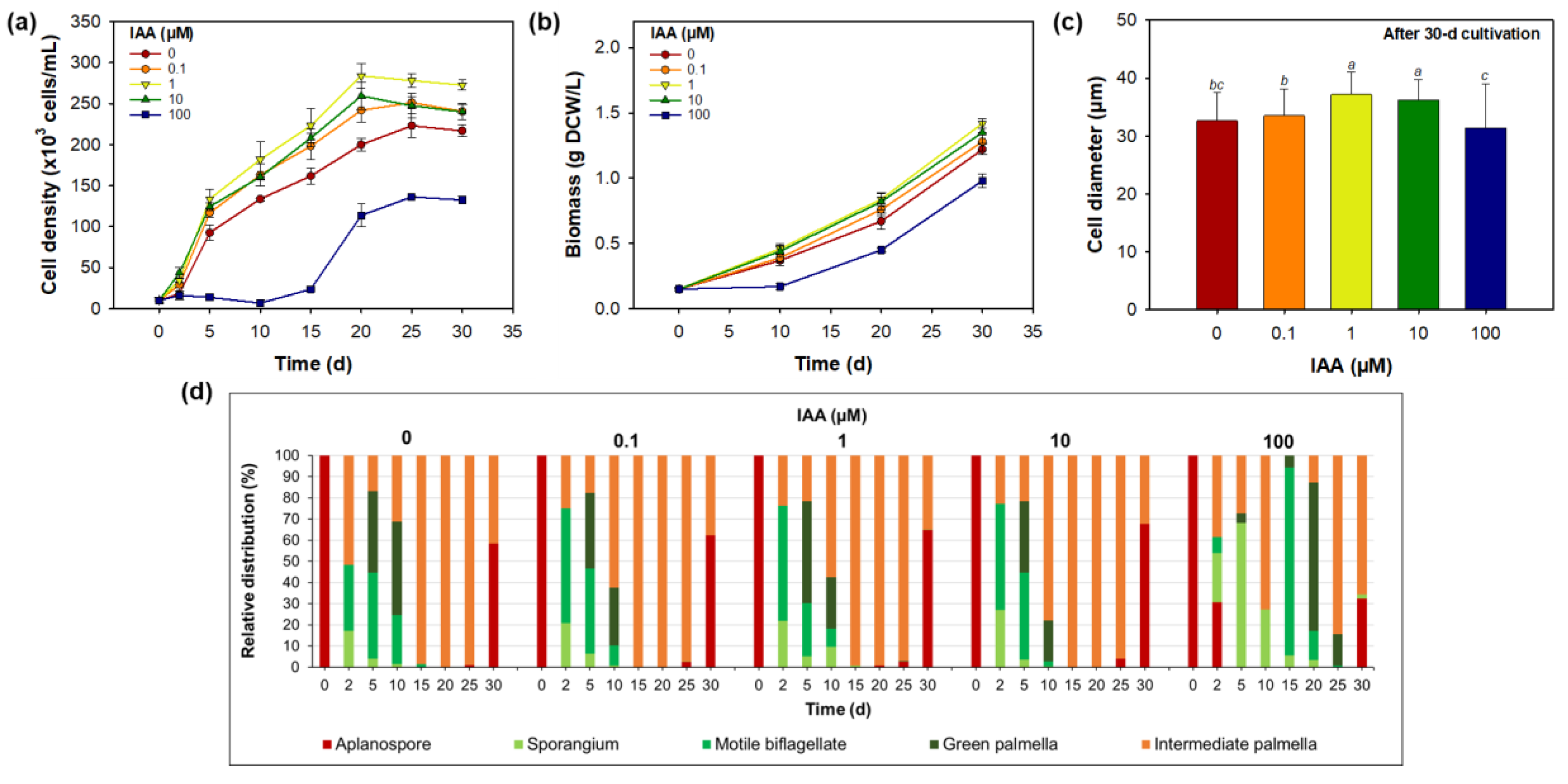
The effects of varying IAA concentrations (0.1, 1.0, 10, and 100 µM) on H. lacustris morphology, growth, and astaxanthin production are shown in Figure 2 and Figure 3. During the 30 d cultivation period, the microalgae exhibited a characteristic three-phase growth pattern: an exponential phase (days 0–5), a linear phase (days 5–20), and a stationary phase (days 20–30) (Figure 2a). These phases were defined based on cell density plotted on a base-10 logarithmic scale over time, as detailed in Supplementary Figure S3. An exception was observed at the highest IAA concentration (100 µM), which showed a pronounced lag phase of approximately 10 d before cell proliferation resumed.
Among the tested concentrations, 1 µM IAA resulted in the highest final cell density (272.5 ± 6.1 × 10³ cells/mL), corresponding to a 26% increase compared to the untreated control (216.9 ± 7.2 × 10³ cells/mL). Moderate growth enhancement was also observed at 0.1 µM and 10 µM. In contrast, 100 µM IAA significantly inhibited cell proliferation, suggesting a biphasic dose–response relationship characteristic of auxin-regulated signaling pathways.
Biomass production followed a similar trend (Figure 2b), peaking at 1.42 ± 0.04 g/L with 1 µM IAA and declining sharply at 100 µM. The inhibition suggests that excessive IAA may disrupt photosynthetic efficiency or cellular metabolic homeostasis [33,34]. Cell diameter data at day 30 (Figure 2c) mirrored these patterns. Larger average cell sizes (36–37 µm) were observed at 1–10 µM, while both 0.1 µM and 100 µM treatments yielded significantly smaller cells (~33 µm and ~31 µm, respectively), suggesting impaired cellular development at suboptimal concentrations.
Morphological analysis (Figure 2d) showed that control and moderate IAA treatments supported normal developmental transitions, including germination, biflagellate formation, and progression into the palmella stage. However, cultures treated with 100 µM IAA delayed germination, with approximately 31% of cells persisting as aplanospores at day 2, indicating an arrest in cell cycle progression [42,49].
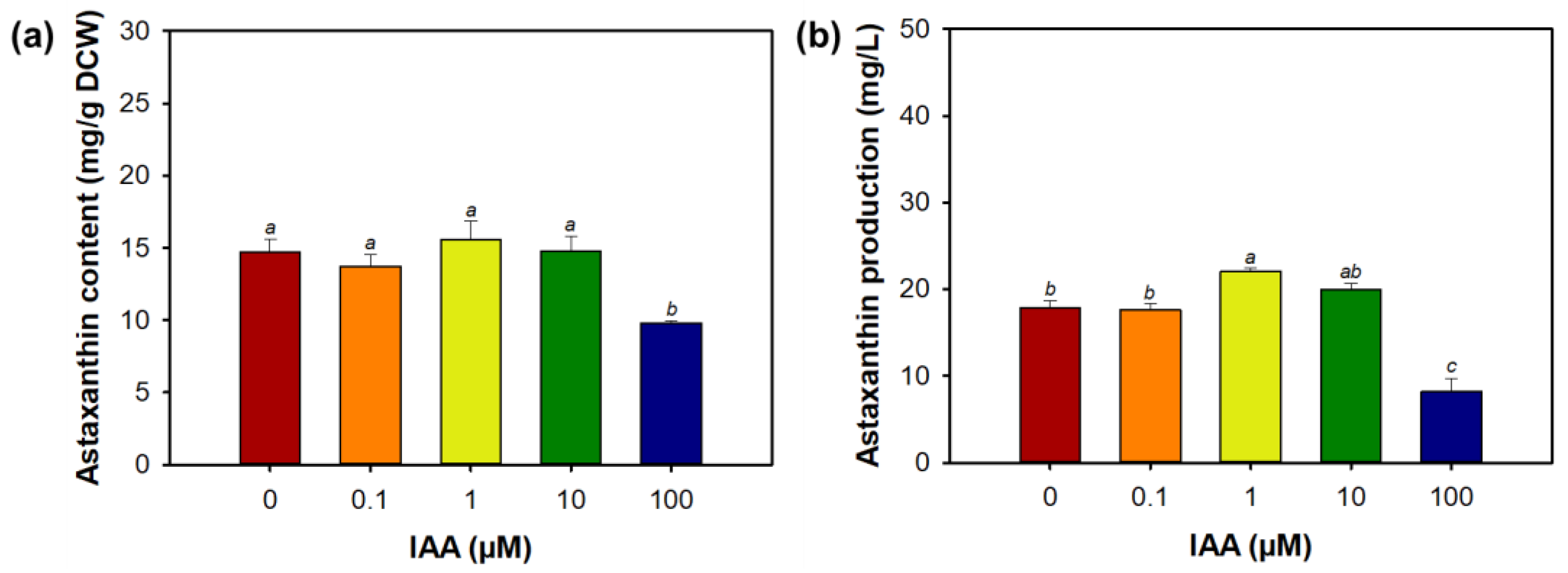
Astaxanthin production peaked at 1 µM IAA, reaching 22.1 ± 0.4 mg/L—24% increase compared to the control (17.9 ± 0.7 mg/L) (Figure 3). This enhancement was primarily driven by increased biomass production, as the intracellular astaxanthin content remained relatively stable across the 0.1–10 µM range (14–16 mg/g DCW). In contrast, at 100 µM IAA, both intracellular astaxanthin content (9.8 mg/g DCW) and total production (8.2 mg/L) were significantly reduced, likely due to stress-related metabolic disruption [50].
These findings are consistent with previous studies demonstrating that auxins regulate microalgal growth through redox modulation and cell cycle control. Piotrowska-Niczyporuk and Bajguz [33] reported that IAA-activated antioxidant enzymes in C. vulgaris promoted growth at low concentrations and inhibited it at higher concentrations. Comparable biphasic responses have been reported in Scenedesmus quadricauda [35] and D. salina [34], where 1 µM IAA optimally enhanced β-carotene accumulation and cell proliferation. Additionally, bacterial-derived IAA has been shown to stimulate H. lacustris growth without significantly altering astaxanthin content [38], supporting the moderate growth-promoting effect observed at 1 µM IAA in the present study. Future studies on IAA in H. lacustris should focus on elucidating the molecular signaling mechanisms by which IAA influences cell cycle regulation, lipid metabolism, and carotenoid biosynthesis. Comprehensive transcriptomic and metabolomic analyses could reveal key pathways modulated by IAA.

3.3. Effect of Abscisic Acid on Morphology, Growth, and Astaxanthin Biosynthesis

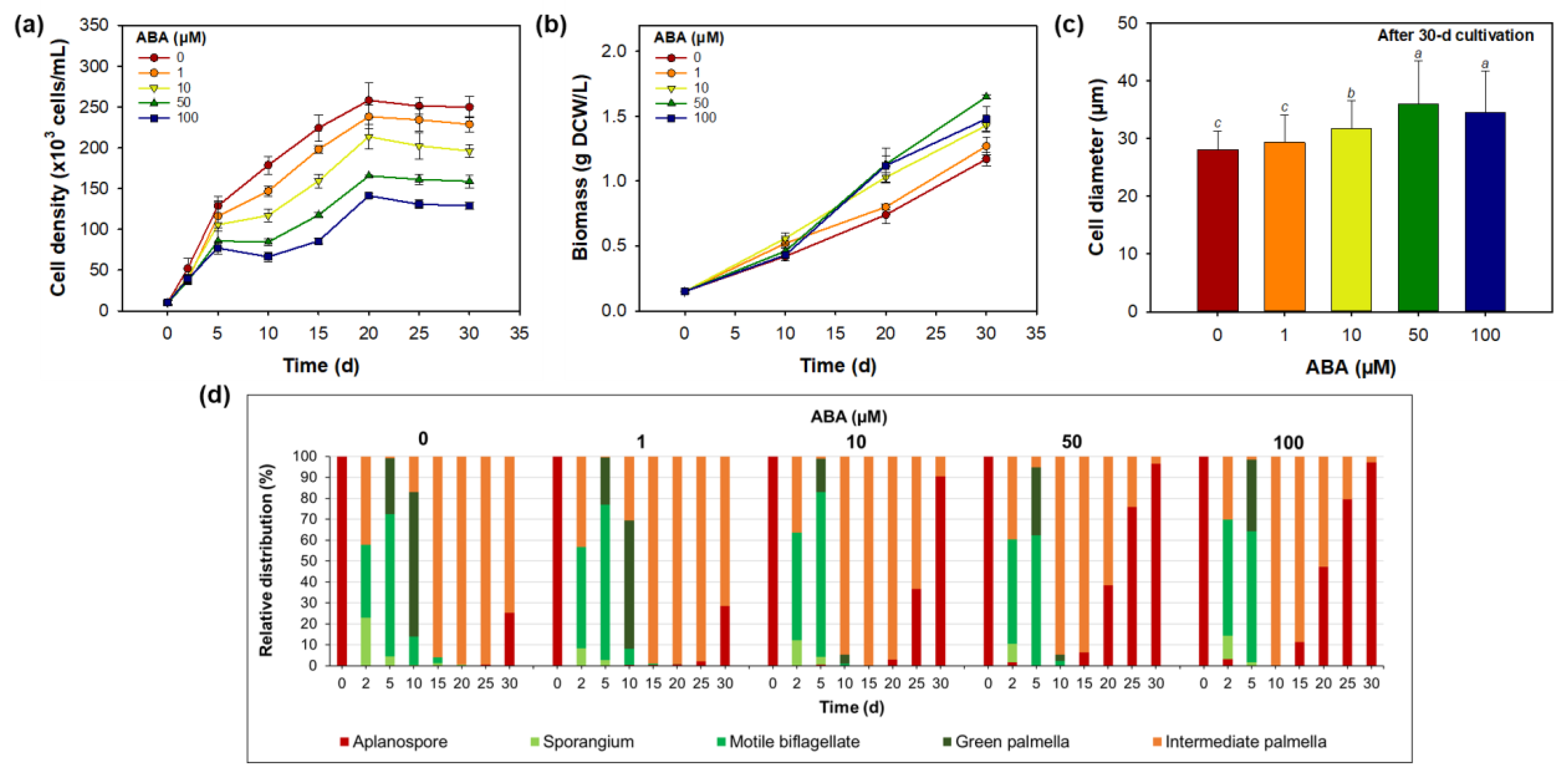
The effects of exogenous ABA at different concentrations (1, 10, 50, and 100 µM) on H. lacustris morphology, growth, and astaxanthin biosynthesis over a 30 d cultivation are summarized in Figure 4 and Figure 5. In contrast to the growth-promoting effect observed with moderate IAA concentrations, ABA exhibited a concentration-dependent response with reduced cell density but enhanced biomass and astaxanthin accumulation.
As shown in Figure 4a, all ABA treatments resulted in lower final cell densities compared to the control, with the most substantial decrease (~49%) occurring at 100 µM. Despite this, biomass production increased with rising ABA concentration, peaking at 1.65 ± 0.02 g DCW/L at 50 µM ABA—representing a 41% increase relative to the control (1.17 ± 0.05 g/L) (Figure 4b). A corresponding increase in average cell diameter was also observed at 50 µM (Figure 4c), indicating enhanced intracellular storage capacity. No further biomass gains were recorded at 100 µM, suggesting a plateau effect at higher ABA levels.
Morphological observations (Figure 4d) revealed that 1 µM ABA-induced developmental changes similar to the control. In contrast, ≥10 µM ABA treatments rapidly promoted differentiation into brownish and brownish-green palmella cells—intermediate morphotypes commonly associated with the onset of astaxanthin accumulation. By day 30, cultures exposed to 10–100 µM ABA exhibited aplanospore formation rates of 90–97%, compared to only 25–29% in the control and 1 µM groups. These results indicate that ABA plays a key regulatory role in promoting encystment and activating carotenogenic pathways.
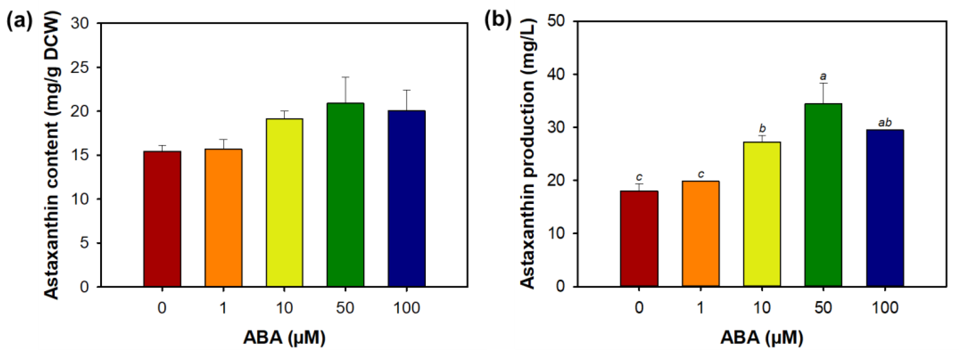
Astaxanthin content and total production were both significantly increased in response to ABA treatment compared to control (Figure 5). The highest intracellular astaxanthin content (20.9 ± 2.9 mg/g DCW) and volumetric production (34.5 ± 3.9 mg/L) were observed at 50 µM ABA, corresponding to increases of 36% and 91% over the control, respectively. No additional improvement was observed at 100 µM, indicating a threshold beyond which further ABA does not enhance biosynthetic output.
These findings align with previous reports demonstrating the role of ABA in promoting carotenoid and lipid biosynthesis in microalgae. For example, Kozlova et al. [35] reported increased metabolite accumulation in C. zofingiensis at 1 µM ABA, while Contreras-Pool et al. [36] observed triacylglycerol enrichment in C. saccharophila within the 1–20 µM ABA range. In H. lacustris, however, effective responses appear to occur at relatively higher concentrations. Gao et al. [39] showed that 95 µM ABA upregulated the expression of key astaxanthin biosynthetic genes, and Kobayashi et al. [40] reported that ABA enhanced carotenoid accumulation in agar-based cultures under desiccation stress, but not in liquid culture—underscoring the importance of cultivation conditions in modulating ABA responsiveness. Future studies on ABA in H. lacustris should aim to elucidate the molecular mechanisms underlying its concentration-dependent effects on growth suppression and astaxanthin induction, particularly its role in triggering encystment and activating carotenogenic gene expression. Transcriptomic or proteomic profiling under various ABA concentrations and cultivation modes could clarify the regulatory networks involved.

3.4. Combined Effect of Indole-3-Acetic Acid and Abscisic Acid on Morphology, Growth, and Astaxanthin Biosynthesis

To further optimize astaxanthin production, the combined application of 1 µM IAA and 50 µM ABA was evaluated by varying the timing of ABA administration, specifically on day 0 (inoculation phase), day 15 (linear growth phase), and day 20 (early stationary phase). This approach aimed to identify the most effective timing for maximizing both growth and carotenoid biosynthesis.
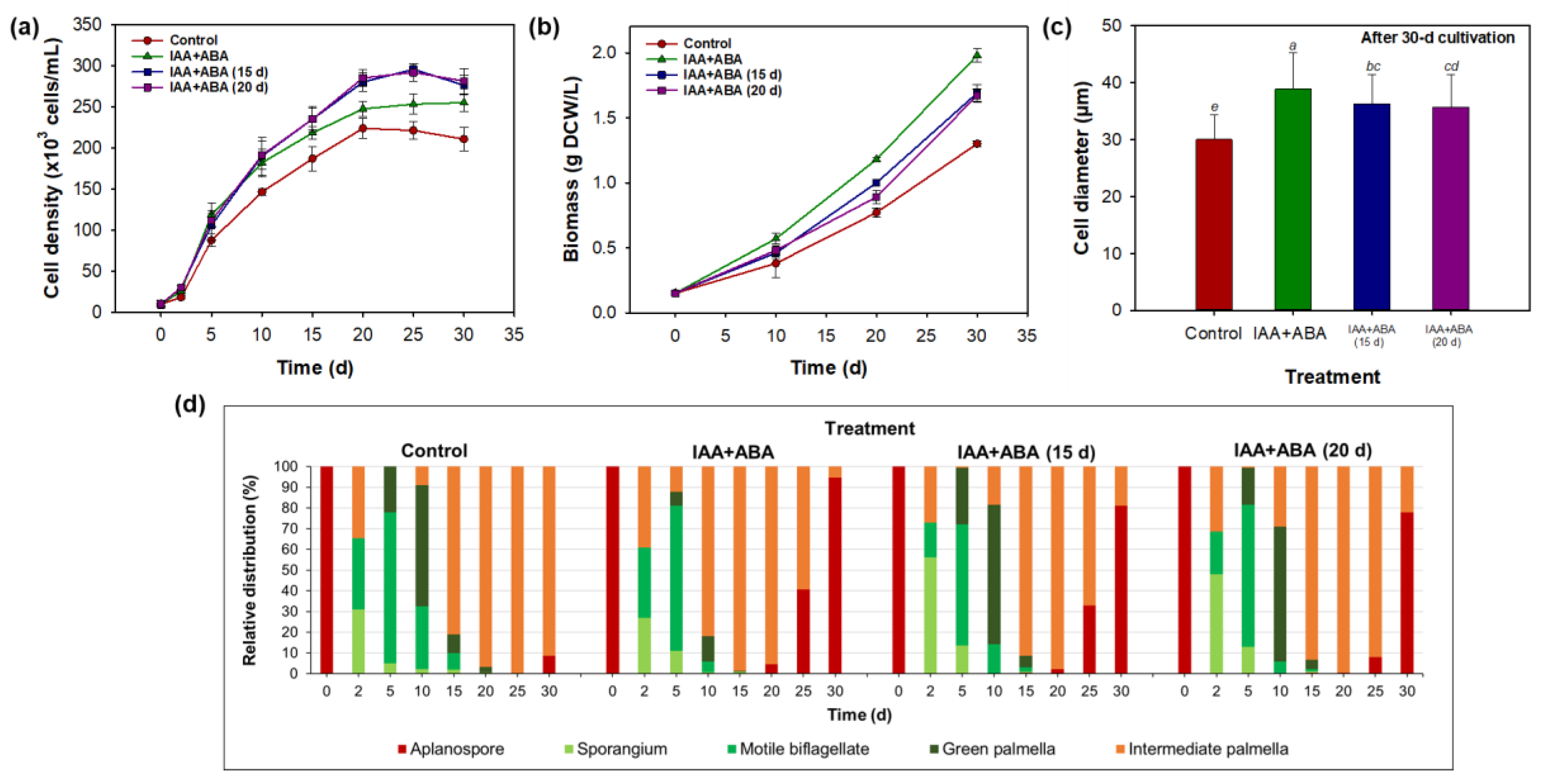
All combined treatments significantly outperformed the control in terms of both cell density and biomass production (Figure 6a,b). The highest cell densities (276.3–281.3 × 10³ cells/mL) were observed when ABA was added on day 15 or 20, representing a ~34% increase compared to the control (210.6 ± 14.2 × 10³ cells/mL). However, peak biomass yield (1.98 ± 0.05 g DCW/L) was achieved when ABA was administered at inoculation (day 0), corresponding to a 52% enhancement relative to the control (1.30 ± 0.02 g/L). Additionally, all combined treatments resulted in a large cell diameter (36–39 µm), suggesting elevated intracellular carbon and metabolite accumulation (Figure 6c).
Morphological observation (Figure 6d) revealed accelerated transitions into intermediate palmella stages in all IAA + ABA groups, consistent with active astaxanthin biosynthesis. Notably, aplanospore formation peaked at 95% in the group receiving ABA at inoculation, followed by 81% and 78% in the day 15 and 20 treatments, respectively—substantially higher than the 9% observed in the control. These findings indicate that early ABA exposure most effectively primes the carotenogenic differentiation pathways. Additionally, the morphological heterogeneity observed under hormonally identical conditions highlights the inherent developmental plasticity and hormone sensitivity of H. lacustris [9].
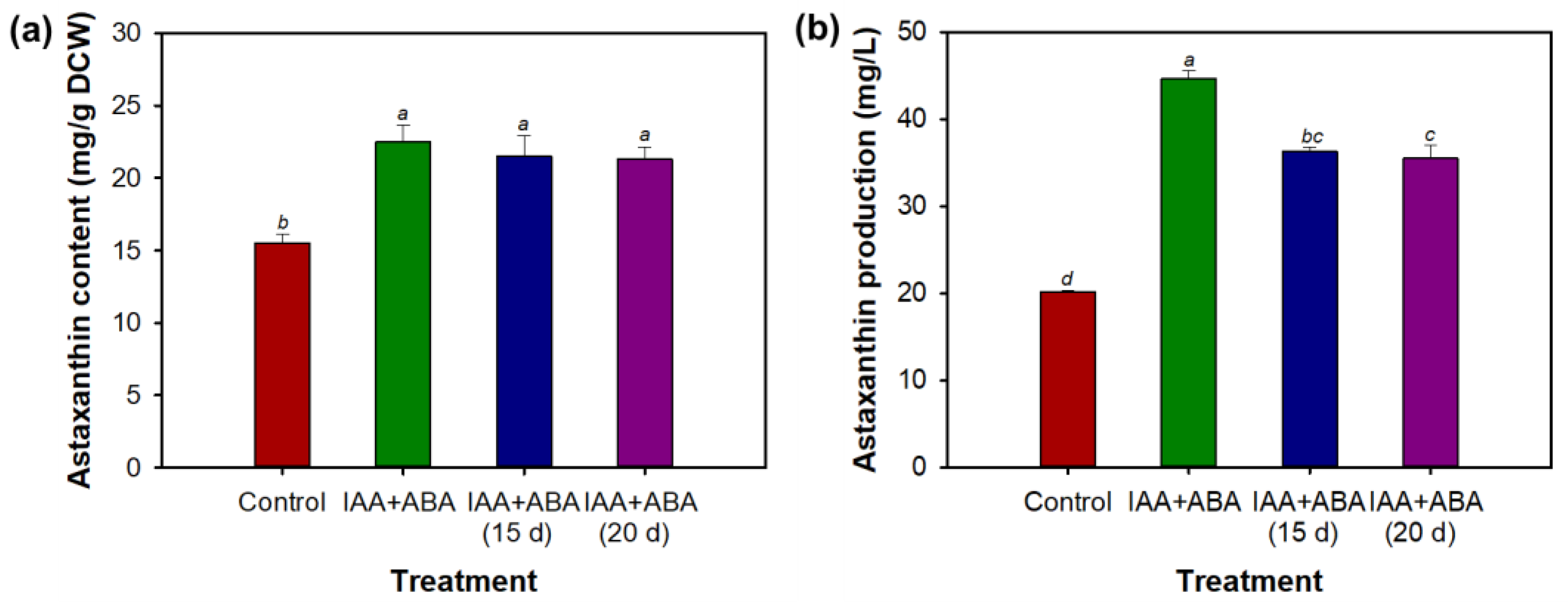
Astaxanthin accumulation was significantly enhanced by all combined treatments (Figure 7). The highest intracellular astaxanthin content (22.5 ± 1.1 mg/g DCW) and total volumetric production (44.7 ± 0.9 mg/L) were achieved when ABA was applied at day 0, representing respective increases of 45% and 122% compared to the control. The improvement appears additive rather than synergistic, indicating that each hormone contributes independently to overall enhancement.
IAA is well known to enhance biomass accumulation by activating auxin-responsive genes and regulating the cell cycle [26,28], while ABA is associated with the induction of stress-related transcriptional pathways, including ABRE-binding factors that play a central role in carotenoid biosynthesis [29,31]. In H. lacustris, early exposure to ABA likely promotes the transcription of key astaxanthin biosynthetic genes such as crtO, crtR-b, and psy [39]. Concurrently, IAA-induced stimulation of cell proliferation may lead to an increase in intracellular precursor pools (e.g., acetyl-CoA and NADPH), facilitating the biosynthesis of secondary metabolites such as astaxanthin.
The combination of IAA and ABA treatments resulted in additive, rather than synergistic, effects on astaxanthin and fatty acid production. This outcome may reflect complex hormonal crosstalk or feedback regulation, in which ABA-induced accumulation of ROS potentially interferes with auxin signaling pathways [51,52], thereby limiting synergistic enhancement. These observations emphasize the importance of timing and dose optimization when applying exogenous hormones to maximize metabolic output in H. lacustris.
Due to the limited number of studies focused on the hormonal regulation of astaxanthin and lipid biosynthesis in H. lacustris, direct genus-level comparisons remain sparse and partially inconsistent [38,39,40,41]. To provide broader mechanistic insight and contextualize the present findings, selected studies involving other microalgal genera—such as Chlorella and Chromochloris—were referenced. For instance, Lin et al. [37] reported enhanced lipid accumulation in C. vulgaris upon combined IAA and ABA treatment, and Mousavi et al. [34] observed improved β-carotene production in D. salina following sequential application of these hormones. Although such inter-genus comparisons must be interpreted with caution, they offer relevant mechanistic parallels that can inform future research in Haematococcus.
Furthermore, while scalability remains a critical consideration for industrial implementation—particularly in the context of continuous photobioreactor operation—it is well known that physiological and biochemical responses of microalgae can shift significantly under large-scale conditions. In the present study, flask-scale cultivation was adopted as a practical and standardized for initial feasibility assessment, given the extended cultivation period required for H. lacustris and the need to systematically test multiple phytohormone treatment combinations. Although this set-up did not include active CO2 supplementation, passive aeration using ambient air was provided, and we acknowledgement that elevated CO2 concentrations has been shown to enhance both biomass accumulation and astaxanthin biosynthesis in Haematococcus species [53]. This important factor will be considered in further studies aimed at process scale-up. In addition, improvements in downstream processing—especially biomass harvesting and astaxanthin extraction—are essential, as these steps frequently represent major bottlenecks in commercial microalgal production [6]. Investigating the mechanistic basis of hormone interactions and refining process integration will be key to developing scalable and economically viable phytohormone-assisted biorefineries.

3.5. Combined Effect of Indole-3-Acetic Acid and Abscisic Acid on Fatty Acid Content and Production

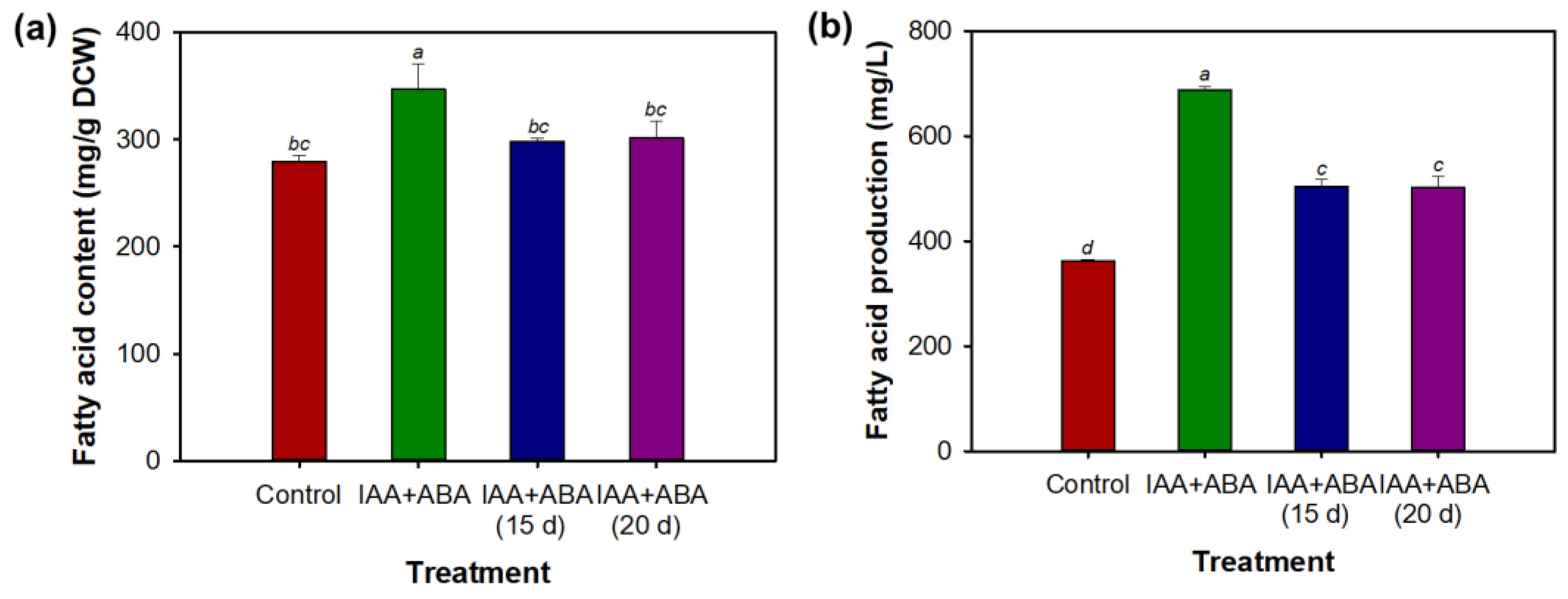
Simultaneous application with 1 µM IAA and 50 µM ABA at inoculation (day 0) yielded the highest fatty acid content and production, reaching 276.8 ± 15.4 mg/g DCW in content and 687.0 ± 7.4 mg/L in volumetric production (Figure 8). These values represent 24% and 90% increases compared to the control (223.8 ± 6.0 mg/g DCW and 362.4 ± 2.3 mg/L). When ABA was supplemented at later time points (days 15 or 20), the fatty acid content remained comparable (~300 mg/g DCW), but total production decreased significantly (~504 mg/L), underscoring the importance of early hormone signaling in activating lipid biosynthesis during metabolically active growth phases [52,54].
Fatty acid composition under the different hormonal treatments is presented in Supplementary Table S1. The predominant species across all conditions were palmitic acid (C16:0), oleic acid (C18:1n9c), linoleic acid (C18:2n6c), and α-linolenic acid (C18:3n3c), collectively accounting for over 64% of total fatty acid content. This compositional profile aligns with previous findings for H. lacustris [42]. Notably, despite the significant increase in total fatty acid production under the combined IAA + ABA treatment, the relative proportions of individual fatty acids remained largely unchanged. This indicates that the hormonal treatment enhanced global lipid biosynthesis without altering fatty acid desaturation or elongation pathways. Such compositional stability is advantageous for downstream applications in the food, nutraceutical, and biofuel industries, where consistency in lipid quality is a critical parameter [34,37]. Future work should focus on optimizing cultivation parameters and elucidating the molecular mechanisms underlying the synergistic effects of combined IAA and ABA to fully realize the potential of phytohormone-assisted coproduction of astaxanthin and lipids.

4. Conclusions

This study demonstrates the potential of phytohormonal regulation—specifically the combined application of indole-3-acetic acid (IAA) and abscisic acid (ABA)—to simultaneously enhance cell growth, astaxanthin biosynthesis, and fatty acid accumulation in Haematococcus lacustris. Moderate concentrations of IAA primarily stimulated cell proliferation and biomass production, while ABA effectively induced the intracellular accumulation of astaxanthin and lipids. Importantly, the co-application of IAA and ABA at the onset of cultivation yielded the highest levels of both astaxanthin and total fatty acids, underscoring the importance of early-stage hormonal signaling in modulating metabolic flux toward high-value bioproducts. These findings may contribute to developing strategies for optimizing microalgal bioprocesses in commercial applications, from nutraceuticals to biofuels.

Supplementary Materials

The following supporting information can be downloaded at: https://www.mdpi.com/article/10.3390/app151810136/s1, Supplementary Figure S1: Time-course pH profiles of Haematococcus lacustris during 30-d photosynthetic cultivation under varying IAA and ABA concentrations; Supplementary Figure S2: Time-course morphological profiles of Haematococcus lacustris during 30-d cultivation under various phytohormone treatments; Supplementary Figure S3: Time-course profiles of cell density plotted on a logarithmic scale; Supplementary Table S1: Fatty acid composition (%) Haematococcus lacustris after 30-d cultivation under combined IAA (1 µM) and ABA (50 µM) treatments.

Author Contributions

Conceptualization, Y.-K.O. and R.M.; methodology, R.M., L.P.S. and A.L.N.; Resources, E.-J.K. and J.Y.C., data curation, R.M., L.P.S. and A.L.N.; writing—original draft preparation, Y.-K.O. and R.M.; writing—review and editing, Y.-K.O., R.M., A.L.N., E.-J.K. and J.Y.C.; visualization, R.M., Y.-K.O., L.P.S. and A.L.N.; supervision, Y.-K.O.; funding acquisition, Y.-K.O., E.-J.K. and J.Y.C. All authors have read and agreed to the published version of the manuscript.

Funding

This study was supported by the Nakdonggang National Institute of Biological Resources (NNIBR; Grant No. NNIBR20253114) funded by the Ministry of Environment (MOE), Republic of Korea; the STEAM Project (Grant No. RS-2024-00459155) of the National Research Foundation of Korea (NRF), funded by the Ministry of Science and ICT (MSIT).

Institutional Review Board Statement

Not applicable.

Informed Consent Statement

Not applicable.

Data Availability Statement

The data presented in this study are available in the article and Supplementary Materials.

Conflicts of Interest

The authors declare no conflicts of interest.

References

  1. Choi, H.; Kim, B.; Jeong, S.H.; Kim, T.Y.; Kim, D.; Oh, Y.; Hahn, S.K. Microalgae-Based Biohybrid Microrobot for Accelerated Diabetic Wound Healing. Small 2023, 19, 2204617. [Google Scholar] [CrossRef]
  2. Diankristanti, P.A.; Ho, N.H.E.; Chen, J.-H.; Nagarajan, D.; Chen, C.-Y.; Hsieh, Y.-M.; Ng, I.-S.; Chang, J.-S. Unlocking the potential of microalgae as sustainable bioresources from up to downstream processing: A critical review. Chem. Eng. J. 2024, 488, 151124. [Google Scholar] [CrossRef]
  3. Yoo, D.; Hong, S.-J.; Yun, S.; Kang, M.-J.; Cho, B.-K.; Lee, H.; Choi, H.-K.; Kim, D.-M.; Lee, C.-G. Metabolic engineering for redirecting carbon to enhance the fatty acid content of Synechocystis sp. Pcc6803. Biotechnol. Bioprocess Eng. 2023, 28, 274–280. [Google Scholar] [CrossRef]
  4. Debnath, T.; Bandyopadhyay, T.K.; Vanitha, K.; Bobby, M.N.; Nath Tiwari, O.; Bhunia, B.; Muthuraj, M. Astaxanthin from microalgae: A review on structure, biosynthesis, production strategies and application. Food Res. Int. 2024, 176, 113841. [Google Scholar] [CrossRef]
  5. Patel, A.K.; Tambat, V.S.; Chen, C.-W.; Chauhan, A.S.; Kumar, P.; Vadrale, A.P.; Huang, C.-Y.; Dong, C.-D.; Singhania, R.R. Recent advancements in astaxanthin production from microalgae: A review. Bioresour. Technol. 2022, 364, 128030. [Google Scholar] [CrossRef]
  6. Kim, B.; Youn Lee, S.; Lakshmi Narasimhan, A.; Kim, S.; Oh, Y.-K. Cell disruption and astaxanthin extraction from Haematococcus pluvialis: Recent advances. Bioresour. Technol. 2022, 343, 126124. [Google Scholar] [CrossRef]
  7. Hata, N.; Ogbonna, J.C.; Hasegawa, Y.; Taroda, H.; Tanaka, H. Production of astaxanthin by Haematococcus pluvialis in a sequential heterotrophic-photoautotrophic culture. J. Appl. Phycol. 2001, 13, 395–402. [Google Scholar] [CrossRef]
  8. Ren, Y.; Deng, J.; Huang, J.; Wu, Z.; Yi, L.; Bi, Y.; Chen, F. Using green alga Haematococcus pluvialis for astaxanthin and lipid co-production: Advances and outlook. Bioresour. Technol. 2021, 340, 125736. [Google Scholar] [CrossRef]
  9. Lakshmi Narasimhan, A.; Lee, N.; Kim, S.; Kim, Y.-E.; Christabel, C.; Yu, H.; Kim, E.-J.; Oh, Y.-K. Enhanced astaxanthin production in Haematococcus lacustris by electrochemical stimulation of cyst germination. Bioresour. Technol. 2024, 411, 131301. [Google Scholar] [CrossRef]
  10. Lee, C.; Choi, Y.-E.; Yun, Y.-S. A strategy for promoting astaxanthin accumulation in Haematococcus pluvialis by 1-aminocyclopropane-1-carboxylic acid application. J. Biotechnol. 2016, 236, 120–127. [Google Scholar] [CrossRef]
  11. Khoo, K.S.; Lee, S.Y.; Ooi, C.W.; Fu, X.; Miao, X.; Ling, T.C.; Show, P.L. Recent advances in biorefinery of astaxanthin from Haematococcus pluvialis. Bioresour. Technol. 2019, 288, 121606. [Google Scholar] [CrossRef]
  12. Han, S.-I.; Chang, S.H.; Lee, C.; Jeon, M.S.; Heo, Y.M.; Kim, S.; Choi, Y.-E. Astaxanthin biosynthesis promotion with pH shock in the green microalga, Haematococcus lacustris. Bioresour. Technol. 2020, 314, 123725. [Google Scholar] [CrossRef]
  13. Samhat, K.; Kazbar, A.; Takache, H.; Gonçalves, O.; Drouin, D.; Ismail, A.; Pruvost, J. Optimization of continuous astaxanthin production by Haematococcus pluvialis in nitrogen-limited photobioreactor. Algal Res. 2024, 80, 103529. [Google Scholar] [CrossRef]
  14. Scibilia, L.; Girolomoni, L.; Berteotti, S.; Alboresi, A.; Ballottari, M. Photosynthetic response to nitrogen starvation and high light in Haematococcus pluvialis. Algal Res. 2015, 12, 170–181. [Google Scholar] [CrossRef]
  15. Zhang, W.; Zhou, X.; Zhang, Y.; Cheng, P.; Ma, R.; Cheng, W.; Chu, H. Enhancing astaxanthin accumulation in Haematococcus pluvialis by coupled light intensity and nitrogen starvation in column photobioreactors. J. Microbiol. Biotechnol. 2018, 28, 2019–2028. [Google Scholar] [CrossRef]
  16. Giannelli, L.; Yamada, H.; Katsuda, T.; Yamaji, H. Effects of temperature on the astaxanthin productivity and light harvesting characteristics of the green alga Haematococcus pluvialis. J. Biosci. Bioeng. 2015, 119, 345–350. [Google Scholar] [CrossRef]
  17. Li, Q.; You, J.; Qiao, T.; Zhong, D.; Yu, X. Sodium chloride stimulates the biomass and astaxanthin production by Haematococcus pluvialis via a two-stage cultivation strategy. Bioresour. Technol. 2022, 344, 126214. [Google Scholar] [CrossRef]
  18. Yao, J.; Kim, H.S.; Kim, J.Y.; Choi, Y.-E.; Park, J. Mechanical stress induced astaxanthin accumulation of H. pluvialis on a chip. Lab Chip 2020, 20, 647–654. [Google Scholar] [CrossRef]
  19. Mahadi, R.; Kim, S.; Ilhamsyah, D.P.A.; Vahisan, L.P.S.; Narasimhan, A.L.; Park, G.W.; Lee, S.Y.; Oh, Y.-K. Rapid accumulation of astaxanthin in Haematococcus pluvialis induced by mild hydrostatic pressure. Biotechnol. Bioprocess Eng. 2023, 28, 345–351. [Google Scholar] [CrossRef]
  20. Li, Q.; Li, L.; Zhang, Y.; Gao, H.; Zhao, Y.; Yu, X. Chemical inducers regulate ROS signalling to stimulate astaxanthin production in Haematococcus pluvialis under environmental stresses: A Review. Trends Food Sci. Technol. 2023, 136, 181–193. [Google Scholar] [CrossRef]
  21. Liu, Y.H.; Alimujiang, A.; Wang, X.; Luo, S.W.; Balamurugan, S.; Yang, W.D.; Liu, J.S.; Zhang, L.; Li, H.Y. Ethanol induced jasmonate pathway promotes astaxanthin hyperaccumulation in Haematococcus pluvialis. Bioresour. Technol. 2019, 289, 121720. [Google Scholar] [CrossRef]
  22. Du, F.; Hu, C.; Sun, X.; Zhang, L.; Xu, N. Transcriptome analysis reveals the promoting effect of trisodium citrate on astaxanthin accumulation in Haematococcus pluvialis under high light condition. Aquaculture 2021, 543, 736978. [Google Scholar] [CrossRef]
  23. Kim, Y.E.; Matter, I.A.; Lee, N.; Jung, M.; Lee, Y.C.; Choi, S.A.; Lee, S.Y.; Kim, J.R.; Oh, Y.K. Enhancement of astaxanthin production by Haematococcus pluvialis using magnesium aminoclay nanoparticles. Bioresour. Technol. 2020, 307, 123270. [Google Scholar] [CrossRef]
  24. Raza, A.; Salehi, H.; Rahman, M.A.; Zahid, Z.; Madadkar Haghjou, M.; Najafi-Kakavand, S.; Charagh, S.; Osman, H.S.; Albaqami, M.; Zhuang, Y.; et al. Plant hormones and neurotransmitter interactions mediate antioxidant defenses under induced oxidative stress in plants. Front. Plant Sci. 2022, 13, 961872. [Google Scholar] [CrossRef]
  25. Yu, X.; Chen, L.; Zhang, W. Chemicals to enhance microalgal growth and accumulation of high-value bioproducts. Front. Microbiol. 2015, 6, 56. [Google Scholar] [CrossRef]
  26. Majda, M.; Robert, S. The role of auxin in cell wall expansion. Int. J. Mol. Sci. 2018, 19, 951. [Google Scholar] [CrossRef]
  27. Ramaih, S.; Guedira, M.; Paulsen, G.M. Relationship of indoleacetic acid and tryptophan to dormancy and preharvest sprouting of wheat. Funct. Plant Biol. 2003, 30, 939. [Google Scholar] [CrossRef]
  28. Li, W.; Wang, D.; Hu, F.; Li, H.; Ma, L.; Xu, L. Exogenous IAA treatment enhances phytoremediation of soil contaminated with phenanthrene by promoting soil enzyme activity and increasing microbial biomass. Environ. Sci. Pollut. Res. 2016, 23, 10656–10664. [Google Scholar] [CrossRef]
  29. Verma, V.; Ravindran, P.; Kumar, P.P. Plant hormone-mediated regulation of stress responses. BMC Plant Biol. 2016, 16, 86. [Google Scholar] [CrossRef]
  30. Liu, Y.; Dong, B.; Zhang, C.; Yang, L.; Wang, Y.; Zhao, H. Effects of exogenous abscisic acid (ABA) on carotenoids and petal color in Osmanthus fragrans ‘Yanhonggui’. Plants 2020, 9, 454. [Google Scholar] [CrossRef]
  31. Kayani, S.-I.; Rahman, S.; Shen, Q.; Cui, Y.; Liu, W.; Hu, X.; Zhu, F.; Huo, S. Molecular approaches to enhance astaxanthin biosynthesis: Future outlook: Engineering of transcription factors in Haematococcus pluvialis. Crit. Rev. Biotechnol. 2024, 44, 514–529. [Google Scholar] [CrossRef]
  32. Zhao, Y.; Wang, H.-P.; Han, B.; Yu, X. Coupling of abiotic stresses and phytohormones for the production of lipids and high-value by-products by microalgae: A review. Bioresour. Technol. 2019, 274, 549–556. [Google Scholar] [CrossRef]
  33. Piotrowska-Niczyporuk, A.; Bajguz, A. The effect of natural and synthetic auxins on the growth, metabolite content and antioxidant response of green alga Chlorella vulgaris (Trebouxiophyceae). Plant Growth Regul. 2014, 73, 57–66. [Google Scholar] [CrossRef]
  34. Mousavi, P.; Morowvat, M.; Montazeri-Najafabady, N.; Abolhassanzadeh, Z.; Mohagheghzadeh, A.; Hamidi, M.; Niazi, A.; Ghasemi, Y. Investigating the effects of phytohormones on growth and beta-carotene production in a naturally isolates stain of Dunaliella salina. J. Appl. Pharm. Sci. 2016, 6, 164–171. [Google Scholar] [CrossRef]
  35. Kozlova, T.A.; Kartashov, A.V.; Zadneprovskaya, E.; Krapivina, A.; Zaytsev, P.; Chivkunova, O.B.; Solovchenko, A.E. Effect of abscisic acid on growth, fatty acid profile, and pigment composition of the Chlorophyte chlorella (Chromochloris) zofingiensis and Its Co-Culture Microbiome. Life 2023, 13, 452. [Google Scholar] [CrossRef]
  36. Contreras-Pool, P.Y.; Peraza-Echeverria, S.; Ku-González, Á.F.; Herrera-Valencia, V.A. The phytohormone abscisic acid increases triacylglycerol content in the green microalga Chlorella saccharophila (Chlorophyta). Algae 2016, 31, 267–276. [Google Scholar] [CrossRef]
  37. Lin, Y.; Dai, Y.; Xu, W.; Wu, X.; Li, Y.; Zhu, H.; Zhou, H. The growth, lipid accumulation and fatty acid profile analysis by abscisic acid and indol-3-acetic acid induced in Chlorella sp. FACHB-8. Int. J. Mol. Sci. 2022, 23, 4064. [Google Scholar] [CrossRef]
  38. Lee, C.; Jeon, M.S.; Kim, J.Y.; Lee, S.H.; Kim, D.G.; Roh, S.W.; Choi, Y.-E. Effects of an auxin-producing symbiotic bacterium on cell growth of the microalga Haematococcus pluvialis: Elevation of cell density and prolongation of exponential stage. Algal Res. 2019, 41, 101547. [Google Scholar] [CrossRef]
  39. Gao, Z.; Hongzheng, G.; Chunxiao, M. Effects of abscisic acid (ABA) carotenogenesis expression and astaxanthin accumulation in Haematococcus pluvialis. Res. J. Biotechnol. 2013, 8, 9–15. [Google Scholar]
  40. Kobayashi, M.; Hirai, N.; Kurimura, Y.; Ohigashi, H.; Tsuji, Y. Abscisic acid-dependent algal morphogenesis in the unicellular green alga Haematococcus pluvialis. Plant Growth Regul. 1997, 22, 79–85. [Google Scholar] [CrossRef]
  41. Kobayashi, M.; Todoroki, Y.; Hirai, N.; Kurimura, Y.; Ohigashi, H.; Tsuji, Y. Biological activities of abscisic acid analogs in the morphological change of the green alga Haematococcus pluvialis. J. Ferment. Bioeng. 1998, 85, 529–531. [Google Scholar] [CrossRef]
  42. Mahadi, R.; Vahisan, L.P.S.; Ilhamsyah, D.P.A.; Kim, S.; Kim, B.; Lee, N.; Oh, Y.-K. Enhancement of astaxanthin and fatty acid production in Haematococcus pluvialis using strigolactone. Appl. Sci. 2022, 12, 1791. [Google Scholar] [CrossRef]
  43. Zhang, C.; Liu, J.; Zhang, L. Cell cycles and proliferation patterns in Haematococcus pluvialis. Chin. J. Oceanol. Limnol. 2017, 35, 1205–1211. [Google Scholar] [CrossRef]
  44. Fábregas, J.; Domínguez, A.; Maseda, A.; Otero, A. Interactions between irradiance and nutrient availability during astaxanthin accumulation and degradation in Haematococcus pluvialis. Appl. Microbiol. Biotechnol. 2003, 61, 545–551. [Google Scholar] [CrossRef] [PubMed]
  45. Li, Q.; Li, B.; Li, J. The dynamic behaviors of photosynthesis during non-motile cell germination in Haematococcus pluvialis. Water 2022, 14, 1280. [Google Scholar] [CrossRef]
  46. Han, D.; Wang, J.; Sommerfeld, M.; Hu, Q. Susceptibility and protective mechanisms of motile and non motile cells of Haematococcus pluvialis (chlorophyceae) to photooxidative stress 1. J. Phycol. 2012, 48, 693–705. [Google Scholar] [CrossRef]
  47. Lee, S.-A.; Lee, N.; Oh, H.-M.; Kim, D.G.; Ahn, C.-Y. Fast-track production of astaxanthin by reduced cultivation time with the “red cell inoculation system” (RCIS) and various chemical cues in Haematococcus lacustris. J. Appl. Phycol. 2020, 32, 41–50. [Google Scholar] [CrossRef]
  48. Ha, N.C.; Tam, L.T.; Hien, H.T.M.; Thu, N.T.H.; Hong, D.D.; Thom, L.T. Optimization of culture conditions for high cell productivity and astaxanthin accumulation in Vietnam’s green microalgae Haematococcus pluvialis HB and a neuroprotective activity of its astaxanthin. Bioengineering 2024, 11, 1176. [Google Scholar] [CrossRef]
  49. Bauer, A.; Minceva, M. Examination of photo-, mixo-, and heterotrophic cultivation conditions on Haematococcus pluvialis cyst cell germination. Appl. Sci. 2021, 11, 7201. [Google Scholar] [CrossRef]
  50. Dao, G.-H.; Wu, G.-X.; Wang, X.-X.; Zhuang, L.-L.; Zhang, T.-Y.; Hu, H.-Y. Enhanced growth and fatty acid accumulation of microalgae Scenedesmus sp. Lx1 by two types of auxin. Bioresour. Technol. 2018, 247, 561–567. [Google Scholar] [CrossRef]
  51. Yu, X.; Niu, X.; Zhang, X.; Pei, G.; Liu, J.; Chen, L.; Zhang, W. Identification and mechanism analysis of chemical modulators enhancing astaxanthin accumulation in Haematococcus pluvialis. Algal Res. 2015, 11, 284–293. [Google Scholar] [CrossRef]
  52. Chen, J.; Wei, D.; Lim, P.-E. Enhanced coproduction of astaxanthin and lipids by the green microalga Chromochloris zofingiensis: Selected phytohormones as positive stimulators. Bioresour. Technol. 2020, 295, 122242. [Google Scholar] [CrossRef]
  53. Cheng, J.; Li, K.; Yang, Z.; Zhou, J.; Cen, K. Enhancing the growth rate and astaxanthin yield of Haematococcus pluvialis by nuclear irradiation and high concentration of carbon dioxide stress. Bioresour. Technol. 2016, 204, 49–54. [Google Scholar] [CrossRef] [PubMed]
  54. Sun, X.-M.; Ren, L.-J.; Zhao, Q.-Y.; Ji, X.-J.; Huang, H. Microalgae for the production of lipid and carotenoids: A review with focus on stress regulation and adaptation. Biotechnol. Biofuels 2018, 11, 272. [Google Scholar] [CrossRef] [PubMed]
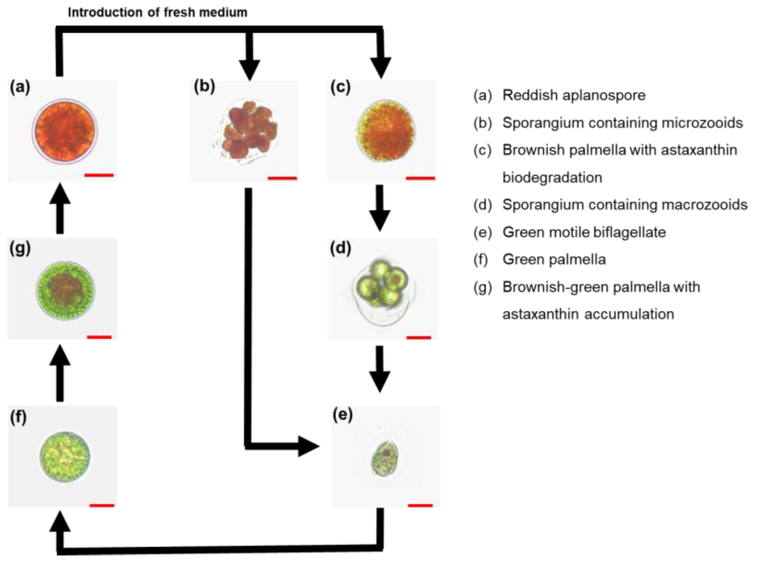
Figure 1. Representative light microscopy images showing key morphological stages in the life cycle of Haematococcus lacustris during 30 d photosynthetic cultivation. Scale bar: 20 µm.
Applsci 15 10136 g001
Figure 2. Time-course profiles of (a) cell density, (b) biomass concentration, (c) cell diameter, and (d) cell-type distribution of Haematococcus lacustris cultivated with different IAA concentrations (0.1, 1, 10, and 100 µM) for 30 d. Morphological analyses were conducted on >200 cells from two independent replicates. In (d), sporangia (microzooids and macrozooids) and intermediate palmella cells (brownish and brownish-green) were grouped as described in Figure 1. Error bars represent mean ± standard deviation (n = 4 for cell density and dry cell weight). Different superscript letters indicate significant difference (p < 0.05). DCW: dry cell weight; IAA: indole-3-acetic acid.
Applsci 15 10136 g002
Figure 3. (a) Intracellular astaxanthin content and (b) volumetric astaxanthin production of Haematococcus lacustris after 30 d of cultivation under varying IAA concentrations (0.1, 1, 10, and 100 µM). Error bars represent mean ± standard deviation (n = 4). Different superscript letters indicate significant differences (p < 0.05). DCW: dry cell weight; IAA: indole-3-acetic acid.
Applsci 15 10136 g003
Figure 4. Time-course profiles of (a) cell density, (b) biomass concentration, (c) cell diameter, and (d) cell-type distribution of Haematococcus lacustris cultivated under different ABA concentrations (1, 10, 50, and 100 µM) for 30 d. Morphological analyses were conducted on >200 cells from two independent replicates. In (d), sporangia (microzooids and macrozooids) and intermediate palmella cells (brownish and brownish-green) were grouped as described in Figure 1. Error bars represent mean ± standard deviation (n = 4 for cell density and dry cell weight). Different superscript letters indicate significant differences (p < 0.05). ABA: abscisic acid; DCW: dry cell weight.
Applsci 15 10136 g004
Figure 5. (a) Intracellular astaxanthin content and (b) volumetric astaxanthin production of Haematococcus lacustris after 30 d cultivation under different ABA concentrations (1, 10, 50, and 100 µM). Error bars represent mean ± standard deviation (n = 4). Different superscript letters indicate significant differences (p < 0.05). ABA: abscisic acid, DCW: dry cell weight.
Applsci 15 10136 g005
Figure 6. Time-course profiles of (a) cell density, (b) biomass concentration, (c) cell diameter, and (d) cell-type distribution of Haematococcus lacustris under combined hormone treatment with 1 µM IAA and 50 µM ABA administered at different time points (day 0, 15, and 20). Morphological analyses were conducted on >200 cells from two independent replicates. In (d), sporangia (microzooids and macrozooids) and intermediate palmella cells (brownish and brownish-green) were grouped as described in Figure 1. Error bars represent mean ± standard deviation (n = 4 cell density and dry cell weight). Different superscript letters indicate significant differences (p < 0.05). ABA: abscisic acid; DCW: dry cell weight, IAA: indole-3-acetic acid.
Applsci 15 10136 g006
Figure 7. (a) Intracellular astaxanthin content and (b) volumetric astaxanthin production of Haematococcus lacustris after 30 d cultivation under combined IAA (1 µM) and ABA (50 µM) treatment. Error bars represent mean ± standard deviation (n = 4). Different superscript letters indicate significant differences (p < 0.05). ABA: abscisic acid; DCW: dry cell weight; IAA: indole-3-acetic acid.
Applsci 15 10136 g007
Figure 8. (a) Total fatty acid content and (b) production of Haematococcus lacustris after 30 d cultivation with combined IAA (1 µM) and ABA (50 µM) treatment. Error bars represent mean ± standard deviation (n = 4). Different superscript letters indicate significant differences (p < 0.05). ABA: abscisic acid; DCW: dry cell weight; IAA: indole-3-acetic acid.
Applsci 15 10136 g008
Disclaimer/Publisher’s Note: The statements, opinions and data contained in all publications are solely those of the individual author(s) and contributor(s) and not of MDPI and/or the editor(s). MDPI and/or the editor(s) disclaim responsibility for any injury to people or property resulting from any ideas, methods, instructions or products referred to in the content.