Article

Physicochemical Stability of the Pigment Produced by Pseudofusicoccum adansoniae: Influence of pH, Temperature, Additives, and Light Exposure

by
Bianca Vilas Boas Alves
1,†,
Letícia Jambeiro Borges
1,†,
Vitor Hugo Moreau
2,
Samira Abdallah Hanna
2 and
Marcelo Andrés Umsza-Guez
1,*
1
Food Science Postgraduate Program, Faculty of Pharmacy, Federal University of Bahia, Salvador 40170-115, BA, Brazil
2
Laboratory of Applied Microbiology of the Health Sciences Institute, Federal University of Bahia, Salvador 40110-100, BA, Brazil
*
Author to whom correspondence should be addressed.
These authors contributed equally to this work.
Appl. Sci. 2025, 15(16), 8800; https://doi.org/10.3390/app15168800
Submission received: 4 July 2025 / Revised: 31 July 2025 / Accepted: 6 August 2025 / Published: 9 August 2025

Abstract

Physicochemical stability is sought after in natural pigments, as their color must remain unchanged throughout the food’s shelf life. Our objective was to evaluate how different process variables affect the physicochemical stability of the pigment produced by Pseudofusicoccum adansoniae. The effects of pH, temperature, ions, additives, storage temperature, and light exposure on color stability were evaluated using colorimetry and/or spectrophotometry. The pigment extract was more stable at acidic pH and at temperatures below 60 °C. Additives and ions had a slight influence on color stability, and there was minimal degradation after three months of storage in the absence of light, but significant degradation when exposed to light. Degradation was characterized by a decrease in the red hue and the appearance of a yellow hue. Even with this hue change under certain conditions, the pigment remains suitable for use in the food industry.

1. Introduction

Colors are vital to perception and fundamental in consumer choice and decision-making [1,2].
The growing concern for healthy foods has increased the demand for natural sources, leading industries to focus their efforts on developing high-quality, fresh-looking products [3,4,5,6,7]. This trend is evident in the annual growth rate of 3.57% in the demand for natural colors, which could reach approximately USD 3.5 billion by 2028 [8,9,10,11].
The demand for healthy, environmentally friendly foods has driven the search for natural alternatives to synthetic dyes, prompting industries to develop high-quality, stable, and visually appealing products [4,5,12,13,14].
The use of microorganisms as a source for pigment production has gained prominence, with an increasing number of patents filed since 1988. Microbial pigments offer several advantages, including greater physicochemical stability and a broad spectrum of color [2,15,16,17]. Compared to other natural pigments, they provide benefits such as lower production costs, easier extraction, higher yields, renewable raw materials, and independence from seasonal variations [18,19,20]. This ensures consistent metabolite production with minimal batch-to-batch variability, making them highly relevant to industries [19,21]. However, for microbial pigments to be used as food additives, their toxicity must be assessed, particularly those produced by fungi [22,23]. The exact reason why some microorganisms produce pigments remains unclear, but Liu & Nizet [1] postulated that most pigments initially evolved as a defense mechanism against reactive oxygen species (ROS). Over time, these compounds have adapted to serve additional functions.
Among the microorganisms used, filamentous fungi are the most common for dye production [15,16,23,24,25]. These pigments are already applied in the fishing industry, for example, to enhance the pink color of farmed salmon. Aspergillus, Penicillium, Paecilomyces, and Monascus are the main fungal genera responsible for pigment production [26,27].
Pigment biosynthesis is directly influenced by fermentation conditions, such as medium composition and process parameters. While synthetic dyes are known for their high physicochemical stability, natural dyes are generally less stable [28]. The primary factors affecting pigment stability include pH, high temperature, oxygen exposure, light, and hydrolytic enzymes, as well as interactions with other food components such as ascorbic acid, metal ions, and sugars [25,29]. Most literature data on fungi that produce pigments of various colors focus on the Monascus genus, whose pigments have already been applied in food products, demonstrating good stability. These pigments generally exhibit stability across a wide pH range (2 to 10), at moderately high temperatures, and under ultraviolet light, in addition to producing a variety of colors [25,30,31]. Previously, Alves et al. demonstrated in another study that the same extract produced by this identified fungus showed no cytotoxicity (tested cell lines: HepG2, SCC4, BJ, and MRC-5) and produced beneficial compounds (diketopiperazines), as observed in other studies [32].
In this study, we aimed to evaluate the physicochemical stability of a new natural biopigment (raw extract) produced by the endophytic fungus Pseudofusicoccum adansoniae, isolated from Manilkara sp., for potential industrial applications. Although Pseudofusicoccum adansoniae has already been isolated and characterized [33], there is a significant gap in the literature regarding the production and characterization of its pigments. Studying these compounds is relevant to broaden the sources of natural dyes, especially those with potential applicability and safety, contributing to overcome limitations observed in other available fungal pigments.

2. Methods

2.1. Submerged Culture and Extract Production

The fungus Pseudofusicoccum adansoniae was isolated from Manilkara salzmannii and maintained in the mycothek of the Laboratory of Applied Microbiology and Bioprospection—LAMAB, located at the Institute of Health Sciences—UFBA. The culture was preserved on Sabouraud dextrose agar (SDA) at 28 ± 2 °C.
The strain was incubated for 7 days in PDA to be used as an inoculum for submerged fermentation. This was conducted according to Alves et al. [34]. After the incubation in plates, 3 mycelial agar discs (5 mm diameter) were transferred to 100 mL of sterile Potato Dextrose Broth (PDB), pH 8.5 in Erlenmeyer Flasks. The flasks were under yellow light (597–577 nm) and kept at 28 ± 2 °C for 21 days. After the incubation, the submerged fermentation was filtered using common paper filter and then using 0.45 µm filter with vacuum pump. The crude pigment extract was then filtered, first on common filter paper and then with a 0.45 µm filter, under low pressure. The pigment extract was then stored in a capped tube at 4 °C.

2.2. Stability of the Crude Pigment Extract

The stability tests of the burgundy crude pigment extract (color similar to that of red wine) were performed with a 1:18 dilution (absorbance between 0.740 and 0.790) in distilled water or in the appropriate buffer (see Section 2.4.1). Crude extracts exhibited absorption spectra with a characteristic peak at 527 nm (see Figure 1A). The absorbance at 520 nm was then used to determine the relative concentration of the crude extract using a UV-Vis Model UV-M51 spectrophotometer (Bell Labs, New York, NY, USA). The stability of the extract over time was analyzed in a Konica Minolta CM-5 colorimeter. The results were expressed on the CIE L* a* b* color space scale, in which the L* coordinate denotes luminosity on a scale of 0–100 from black to white; a*, red (+) to green (−) components; and b*, yellow (+) to blue (−) components. All analyses were performed in triplicate. The results obtained in the spectrophotometer are expressed as a percentage of the initial absorbance.

2.3. Temperature Stability

The pigment stability of the crude extract was evaluated in the temperature range between 30 and 90 °C, at pH 4.5 (the natural pH of the extract). The crude pigment extract was diluted in water (1:18) and incubated at the desired temperature in an incubator. Spectrophotometric and colorimetric measurements were performed at 0, 1, 3, 6, 9, 12, and 24 h, with the time intervals determined based on preliminary testing.

2.4. pH Stability

The effect of different pHs (from 3 to 10) on the color stability of the crude pigment extract was evaluated. For this, citrate/phosphate buffer (pH 3 to 7), phosphate buffer (pH 8), and glycine sodium hydroxide buffer (pH 9 and 10) were used. The crude extracts were diluted in buffers, depending on the pH value, and the control was diluted in water at 25 °C. The samples were kept free of light and stability was evaluated both by spectrophotometry and by colorimetry, over time, for 24 h, as described above.

2.4.1. Effect of Additives and Ions

The interaction of pigment from the crude extract with additives and ions was evaluated at 25 °C for 24 h, as described above. The evaluated additives were weighed and diluted directly in the crude extract to reach the desired concentrations. The concentrations used were established for dietary supplements, in accordance with current Brazilian legislation [35] (ANVISA, 2018): aspartame (75 mg/100 g), saccharin (30 mg/100 g), sodium cyclamate (130 mg/100 g), calcium propionate (0.40 g/100 g), potassium sorbate (0.2 g/100 mL), acesulfame (0.2 g/100 mL), maltitol (0.2 g/100 mL), inulin (0.2 g/100 mL), potassium benzoate (0.2 g/100 mL), calcium chloride (Synth), sodium nitrate (Vetec, Caxias do Sul, Brazil), magnesium sulfate PA (scientific exodus), and zinc sulfate PA (Synth, Diadema, Brazil), at concentrations of 0.1 M and 0.5 M. The samples were kept free from incidence of light and measurements were performed as described above. All chemicals used were of analytical grade of purity.

2.4.2. Effect of the Storage

The color stability of the burgundy pigment over time was performed with samples stored at freezer (−20 °C) or refrigerator (4 °C) temperature. The samples were diluted in water and stored for three months without light. Additional tests were performed on samples with the incidence of a constant light source, at 4 °C. The crude extract of the pigment was diluted (1:18) in distilled water in glass tubes and stored in a cold room illuminated by a 12 W LED cold light, 1350 lumens. The control was completely wrapped in aluminum foil. Measurements were taken at times 0, 7, 15, 30, 45, 60, and 90 days.

2.5. Statistical Analysis

Graphs and calculations displayed in Figures were made with Grace software 5.0.3 (https://plasma-gate.weizmann.ac.il/Grace/ accessed on 12 December 2023) and are detailed according to figure captions.

3. Results

3.1. Effect of Temperature

Temperature has a notable influence on the stability of most pigments. Figure 1 shows the effect of temperature on pigment degradation in the crude extract produced by Pseudofusicoccum adansoniae. The absorbance at 520 nm was chosen as a parameter to assess pigment color degradation, since the spectral analysis of the absorbance of the crude extract showed a large decrease in the 520 nm peak as the pigment color faded (Figure 1A). Figure 1, panel A, also shows the spectral change observed within the thermal degradation of the pigment. Absorption spectra clearly display a monomorphic pattern, with no expressive peak shift nor presence of secondary peaks. It was possible to notice, in Figure 1B, that the increase in temperature above 30 °C promotes a faster degradation of the burgundy pigment, evaluated by the decrease in absorbance at 520 nm. Almost 50% of the original absorbance is lost after 24 h at 50 °C and in less than 5 h at temperatures above 70 °C (Figure 1C).
The stability of the burgundy pigment of the crude extract produced by Pseudofusicoccum adansoniae. was also evaluated by colorimetric analysis (Figure 1D). As shown above, incubation for 24 h at higher temperatures caused the a* component to decrease as the L* and b* components increased, suggesting a loss of red color, while the luminosity and yellow components increased. This difference can be seen in Figure 2. The Burgundy pigment with an intense red hue becomes faded and lighter after 1 h at 90 °C. It is possible to notice the replacement of the red hue by a more yellow one, corroborating the results of Figure 1D, where the a* (red) component decreased as the L* (brightness) and b* (yellow) increased after 24 h at higher temperatures.

3.2. Effect of pH

pH stability is one of the most important characteristics of industrial pigments. The stability of the burgundy pigment of Pseudofusicoccum adansoniae between pHs 3 to 10 was evaluated in this study. Figure 3A shows the residual absorbance at 520 nm of the burgundy pigment after 24 h of incubation at each studied pH. As can be noticed, the pigment showed greater stability at low pHs up to 5 and suffered faster degradation at pHs above 6. The acid stability of the burgundy pigment is reinforced by colorimetric analysis. Figure 3B shows that component a* is quite stable at pH 3 but decreases more pronouncedly at pH 4 and above. The more alkaline the medium, the faster the pigment degradation. In fact, at pH 10, all changes in color components as well as in absorbance at 520 nm could be observed in less than 10 h of incubation.

3.3. Effect of Additives and Ions on Color Stability

Chemical additives in industry are commonly used to increase the stability of pigments and can therefore interact with them. The effect on stability of the burgundy pigment produced by Pseudofusicoccum adansoniae of some additives commonly used in the food industry were evaluated, including ionic and organic additives, synthetic sweeteners, preservatives, salting agents, and others (Table 1). A very slight effect on the extent of degradation can be seen for aspartame, sodium cyclamate, calcium propionate, potassium sorbate, maltitol, inulin, or potassium benzoate when compared to the control. However, a considerable decrease in the amount of pigment degradation can be observed in the presence of acesulfame (Table 1).
It was found that acesulfame was the most influential additive for the burgundy pigment extract of Pseudofusicoccum adansoniae, with a 16% reduction in absorbance at 520 nm, in 12 h, when compared to the control. This effect may be associated with changes in water activity, similar to that exerted by sugars and other organic osmolytes.
Inorganic ions are often present in food and can affect its processing and storage. Ions can increase the oxidation of some compounds and change their characteristics [36]. The influence of some inorganic ions on the stability of the burgundy pigment of the Pseudofusicoccum adansoniae extract was also evaluated. Table 1 shows the influence of inorganic salts on the residual absorbance at 520 nm of the raw pigment. All ions were added to final concentrations of 0.1 and 0.5 M, and observations were made after 12 and 24 h of incubation. Calcium chloride, sodium nitrate, and magnesium sulphate have no appreciable effect on pigment stability when compared to the control. Zinc sulfate has been shown to exert a mild effect on pigment stability when added at a final concentration of 0.1 M, but not at 0.5 M. These results suggest that the addition of inorganic ions at concentrations of 0.1 and 0.5 M did not exert any statistically considerable effect on color stability when compared to the control, measured by absorbance at 520 nm. In contrast, some mild protective effect can be observed in the residual absorbance of the pigment in the presence of 0.5 M of the ions, when compared to the lowest tested concentration of 0.1 M, as well as for the control (Table 1). This effect may be due to a possible increase in the conservative activity performed by these salts, attributed to the saturation of the medium at high concentrations.

3.4. Effect of Storage

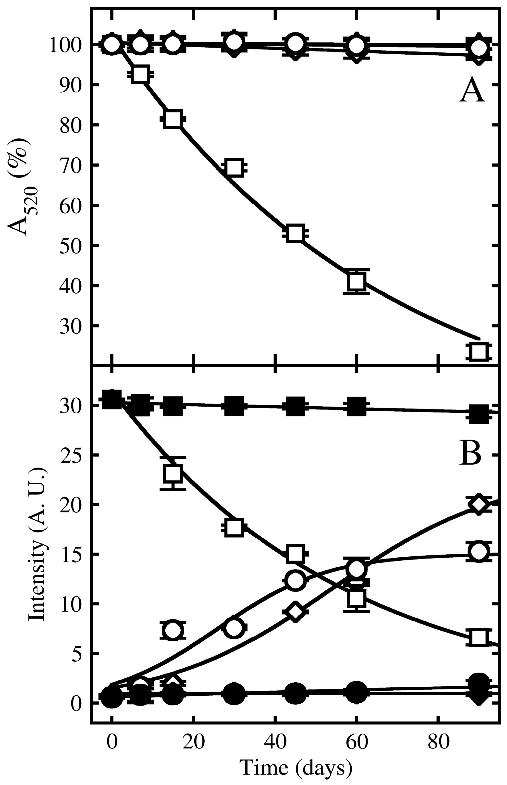
Among the forms of preservation used commercially, storage temperature is the main factor that affects food quality. Low temperatures are the most used conservation method, as it retards biochemical reactions, moisture activity, and the growth of microorganisms [37]. Despite the increase in the cost of maintaining cold chains, it is a viable alternative for the growing consumer market that seeks natural and preservative-free products [38]. The burgundy color of the pigment was preserved during 90 days of storage in a refrigerator or freezer, both without light incidence, with small changes in its stability when compared to room temperature (Figure 4). As shown, no change in absorbance at 520 nm was observed if the pigment extract was kept in the freezer or refrigerator, without light incidence, even after 90 days (Figure 4A). Similar results were observed for the colorimetric properties of the pigment. Any significant changes could be observed in the color pattern of the pigment sample stored in a freezer or refrigerator, without light, even after 90 days (Figure 4B). On the other hand, when the pigment extract samples were stored at refrigerator temperature and exposed to light, a significant loss of absorbance at 520 nm was observed in an exponential decay mode (Figure 4A). In addition, a decrease in the a* component can be observed, followed by an increase in L* and b*, denoting the degradation of the red color tone and the appearance of the yellow color component (Figure 4B).

4. Discussion

Regarding pigment thermostability, Carvalho et al. [39] reported a similar pattern in the thermal stability of the pigment produced by the Monascus LPB 31 strain, verifying the reduction in the residual component of the red color from 57% to 16% at temperatures of 60 and 100 °C, respectively, after 6 h of incubation. The stability of the brown pigment produced by Aspergillus ustus was minimally affected by temperature (0 to 100 °C) [25]; Arcopilus aureus isolated from grapevine produced a yellow pigment with a minimum stability point, corresponding to a temperature of 55.7 °C [31].
The color change (L, a*, b*) can be verified in the extract of Monascus ruber at 100 °C [40]. This suggests that the yellow component present in the degraded extract is stable, and this yellow pigment may be a by-product of red degradation.
Observing the thermal degradation patterns of the components suggests the existence of color intermediates in the pigment degradation pathway (Figure 2), although more refined kinetic studies can be performed to confirm this.
Regarding the change in color of the extract due to exposure to different temperatures, as observed for the thermal degradation of the pigment, the decrease in the a* component in colorimetry is followed by an increase in the L* and b* components (Figure 3B), suggesting that a similar color change is being observed in both temperature and pH degradation processes.
The color stability of the burgundy pigment was considerably affected by both high temperature and alkaline pH. For pH values above 5, stability decreases drastically. Temperature stability was also evaluated at the natural pH of the crude extract (i.e., 4.5), where the stability of the pigment is already impaired (see Figure 3). The results suggest that pHs 3 to 5 are ideal for maintaining the stability of the pigment, even if the temperature is high, as it can maintain the original color tone of the pigment.
As the values of the b* component only increased with the degradation of the burgundy pigment, remaining low at acidic pH, where the red pigment remained stable, it can be concluded that the yellow component is probably formed as the red hue component of the degraded pigment.
The greatest burgundy color stability of the raw pigment was observed in an acidic medium. After submerged fermentation with Pseudofusicoccum adansoniae, the final pH of the growth medium remained around 4.5. This can explain the greater stability of the pigment in acidic pHs than in alkaline ones. Vendruscolo et al. [41] studied the stability of orange and red pigments from Monascus ruber—produced in growth medium at pH 3 in a temperature range of 53.8 to 96.2 °C, as well as at pHs between 4.08 and 6.91. The orange pigment showed an increase in its half-life as the pH decreased from 5.50 to 4.08, although the red pigment showed a loss of stability at pH 4.08. In contrast, a red dye produced by a new Penicillium purpurogenum showed greater stability at pH 8, maintaining 91% of the original color after 24 h of incubation, while showing a higher degradation rate at pH 3 and 4 [42]. Even with the decrease in the color component of the burgundy pigment of Pseudofusicoccum adansoniae, when subjected to alkaline pHs (Figure 3B), it is still more stable than that of the new Penicillium purpurogenum studied by Santos-Ebinuma et al. [42].
De Faria Silva et al. [31] observed that the original color of the pigment solution produced by the Arcopilus aureus is maintained at pH 8.4. The change in color of the pigment solution (weakly yellow to intense yellow and red) was observed when the pH of the solution decreased. This color change can be explained by the variation in the oxidation status of the major compound (cochlioquinol II). On the other hand, the decrease in color may occur due to the increase in pH due to the oxidation of cochlioquinol. The bathochromic effect may also be responsible for these changes.
With a possible application of this extract as a food additive (dye), the interaction of these pigments with additives (sugaras, conservants, sweeteners, and ions) commonly used in the food area was verified and compared with data already existing in the scientific literature. Amr & Al-Tamimi [43] found that the addition of sugars to the medium decreased the stability of the crude extract of anthocyanins from Ranunculus asiaticus at a high temperature. The role that sugars play in the stability of anthocyanin extracts is unclear and may have a protective effect, reducing water activity or accelerating degradation.
In another study, Righetto, Beleia & Ferreira [44] also concluded that the addition of sucrose helped in the stabilization of β-carotene, preserving the color in the storage of frozen passion fruit juice, although the cause is this unknown effect. Mapari, Meyer & Thrane [45] tested the photostability of the Penicillium aculeatum fungus extract using a liquid food system (soft drinks and citrate buffer) and observed a relatively slower discoloration of the fungal pigment extract in the soft drink. They concluded that this stabilization may be due to some ingredients present in the soft drink, for example, sucrose, sodium benzoate, or potassium sorbate.
On the other hand, Reynoso et al. [36] evaluated the effect of iron chromate and potassium chromate on the stability of betalain pigments in storage of Cactaceae fruits after 4 days and verified the degradation of 52% and 32% of the pigment, respectively. The observed degradation effect for iron ions may occur because they attack the electrophilic center of betalain, causing color loss by degradation of the chromophore.
Herbach, Stintzing & Carle [46] found that the presence of metal ions such as Fe2+, Fe3+, Al3+, and Cu2+ facilitates or accelerates betalain degradation. Zhou et al. [25] produced a pigment by Aspergillus ustus, which was sensitive to Zn2+, Fe3+, Fe2+, and Cu2+.
Furthermore, the physical state of the environment in which it is inserted interferes with the concentration necessary to cause this effect. No data were found in the literature using microbial pigments.
These color changes are similar to those observed for the thermal and pH degradation of the pigment extract shown above and characterize the degradation of the burgundy pigment. The replacement of the red by yellow hue component is visually verified in Figure 2 and was previously observed for the red pigment produced by Monascus ruber [40]. However, different results were reported for the extract of Ranunculus asiaticus, when exposed to a light source for 10 days, in the presence of 13% sugar, where the authors found that there was a decrease of 16 and 60% of the sugar component’s red color even without light incidence, both in the presence and absence of sugar, respectively. Samples kept under light lost about 60% of their red color with or without the presence of sugar [43].
The effect of light on pigment degradation was observed, even at refrigeration temperatures. Light radiation induces changes in the structure of many color compounds, possibly in the arrangement of conjugated double bonds, leading to color loss. This effect may affect some unstable compounds more pronouncedly while exerting less influence on some others, with greater stability. Such a mechanism has been proposed for the Penicillium aculeatum fungus extract and such discriminant destabilization can affect the resulting pigment color [45].
The pigment of Pseudofusicoccum adansoniae represents a promising alternative to existing natural dyes, as it exhibits reasonable stability at acidic pH, controlled light resistance, lack of cytotoxicity, and a distinctive burgundy color that is still scarcely explored among fungal pigments. A possible way to increase its stability under other conditions would be the encapsulation process, which could protect it from oxidation, heat, light, or other conditions that reduce its color [47]. This strategy was adopted for the Monascus pigment to increase its stability [48].

5. Conclusions

In this study, temperature was the factor that had the greatest influence on pigment stability. The results showed that the pigment can be stored for 90 days at refrigeration temperature and without light exposure, with little to no change in color and absorbance. However, it undergoes severe degradation when subjected to 90 °C for 3 h. Light exposure was found to reduce stability over storage time. pH also had a significant impact on pigment stability, with color and absorbance being more stable at acidic pHs (3 and 4) than at alkaline pHs (7 to 10). Under all tested conditions, the color followed the same pattern, with the formation of a yellow pigment as the red pigment degraded. The presence of additives or ions did not affect the pigment’s color stability. Despite its lower stability under certain conditions, the crude extract of Pseudofusicoccum adansoniae produces a unique burgundy pigment, distinct from the red hues typically found in fungal pigments. This characteristic makes it a promising natural alternative to artificial colorants in the food industry.

Author Contributions

Conceptualization, M.A.U.-G., B.V.B.A., L.J.B. and S.A.H.; methodology, M.A.U.-G., S.A.H. and B.V.B.A.; validation, M.A.U.-G., B.V.B.A., L.J.B., S.A.H. and V.H.M.; formal analysis, B.V.B.A., L.J.B., M.A.U.-G., S.A.H. and V.H.M.; investigation, M.A.U.-G., B.V.B.A., L.J.B. and S.A.H.; data curation, M.A.U.-G., B.V.B.A., L.J.B., S.A.H. and V.H.M.; writing—original draft preparation, M.A.U.-G., B.V.B.A., L.J.B., S.A.H. and V.H.M.; writing—review and editing, M.A.U.-G. and V.H.M.; visualization, M.A.U.-G. and V.H.M.; supervision, M.A.U.-G.; project administration, M.A.U.-G. All authors have read and agreed to the published version of the manuscript.

Funding

B.V.B.A. was supported by the Coordination on High Education Personnel Improvement (CAPES) and L.J.B. was supported by the Council on Scientific and Technological Development (CNPq) fellowships. M.A.U.-G. (CNPq-Proc. 304747/2020-3) is a Technological Development fellow from CNPq, Brazil.

Data Availability Statement

The original contributions presented in the study are included in the article, further inquiries can be directed to the corresponding author.

Acknowledgments

Coordination on High Education Personnel Improvement (CAPES),Council on Scientific and Technological Development (CNPq) and Federal University of Bahia (UFBA).

Conflicts of Interest

The authors declare no conflicts of interest.

References

  1. Liu, G.Y.; Nizet, V. Color me bad: Microbial pigments as virulence factors. Trends Microbiol. 2009, 17, 406–413. [Google Scholar] [CrossRef]
  2. Dufossé, L. Biotechnological approaches in the production of fungal pigments. In Fungal Biotechnology; Elsevier: Amsterdam, The Netherlands, 2025; pp. 449–466. [Google Scholar] [CrossRef]
  3. Sivakumar, B.; Rao, N.R.; Poornamath, B.P.; Jayaram, S.; Sarojini, S. Multifarious pigment producing fungi of Western Ghats and their potential. Plant Sci. Today 2022, 9, 733–747. [Google Scholar] [CrossRef]
  4. Aruldass, C.A.; Dufossé, L.; Ahmad, W.A. Current perspective of yellowish-orange pigments from microorganisms—A review. J. Clean. Prod. 2018, 180, 168–182. [Google Scholar] [CrossRef]
  5. Aguilar, D.; Morales-Oyervides, L.; Contreras-Esquivel, J.C.; Méndez-Zavala, A.; Raso, J.; Montañez, J. Effect of ozone processing conditions on stability of fungal pigments. Innov. Food Sci. Emerg. Technol. 2018, 45, 255–263. [Google Scholar] [CrossRef]
  6. Kour, D.; Yadav, N.; Yadav, A.N. Endophytic Fungi as Emerging Bioresources for Bioactive Compounds for Sustainable Development. J. Appl. Biol. Biotechnol. 2023, 11, i–iii. [Google Scholar] [CrossRef]
  7. Umar, A.; Darwish, D.B.E.; Alenezi, M.A. Fungal pigments: Secondary metabolites and their application. In Fungal Secondary Metabolites: Synthesis and Applications in Agroecosystem; Elsevier: Amsterdam, The Netherlands, 2024; pp. 173–195. [Google Scholar] [CrossRef]
  8. Chatragadda, R.; Dufossé, L. Ecological and biotechnological aspects of pigmented microbes: A way forward in development of food and pharmaceutical grade pigments. Microorganisms 2021, 9, 637. [Google Scholar] [CrossRef]
  9. Downham, A.; Collins, P. Colouring our foods in the last and next millennium. Int. J. Food Sci. Technol. 2000, 35, 5–22. [Google Scholar] [CrossRef]
  10. Global Natural Colorants Market Research Report 2022 (Status and Outlook), 2022. Available online: https://www.databridgemarketresearch.com/reports/global-colorants-market (accessed on 12 December 2023).
  11. Anshi; Kapil, S.; Goswami, L.; Sharma, V. Unveiling the Intricacies of Microbial Pigments as Sustainable Alternatives to Synthetic Colorants: Recent Trends and Advancements. Micro 2024, 4, 621–640. [Google Scholar] [CrossRef]
  12. Abel, G.; Amobonye, A.; Bhagwat, P.; Pillai, S. Diversity, stability and applications of mycopigments. Process Biochem. 2023, 133, 270–284. [Google Scholar] [CrossRef]
  13. Nigam, P.S.; Luke, J.S. Food additives: Production of microbial pigments and their antioxidant properties. Curr. Opin. Food Sci. 2016, 7, 93–100. [Google Scholar] [CrossRef]
  14. Lee, S.Y.; Coutinho, J.A.P.; Weingarten, M. Sustainable recovery of microbial-derived natural pigments using deep eutectic solvents: Advances, potential, and challenges. Sep. Purif. Technol. 2025, 361, 131413. [Google Scholar] [CrossRef]
  15. Gonçalves, B.R.P.; Machado, B.A.S.; Hanna, S.A.; Umsza-Guez, M.A. Prospective Study of Microbial Colorants under the Focus of Patent Documents. Recent Pat. Biotechnol. 2020, 14, 184–193. [Google Scholar] [CrossRef]
  16. de Souza, T.L.S.; de Souza, C.O.; Umsza-Guez, M.A. Technological and Scientific Prospection on Pigments Produced by Microorganisms. Recent Pat. Biotechnol. 2022, 17, 364–375. [Google Scholar] [CrossRef]
  17. Mishra, A.; Srivastava, P.; Singh, M.; Joshi, D.; Soni, R.; Suyal, D.C. Microbial pigments: Overview and industrial perspective. In Microbial Bioactive Compounds: Industrial and Agricultural Applications; Springer Nature: Berlin/Heidelberg, Germany, 2023; pp. 291–297. [Google Scholar] [CrossRef]
  18. Malik, K.; Tokas, J.; Tokkas, J.; Goyal, S.; Pigments, M. 2012. Available online: http://ijmrt.inpressco.com (accessed on 12 December 2023).
  19. Lin, L.; Xu, J. Production of Fungal Pigments: Molecular Processes and Their Applications. J. Fungi 2023, 9, 44. [Google Scholar] [CrossRef]
  20. Sen, T.; Barrow, C.J.; Deshmukh, S.K. Microbial Pigments in the Food Industry—Challenges and the Way Forward. Front. Nutr. 2019, 6, 7. [Google Scholar] [CrossRef]
  21. Torres, F.A.E.; Zaccarim, B.R.; de Lencastre Novaes, L.C.; Jozala, A.F.; Santos, C.A.D.; Teixeira, M.F.S.; Santos-Ebinuma, V.C. Natural colorants from filamentous fungi. Appl. Microbiol. Biotechnol. 2016, 100, 2511–2521. [Google Scholar] [CrossRef]
  22. Dufossé, L.; Fouillaud, M.; Caro, Y.; Mapari, S.A.S.; Sutthiwong, N. Filamentous fungi are large-scale producers of pigments and colorants for the food industry. Curr. Opin. Biotechnol. 2014, 26, 56–61. [Google Scholar] [CrossRef]
  23. Molelekoa, T.B.J.; Augustyn, W.; Regnier, T.; da Silva, L.S. Chemical characterization and toxicity evaluation of fungal pigments for potential application in food, phamarceutical and agricultural industries. Saudi J. Biol. Sci. 2023, 30, 103630. [Google Scholar] [CrossRef] [PubMed]
  24. de Melo Pereira, D.Í.; Gurgel, R.S.; de Souza, A.T.F.; Matias, R.R.; de Souza Falcão, L.; Chaves, F.C.M.; da Silva, G.F.; Martínez, J.G.; de Lima Procópio, R.E.; Fantin, C.; et al. Isolation and Identification of Pigment-Producing Endophytic Fungi from the Amazonian Species Fridericia chica. J. Fungi 2024, 10, 77. [Google Scholar] [CrossRef] [PubMed]
  25. Zhou, M.; Chen, Y.; Fang, X.; Wu, L.; Zhang, Y. Isolation and identification of pigment-producing filamentous fungus DBFL05 and its pigment characteristics and chemical structure. CyTA-J. Food 2023, 21, 374–385. [Google Scholar] [CrossRef]
  26. Venil, C.K.; Lakshmanaperumalsamy, P.; Venil, C.K.; Lakshmanaperumalsamy, P. An Insightful Overview on Microbial Pigment, Prodigiosin, An Insightful Overview on Microbial Pigment, Prodigiosin. Electron. J. Biol. 2013, 5, 49–61. [Google Scholar]
  27. Venil, C.K.; Lakshmanaperumalsamy, P. Application of response surface methodology in medium optimization for protease production by the new strain of Serratia marcescens SB08. Pol. J. Microbiol. 2009, 58, 117–124. [Google Scholar]
  28. Parmar, R.S.; Singh, C. A comprehensive study of eco-friendly natural pigment and its applications. Biochem. Biophys. Rep. 2018, 13, 22–26. [Google Scholar] [CrossRef]
  29. Maeda, R.N.; Pantoja, L.; Kiyoko, L.; Yuyama, O.; Chaar, J.M. Stability of ascorbic acid and anthocyanin on camu-camu (Myrciaria dubia McVaugh) nectar. Ciênc. Tecnol. Aliment. 2007, 27, 313–316. [Google Scholar] [CrossRef]
  30. Mapari, S.A.S.; Meyer, A.S.; Thrane, U. Colorimetric Characterization for Comparative Analysis of Fungal Pigments and Natural Food Colorants. J. Agric. Food Chem. 2006, 54, 7027–7035. [Google Scholar] [CrossRef] [PubMed]
  31. De Faria Silva, L.A.; Alves, M.F.; Filho, D.F.; Takahashi, J.A.; Santos, L.S.; De Carvalho, S.A. Pigment produced from Arcopilus aureus isolated from grapevines: Promising natural yellow colorants for the food industry. Food Chem. 2022, 389, 132967. [Google Scholar] [CrossRef] [PubMed]
  32. Gudepu, R.; Thirunahari, U. Exploring the Therapeutic Potential of Microbial Pigments: A Comprehensive Review. Int. J. Sci. Res. (IJSR) 2024, 13, 1233–1240. [Google Scholar] [CrossRef]
  33. Pavlic, D.; Wingfield, M.J.; Barber, P.; Slippers, B.; Hardy, G.E.S.J.; Burgess, T.I. Seven new species of the Botryosphaeriaceae from baobab and other native trees in Western Australia. Mycologia 2008, 100, 851–866. [Google Scholar] [CrossRef]
  34. Alves, B.V.B.; Borges, L.J.; Hanna, S.A.; Soares, M.B.P.; Bezerra, D.P.; Moreira, L.L.P.F.; Borges, W.d.S.; Portela, R.W.D.; Fernandez, C.C.; Umsza-Guez, M.A. Pigment Production by Pseudofusicoccum sp.: Extract Production, Cytotoxicity Activity, and Diketopiperazines Identified. Microorganisms 2025, 13, 277. [Google Scholar] [CrossRef]
  35. Ministério da Saúde-MS Agência Nacional de Vigilância Sanitária-ANVISA. Available online: https://www.gov.br/anvisa/pt-br (accessed on 12 December 2023).
  36. Reynoso, R.; Garcia, F.A.; Morales, D.; de Mejia, E.G. Stability of Betalain Pigments from a Cactacea Fruit. J. Agric. Food Chem. 1997, 45, 2884–2889. [Google Scholar] [CrossRef]
  37. Luo, W.; Xue, H.; Xiong, C.; Li, J.; Tu, Y.; Zhao, Y. Effects of temperature on quality of preserved eggs during storage. Poult. Sci. 2020, 99, 3144–3157. [Google Scholar] [CrossRef] [PubMed]
  38. Zanoni, S.; Zavanella, L. Chilled or frozen? Decision strategies for sustainable food supply chains. Int. J. Prod. Econ. 2012, 140, 731–736. [Google Scholar] [CrossRef]
  39. De Carvalho, J.C.; Oishi, B.O.; Pandey, A.; Soccol, C.R. Biopigments from Monascus: Strains Selection, Citrinin Production and Color Stability. Braz. Arch. Biol. Technol. 2005, 48, 885–894. [Google Scholar] [CrossRef]
  40. Fabre, C.E.; Santerre, A.L.; Loret, M.O.; Baberian, R.; Pareilleux, A.; Goma, G.; Blanc, P.J. Production and Food Applications of the Red Pigments of Monascus ruber. J. Food Sci. 1993, 58, 1099–1102. [Google Scholar] [CrossRef]
  41. Vendruscolo, F.; Müller, B.L.; Moritz, D.E.; De Oliveira, D.; Schmidell, W.; Ninow, J.L. Thermal stability of natural pigments produced by Monascus ruber in submerged fermentation. Biocatal. Agric. Biotechnol. 2013, 2, 278–284. [Google Scholar] [CrossRef]
  42. Santos-Ebinuma, V.C.; Roberto, I.C.; Teixeira, M.F.S.; Pessoa, A. Improving of red colorants production by a new Penicillium purpurogenum strain in submerged culture and the effect of different parameters in their stability. Biotechnol. Prog. 2013, 29, 778–785. [Google Scholar] [CrossRef]
  43. Amr, A.; Al-Tamimi, E. Stability of the crude extracts of Ranunculus asiaticus anthocyanins and their use as food colourants. Int. J. Food Sci. Technol. 2007, 42, 985–991. [Google Scholar] [CrossRef]
  44. Righetto, A.M.; Beleia, A.; Helena, S.; Ferreira, P. Physicochemical Stability of Natural or Pre-Sweetened Frozen Passion Fruit Juice. Braz. Arch. Biol. Technol. 1999, 42, 1–4. [Google Scholar] [CrossRef]
  45. Mapari, S.A.S.; Meyer, A.S.; Thrane, U. Photostability of Natural Orange−Red and Yellow Fungal Pigments in Liquid Food Model Systems. J. Agric. Food Chem. 2009, 57, 6253–6261. [Google Scholar] [CrossRef]
  46. Herbach, K.M.; Stintzing, F.C.; Carle, R. Betalain Stability and Degradation—Structural and Chromatic Aspects. J. Food Sci. 2006, 71, R41–R50. [Google Scholar] [CrossRef]
  47. Santos, D.T.; Angela, M.; Meireles, A. Carotenoid Pigments Encapsulation: Fundamentals. Tech. Recent Trends 2010, 4, TOCENGJ-4-42. [Google Scholar] [CrossRef]
  48. Priatni, S. Encapsulation and Stability Study of Monascus Fermented Rice Extract. Procedia Chem. 2015, 17, 189–193. [Google Scholar] [CrossRef]
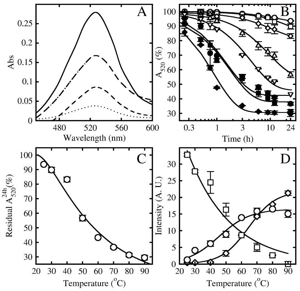
Figure 1. Temperature stability of the burgundy pigment from Pseudofusicoccum adansoniae. (A): absorbance spectra of the crude extract after 24 h of incubation at 25 (solid line), 50 (larger-traced line), 70 (shorter-traced line) and 90 °C (dotted line). (B): isotherms of degradation of the crude extract measured by the decreasing absorbance at 520 nm. Temperatures are 25 (open circles), 30 (open squares), 40 (open diamonds), 50 (triangles up), 60 (triangles down), 70 (closed circles), 80 (closed squares), and 90 °C (closed diamonds). Isotherms were fit into double exponential decay equations (lines) and x axis is in log scale to ease the visualization. (C): residual absorbance at 520 nm of the crude extract after 24 h incubation at the studied temperatures. (D): colorimetric intensity of L* (circles) a* (squares) and b* (diamonds) components of the burgundy pigment present in the crude extract as a function of the 24 h temperature incubation. Lines in (C,D) were drawn merely to guide the eyes. Error bars represent SMD of triplicates.
Applsci 15 08800 g001
Figure 2. Visual appearance of the burgundy pigment crude extract and dilution of 1:18 (A). Visual changes in thermal degradation at 90 °C (B) at different times.
Applsci 15 08800 g002
Figure 3. pH stability of the burgundy pigment in the crude extract produced by Pseudofusicoccum adansoniae. Residual absorbance at 520 nm (A), as well as the colorimetric pattern of L* (circles), a* (squares) and b* (diamonds) components at each pH, after 24 h incubation (B). Lines were drawn merely to guide the eyes. Error bars represent SMD of triplicates.
Applsci 15 08800 g003
Figure 4. Degradation of the burgundy pigment produced by Pseudofusicoccum adansoniae subjected to storage under light or without light, in a refrigerator and freezer. (A): absorbance at 520 nm of pigment extract samples stored for 90 days without light, both in a freezer (open circles) or refrigerator (open triangles), as well as under a light source in the refrigerator (open squares), as described in Materials and Methods. (B): colorimetric analysis of pigment extract samples stored for 90 days in the refrigerator without light (closed symbols) or under light (open symbols), as described in Materials and Methods. The graph displays the a* (squares), b* (diamonds), and L* (circles) color components for both conditions. Error bars represent triplicate SMD.
Applsci 15 08800 g004
Table 1. Residual absorbance of the burgundy pigment (BCR) crude extract produced by Pseudofusicoccum adansoniae subjected to additives and ions. Values are shown as a percentage of the initial value. Errors represent MSD of triplicates.
Additives
(BCR (%)/Standard Deviation)
Time (h)
01224
Control100 ± 0.0695.86 ± 0.0693.59 ± 0.15
Aspartame100 ± 0.4694.86 ± 0.9991.72 ± 1.36
Sodium cyclamate100 ± 1.5695.69 ± 0.8292.38 ± 1.46
Calcium propionate100 ± 1.5295.59 ± 0.9794.40 ± 1.23
Potassium sorbate100 ± 2.0296.05 ± 1.6794.12 ± 1.46
Acesulfame100 ± 1.9587.74 ± 0.9784.50 ± 0.83
Maltitol100 ± 2.2995.81 ± 1.4994.68 ± 1.53
Inulin100 ± 1.2496.19 ± 1.3094.05 ± 1.42
Potassium benzoate100 ± 1.3693.62 ± 2.0691.01 ± 1.78
Ions
(BCR (%)/Standard Deviation)
Control100 ± 0.0695.86 ± 0.0693.59 ± 0.15
Calcium chloride 0.1 M100 ± 1.3994.90 ± 1.5292.42 ± 1.35
Calcium chloride 0.5 M100 ± 1.4197.53 ± 1.3096.17 ± 1.28
Sodium nitrate 0.1 M100 ± 1.7494.33 ± 1.5392.89 ± 1.46
Sodium nitrate 0.5 M100 ± 1.7898.16 ± 0.4196.86 ± 0.50
Magnesium sulfate 0.1 M100 ± 1.1894.81 ± 1.3493.00 ± 1.49
Magnesium sulfate 0.5 M100 ± 0.7494.16 ± 1.7292.85 ± 1.64
Zinc sulfate 0.1 M100 ± 4.1791.92 ± 3.1389.81 ± 2.90
Zinc sulfate 0.5 M100 ± 1.2094.01 ± 0.9793.03 ± 1.01
Disclaimer/Publisher’s Note: The statements, opinions and data contained in all publications are solely those of the individual author(s) and contributor(s) and not of MDPI and/or the editor(s). MDPI and/or the editor(s) disclaim responsibility for any injury to people or property resulting from any ideas, methods, instructions or products referred to in the content.