Article

Magnetic Properties of Commercial Cornflakes

1
Departamento de Ciencias Naturales y Exactas, Centro Universitario de los Valles, Universidad de Guadalajara, Carretera Guadalajara—Ameca Km. 45.5, Ameca 46600, Jalisco, Mexico
2
División de Desarrollo Biotecnológico, Centro Universitario de la Ciénega, Universidad de Guadalajara, Av. Universidad, 1115, Ocotlan 47820, Jalisco, Mexico
3
Departamento de Métodos Cuantitativos, Centro Universitario de Ciencias Económico Administrativas, Universidad de Guadalajara, Periférico Norte 799, Col. Los Belenes, Zapopan 45100, Jalisco, Mexico
4
Division de Ciencias e Ingenierias Campus Leon, Universidad de Guanajuato, Loma del Bosque #103, Lomas del Campestre, Leon 37150, Guanajuato, Mexico
*
Author to whom correspondence should be addressed.
Appl. Sci. 2025, 15(15), 8652; https://doi.org/10.3390/app15158652
Submission received: 10 May 2025 / Revised: 25 July 2025 / Accepted: 2 August 2025 / Published: 5 August 2025
(This article belongs to the Section Food Science and Technology)

Abstract

This study reports on the magnetic properties of commercial cornflakes, which are primarily influenced by the iron content. An initial analysis of X-ray fluorescence on a brand of cornflakes evidenced the presence of a high concentration of Cl and up to 10.9 mg/100 g of Fe. After the extraction of iron from the cornflakes of two different brands, as iron filings, X-ray diffraction measurements indicate the presence of crystals of elemental iron, and no traces of other crystals of iron-derived compounds were found. The Fourier Transform Infrared analysis on the iron filings does not show any binding between iron and oxygen, which further discards the presence of iron oxides. The magnetic hysteresis loops of whole powdered cornflakes exhibit weak Langevin-like magnetizations, which principally correspond to the iron used as a fortification element. The diamagnetic behavior of the higher organic material content significantly attenuates this magnetic response. The hysteresis loops of the iron filings reached magnetic saturations 1% and 5% lower than those of a pure iron sample. Additionally, the indirect measurement of magnetic susceptibility of the iron filings by magneto-thermograms revealed only one Curie transition very close to 771 °C, which corresponds to pure elemental iron.

1. Introduction

Since the last century, food has been fortified to combat global malnutrition. Iron, zinc, and vitamins are the primary elements added to processed foods, which are primarily composed of carbohydrates. Iron is the most essential element in preventing anemias or related diseases caused by a deficit in daily intake or intestinal malabsorption [1,2]. Flour and flour-derived foods, as well as cereals, are fortified to ensure an efficient absorption of essential nutrients when these are lacking in the traditional diet [3].
The most popular cereals, eaten mainly for breakfast in millions of households, are made from toasted rice, oats, wheat, or maize. The type and concentration of iron added to foods depend on the manufacturer’s criteria and the health legislation of each country [4]. Iron may be added in a water-soluble form (ferrous sulfate, gluconate, lactate, or ferric citrate), in a dilute acid-soluble form (ferrous fumarate, succinate), in an insoluble form (different ferric phosphates), or as protected iron compounds (Fe-ethylenediaminetetraacetic acid) [5].
On the other hand, using iron oxides like magnetite, maghemite, or hematite as fortification elements is not entirely standardized. Some side effects have been observed because of their nanometric sizes, which are related to an accumulative effect in the organism and cytotoxic properties [6]. In contrast, several authors have emphasized the feasibility of using iron oxides, such as magnetite, as a fortification complement, for example, in wheat biscuits [7,8,9] or yogurt fortified with maghemite [10]. Other important applications related to the food industry include antimicrobial activity, the production of artificial enzymes, and food preservation [11].
Frequently, it is difficult to identify the type of fortifying iron added to the foods. Also, when it is indicated, the oxidation state of iron may be confusing because it depends on how it was obtained. Thus, metallic iron could be described as elemental, electrolytic, or reduced iron [5]. Other iron-containing food additives are colorants. In particular, the pigment E172 contains three different chemical compounds: yellow Fe2O3(H2O), red Fe2O3, and black Fe3O4 [12,13]. From the perspectives of physical chemistry and biochemistry, the toxicological properties of these colorants have been widely discussed [14,15]. Consequently, the European Food Safety Authority (EFSA) considered the maximum recommended concentrations of E172 for coloring foods, indicating a no observable adverse effect level (NOAEL) of 1 g/kg*d for microsized iron oxides. In contrast, nanosized iron oxides at this concentration exhibited severe toxic symptoms in rats [16].
On the other hand, in 2020, Lermyte et al. analyzed the iron content of a popular brand of cornflakes in the United Kingdom [17]. Using high-precision spectroscopy, the crystal structure was found to correspond to body-centered cubic (BCC) metallic iron (also known as alpha iron), with no detectable traces of iron oxide.
This work aims to determine the magnetic properties of the most popular cereal on the American continent [18] and investigate the presence of oxidized iron. We have observed a lack of information in the nutritional information table displayed for many foods, specifically the omission of specifying the type of iron included.

2. Materials and Methods

2.1. Extraction of Iron

Four packages, each containing 275 g of the most popular brand of cornflakes in Mexico, were bought from different supermarkets to extract as much iron as possible for subsequent physicochemical characterization. The ingredients published on the package are sugar, malt extract, iodized salt, calcium carbonate, ascorbic acid, maltodextrin, iron (11.7 mg/100 g), niacinamide, α-tocopherol acetate, zinc oxide, retinyl palmitate, cobalamin, pyridoxine hydrochloride, thiamine mononitrate, riboflavin, folic acid, vitamin C, vitamin A, and BHT antioxidant. Additionally, the absence of artificial colorants is highlighted. After reading this nutritional information, the question arises about the kind of iron mentioned and whether any magnetic colorant is included.
The iron was extracted under aseptic conditions using a method similar to that described by Lermyte et al. [17]. In total, 1 kg of cornflakes was manually ground with a mortar, and the resulting fine powder was mixed with pure Milli-Q water and homogenized to a soft paste. This paste was transferred separately into five clean nylon bags, and in each bag, the paste was spread out to form a thin layer. Then, neodymium magnets were passed over the bags, whereby the attracted iron attached to their inner walls became visible as small, dark, and wet pellets, which were gently dragged and deposited into Eppendorf tubes. The procedure was repeated until no more iron remained. Finally, all the collected iron was dried at room temperature for 12 h without a controlled atmosphere. At the end of the extraction process, surprisingly, the initially black iron filings changed to a red–brown color (RBIFs). In total, ≈70 mg of RBIFs were collected. But this color change of iron was not reported in [17]; therefore, we conclude that this extraction procedure alters the initial type of iron content in the cornflakes.
Because of the unexpected color change observed in the last step of the previous method, a second extraction method was used to obtain the iron filings. Five packages with 0.275 kg (of the same cornflakes brand) were ground using a domestic electrical grinder at maximum grinding power (350 W) for 30 s. In total, 1.275 kg of powdered cereal was then sifted and transferred into six clean nylon bags, where it was spread out to form thin layers. As in the first method, neodymium magnets were passed over the bags, and the attracted gray iron filings on the inner face of each bag were collected into 50 mL polypropylene tubes. This step was repeated until no more iron remained. In contrast to the first method, these iron filings did not change their color over time, which is more consistent with the findings reported in [17]. The next step was to remove the organic material adhered to the iron using a method that involved a N2 atmosphere. Then, the iron filings were deposited into an Erlenmeyer flask, mixed with 250 mL of ultra-purified water (previously bubbled with N2 gas), and agitation at 200 rev/min was applied for 5 min. The iron filings were decanted using a magnet to remove the supernatant, which was then replaced with 200 mL of fresh water; immediately, agitation was applied again for 10 min. Two additional series of agitation and washings were performed, resulting in clear water that appeared to be free of organic residues. After that, the flask was subjected to sonication for 1 h, maintaining the inert atmosphere throughout. Later, the iron filings were decanted and washed twice. The resultant dark powder was then transferred and dried on absorbent paper while a direct saturant flux of N2 was ventilated. Finally, the dried powder was deposited into two sealed bags using a magnet. Using this second extraction procedure, only ≈110 mg of dark iron filings (DIFs, brand 1) from the first brand of cornflakes were obtained. Furthermore, this extraction procedure was used to extract the contained DIFs from 1 kg of cornflakes from a second brand (DIFs, brand 2), reaching ≈100 mg.

2.2. Experimental Characterizations

Before explaining all the physico-chemical characterization carried out, it is essential to emphasize our specific interest in the properties of DIFs, as the type of iron is identical to that initially present in cornflakes.
X-ray fluorescence (XRF) analysis was performed on both brands of cornflakes. A high-definition XRF spectrometer (XOS, HOS-HD Prime, New York, NY, USA) was used to determine the types and concentrations of metallic elements present in each brand of cornflakes. A sample of 1 g of each brand was compressed, forming thin capsules that were deposited in special sample holders. The spectrometer was programmed to detect all possible elements.
X-ray diffraction (XRD) measurements were conducted using a Grazing Incidence X-Ray Diffractometer (Malvern Panalytical, GIXRD, Wo, Malvern, UK) to determine the crystal structure of the iron in the DIFs. The XRD was operated with Cu Kα (λ = 0.154 nm) radiation, covering the interval 5° < 2θ < 100° with a resolution of 0.2°/step. Two samples of approximately 30 mg of DIFs (one for each brand) were deposited and compacted onto the corresponding sample holders. The XRD patterns were stored on a computer and analyzed using the database software. A third pattern was obtained using a sample of pure iron (99.9%).
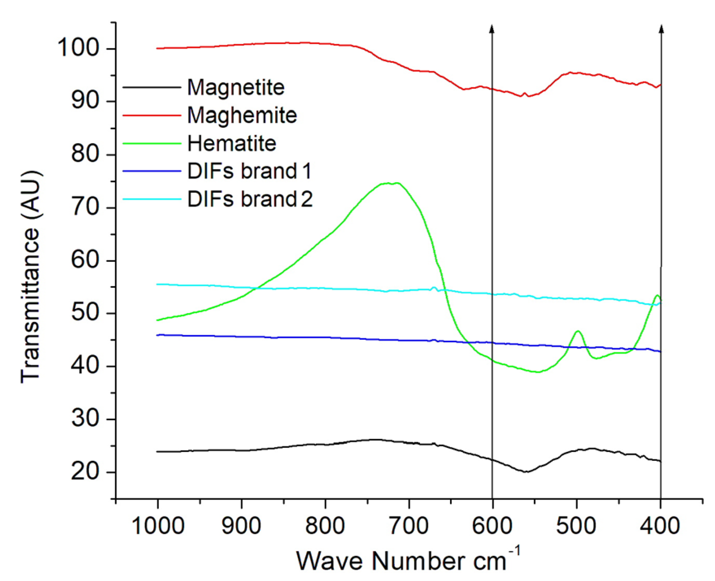
Fourier transform infrared spectroscopy (FTIR) measurements were performed on the DIFs of both brands. A Nicolet IR200 spectrometer (Thermo Fisher Scientific, Waltham, MA, USA) was used. The powders (5 mg) were attached to the surface of KBr windows using tetrahydrofuran; the bands of resonances were analyzed by measuring the transmittance, covering the range of 1000 to 400 cm−1. Other samples of Fe3O4 (Sigma Aldrich CAS 1317-61-9), γ-Fe2O3 (Sigma Aldrich CAS 1309-37-1), α-Fe2O3 (oxidizing γ-Fe2O3 at 600 °C within oxygen flow), and pure iron (Sigma Aldrich CAS 7439-89-6) were also analyzed for use as references.
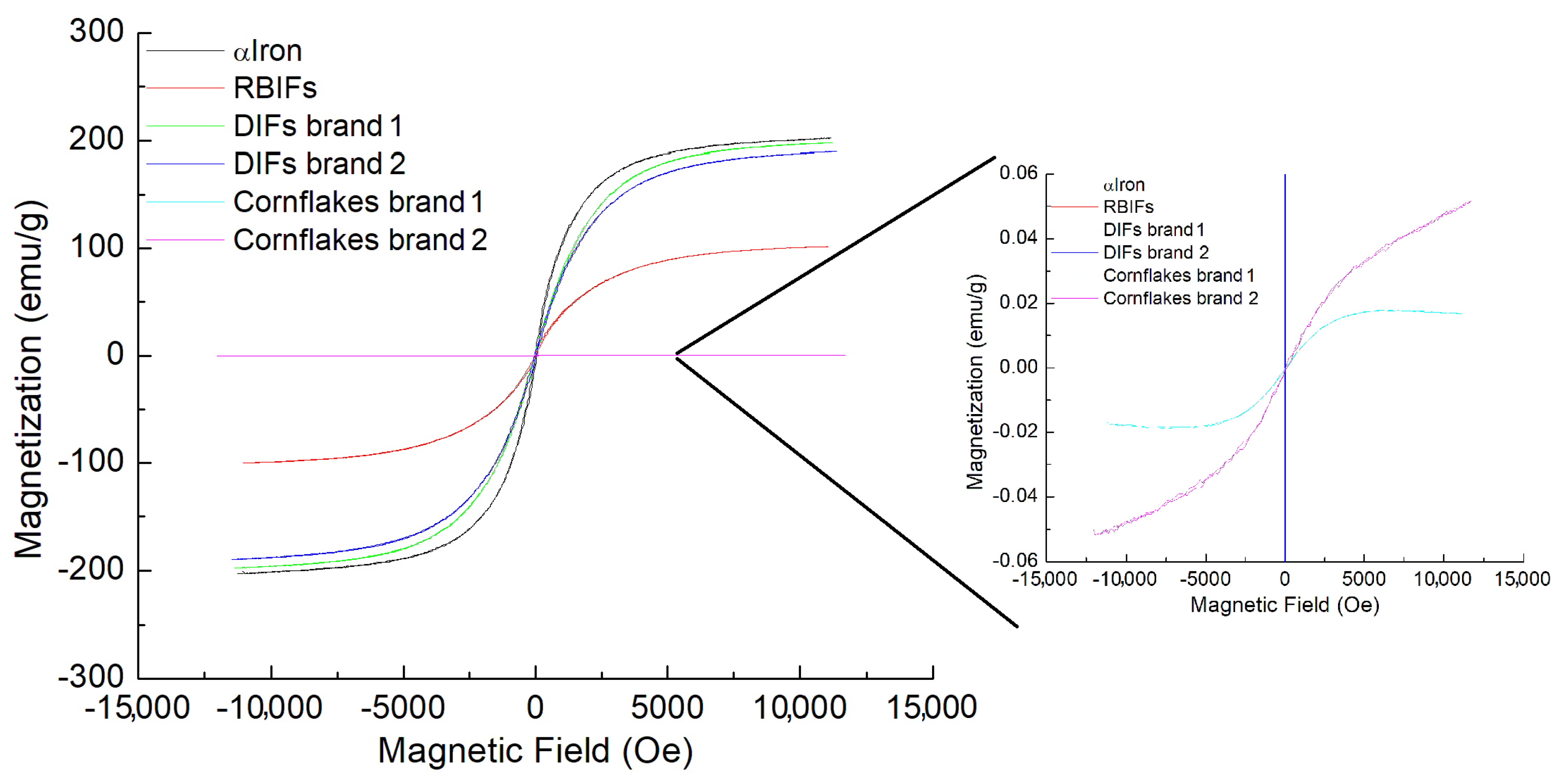
The magnetizations of six samples were measured by determining their hysteresis loop at room temperature, as explained below. Approximately 100 mg of cornflakes brand 1 (sample 1), 100 mg of cornflakes brand 2 (sample 2), 20 mg of DIFs brand 1 (sample 3), 20 mg of DIFs brand 2 (sample 4), 20 mg of RBIFs (sample 5), respectively, were placed into diamagnetic polypropylene sample holders of a vibrating sample magnetometer (VSM 9500, LDJ Electronics, Troy, MI, USA) and a magnetic field H in the range of −12 < H < +12 kOe, with a resolution of 50 μemu was applied. The measurements were compared with the magnetization obtained from a pure iron sample.
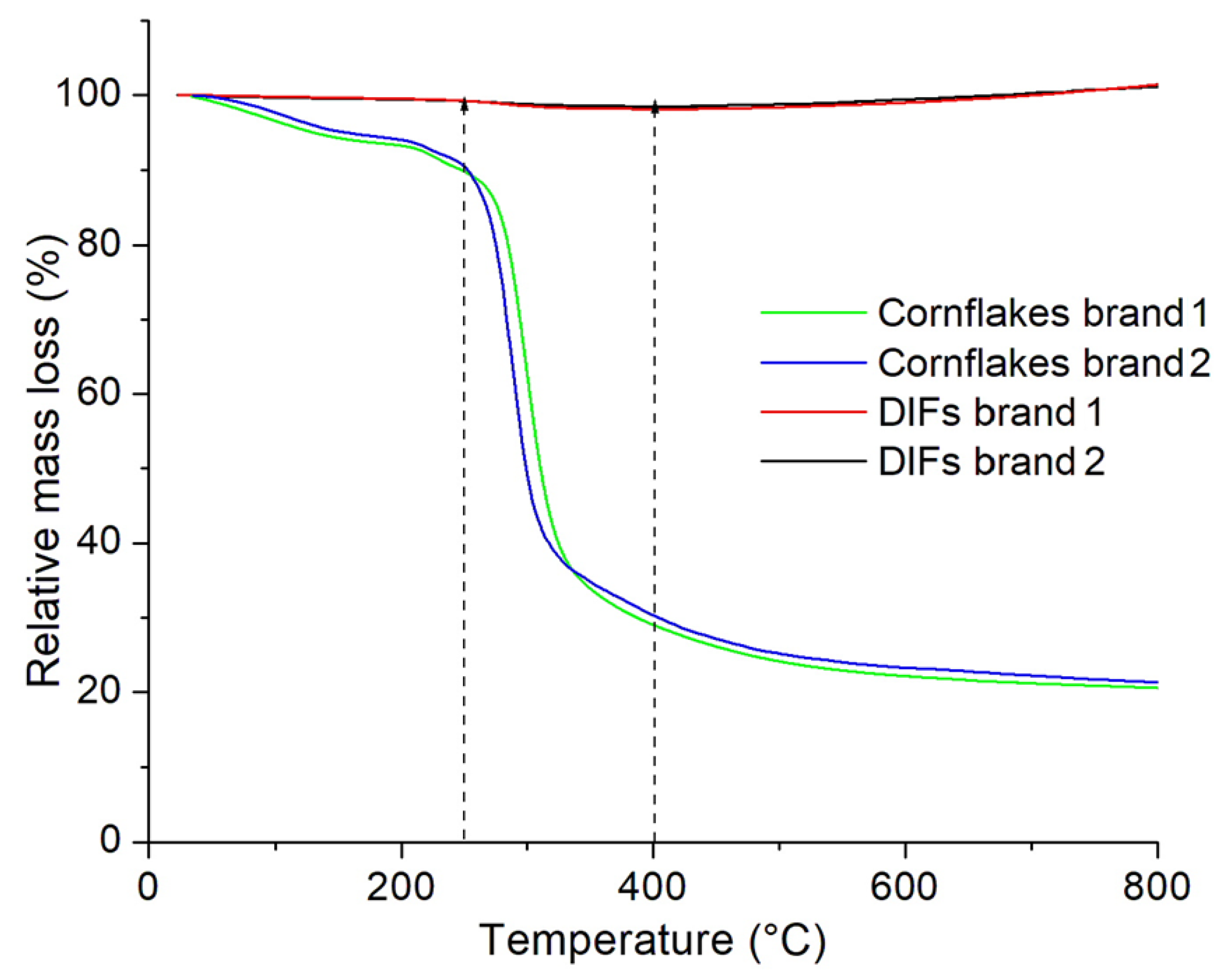
Thermal gravimetric analysis (TGA) was performed on the two same samples of DIFs measured by VSM to determine if any residual material was attached to the surface of the iron. The DIFs were deposited onto a platinum sample holder of a thermogravimetric analyzer (TGA 1000, ISI, Twin Lakes, WI, USA), and a heating rate of 10 K/min was applied under an inert N2 atmosphere. Additionally, two more thermograms were obtained, this time of the entire cornflakes of both brands analyzed.
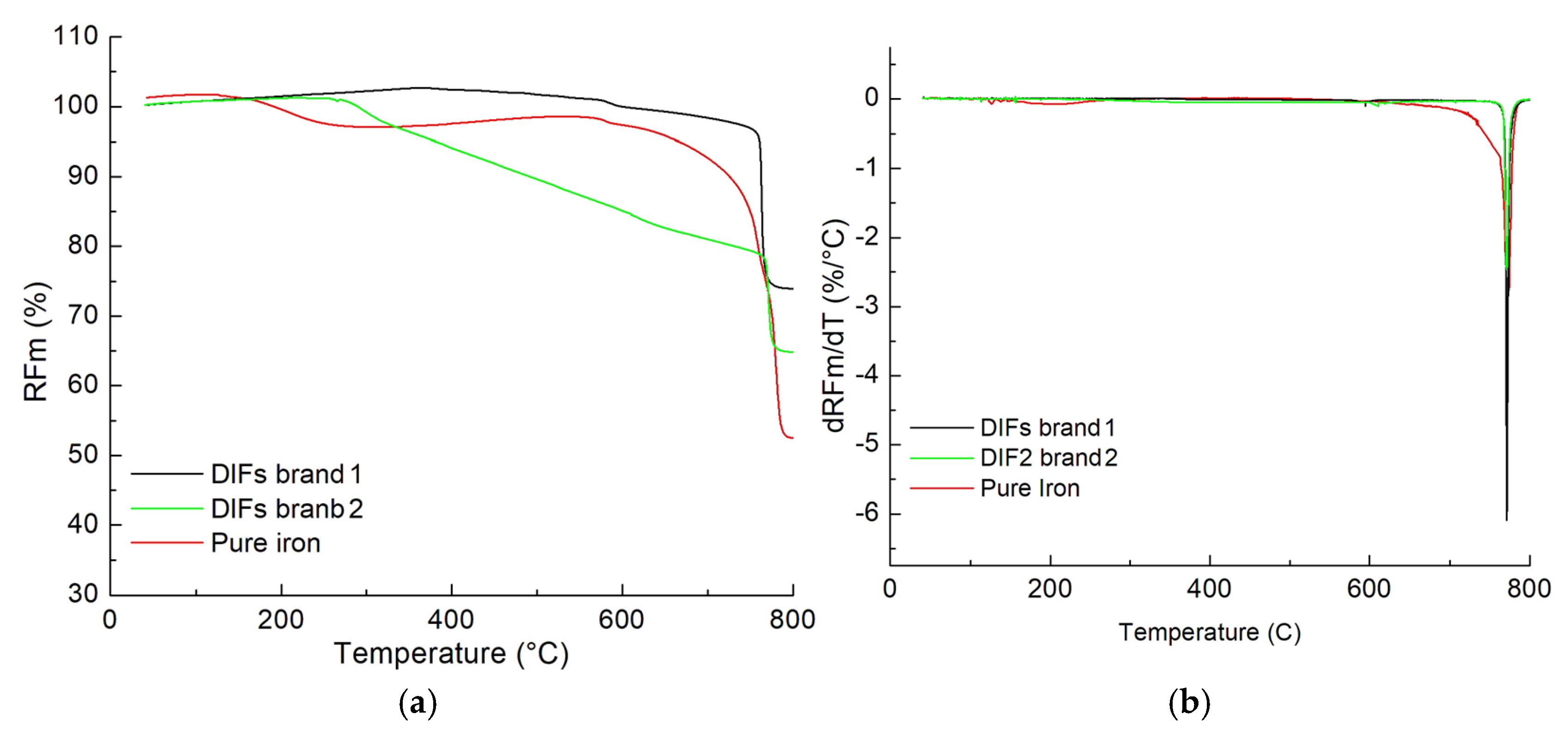
The Curie temperatures of both types of DIFs extracted were determined using the TGA, complemented by a magnetic accessory constructed in-house. An additional measurement of the Curie temperature of a standard sample of pure iron was performed. A similar setup was previously described by Lisenko et al. [19]. The magnetic interaction force (Fm) between the sample and the external magnetic field (H) was registered by the balance of the TGA. This force, Fm, is directly proportional to the magnetization of the sample, which in turn depends on its magnetic susceptibility (χ). By heating the sample, the parameter χ of the sample itself diminishes. This type of measurement is also known as a magneto-thermogram [9]. It is crucial to note that, ideally, all changes registered by the balance are associated with the “fictitious weight”, i.e., changes in the magnetization of the sample, and not changes in the mass of the sample, e.g., by evaporation of associated water or decomposition of residual organic material. The samples (5 mg) were deposited onto the TGA sample holder and magnetized in a magnetic field of 200 Oe. Then, the magneto-thermograms of the samples DIFs (of both brands) were registered, encompassing the temperature interval of 30 °C < T < 800 °C.

3. Results

3.1. The XRF Spectra

In the measurement of the elemental composition of cornflakes (only brand 1), a total of 37 elements were observed. Table 1 summarizes the elements with higher concentrations, expressed in mg/100 g units. As can be observed, Cl is the element most commonly detected, with a concentration of 271.0 mg/100 g, while the concentration of Fe is 10.9 mg/100 g. The presence of Cl and K is related to the salts used to increase the flavor. It might be interesting to try to understand the origin of the high concentration of Ba observed.

3.2. XRD Spectrum

The XRD patterns (with a copper source) of both types of DIFs and that of the reference sample are displayed in Figure 1. All the patterns are completely collapsed and are characterized by four peaks at 2θ = 44.7°, 65.2°, 82.3°, and 98.9°, corresponding to the ferromagnetic BCC metallic iron (α-iron) [17,20]. According to the Bragg law and the JCPDS/ICDD card 06-0696, the corresponding crystallographic planes are (110), (200), (211), and (220), respectively. The absence of other peaks below 45° suggests the lack of the most common iron oxides, such as hematite (α-Fe2O3), maghemite (γ-Fe2O3), or magnetite (Fe3O4) [21]. Those spectra are concordant with the one reported by Lermyte et al. [17]. Additionally, the main peak (2θ = 28.6°) associated with the crystals of Ba does not appear in the pattern. Nevertheless, other amorphous iron oxide species used to fortify aliments could be present in the measured sample, such as Fe(OH)3 and Fe5O8 · 4H2O, which are undetectable by XRD.

3.3. The FTIR Spectra

The FTIR patterns of the typical iron oxides, magnetite, hematite, and maghemite, are used as reference signals to compare with the patterns obtained from both DIFs extracted. The typical resonance bands corresponding to the Fe-O binding in the interval 1000 to 400 cm−1 are evidenced in Figure 2, which are associated with the ferromagnetic or antiferromagnetic behavior of the iron oxides. In contrast, no IR resonant signals are observed when the samples with both DIFs are analyzed; in those two patterns, only a monotone decrease can be highlighted in the interval from 600 to 400 cm−1. Then, no oxidation of Fe occurs during the second extraction procedure, and the presence of other amorphous iron ions can also be discarded. Nevertheless, some organic materials may remain attached to the iron contained in the DIFs.

3.4. Magnetization Loops

The magnetization of both brands of cornflakes exhibits a Langevin-like behavior typical of paramagnetic materials, characterized by low intensity and no trend towards magnetic saturation (Inset of Figure 3, cyan and pink lines). However, most of the mass of cornflakes is diamagnetic, which tends to silence the magnetization. The small concentrations of iron (see Table 1) are sufficient to allow a paramagnetic-like behavior with a very weak hysteresis loop, which requires further analysis, including measurement of the DIFs.
The magnetization of the DIFs from brands 1 and 2 (green line and blue line, respectively) reveals magnetic saturation at 198 and 190 emu/g, respectively, with a coercivity of 20 Oe. Additionally, the saturation of the sample of pure iron reached 202 emu/g. This indicates that there are differences ranging from 2% to 6% between the saturation of the DIFs and that of pure iron. However, the saturation of ultrapure bulk α-iron is 217.4 emu/g [22], which is much higher than the saturations of magnetite (92 emu/g) and maghemite (72 emu/g) [23,24]. In both cases, the DIFs reach a similar saturation to that reported by Lermyte et al. [17] (196 emu/g). However, a thermogravimetric analysis is necessary to determine if some organic material (for example, starch or glucose) is attached to the DIFs. On the other hand, the RBIFs exhibit a relatively diminished saturation (red line, 100.0 emu/g), which is associated with the iron oxidation induced in the first extraction procedure.

3.5. Thermograms

In the thermogravimetric analysis (TGA), the changes in the relative mass (RM) of the samples, either by evaporation, sublimation, or thermal decomposition, by raising the temperature to 800 °C, can be determined. In the case of the DIFs measured by VSM (depicted in Figure 3), RML = 1.5% was the lowest relative mass loss detected (Figure 4, red and black lines), while for the cornflakes of both brands, approximately RML = 79% was determined. The cornflakes reached two different thermal transition points. The first weak transition point occurs within the temperature interval of 30–250 °C. It is associated with the humidity induced by the environment (0.5% evaporation up to 150 °C), as well as the decomposition of sugars and the evaporation of residual moisture in the samples (1% of decomposition between 200 °C and 150 °C). The second strong transition point occurs within the 250–400 °C temperature range. This slope corresponds to the thermolysis of starch [25]. Using the determined RML value, the magnetic saturations of DIFs for brand 1 and brand 2, as displayed in Figure 3, increased to 200.1 and 192.9 emu/g, respectively. These two magnitudes are closer to the reference sample with pure iron.

3.6. Magneto-Thermograms

The magneto-thermograms for both types of DIFs and the pure iron sample were recorded within a 30–800 °C interval, and the curves are shown in Figure 5a. The DIFs of brand 1 exhibit only one rapid inflection point, similar to a step, in the RFm below 700 °C. The DIFs of brand 2 also exhibit only a rapid inflection point below 700 °C, which is shorter than that of the previous step. The reference sample of pure iron also exhibits an initial decrease below 300 °C and a fast, longer inflection point below 700 °C. The critical points were calculated by analyzing the derivative of each RFm, and the corresponding plots are displayed in Figure 5b. The peaks obtained are 770.2 °C (DIFs brand 1), 770.9 °C (DIFs brand 2), and 772 °C (pure iron). These points correspond to the Curie temperature of metallic alpha iron [26,27], which is approximately 771 °C. Remarkably, there is an absence of critical points below 700 °C, which could usually be associated with traces of some iron oxides.

4. Discussion

It is not easy to determine the origin of the high concentration of Ba in the cornflakes, as determined by XRF, which is 32.8 mg/100 g. Using this spectrometry, we cannot determine the presence of anions, cations, or pure Ba in the cornflakes. Nevertheless, in this context, several scientific reports have analyzed the fatal toxic effects of Ba compounds [28,29,30]. Probably, the origin of this element in cornflakes could be associated with the cultivation process of corn, the nutrients in the soil, and the use of pesticides [31].
On the other hand, according to the XRF and Table 1, the concentration of Fe was 7% lower than the content specified in the corresponding packaging. After analyzing the DIFs using XRD, no traces of any magnetic crystalline material other than iron were found. The type of iron crystals corresponds to a BCC structure, which is associated with α-iron, and no crystals of any iron oxide were found.
Using the FTIR spectra, the presence of iron oxides (crystal or amorphous materials) can also be discarded because of the absence of Fe-O bonds. Regarding our RBIFs, a significant fraction of alpha iron was probably oxidized during the first extraction procedure and subsequent storage. This would also explain the color differences between the DIFs and RBIFs. When the first extraction method was employed, the elemental iron was highly oxidized, resulting in the probable formation of hematite in RBIFs. This hypothesis is reinforced because the water solubilizes the content of NaCl and/or KCl, which work as electrolytes and promote the reaction of Equation (1) [32], on the surface of the metallic iron in the presence of O2. Probably, the concentration of NaCl or KCl in the cornflakes analyzed in [17] was not sufficient to reach a fast oxidation of the iron extracted.
4 F e + 6 H 2 O + 3 O 2 4 F e ( O H ) 3 2 F e 2 O 3 + 6 H 2 O
When the second method to extract the DIFs was applied, exhaustive washing of the iron filings in a controlled inert atmosphere allowed us to avoid oxidation of the iron and remove 99% of the organic and inorganic material attached to its surface. This is confirmed with the lack of resonances in the interval of 600 to 400 cm−1 observed in the FTIR spectra [33]. Nevertheless, the RML and decomposition temperatures observed in the TGA measurements suggest the presence of 1% starch attached to the DIFs.
The XRD and FTIR analyses using the respective reference samples suggest that the DIFs are composed principally of pure α-iron. Additionally, the magnetic hysteresis loops confirm the ferromagnetic behavior with magnetic saturation very close to the reference sample of iron with 99.9% purity. For example, the magnetic saturation of the DIFs extracted from the brand 1 is 1% lower than that of the reference sample. Additionally, the Curie temperatures reached very close to 771 °C, together with the lack of other magnetic critical points associated with iron oxides, support this statement.
It is crucial to discard the presence of ion oxides in fortified foods. For example, for decades, the cornflakes have been a leading source of fast food on the American continent. Millions of people, mainly children, eat breakfast of fortified cereal with milk every morning, unaware that the cereals contain a non-standardized type of iron, which must be widely studied for its interactions within the human body. It is well known that iron oxide particles can induce a high production of ROS in vitro experiments. It was observed using cell lines of the digestive tract, such as CACO-2 [34] or HT29 [35]. The ROS are directly related to apoptotic activity in cells, but uncontrolled oxidation can induce noxious effects on health.

5. Conclusions

The methodology described in this work is suitable for analyzing mixtures of organic materials with iron or various iron oxides. Nevertheless, we have focused our application on fortified cereals, as they represent an attractive physical system. Other reasons include the lack of specifications in the packaging regarding the real content of elements. For instance, XRF analysis revealed a high concentration of Ba, yet it is not specified in the analyzed cornflakes. Further investigations are needed to determine the role of Ba in cornflakes and its potential health effects.
Furthermore, determining iron oxides in fortified cereals is crucial because iron can undergo significant changes depending on its immediate environment. The conversion from iron to iron oxide could be a delicate issue, given the actual lack of regulation on the concentration of iron oxides permitted for human consumption and the deeply ingrained culture of consuming fortified foods worldwide.
Following the sequence of experiments of XRD and FTIR, it was possible to discard the presence of iron oxides, whether crystalline or amorphous phases. Subsequent experiments on the magnetic properties of DIFs, conducted at room temperature and under a magnetic field, confirmed that the iron content exhibits magnetic saturation, approaching that of the pure iron reference sample. Finally, when DIFs are submitted to a static field and exposed to a wide range of temperatures, the measured Curie temperatures confirm the ferromagnetic BCC structure of α-iron.

Author Contributions

Conceptualization, L.H.Q.; methodology, J.A.P.; validation, C.A.V.; formal analysis, F.A.C.; investigation A.A.O.; writing—original draft preparation, P.K.; writing—review and editing, Z.L.; data curation, J.d.J.B.; supervision, M.E.C. All authors have read and agreed to the published version of the manuscript.

Funding

This research received no external funding.

Data Availability Statement

Data are available from the corresponding author upon request.

Acknowledgments

The authors thank the Mexican institution CONAHCYT for the scholarships for undergraduate and graduate students and their financial support.

Conflicts of Interest

The authors declare that they have no conflicts of interest.

Abbreviations

The following abbreviations are used in this manuscript:
EFSAThe European Food Safety Authority
NOAELNo observable adverse effect level
DIFsDark iron filings
BCCBody-centered cubic
RBIFsRed/brown iron filings

References

  1. Latunde-Dada, G.O. Iron Intake and Human Health. Nutrients 2024, 16, 206. [Google Scholar] [CrossRef] [PubMed]
  2. McMillen, S.A.; Dean, R.; Dihardja, E.; Ji, P.; Lönnerdal, B. Benefits and risks of early life iron supplementation. Nutrients 2022, 14, 4380. [Google Scholar] [CrossRef] [PubMed]
  3. Hurrell, R.F. Ensuring the efficacious iron fortification of foods: A tale of two barriers. Nutrients 2022, 14, 1609. [Google Scholar] [CrossRef] [PubMed]
  4. Allen, L.; De Benoist, B.; Dairy, O.; Hurrel, R. Guidelines on Food Fortification with Micronutrients. World Health Organization, 2006. Available online: https://citeseerx.ist.psu.edu/document?repid=rep1&type=pdf&doi=bbb90acf54fdd30ed9ebfc018b298e3515aefdca (accessed on 5 October 2024).
  5. Lee, K.; Clydesdale, F.M.; Tannenbaum, S.R. Iron sources used in food fortification and their changes due to food processing. Crit. Rev. Food Sci. Nutr. 1979, 11, 117–153. [Google Scholar] [CrossRef]
  6. Patil, U.S.; Adireddy, S.; Jaiswal, A.; Mandava, S.; Lee, B.R.; Chrisey, D.B. In vitro/in vivo toxicity evaluation and quantification of iron oxide nanoparticles. Int. J. Mol. Sci. 2015, 16, 24417–24450. [Google Scholar] [CrossRef]
  7. Sakhare, D.T. Green Synthesis, Characterization And Antimicrobial Activity of Iron Nanoparticles Using Hibiscus Leaf Extract. SN Appl. Sci. 2020, 2, 898. [Google Scholar] [CrossRef]
  8. Osman, A.G.; El-Desouky, A.I.; Morsy, M.K.; Aboud, A.A.; Mohamed, M.H. Production and evaluation of fortified biscuit with iron nanoparticles for anemia as functional food. Egypt. J. Chem. 2022, 65, 181–194. [Google Scholar] [CrossRef]
  9. Salaheldin, T.A.; Regheb, E.M. In-Vivo Nutritional and Toxicological Evaluation of Nano Iron Fortified Biscuits as Food Supplement for Iron Deficient Anemia. J. Nanomed. Res. 2016, 3, 00049. [Google Scholar] [CrossRef]
  10. Garcés, V.; González, A.; Sabio, L.; Sánchez-Arévalo, C.M.; Gálvez, N.; Dominguez-Vera, J.M. Magnetic and golden yogurts. Foods a potential nanomedicine carrier. Materials 2020, 13, 481. [Google Scholar] [CrossRef]
  11. Góral, D.; Marczuk, A.; Góral-Kowalczyk, M.; Koval, I.; Andrejko, D. Application of iron nanoparticle-based materials in the food industry. Materials 2023, 16, 780. [Google Scholar] [CrossRef]
  12. Sieg, H.; Schaar, C.; Fouquet, N.; Böhmert, L.; Thünemann, A.F.; Braeuning, A. Particulate iron oxide food colourants (E 172) during artificial digestion and their uptake and impact on intestinal cells. Toxicol. Vitr. 2024, 96, 105772. [Google Scholar] [CrossRef]
  13. Voss, L.; Hsiao, I.L.; Ebisch, M.; Vidmar, J.; Dreiack, N.; Böhmert, L.; Stock, V.; Braeuning, A.; Loeschner, K.; Laux, P.; et al. The presence of iron oxide nanoparticles in the food pigment E172. Food Chem. 2020, 327, 127000. [Google Scholar] [CrossRef]
  14. Askri, D.; Ouni, S.; Galai, S.; Chovelon, B.; Arnaud, J.; Sturm, N.; Lehmann, S.G.; Sakly, M.; Amara, S.; Sève, M. Nanoparticles in foods? A multiscale physiopathological investigation of iron oxide nanoparticle effects on rats after an acute oral exposure: Trace element biodistribution and cognitive capacities. Food Chem. Toxicol. 2019, 127, 173–181. [Google Scholar] [CrossRef]
  15. Kumari, A.; Chauhan, A.K. Iron nanoparticles as a promising compound for food fortification in iron deficiency anemia: A review. J. Food Sci. Technol. 2022, 59, 3319–3335. [Google Scholar] [CrossRef]
  16. EFSA Panel on Food Additives and Nutrient Sources Added to Food (ANS). Scientific Opinion on the re-evaluation of iron oxides and hydroxides (E 172) as food additives. EFSA J. 2015, 13, 4317. [Google Scholar] [CrossRef]
  17. Lermyte, F.; Zhang, W.Y.; Brooks, J.; Huband, S.; Collingwood, J.F.; Lees, M.R.; Rayman, M.P.; Sadler, P.J. Metallic iron in cornflakes. Food Funct. 2020, 11, 2938–2942. [Google Scholar] [CrossRef] [PubMed]
  18. Myers, P.; Parcell, L.M. Beauty and the Bran: Kellogg’s Campaign to “Correct Faulty Elimination” and Conquer the Cereal Industry. J. Hist. 2022, 48, 324–348. [Google Scholar] [CrossRef]
  19. Lysenko, E.; Surzhikov, A.; Nikolaev, E.; Starý, O. Curie temperature control of magnetic materials using thermogravimetric measurements in a magnetic field. In Recent Developments in the Field of Non-Destructive Testing, Safety and Materials Science, Proceedings of the ICMTNT 2021, Tomsk, Russia, 9–11 November 2021; Studies in Systems, Decision and Control; Springer: Cham, Switzerland, 2021; Volume 433, pp. 195–203. [Google Scholar] [CrossRef]
  20. Hull, A.W. A New Method of X-Ray Crystal Analysis. Phys. Rev. 1917, 10, 661–696. [Google Scholar] [CrossRef]
  21. Sun, J.; Zhou, S.; Hou, P.; Yang, Y.; Weng, J.; Li, X.; Li, M. Synthesis and characterization of biocompatible Fe3O4 nanoparticles. J. Biomed. Mater. Res. Part A 2007, 80, 333–341. [Google Scholar] [CrossRef]
  22. Graham, C.D. Iron and nickel as magnetization standards. J. Appl. Phys. 1982, 53, 2032–2034. [Google Scholar] [CrossRef]
  23. Rebodos, R.L.; Vikesland, P.J. Effects of oxidation on the magnetization of nanoparticulate magnetite. Langmuir 2010, 26, 16745–16753. [Google Scholar] [CrossRef]
  24. Millan, A.; Urtizberea, A.; Silva, N.J.; Palacio, F.; Amaral, V.S.; Snoeck, E.; Serin, V. Surface effects in maghemite nanoparticles. J. Magn. Magn. Mater. 2007, 312, L5–L9. [Google Scholar] [CrossRef]
  25. Liu, X.; Yu, L.; Liu, H.; Chen, L.; Li, L. Thermal decomposition of corn starch with different amylose/amylopectin ratios in open and sealed systems. Cereal Chem. 2009, 86, 383–385. [Google Scholar] [CrossRef]
  26. Chakraborty, A. Kinetics of the reduction of hematite to magnetite near its Curie transition. J. Magn. Magn. Mater. 1999, 204, 57–60. [Google Scholar] [CrossRef]
  27. Laughlin, D.E. The β iron controversy revisited. J. Phase Equilibria Diffus. 2018, 39, 274–279. [Google Scholar] [CrossRef]
  28. Copeland, C.S.; Rock, K.L.; Pinhal, A.; Chapman, R.C.; Chilcott, R.P. A fatal case report of barium chloride toxicity. J. Anal. Toxicol. 2023, 47, e33–e41. [Google Scholar] [CrossRef]
  29. Johnson, C.H.; Van Tassell, V.J. Acute barium poisoning with respiratory failure and rhabdomyolysis. Ann. Emerg. Med. 1991, 20, 1138–1142. [Google Scholar] [CrossRef]
  30. Boyd, E.M.; Abel, M. The acute toxicity of barium sulfate administered intragastrically. Can. Med. Assoc. J. 1966, 94, 849–853. [Google Scholar]
  31. Aziz, H.A.; Ghazali, M.F.; Hung, Y.T.; Wang, L.K. Toxicity, source, and control of barium in the environment. In Handbook of Advanced Industrial and Hazardous Wastes Management; CRC Press: Boca Raton, FL, USA, 2017; pp. 463–482. [Google Scholar] [CrossRef]
  32. Leeuw, N.H.; Cooper, T.G. Surface simulation studies of the hydration of white rust Fe (OH) 2, goethite α-FeO (OH) and hematite α-Fe2O3. Geochim. Cosmochim. Acta 2007, 71, 1655–1673. [Google Scholar] [CrossRef]
  33. Nasrazadani, S.; Raman, A. The application of infrared spectroscopy to the study of rust systems—II. Study of cation deficiency in magnetite (Fe3O4) produced during its transformation to maghemite (γ-Fe2O3) and hematite (α-Fe2O3). Corros. Sci. 1993, 34, 1355–1365. [Google Scholar] [CrossRef]
  34. Voss, L.; Hoché, E.; Stock, V.; Böhmert, L.; Braeuning, A.; Thünemann, A.F.; Sieg, H. Intestinal and hepatic effects of iron oxide nanoparticles. Arch. Toxicol. 2021, 95, 895–905. [Google Scholar] [CrossRef] [PubMed]
  35. Ranjbary, A.G.; Saleh, G.K.; Azimi, M.; Karimian, F.; Mehrzad, J.; Zohdi, J. Superparamagnetic Iron Oxide Nanoparticles Induce Apoptosis in HT-29 Cells by Stimulating Oxidative Stress and Damaging DNA. Biol. Trace Elem. Res. 2023, 201, 1163–1173. [Google Scholar] [CrossRef] [PubMed]
Figure 1. XRD patterns of DIFs brand 1 (black line), DIFs brand 2 (red line), and 99.9% pure iron (green line).
Applsci 15 08652 g001
Figure 2. FTIR spectra of DIFs brand 1 (blue line), DIFs brand 2 (cyan line), and other reference iron oxides, including magnetite (dark line), maghemite (red line), and hematite (green line).
Applsci 15 08652 g002
Figure 3. Magnetization loops of samples of cornflakes brands 1 and 2 (cyan and pink lines), DIFs of brands 1 and 2 (green and blue lines), RBIFs (red line), and pure iron (dark line).
Applsci 15 08652 g003
Figure 4. Relative mass loss (RML) of cornflakes and DIFs of brand 1 and brand 2 over the interval of 30–800 °C.
Applsci 15 08652 g004
Figure 5. (a) Magneto-thermograms of both types of DIFs and pure iron samples, and (b) the corresponding derivatives of the relative magnetic forces.
Applsci 15 08652 g005
Table 1. Principal elements composing the cornflakes.
ElementConcentration (mg/100 g)
Cl271.0
K117.8
P92.4
Ca44.2
S41.3
Fe10.9
Ba3.28
Zn0.46
Disclaimer/Publisher’s Note: The statements, opinions and data contained in all publications are solely those of the individual author(s) and contributor(s) and not of MDPI and/or the editor(s). MDPI and/or the editor(s) disclaim responsibility for any injury to people or property resulting from any ideas, methods, instructions or products referred to in the content.