Review

The Potential Therapeutic Role of Bruton Tyrosine Kinase Inhibition in Neurodegenerative Diseases

by
Francesco D’Egidio
1,
Housem Kacem
1,
Giorgia Lombardozzi
1,
Michele d’Angelo
1,2,*,
Annamaria Cimini
1,2 and
Vanessa Castelli
1,*
1
Department of Life, Health and Environmental Sciences, University of L’Aquila, 67100 L’Aquila, Italy
2
Sbarro Institute for Cancer Research and Molecular Medicine, Temple University, Philadelphia, PA 19122, USA
*
Authors to whom correspondence should be addressed.
Appl. Sci. 2025, 15(15), 8239; https://doi.org/10.3390/app15158239
Submission received: 21 June 2025 / Revised: 14 July 2025 / Accepted: 22 July 2025 / Published: 24 July 2025
(This article belongs to the Section Applied Biosciences and Bioengineering)

Abstract

Bruton Tyrosine Kinase (BTK) has emerged as a critical mediator in the pathophysiology of neuroinflammation associated with neurodegenerative diseases. BTK, a non-receptor tyrosine kinase predominantly expressed in cells of the hematopoietic lineage, modulates B-cell receptor signaling and innate immune responses, including microglial activation. Recent evidence implicates aberrant BTK signaling in the exacerbation of neuroinflammatory cascades contributing to neuronal damage in disorders such as Alzheimer’s disease, Parkinson’s disease, multiple sclerosis, ischemic stroke, and Huntington’s disease. Pharmacological inhibition of BTK has shown promise in attenuating microglial-mediated neurotoxicity, reducing pro-inflammatory cytokine release, and promoting neuroprotection in preclinical models. BTK inhibitors, originally developed for hematological malignancies, demonstrate favorable blood–brain barrier penetration and immunomodulatory effects relevant to central nervous system pathology. This therapeutic approach may counteract detrimental neuroimmune interactions without broadly suppressing systemic immunity, thus preserving host defense. Ongoing clinical trials are evaluating the safety and efficacy of BTK inhibitors in patients with neurodegenerative conditions, with preliminary results indicating potential benefits in slowing disease progression and improving neurological outcomes. This review consolidates current knowledge on BTK signaling in neurodegeneration and highlights the rationale for BTK inhibition as a novel, targeted therapeutic strategy to modulate neuroinflammation and mitigate neurodegenerative processes.

1. Introduction

Tyrosine kinases constitute a large family of enzymes that catalyze the phosphorylation of tyrosine residues on target proteins, serving as pivotal regulators of intracellular signaling pathways. Tyrosine kinases can be divided into receptor tyrosine kinases, which promote transmembrane signaling by auto-phosphorylating at the cell membrane level responding to extracellular ligands, and non-receptor or cytoplasmic tyrosine kinases, which mediate downstream signaling cascades intracellularly [1]. Tyrosine phosphorylation regulates numerous cellular processes including proliferation, differentiation, migration, survival, and immune responses. Dysregulation of tyrosine kinase activity has been implicated in various pathological conditions such as cancer, immune disorders, and neurodegenerative diseases [1,2,3]. Hence, tyrosine kinases have become attractive therapeutic targets, with numerous inhibitors already approved for clinical use in oncology and inflammatory diseases [4,5]. Among the non-receptor tyrosine kinases, Bruton Tyrosine Kinase (BTK) occupies a critical role in hematopoietic signaling networks [6,7,8]. BTK is predominantly expressed in B cells, myeloid cells, and platelets, where it transduces signals from the B-cell receptor (BCR), Fc receptors, and toll-like receptors (TLRs), thereby regulating cellular activation, proliferation, differentiation, and survival [6]. Germline mutations in the BTK gene cause X-linked agammaglobulinemia (XLA), a primary immunodeficiency characterized by profound B-cell developmental arrest and consequent antibody deficiency, underscoring BTK’s essential role in adaptive immunity [9]. While BTK’s canonical functions have been extensively studied in hematopoietic cells, growing evidence has expanded its relevance to neuroimmune interactions, particularly in the context of neurodegenerative diseases. Conditions such as Alzheimer’s disease (AD), Parkinson’s disease (PD), multiple sclerosis (MS), ischemic stroke (IS), and Huntington’s disease are characterized by progressive loss of neuronal structure and function, leading to cognitive and motor impairments [10]. The common thread across these conditions is neuroinflammation, primarily driven by the activation of resident immune cells in the central nervous system (CNS), especially microglia, and the eventual infiltration of peripheral immune cells. Microglia are the CNS’s innate immune sentinels, playing dual roles in neuroprotection and neurotoxicity depending on their activation state [11]. Dysregulated microglial activation contributes to sustained neuroinflammatory responses and the release of pro-inflammatory cytokines, reactive oxygen species (ROS), and other neurotoxic mediators, thereby exacerbating neuronal damage. Recent studies have demonstrated that BTK is expressed in microglia and myeloid-derived cells within the CNS, where it modulates signaling pathways triggered by TLRs and Fc receptors, key receptors involved in innate immune activation [12,13]. BTK activation in microglia promotes inflammatory signaling cascades including NF-κB and inflammasome pathways, which amplify neuroinflammatory responses [2]. This mechanistic insight has fueled interest in targeting BTK to modulate neuroinflammation in neurodegenerative disorders. Pharmacological BTK inhibitors (BTKis), originally developed for B-cell malignancies such as chronic lymphocytic leukemia and mantle cell lymphoma, exhibit potent immunomodulatory properties but also the ability to cross the blood–brain barrier (BBB), exerting modulatory effects over the CNS-resident immune cells [14]. Preclinical models of neurodegenerative diseases treated with BTKis have shown reductions in microglial activation, decreased production of pro-inflammatory mediators, and attenuation of neurodegeneration, suggesting neuroprotective potential [12,13]. Moreover, targeting BTK may circumvent some limitations of broad-spectrum immunosuppressive therapies by selectively modulating pathogenic immune pathways while preserving essential host defense mechanisms [15]. This feature may translate into improved safety profiles and tolerability, crucial for chronic neurodegenerative diseases requiring long-term treatment. Current clinical trials are actively evaluating BTKis in neurodegenerative indications, assessing their efficacy, safety, and impact on clinical outcomes [5]. Thus, tyrosine kinases represent critical regulators of cellular signaling with extensive implications in health and disease. Among them, Bruton Tyrosine Kinase has emerged as a pivotal mediator of neuroimmune crosstalk, particularly in microglial activation and neuroinflammation, processes central to the pathogenesis of neurodegenerative diseases. Considering the therapeutic potential of the pharmacological inhibition of BTK in modulating detrimental immune responses within the CNS, the aim of this review article is to provide a comprehensive overview of BTK signaling pathways in neuroinflammation, critically examine preclinical and clinical evidence supporting BTK inhibition in various neurodegenerative diseases, and discuss current challenges and future directions for its translation into effective neurotherapeutic strategies.

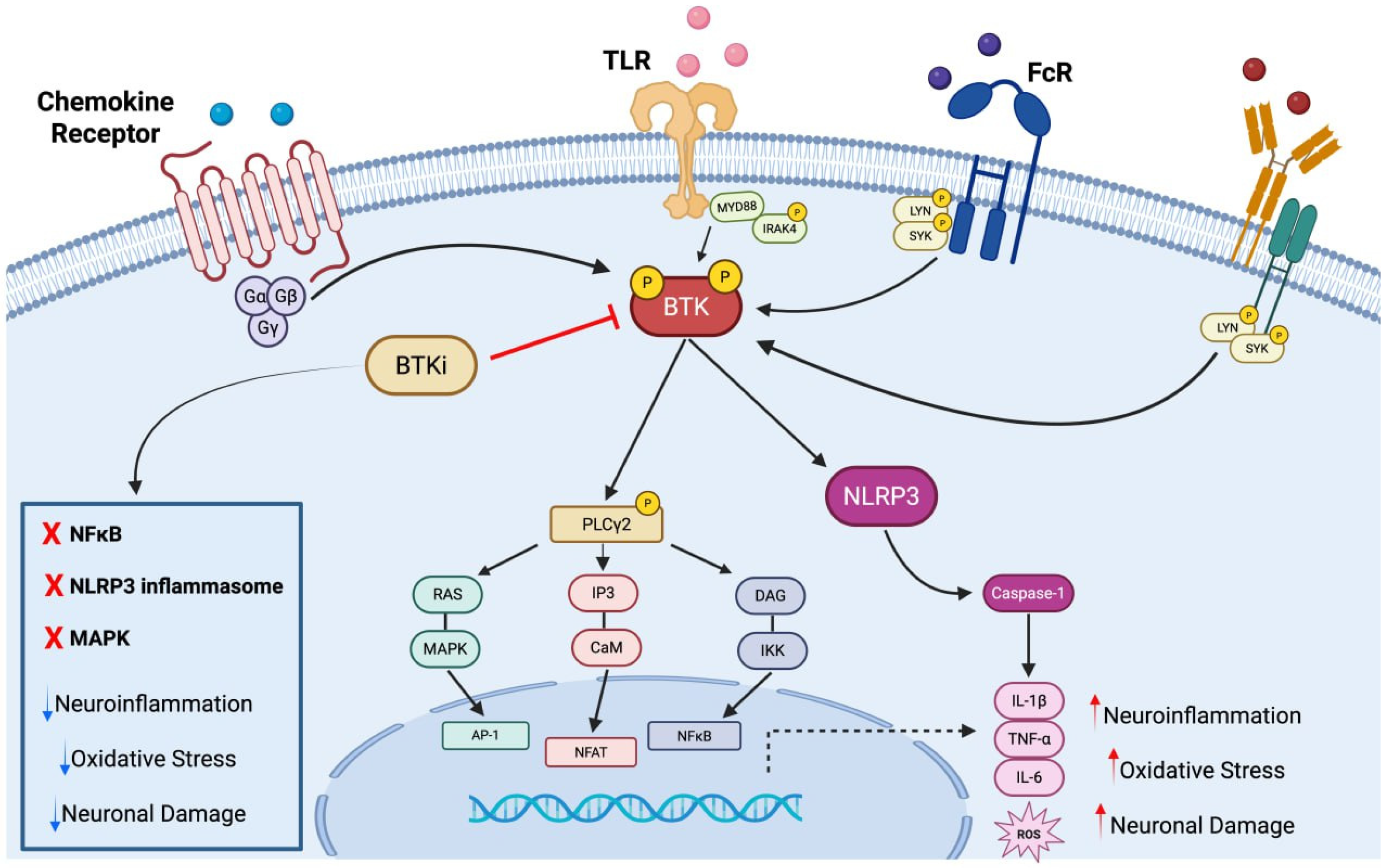
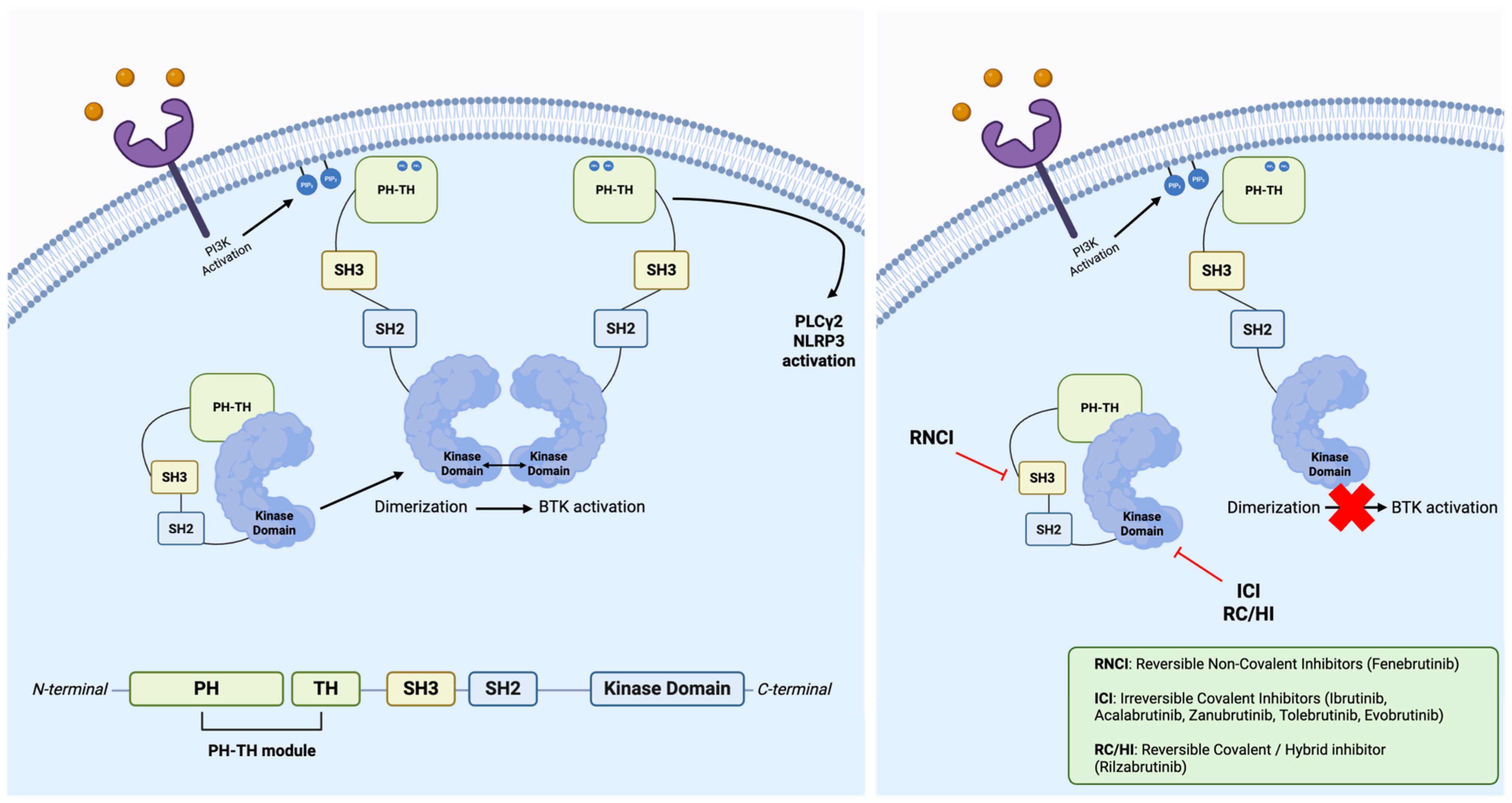
2. Bruton Tyrosine Kinase

BTK is a member of the Tec family of non-receptor tyrosine kinases, playing a pivotal role in signal transduction in various hematopoietic cell types, as previously mentioned. Structurally, BTK comprises five functional domains: a pleckstrin homology (PH) domain, a Tec homology (TH) domain, Src homology 3 (SH3) and SH2 domains, and a catalytic kinase domain (SH1) [16]. Each domain contributes to BTK’s localization, interaction with adaptor proteins, and enzymatic activity. For instance, the PH domain enables membrane recruitment through binding to phosphatidylinositol-3,4,5-trisphosphate (PIP3), a process essential for BTK activation upon receptor stimulation [17]. Upon membrane translocation, BTK is phosphorylated at Tyr551 by upstream Src-family kinases, followed by autophosphorylation at Tyr223, a critical step for full catalytic activation [18]. BTK is most prominently expressed in B lymphocytes, where it mediates BCR activation triggering multiple pathways such as PLCγ2-mediated calcium mobilization, PI3K-AKT signaling, and MAPK cascades, leading to B-cell proliferation, differentiation, and survival [19]. In particular, the antigen interaction with BCR causes activation of Src-family kinases, among which Lyn phosphorylates immunoreceptor tyrosine-based activation motifs (ITAMs) on Igα/Igβ, leading to recruitment of BTK via its SH2 domain [20,21]. Membrane-bound BTK is activated through phosphorylation at Tyr551 and proceeds to phosphorylate PLCγ2, a crucial step that leads to intracellular calcium influx and the activation of downstream transcription factors such as NF-κB, NFAT, and AP-1 [19]. These events drive gene expression programs involved in B-cell activation and antibody production. However, BTK expression is not limited to B cells. As mentioned above, it is also present in myeloid cells including monocytes, macrophages, dendritic cells, and microglia, the resident macrophages of the CNS, where it regulates innate immune responses. In these cells, BTK acts downstream of Fcγ receptors and TLRs, mediating activation of NF-κB and MAPKs, which promote pro-inflammatory gene expression [22]. Upon ligand recognition, these receptors recruit adaptor proteins such as MyD88 and SYK, which in turn activate BTK. BTK then participates in the formation of signalosomes that activate the IKK complex, leading to NF-κB nuclear translocation and transcription of inflammatory mediators such as tumor necrosis factor α (TNF-α) and interleukins (ILs) 1β (IL-1β) and 6 (IL-6) [23,24]. Additionally, BTK is implicated in NLRP3 inflammasome activation in microglia and macrophages, further linking it to a neuroinflammatory pathology [25]. In particular, BTK signaling promotes the cytoskeletal rearrangements required for phagocytosis, as well as microglial metabolic polarization, favoring glycolysis and pentose phosphate pathway activity, thereby enhancing ROS generation and sustaining the microglial inflammatory phenotype. By standing at the intersection of innate immune signaling and metabolic control, BTK amplifies and maintains microglial activation [26]. The involvement of BTK in these signaling networks suggests that its dysregulation can amplify maladaptive immune responses. Indeed, hyperactive BTK signaling has been associated with heightened inflammatory cytokine production, ROS generation, and microglial priming, all features commonly observed in neurodegenerative diseases [27,28]. The convergence of BTK signaling with key inflammatory pathways such as NF-κB, MAPK, and inflammasome activation underscores its role as a central node in neuroimmune signaling. Importantly, these pathways are dysregulated in neurodegenerative diseases, where persistent activation of microglia sustains a chronic pro-inflammatory environment. Thus, targeting BTK may disrupt multiple pathogenic cascades simultaneously, offering a multifaceted approach to neuroinflammatory modulation (Figure 1).

Bruton Tyrosine Kinase’s Inhibitors

BTKis represent a promising class of small molecules capable of selectively modulating immune signaling pathways implicated in both peripheral and central inflammation (Figure 1) [29,30]. Initially developed for B-cell malignancies, BTKis are now being investigated for their potential to attenuate neuroinflammation, a pathological hallmark shared across multiple neurodegenerative disorders [31]. These inhibitors can be categorized by their mechanism of binding (Figure 2), which can be irreversible (covalent) or reversible (non-covalent), and have evolved through successive generations to optimize selectivity, pharmacokinetics, and CNS accessibility, broadening their applicability beyond hematologic malignancies to autoimmune and neurodegenerative conditions [32].
First-generation BTKis, such as ibrutinib, irreversibly bind to the conserved cysteine residue (Cys481) within BTK’s ATP-binding pocket via covalent Michael addition, displaying potent inhibition but limited selectivity due to off-target binding to other kinases. Indeed, although effective with modest BBB penetration, ibrutinib exhibits off-target binding to kinases like tyrosine-protein kinase (TEC), epidermal growth factor receptor (EGFR), and interleukin-2-inducible T-cell kinase (ITK), leading to a broad side-effect profile that includes atrial fibrillation, hypertension, diarrhea, and bleeding due to impaired platelet aggregation [29,33]. Second-generation inhibitors, including acalabrutinib and zanubrutinib, retain irreversible covalent binding to Cys481 but were designed for enhanced selectivity and reduced off-target toxicity. Acalabrutinib, for instance, does not inhibit Src-family kinases, a major advancement over ibrutinib in terms of platelet safety. Zanubrutinib similarly demonstrates minimal inhibition of ITK and is associated with lower incidence of cardiac events, which positions it as a safer alternative in B-cell malignancies such as mantle cell lymphoma and chronic lymphocytic leukemia. Both compounds maintain high BTK occupancy and plasma stability, allowing effective B-cell inhibition with a better side-effect profile. However, their limited CNS penetration curtails their utility in neuroinflammatory disorders [34,35]. Third-generation BTKis include reversible and hybrid covalent–reversible inhibitors such as rilzabrutinib and fenebrutinib [36,37]. These agents bind either non-covalently or via transient covalent bonds to BTK, thus offering the potential to overcome resistance mutations such as C481S, which limit the efficacy of irreversible BTKis. Reversible inhibitors like fenebrutinib interact with alternative residues in the SH3 domain (e.g., K430, M477), avoiding reliance on Cys481 for activity and providing utility in autoimmune indications such as MS [38]. Rilzabrutinib, a hybrid inhibitor, demonstrates high target occupancy in vivo and favorable pharmacokinetics with minimal systemic exposure, suggesting potential for chronic use with reduced toxicity [36]. The other third-generation inhibitors are orelabrutinib and remibrutinib, able to further enhance specificity and bioavailability, with minimal inhibition of kinases unrelated to BTK and promising CNS exposure [39,40]. The adverse effects vary by generation: first-generation compounds are associated with systemic immunosuppression and cardiovascular events, whereas newer agents show reduced systemic toxicity [34]. CNS-penetrant BTKis, such as tolebrutinib and evobrutinib, have emerged as leading candidates for the treatment of neurodegenerative disorders. These compounds possess optimized physicochemical properties, allowing efficient BBB crossing and central BTK engagement [41]. Tolebrutinib, for instance, achieves therapeutic concentrations in CNS tissue and modulates microglial responses without broadly suppressing systemic immunity [42]. Importantly, BTK inhibition may achieve a desirable therapeutic balance by preserving essential immune functions while selectively attenuating pathologic inflammation. Unlike broad-spectrum immunosuppressants, BTKis act downstream of specific immune receptors and modulate signaling cascades without completely ablating immune cell function. This may reduce the risk of infections or systemic immune dysregulation, which is crucial in chronic conditions where long-term therapy is often necessary.

3. Bruton Tyrosine Kinase’s Inhibitors in Neurodegenerative Disease

Neuroinflammation in neurodegenerative diseases is characterized by chronic microglial activation, elevated production of pro-inflammatory cytokines and ROS, and sustained activation of transcription factors like NF-κB. BTK acts as a proximal transducer of these signals, linking receptor engagement to the activation of pathways that promote cytokine transcription, inflammasome assembly (notably NLRP3), and phagocytic responses [11]. In particular, when BTK is inhibited, NLRP3 inflammasome assembly is disrupted, reducing caspase-1 activation and subsequent maturation of IL-1β and IL-18. In this context, BTK inhibitors exert their effects by blocking BTK’s kinase activity, thus dampening the amplification of detrimental neuroinflammatory loops. Evidence of this, for instance, can be found in preclinical studies that have shown BTKi-dependent reductions in microglial pro-inflammatory markers in models of AD and MS, promoting a microglial shift toward neuroprotective phenotypes [12,43]. Taken together, these data underscore the potential of BTK inhibitors to act as disease-modifying agents in neurodegenerative disorders, not merely through immunosuppression but by reprogramming maladaptive neuroimmune interactions. Their pleiotropic effects on microglial biology, cytokine signaling, and inflammasome regulation position BTKis as a targeted approach to mitigate neuroinflammation and its downstream neurodegenerative consequences.

3.1. Alzheimer’s Disease

AD is the most prevalent form of dementia, affecting over 50 million individuals globally [44]. It is characterized by progressive memory impairment, executive dysfunction, and behavioral changes, ultimately culminating in loss of independence and death. Histopathologically, the disease is marked by extracellular deposits of amyloid-β (Aβ) peptides forming plaques and intracellular aggregates of hyper-phosphorylated tau protein as neurofibrillary tangles [45]. In recent decades, chronic neuroinflammation has emerged as a third critical pillar in the pathogenesis of AD, intricately interacting with amyloid and tau pathologies. Microglia, the brain’s resident macrophages, play a central role in neuroinflammatory responses in AD [46]. Upon encountering Aβ aggregates, microglia become activated and assume a pro-inflammatory phenotype, characterized by increased secretion of pro-inflammatory cytokines (e.g., IL-1β, TNF-α), chemokines, and ROS. Chronic activation contributes to synaptic loss, impaired phagocytic clearance of Aβ, and further recruitment of peripheral immune cells through BBB disruption. Within this context, BTK is increasingly recognized as a central signaling molecule modulating microglial activity. BTK is upregulated in activated microglia in post-mortem AD brains, particularly in proximity to Aβ plaques [12]. Mechanistically, BTK is involved in TLR and FcR signaling cascades, which are activated upon recognition of damage-associated molecular patterns (DAMPs) such as aggregated Aβ. BTK activation leads to phosphorylation of PLCγ2, initiating calcium mobilization, NF-κB translocation, and inflammasome priming [47]. For instance, PLCγ2 acts downstream of AD-related TREM2, TLR4, BTK, and CSF1R, modulating microglial responses. Moreover, PLCγ2 variants have been linked to AD, showing that PLCγ2 variant M1141K, which is associated with ibrutinib resistance, greatly increases PLC activity by disrupting its autoinhibitory activity, whereas the AD-protective P522R variant enhances BTK-mediated signaling without affecting PLC basal activity [48]. BTK is also implicated in NLRP3 inflammasome activation, a key driver of IL-1β maturation and release in AD models [49]. Upon priming through TLRs or Fcγ receptors, BTK is activated and translocates to NLRP3-containing complexes, where it phosphorylates NLRP3 on specific tyrosine residues promoting conformational rearrangement and interaction with the adaptor ASC, thereby facilitating inflammasome oligomerization, caspase-1 activation, and subsequent maturation of IL-1β and IL-18. BTK also enhances potassium efflux and mitochondrial ROS production, which are two critical signals for full inflammasome activation [50]. Preclinical studies have demonstrated the efficacy of BTK inhibitors in modulating these inflammatory processes. In APP/PS1 transgenic mice, ibrutinib administration reduced microglial activation, decreased pro-inflammatory cytokine levels, and significantly improved cognitive performance in behavioral tests [51]. These effects were accompanied by lower Aβ plaque burden, suggesting that BTK inhibition may restore microglial phagocytic function. Importantly, BTK inhibition appears to selectively modulate pathogenic immune pathways without impairing homeostatic microglial functions. Keaney and collaborators demonstrated in vitro, using primary rodent microglia, that ibrutinib reduces pro-inflammatory gene expression while preserving phagocytic capacity and neurotrophic support [12]. These features are critical for long-term safety in chronic diseases like AD. In addition to its role in innate immunity, BTK may influence adaptive immune responses in AD. B cells are found in the meninges and choroid plexus in AD patients, where they may contribute to local inflammation through antigen presentation and antibody production [52]. BTK inhibition can reduce B-cell activation and survival, potentially curtailing aberrant immune responses at CNS interfaces. Currently, no BTKi is approved for AD treatment. Nonetheless, given the multifaceted role of BTK in microglial signaling, Aβ processing, and B-cell-mediated immunity, BTK inhibition represents a compelling disease-modifying strategy for AD that warrants further clinical development.

3.2. Parkinson’s Disease

PD is the second most common neurodegenerative disorder, affecting 2–3% of the population over 65 years, and is characterized by the progressive degeneration of dopaminergic neurons in the substantia nigra pars compacta, resulting in the hallmark motor symptoms of bradykinesia, rigidity, resting tremor, and postural instability. In addition to motor dysfunction, PD involves a wide array of non-motor symptoms, including cognitive decline, depression, autonomic dysfunction, and sleep disturbances. Pathologically, PD is defined by the accumulation of misfolded α-synuclein in Lewy bodies and Lewy neurites, as well as widespread neuroinflammation [53,54]. Microglial activation is observed early in PD progression and is exacerbated in the presence of α-synuclein aggregates, leading to the release of pro-inflammatory mediators such as IL-1β, TNF-α, and prostaglandins and contributing to neuronal injury by promoting a self-perpetuating cycle of inflammation and neurodegeneration [55]. BTK, which is expressed in microglia and peripheral monocytes, could be involved in mediating TLR4 and Fcγ receptor signaling in response to aggregated α-synuclein [56]. A recent bioinformatic analysis of PD’s gene datasets revealed upregulation of BTK [57]. In particular, B lymphocyte populations are altered in PD, with reduced proliferation, decreased regulatory B cells, and increased pro-inflammatory subsets [58]. Experimental models link B-cell depletion and impaired meningeal drainage to worsened α-synuclein pathology. BTK expression has been proposed as a PD biomarker, supporting the rationale for B-cell-targeted therapies, which may modulate broader immune networks and offer new treatment avenues. Even though BTK is pivotal in B-cell maturation, BTK signaling enhances NF-κB activation and promotes the assembly of the NLRP3 inflammasome, leading to caspase-1 activation and IL-1β secretion, hallmarks of PD-associated neuroinflammation [59]. The NLRP3 inflammasome is expressed in microglia, astrocytes, and neurons; in PD, α-synuclein and dopaminergic neuron damage activates NLRP3 in microglia, while Parkin normally suppresses NLRP3 in neurons via ubiquitination [50]. BTK’s role in PD suggests that its inhibition could modulate peripheral immune responses implicated in PD progression, as well as detrimental central events: BTK inhibition may limit peripheral immune cell trafficking into the CNS and reduce peripheral cytokine production, providing a dual mechanism of action. Although BTKi evaluations in the PD context show relevant gaps, these preclinical findings provide a strong rationale for their investigation. Given the need for disease-modifying therapies in PD and the role of inflammation in its pathogenesis, BTK inhibition may offer a novel therapeutic avenue with both neuroprotective and immunomodulatory properties.

3.3. Multiple Sclerosis

MS is a chronic autoimmune demyelinating disease of the CNS characterized by recurrent episodes of neurological dysfunction due to immune-mediated damage to the myelin sheath, axonal degeneration, and neuroinflammation [60]. The disease predominantly affects young adults and is a leading cause of non-traumatic neurological disability in this population [61]. MS is considered a multifactorial disease involving genetic susceptibility and environmental triggers such as Epstein–Barr virus infection and vitamin D deficiency [62,63]. Immune dysregulation lies at the core of MS pathogenesis. The adaptive immune response, particularly autoreactive CD4+ T cells, plays a central role in initiating inflammation [60]. However, emerging evidence has highlighted the importance of B cells and myeloid cells, including microglia and monocyte-derived macrophages, in perpetuating CNS injury [64]. B cells contribute through antigen presentation, pro-inflammatory cytokine release, and the formation of ectopic follicle-like structures within the meninges, and BTK is a crucial signaling mediator in these cell types [24]. In MS lesions, BTK expression is increased in microglia and infiltrating macrophages, where it promotes inflammatory responses via activation of the NF-κB and NLRP3 inflammasome pathways [65]. In vitro studies using human microglia have shown that BTK inhibition reduces LPS-induced TNF-α, IL-6, and IL-1β production, highlighting its role in modulating innate immune responses [27,43]. Preclinical studies using experimental autoimmune encephalomyelitis, the prototypical animal model of MS, have demonstrated the efficacy of BTK inhibitors such as ibrutinib and evobrutinib in reducing disease severity [43,66]. In a recent study, the effects of three BTK inhibitors (i.e., evobrutinib, fenebrutinib, and tolebrutinib) were evaluated on neutrophil activity [67]. All inhibitors attenuated neutrophil activation triggered by the bacterial peptide fMLF and CXCL8, reducing reactive oxygen species production, neutrophil extracellular trap formation, and the secretion of CXCL8 and IL-1β in response to inflammatory stimuli. These effects were independent of toxicity and were accompanied by prolonged neutrophil survival in inflammatory conditions. These findings highlight a novel role for BTK inhibitors in modulating innate immune functions beyond B cells [67]. Also, treatment with fenebrutinib leads to reduced spinal cord inflammation, demyelination, and axonal damage [13]. Importantly, BTK inhibition dampens both peripheral and CNS-resident immune responses [68]. Inhibition of BTK in B cells limits the production of granulocyte–macrophage colony-stimulating factor (GM-CSF) and IL-6, crucial cytokines in T-cell activation, while microglial inhibition reduces local cytokine storms and cytotoxicity [69]. Moreover, in vitro BTK inhibition with evobrutinib in iron-loaded human microglia reduces proinflammatory gene expression and iron importer levels while increasing ferroportin, suggesting that BTK blockade modulates both inflammation and iron homeostasis in microglia, key features of chronic lesions in MS [70]. Clinical translation of these findings is underway. Evobrutinib has been evaluated in a phase II trial (ID: NCT02975349) involving 267 patients with relapsing–remitting MS, demonstrating significant reductions in gadolinium-enhancing lesions on MRI and favorable safety and tolerability profiles [71]. These findings have prompted phase III trials (IDs: NCT04338022 and NCT04338061) to assess long-term clinical benefits. However, evobrutinib did not distinguish itself from teriflunomide, an MS drug, in reducing the annualized relapse rate, causing the termination of these studies with no further support for evobrutinib application in MS [72]. Other BTK inhibitors are also in advanced stages of clinical development and exhibit promising CNS penetrance and immunomodulatory activity. For instance, fenebrutinib is currently under evaluation in different MS trials, including studies on primary progressive MS (FENtrepid, ID: NCT04544449) and relapsing MS (FENhance 1 and 2, IDs: NCT04586010 and NCT04586023; FENopta, ID: NCT05119569), comparing it to ocrelizumab and teriflunomide. In particular, in FENopta, fenebrutinib showed maintenance of low disease activity in relapsing MS with a favorable safety profile, supporting phase III clinical trials [73]. Regarding FENtrepid and FENhance 1 and 2, the first data from these studies are expected at the end of 2025 and will be pivotal in characterizing fenebrutinb’s effects on disease progression across the MS spectrum. Orelabrutinib is in phase 2 testing for relapsing–remitting MS (ID: NCT04711148), where it caused significant lesion reduction in all active treatment groups compared to placebo, while remibrutinib is being compared to teriflunomide in a large phase 3 relapsing MS study (ID: NCT05147220). Lastly, tolebrutinib showed efficacy in reducing brain lesions in RMS but had all trials paused due to liver toxicity concerns [74]. Unlike existing MS therapies that broadly suppress immune function and increase infection risk, BTK inhibitors offer a more targeted approach by modulating pathogenic immune pathways while sparing protective immunity. This mechanism may be particularly valuable in progressive forms of MS, where inflammation is compartmentalized within the CNS and resistant to peripheral immunosuppression [75]. In summary, BTK plays a critical role in MS pathogenesis through its effects on B cells and myeloid cells. BTK inhibition offers a rational, targeted therapeutic strategy with the potential to ameliorate disease activity, prevent progression, and reduce treatment-related toxicity in MS patients.

3.4. Ischemic Stroke

IS results from the sudden interruption of cerebral blood flow due to arterial occlusion, leading to acute neuronal injury and rapid activation of inflammatory pathways [76]. Stroke is a leading cause of death and disability worldwide, with current treatment options limited to thrombolytic agents and mechanical thrombectomy, both constrained by narrow therapeutic windows. Neuroinflammation is now recognized as a critical determinant of secondary brain injury following ischemic insult [77]. Within minutes of ischemia, DAMPs released from dying cells activate resident microglia and astrocytes, triggering the production of pro-inflammatory cytokines, chemokines, and matrix metalloproteinases (MMPs), leading to BBB disruption, leukocyte infiltration, and further neuronal injury [78]. Peripheral immune cells, particularly neutrophils, monocytes, and B cells, are rapidly recruited and contribute to post-ischemic inflammation. In murine models of stroke, BTK expression is significantly upregulated in infiltrating immune cells and perilesional microglia [79]. Pharmacological inhibition of BTK has shown neuroprotective effects in rodent models of cerebral ischemia. In mice subjected to middle cerebral artery occlusion (MCAO), ibrutinib administration reduced brain damage and improved neurological outcomes in diabetic mice with ischemia/reperfusion injury [80]. It enhanced cell viability, decreased oxidative stress, and activated the PI3K/Akt/mTOR pathway, suggesting neuroprotection via reduced inflammation and improved autophagy. These effects were associated with reduced oxidative stress, lower levels of IL-1β and TNF-α, and decreased peripheral cell infiltration. Similar benefits were observed with tolebrutinib, which showed enhanced neuroprotection in a model of chronic cerebral hypoperfusion, with reduced white matter damage and cognitive deficits, by suppressing microglial activation, inflammation, oxidative stress, and ferroptosis, highlighting its therapeutic potential in ischemic demyelination [81]. BTK inhibitors may also exert beneficial effects on stroke recovery by limiting chronic inflammation, inflammasome activation, and secondary injury. Indeed, NLRP3 expression increases up to 20–30 fold within 24 h after stroke, and inhibition of BTK, an upstream regulator of NLRP3, showed reduced infarct volume and related neurological disruption after ischemic stroke [79,82]. In subacute stroke models, evobrutinib treatment alleviated ischemic injury, reduced gliosis, and promoted anti-inflammatory microglial polarization, suggesting a role for BTKis in promoting CNS repair [83]. Importantly, BTK inhibition does not interfere with clot formation or dissolution, unlike anticoagulants or antiplatelet agents, making it potentially safer in the context of acute ischemia [84]. Additionally, BTKis may be useful adjuncts to reperfusion therapies, extending the therapeutic window and reducing reperfusion injury. Although clinical trials of BTKis in stroke are currently lacking, the mechanistic rationale and robust preclinical data justify their evaluation in human studies. Given the central role of innate immune activation in post-stroke injury, BTK inhibition may represent a promising strategy to mitigate neuroinflammation and improve functional outcomes.

3.5. Huntington’s Disease

HD is an autosomal dominant neurodegenerative disorder caused by an expanded CAG repeat in the huntingtin (HTT) gene, resulting in mutant huntingtin (mHTT) protein with a toxic polyglutamine tract. Clinically, HD manifests with progressive motor dysfunction (chorea, dystonia), psychiatric symptoms (depression, irritability), and cognitive decline. Neuropathologically, HD is characterized by neuronal loss in the striatum and cortex, accompanied by widespread microgliosis and neuroinflammation [85]. Microglial activation is an early and sustained feature of HD, observed even before clinical onset [86]. PET imaging studies using TSPO ligands have demonstrated increased microglial activity in pre-symptomatic mutation carriers, correlating with disease progression [87]. In HD brains, microglia exhibit a pro-inflammatory phenotype with increased expression of IL-6, TNF-α, and C1q, which could contribute to synaptic pruning, glutamate excitotoxicity, and neuronal apoptosis [88]. BTK is expressed in CNS-resident microglia and peripheral monocytes and modulates key inflammatory pathways that are involved in HD pathogenesis: TLR signaling through BTK leads to NF-κB activation and the production of pro-inflammatory mediators [89]. Given BTK’s role downstream of TLRs and upstream of both NF-κB and NLRP3, it likely functions as a central amplifier of inflammation in HD. Even though BTK inhibition has not yet been tested on this condition, several hallmarks shared with other discussed diseases support the concept of BTKi application in HD. BTKis could be able to prevent neuroinflammatory escalation by acting on microglial signaling. Also, BTK inhibition may modulate astrocyte reactivity and peripheral immune dysfunction in HD. Indeed, peripheral immune cells from HD patients exhibit a pro-inflammatory profile, with increased IL-1β and TNF-α secretion [90]. Given the genetic clarity and consistent inflammatory features of HD, BTK inhibition offers a compelling therapeutic avenue.

4. Discussion

The involvement of BTK in a spectrum of neuroinflammatory pathways has prompted increasing interest in its therapeutic targeting across neurodegenerative diseases [22]. As reviewed here, BTK not only is integral to B-cell development and function but also plays a pivotal role in modulating innate immune responses via microglia and myeloid cells in the CNS. This dual functionality uniquely positions BTK at the crossroads of adaptive and innate immunity, a feature that is particularly relevant in chronic neurodegenerative conditions characterized by both systemic and CNS-compartmentalized inflammation.
Several key themes emerge from the current evidence. First, BTK activation amplifies deleterious neuroimmune interactions that contribute to neuronal loss [19]. In microglia, BTK signaling propagates pro-inflammatory pathways including the NF-κB axis and the NLRP3 inflammasome, which are implicated in the production of cytokines such as IL-1β and TNF-α. These mediators, in turn, exacerbate oxidative stress, synaptic dysfunction, and neuronal death. Second, BTKis, initially developed for hematologic malignancies, exhibit immunomodulatory effects that extend beyond the hematopoietic system. Their ability to penetrate the BBB and selectively modulate pathological immune responses in the CNS without inducing global immunosuppression is a critical therapeutic advantage [14].
The therapeutic rationale for BTK inhibition in neurodegeneration is underscored by the convergence of neuroinflammatory mechanisms across multiple disorders. In AD, BTK appears to mediate microglial phagocytic dysfunction and pro-inflammatory activation in response to amyloid pathology [51]. In PD, BTK amplifies dopaminergic neurotoxicity via TLR and α-synuclein-triggered pathways [57]. In MS, BTK orchestrates both B-cell-driven autoimmunity and microglial activation, supporting its targeting in both relapsing and progressive forms [24]. In ischemic stroke, the timing and extent of BTK activation shape the extent of secondary injury and inflammation-driven neuronal damage [79]. Finally, in HD, early and sustained microglial activation, potentially BTK-dependent, has emerged as a tractable contributor to disease onset and progression [88].
Preclinical studies across diverse animal models consistently demonstrate, as mentioned above, that BTK inhibition attenuates neuroinflammation, preserves neuronal integrity, and improves behavioral outcomes [29]. Notably, BTKis have been shown to reduce microglial reactivity, limit the production of neurotoxic cytokines, and promote protective phenotypes. These findings support the notion that BTK inhibition not only reduces immune-driven damage but may also foster a reparative environment conducive to neuroregeneration.
The clinical pipeline for BTKis in neurodegeneration is rapidly expanding. Several compounds, including evobrutinib and fenebrutinib, are currently being tested for MS. Safety and tolerability data from oncology and autoimmune contexts are encouraging, particularly with respect to long-term administration and immune preservation [91]. Importantly, emerging data suggest that BTKis may synergize with existing therapies, potentially enhancing their therapeutic efficacy and broadening their clinical utility [92,93].
Nevertheless, several limitations and challenges must be addressed. The pleiotropic functions of BTK across different immune compartments raise concerns regarding off-target effects and unintended immune modulation [14]. Furthermore, the extent of BTK involvement may differ by disease stage and CNS region, requiring precise characterization of the patient subgroups that may benefit most from BTK-targeted therapies. In addition, the pharmacokinetics, CNS penetrance, and selectivity profiles of different BTKis vary substantially, influencing therapeutic windows and clinical outcomes. In particular, first-generation inhibitors like ibrutinib are hampered by broad kinase inhibition and systemic toxicity, limiting their usability in chronic neurologic conditions [94]. Second-generation CNS-penetrant BTKis, such as tolebrutinib, evobrutinib, fenebrutinib, and orelabrutinib, offer the potential for direct modulation of compartmentalized neuroinflammation. Tolebrutinib, in the 2025 HERCULES phase III trial involving non-relapsing secondary progressive MS, reduced the risk of disability progression [74]. However, serious liver-related adverse events occurred among treated patients versus placebo, one of which led to liver transplantation and death before intensified monitoring was implemented. Indeed, this has prompted updated recommendations for weekly liver enzyme monitoring during the first 12 weeks of therapy. Fenebrutinib shows favorable in vitro microglial modulation and is undergoing evaluation in FENhance and FENtrepid, with preliminary data suggesting improved tolerability and lower off-target toxicity [13,73]. Evobrutinib showed consistent efficacy in reducing gadolinium-enhancing lesions in RMS patients and a favorable safety profile in the phase III EVOLUTION trials, with no significant liver toxicity or serious infections reported in over 700 patient-years of exposure [72]. Orelabrutinib, currently in phase II trials for MS, has shown promising CNS penetrance and BTK occupancy in cerebrospinal fluid, along with a clean hepatic safety profile in preliminary reports [95]. Nevertheless, the long-term comparative safety profiles, CNS pharmacokinetics, and off-target risks of these agents remain to be defined. Indeed, while several BTKis have shown promising preclinical and early clinical results, some clinical trials have failed to meet primary endpoints, underscoring the need for a deeper understanding of the underlying causes. One critical factor is the differential ability of BTKis to cross the BBB. Even among newer-generation agents, significant variability exists in their cerebrospinal fluid concentrations and BTK occupancy, which may explain the divergent clinical outcomes despite similar peripheral pharmacodynamics [42,96,97]. Moreover, the timing of administration relative to the disease course appears crucial; administration during late-stage neurodegeneration, when irreversible neuronal loss predominates, may not yield significant functional recovery despite the modulation of inflammation. Thus, future clinical trials should incorporate stratified patient selection based on both the disease stage and markers of active inflammation. Pharmacogenomic profiling, including BTK expression patterns and immune signatures, may also help identify the subgroups most likely to benefit from BTK inhibition. In addition, head-to-head trials and rigorous biomarker-driven monitoring must guide optimal BTKi selection standardizing CNS exposure assessments and considering adaptive designs that account for interindividual variability in drug penetration and target engagement [98]. These measures may enhance the translational success of BTKis and clarify their true therapeutic potential across distinct neurodegenerative phenotypes. Future research, therefore, should aim to delineate disease- and context-specific roles of BTK within the neuroimmune axis. Integration of single-cell transcriptomic profiling, spatial transcriptomics, and advanced imaging modalities may offer insight into BTK’s cell-specific functions in vivo.
In summary, BTK inhibition represents a promising avenue in the modulation of neuroinflammation across diverse neurodegenerative diseases. The expanding body of mechanistic and translational data strongly supports the continued exploration of BTKis as part of next-generation, targeted neurotherapeutic interventions.

5. Conclusions

Overall, these data underscore the potential of BTK inhibitors to act as disease-modifying agents in neurodegenerative disorders, not merely through immunosuppression but by reprogramming maladaptive neuroimmune interactions. Their pleiotropic effects on microglial biology, cytokine signaling, and inflammasome regulation position BTKis as a targeted and rational approach to mitigate neuroinflammation and its downstream neurodegenerative consequences.

Author Contributions

Conceptualization, V.C. and F.D.; software, H.K.; writing—original draft preparation, F.D. and M.d.; writing—review and editing, A.C., V.C. and G.L.; supervision, M.d. and V.C. All authors have read and agreed to the published version of the manuscript.

Funding

This research received no external funding.

Conflicts of Interest

The authors declare no conflicts of interest.

References

  1. Bhanumathy, K.K.; Balagopal, A.; Vizeacoumar, F.S.; Vizeacoumar, F.J.; Freywald, A.; Giambra, V. Protein Tyrosine Kinases: Their Roles and Their Targeting in Leukemia. Cancers 2021, 13, 184. [Google Scholar] [CrossRef] [PubMed]
  2. Stevenson, M.; Algarzae, N.K.; Moussa, C. Tyrosine Kinases: Multifaceted Receptors at the Intersection of Several Neurodegenerative Disease-Associated Processes. Front. Dement. 2024, 3, 1458038. [Google Scholar] [CrossRef] [PubMed]
  3. Szilveszter, K.P.; Németh, T.; Mócsai, A. Tyrosine Kinases in Autoimmune and Inflammatory Skin Diseases. Front. Immunol. 2019, 10, 1862. [Google Scholar] [CrossRef] [PubMed]
  4. Thomson, R.J.; Moshirfar, M.; Ronquillo, Y. Tyrosine Kinase Inhibitors. In StatPearls; StatPearls Publishing: Treasure Island, FL, USA, 2025. [Google Scholar]
  5. Boskabadi, S.J.; Dashti, A.; Karevan, S.; Kargar-Soleimanabad, S.; Salehifar, E. Clinical Uses and Safety Concerns of Tyrosine Kinase Inhibitors with a Focus on Novel Drugs: A Narrative Review. J. Oncol. Pharm. Pract. 2023, 12, 10781552231174790. [Google Scholar] [CrossRef] [PubMed]
  6. Rawlings, D.J.; Witte, O.N. The Btk Subfamily of Cytoplasmic Tyrosine Kinases: Structure, Regulation and Function. Semin. Immunol. 1995, 7, 237–246. [Google Scholar] [CrossRef] [PubMed]
  7. Bender, A.T.; Gardberg, A.; Pereira, A.; Johnson, T.; Wu, Y.; Grenningloh, R.; Head, J.; Morandi, F.; Haselmayer, P.; Liu-Bujalski, L. Ability of Bruton’s Tyrosine Kinase Inhibitors to Sequester Y551 and Prevent Phosphorylation Determines Potency for Inhibition of Fc Receptor but Not B-Cell Receptor Signaling. Mol. Pharmacol. 2017, 91, 208–219. [Google Scholar] [CrossRef] [PubMed]
  8. Konen, F.F.; Möhn, N.; Witte, T.; Schefzyk, M.; Wiestler, M.; Lovric, S.; Hufendiek, K.; Jendretzky, K.F.; Gingele, S.; Schwenkenbecher, P.; et al. Disease-Modifying Strategies: Targeting Protein Kinases in Multiple Sclerosis and Other Autoimmune Disorders. Autoimmun. Rev. 2025, 24, 103754. [Google Scholar] [CrossRef] [PubMed]
  9. Brunner, C.; Müller, B.; Wirth, T. Bruton’s Tyrosine Kinase Is Involved in Innate and Adaptive Immunity. Histol. Histopathol. 2005, 20, 945–955. [Google Scholar] [CrossRef] [PubMed]
  10. Wilson, D.M.; Cookson, M.R.; Bosch, L.V.D.; Zetterberg, H.; Holtzman, D.M.; Dewachter, I. Hallmarks of Neurodegenerative Diseases. Cell 2023, 186, 693–714. [Google Scholar] [CrossRef] [PubMed]
  11. D’Egidio, F.; Castelli, V.; d’Angelo, M.; Ammannito, F.; Quintiliani, M.; Cimini, A. Brain Incoming Call from Glia during Neuroinflammation: Roles of Extracellular Vesicles. Neurobiol. Dis. 2024, 201, 106663. [Google Scholar] [CrossRef] [PubMed]
  12. Keaney, J.; Gasser, J.; Gillet, G.; Scholz, D.; Kadiu, I. Inhibition of Bruton’s Tyrosine Kinase Modulates Microglial Phagocytosis: Therapeutic Implications for Alzheimer’s Disease. J. Neuroimmune Pharmacol. 2019, 14, 448–461. [Google Scholar] [CrossRef] [PubMed]
  13. Langlois, J.; Lange, S.; Ebeling, M.; Macnair, W.; Schmucki, R.; Li, C.; DeGeer, J.; Sudharshan, T.J.J.; Yong, V.W.; Shen, Y.-A.; et al. Fenebrutinib, a Bruton’s Tyrosine Kinase Inhibitor, Blocks Distinct Human Microglial Signaling Pathways. J. Neuroinflamm. 2024, 21, 276. [Google Scholar] [CrossRef] [PubMed]
  14. Shirley, M. Bruton Tyrosine Kinase Inhibitors in B-Cell Malignancies: Their Use and Differential Features. Target. Oncol. 2022, 17, 69–84. [Google Scholar] [CrossRef] [PubMed]
  15. Ruiz, R.; Kirk, A.D. Long-Term Toxicity of Immunosuppressive Therapy. Transplant. Liver 2015, 1354–1363. [Google Scholar] [CrossRef]
  16. Wang, Q.; Pechersky, Y.; Sagawa, S.; Pan, A.C.; Shaw, D.E. Structural Mechanism for Bruton’s Tyrosine Kinase Activation at the Cell Membrane. Proc. Natl. Acad. Sci. USA 2019, 116, 9390–9399. [Google Scholar] [CrossRef] [PubMed]
  17. Wang, Q.; Vogan, E.M.; Nocka, L.M.; Rosen, C.E.; Zorn, J.A.; Harrison, S.C.; Kuriyan, J. Autoinhibition of Bruton’s Tyrosine Kinase (Btk) and Activation by Soluble Inositol Hexakisphosphate. eLife 2015, 4, e06074. [Google Scholar] [CrossRef] [PubMed]
  18. Kurosaki, T.; Kurosaki, M. Transphosphorylation of Bruton’s Tyrosine Kinase on Tyrosine 551 Is Critical for B Cell Antigen Receptor Function. J. Biol. Chem. 1997, 272, 15595–15598. [Google Scholar] [CrossRef] [PubMed]
  19. Tavakoli, G.M.; Yazdanpanah, N.; Rezaei, N. Targeting Bruton’s Tyrosine Kinase (BTK) as a Signaling Pathway in Immune-Mediated Diseases: From Molecular Mechanisms to Leading Treatments. Adv. Rheumatol. 2024, 64, 61. [Google Scholar] [CrossRef] [PubMed]
  20. Duarte, D.P.; Lamontanara, A.J.; La Sala, G.; Jeong, S.; Sohn, Y.-K.; Panjkovich, A.; Georgeon, S.; Kükenshöner, T.; Marcaida, M.J.; Pojer, F.; et al. Btk SH2-Kinase Interface Is Critical for Allosteric Kinase Activation and Its Targeting Inhibits B-Cell Neoplasms. Nat. Commun. 2020, 11, 2319. [Google Scholar] [CrossRef] [PubMed]
  21. L’Estrange-Stranieri, E.; Gottschalk, T.A.; Wright, M.D.; Hibbs, M.L. The Dualistic Role of Lyn Tyrosine Kinase in Immune Cell Signaling: Implications for Systemic Lupus Erythematosus. Front. Immunol. 2024, 15, 1395427. [Google Scholar] [CrossRef] [PubMed]
  22. Das, D.; Ghosh, A.; Greco, D.; Michaličková, D.; Slanař, O. Bruton’s Tyrosine Kinase: A Potential Novel Target for Neurological Disorders. Physiol. Res. 2025, 74, 233–248. [Google Scholar] [CrossRef] [PubMed]
  23. Munshi, M.; Liu, X.; Chen, J.G.; Xu, L.; Tsakmaklis, N.; Demos, M.G.; Kofides, A.; Guerrera, M.L.; Jimenez, C.; Chan, G.G.; et al. SYK Is Activated by Mutated MYD88 and Drives Pro-Survival Signaling in MYD88 Driven B-Cell Lymphomas. Blood Cancer J. 2020, 10, 12. [Google Scholar] [CrossRef] [PubMed]
  24. Krämer, J.; Bar-Or, A.; Turner, T.J.; Wiendl, H. Bruton Tyrosine Kinase Inhibitors for Multiple Sclerosis. Nat. Rev. Neurol. 2023, 19, 289–304. [Google Scholar] [CrossRef] [PubMed]
  25. Ghosh, S.; Mohammed, Z.; Singh, I. Bruton’s Tyrosine Kinase Drives Neuroinflammation and Anxiogenic Behavior in Mouse Models of Stress. J. Neuroinflamm. 2021, 18, 289. [Google Scholar] [CrossRef] [PubMed]
  26. Vermersch, P.; Airas, L.; Berger, T.; Deisenhammer, F.; Grigoriadis, N.; Hartung, H.-P.; Magyari, M.; Popescu, V.; Pozzilli, C.; Pugliatti, M.; et al. The Role of Microglia in Multiple Sclerosis: Implications for Treatment with Bruton’s Tyrosine Kinase Inhibitors. Front. Immunol. 2025, 16, 1495529. [Google Scholar] [CrossRef] [PubMed]
  27. Gruber, R.C.; Wirak, G.S.; Blazier, A.S.; Lee, L.; Dufault, M.R.; Hagan, N.; Chretien, N.; LaMorte, M.; Hammond, T.R.; Cheong, A.; et al. BTK Regulates Microglial Function and Neuroinflammation in Human Stem Cell Models and Mouse Models of Multiple Sclerosis. Nat. Commun. 2024, 15, 10116. [Google Scholar] [CrossRef] [PubMed]
  28. Karati, D.; Kumar, D. Tyrosine Kinase as Therapeutic Target of Neurodegenerative Disorders. Brain Disord. 2025, 17, 100193. [Google Scholar] [CrossRef]
  29. Rozkiewicz, D.; Hermanowicz, J.M.; Kwiatkowska, I.; Krupa, A.; Pawlak, D. Bruton’s Tyrosine Kinase Inhibitors (BTKIs): Review of Preclinical Studies and Evaluation of Clinical Trials. Molecules 2023, 28, 2400. [Google Scholar] [CrossRef] [PubMed]
  30. Tang, T.; Luo, J.; Zhang, D.; Lu, Y.; Liao, W.; Zhang, J. Innovative Design and Potential Applications of Covalent Strategy in Drug Discovery. Eur. J. Med. Chem. 2025, 284, 117202. [Google Scholar] [CrossRef] [PubMed]
  31. Zain, R.; Vihinen, M. Structure-Function Relationships of Covalent and Non-Covalent BTK Inhibitors. Front. Immunol. 2021, 12, 694853. [Google Scholar] [CrossRef] [PubMed]
  32. Burger, J.A. BTK Inhibitors: Present and Future. Cancer J. 2019, 25, 386–393. [Google Scholar] [CrossRef] [PubMed]
  33. Honigberg, L.A.; Smith, A.M.; Sirisawad, M.; Verner, E.; Loury, D.; Chang, B.; Li, S.; Pan, Z.; Thamm, D.H.; Miller, R.A.; et al. The Bruton Tyrosine Kinase Inhibitor PCI-32765 Blocks B-Cell Activation and Is Efficacious in Models of Autoimmune Disease and B-Cell Malignancy. Proc. Natl. Acad. Sci. USA 2010, 107, 13075–13080. [Google Scholar] [CrossRef] [PubMed]
  34. Estupiñán, H.Y.; Berglöf, A.; Zain, R.; Smith, C.I.E. Comparative Analysis of BTK Inhibitors and Mechanisms Underlying Adverse Effects. Front. Cell Dev. Biol. 2021, 9, 630942. [Google Scholar] [CrossRef] [PubMed]
  35. Lipsky, A.H.; Lamanna, N. Novel Combination Approaches with Targeted Agents in Frontline Chronic Lymphocytic Leukemia. Cancer 2023, 129, 18–31. [Google Scholar] [CrossRef] [PubMed]
  36. Langrish, C.L.; Bradshaw, J.M.; Francesco, M.R.; Owens, T.D.; Xing, Y.; Shu, J.; LaStant, J.; Bisconte, A.; Outerbridge, C.; White, S.D.; et al. Preclinical Efficacy and Anti-Inflammatory Mechanisms of Action of the Bruton Tyrosine Kinase Inhibitor Rilzabrutinib for Immune-Mediated Disease. J. Immunol. 2021, 206, 1454–1468. [Google Scholar] [CrossRef] [PubMed]
  37. Rotstein, D.L. All Bruton’s Tyrosine Kinase Inhibitors Have Similar Efficacy and Risks: No. Mult. Scler. 2022, 28, 1500–1502. [Google Scholar] [CrossRef] [PubMed]
  38. Elamin, G.; Aljoundi, A.; Alahmdi, M.I.; Abo-Dya, N.E.; Soliman, M.E.S. Battling BTK Mutants with Noncovalent Inhibitors That Overcome Cys481 and Thr474 Mutations in Waldenström Macroglobulinemia Therapy: Structural Mechanistic Insights on the Role of Fenebrutinib. J. Mol. Model. 2022, 28, 355. [Google Scholar] [CrossRef] [PubMed]
  39. Robak, P.; Witkowska, M.; Wolska-Washer, A.; Robak, T. The Preclinical Discovery and Development of Orelabrutinib as a Novel Treatment Option for B-Cell Lymphoid Malignancies. Expert. Opin. Drug Discov. 2023, 18, 1065–1076. [Google Scholar] [CrossRef] [PubMed]
  40. Maurer, M.; Berger, W.; Giménez-Arnau, A.; Hayama, K.; Jain, V.; Reich, A.; Haemmerle, S.; Lheritier, K.; Walsh, P.; Xia, S.; et al. Remibrutinib, a Novel BTK Inhibitor, Demonstrates Promising Efficacy and Safety in Chronic Spontaneous Urticaria. J. Allergy Clin. Immunol. 2022, 150, 1498–1506.e2. [Google Scholar] [CrossRef] [PubMed]
  41. Turner, T.J.; Brun, P.; Gruber, R.C.; Ofengeim, D. Comparative CNS Pharmacology of the Bruton’s Tyrosine Kinase (BTK) Inhibitor Tolebrutinib Versus Other BTK Inhibitor Candidates for Treating Multiple Sclerosis. Drugs R&D 2024, 24, 263–274. [Google Scholar] [CrossRef] [PubMed]
  42. Cabanis, M.; Nicolas, O.; Vitse, O.; Jan, C.; Brun, P.; Soubayrol, P.; Smith, W.B.; Turner, T.J.; Krupka, E. A Phase I Trial Assessing the Safety, Pharmacokinetics, Cerebrospinal Fluid Penetrance, and Food Effect of BTK Inhibitor Tolebrutinib in Healthy Volunteers. Clin. Transl. Sci. 2024, 17, e13693. [Google Scholar] [CrossRef]
  43. Geladaris, A.; Torke, S.; Saberi, D.; Alankus, Y.B.; Streit, F.; Zechel, S.; Stadelmann-Nessler, C.; Fischer, A.; Boschert, U.; Häusler, D.; et al. BTK Inhibition Limits Microglia-Perpetuated CNS Inflammation and Promotes Myelin Repair. Acta Neuropathol. 2024, 147, 75. [Google Scholar] [CrossRef] [PubMed]
  44. Zhang, X.-X.; Tian, Y.; Wang, Z.-T.; Ma, Y.-H.; Tan, L.; Yu, J.-T. The Epidemiology of Alzheimer’s Disease Modifiable Risk Factors and Prevention. J. Prev. Alzheimer’s Dis. 2021, 8, 313–321. [Google Scholar] [CrossRef] [PubMed]
  45. Long, J.M.; Holtzman, D.M. Alzheimer Disease: An Update on Pathobiology and Treatment Strategies. Cell 2019, 179, 312–339. [Google Scholar] [CrossRef] [PubMed]
  46. Merighi, S.; Nigro, M.; Travagli, A.; Gessi, S. Microglia and Alzheimer’s Disease. Int. J. Mol. Sci. 2022, 23, 12990. [Google Scholar] [CrossRef] [PubMed]
  47. Li, K.; Ran, B.; Wang, Y.; Liu, L.; Li, W. PLCγ2 Impacts Microglia-Related Effectors Revealing Variants and Pathways Important in Alzheimer’s Disease. Front. Cell Dev. Biol. 2022, 10, 999061. [Google Scholar] [CrossRef] [PubMed]
  48. Bunney, T.D.; Kampyli, C.; Gregory, A.; Katan, M. Characterisation of Molecular Mechanisms for PLCγ2 Disease-Linked Variants. Adv. Biol. Regul. 2024, 94, 101053. [Google Scholar] [CrossRef] [PubMed]
  49. Weber, A.N.R. Targeting the NLRP3 Inflammasome via BTK. Front. Cell Dev. Biol. 2021, 9, 630479. [Google Scholar] [CrossRef] [PubMed]
  50. Xu, W.; Huang, Y.; Zhou, R. NLRP3 Inflammasome in Neuroinflammation and Central Nervous System Diseases. Cell Mol. Immunol. 2025, 22, 341–355. [Google Scholar] [CrossRef] [PubMed]
  51. Lee, H.; Jeon, S.G.; Kim, J.; Kang, R.J.; Kim, S.; Han, K.; Park, H.; Kim, K.; Sung, Y.M.; Nam, H.Y.; et al. Ibrutinib Modulates Aβ/Tau Pathology, Neuroinflammation, and Cognitive Function in Mouse Models of Alzheimer’s Disease. Aging Cell 2021, 20, e13332. [Google Scholar] [CrossRef] [PubMed]
  52. Gião, T.; Teixeira, T.; Almeida, M.R.; Cardoso, I. Choroid Plexus in Alzheimer’s Disease—The Current State of Knowledge. Biomedicines 2022, 10, 224. [Google Scholar] [CrossRef] [PubMed]
  53. Calabresi, P.; Mechelli, A.; Natale, G.; Volpicelli-Daley, L.; Di Lazzaro, G.; Ghiglieri, V. Alpha-Synuclein in Parkinson’s Disease and Other Synucleinopathies: From Overt Neurodegeneration Back to Early Synaptic Dysfunction. Cell Death Dis. 2023, 14, 1–16. [Google Scholar] [CrossRef] [PubMed]
  54. Poewe, W.; Seppi, K.; Tanner, C.M.; Halliday, G.M.; Brundin, P.; Volkmann, J.; Schrag, A.-E.; Lang, A.E. Parkinson Disease. Nat. Rev. Dis. Primers 2017, 3, 1–21. [Google Scholar] [CrossRef] [PubMed]
  55. Oksanen, M.; Lehtonen, S.; Jaronen, M.; Goldsteins, G.; Hämäläinen, R.H.; Koistinaho, J. Astrocyte Alterations in Neurodegenerative Pathologies and Their Modeling in Human Induced Pluripotent Stem Cell Platforms. Cell. Mol. Life Sci. 2019, 76, 2739–2760. [Google Scholar] [CrossRef] [PubMed]
  56. Brunner, C.; Betzler, A.C.; Brown, J.R.; Andreotti, A.H.; Grassilli, E. Editorial: Targeting Bruton Tyrosine Kinase. Front. Cell Dev. Biol. 2022, 10, 909655. [Google Scholar] [CrossRef] [PubMed]
  57. Sun, Q.; Li, Y.-J.; Ning, S.-B. Investigating the Molecular Mechanisms Underlying the Co-Occurrence of Parkinson’s Disease and Inflammatory Bowel Disease through the Integration of Multiple Datasets. Sci. Rep. 2024, 14, 17028. [Google Scholar] [CrossRef] [PubMed]
  58. Stevens, C.H.; Rowe, D.; Morel-Kopp, M.-C.; Orr, C.; Russell, T.; Ranola, M.; Ward, C.; Halliday, G.M. Reduced T Helper and B Lymphocytes in Parkinson’s Disease. J. Neuroimmunol. 2012, 252, 95–99. [Google Scholar] [CrossRef] [PubMed]
  59. Isik, S.; Yeman Kiyak, B.; Akbayir, R.; Seyhali, R.; Arpaci, T. Microglia Mediated Neuroinflammation in Parkinson’s Disease. Cells 2023, 12, 1012. [Google Scholar] [CrossRef] [PubMed]
  60. Filippi, M.; Bar-Or, A.; Piehl, F.; Preziosa, P.; Solari, A.; Vukusic, S.; Rocca, M.A. Multiple Sclerosis. Nat. Rev. Dis. Primers 2018, 4, 1–27. [Google Scholar] [CrossRef] [PubMed]
  61. Waubant, E.; Lucas, R.; Mowry, E.; Graves, J.; Olsson, T.; Alfredsson, L.; Langer-Gould, A. Environmental and Genetic Risk Factors for MS: An Integrated Review. Ann. Clin. Transl. Neurol. 2019, 6, 1905–1922. [Google Scholar] [CrossRef] [PubMed]
  62. Bjornevik, K.; Münz, C.; Cohen, J.I.; Ascherio, A. Epstein–Barr Virus as a Leading Cause of Multiple Sclerosis: Mechanisms and Implications. Nat. Rev. Neurol. 2023, 19, 160–171. [Google Scholar] [CrossRef] [PubMed]
  63. Sintzel, M.B.; Rametta, M.; Reder, A.T. Vitamin D and Multiple Sclerosis: A Comprehensive Review. Neurol. Ther. 2017, 7, 59–85. [Google Scholar] [CrossRef] [PubMed]
  64. Bassani, C.; Molinari, M.; Romeo, V.; Martinelli, V.; Boschert, U.; Martino, G.; Muzio, L.; Farina, C. The Contribution of BTK Signaling in Myeloid Cells to Neuroinflammation. Front. Immunol. 2025, 16, 1595069. [Google Scholar] [CrossRef] [PubMed]
  65. Li, C.; Morch, M.T.; Gorter, R.; Lozinski, B.; Ghorbani, S.; Dong, Y.; Shen, Y.-A.; Harp, C.; Zandee, S.; Klement, W.; et al. Bruton Tyrosine Kinase in Lesions of Multiple Sclerosis and 3 of Its Models. Neurol. Neuroimmunol. Neuroinflamm. 2025, 12, e200413. [Google Scholar] [CrossRef] [PubMed]
  66. Evonuk, K.S.; Wang, S.; Mattie, J.; Cracchiolo, C.J.; Mager, R.; Ferenčić, Ž.; Sprague, E.; Carrier, B.; Schofield, K.; Martinez, E.; et al. Bruton’s Tyrosine Kinase Inhibition Reduces Disease Severity in a Model of Secondary Progressive Autoimmune Demyelination. Acta Neuropathol. Commun. 2023, 11, 115. [Google Scholar] [CrossRef] [PubMed]
  67. De Bondt, M.; Renders, J.; Petit de Prado, P.; Berghmans, N.; Pörtner, N.; Vanbrabant, L.; de Oliveira, V.L.S.; Duran, G.; Baeten, P.; Broux, B.; et al. Effect on Neutrophil Migration and Antimicrobial Functions by the Bruton’s Tyrosine Kinase Inhibitors Tolebrutinib, Evobrutinib, and Fenebrutinib. J. Leukoc. Biol. 2025, 117, qiae160. [Google Scholar] [CrossRef] [PubMed]
  68. Reidy, M.; Khan, M.; Mills, E.A.; Wu, Q.; Garton, J.; Draayer, D.E.; Zahoor, I.; Giri, S.; Axtell, R.C.; Mao-Draayer, Y. New Frontiers in Multiple Sclerosis Treatment: From Targeting Costimulatory Molecules to Bispecific Antibodies. Int. J. Mol. Sci. 2025, 26, 3880. [Google Scholar] [CrossRef] [PubMed]
  69. Damiani, G.; McCormick, T.S.; Leal, L.O.; Ghannoum, M.A. Recombinant Human Granulocyte Macrophage-Colony Stimulating Factor Expressed in Yeast (Sargramostim): A Potential Ally to Combat Serious Infections. Clin. Immunol. 2020, 210, 108292. [Google Scholar] [CrossRef] [PubMed]
  70. Steinmaurer, A.; Riedl, C.; König, T.; Testa, G.; Köck, U.; Bauer, J.; Lassmann, H.; Höftberger, R.; Berger, T.; Wimmer, I.; et al. The Relation between BTK Expression and Iron Accumulation of Myeloid Cells in Multiple Sclerosis. Brain Pathol. 2024, 34, e13240. [Google Scholar] [CrossRef] [PubMed]
  71. Papasouliotis, O.; Mitchell, D.; Girard, P.; Dangond, F.; Dyroff, M. Determination of a Clinically Effective Evobrutinib Dose: Exposure-Response Analyses of a Phase II Relapsing Multiple Sclerosis Study. Clin. Transl. Sci. 2022, 15, 2888–2898. [Google Scholar] [CrossRef] [PubMed]
  72. Montalban, X.; Vermersch, P.; Arnold, D.L.; Bar-Or, A.; Cree, B.A.C.; Cross, A.H.; Kubala Havrdova, E.; Kappos, L.; Stuve, O.; Wiendl, H.; et al. Safety and Efficacy of Evobrutinib in Relapsing Multiple Sclerosis (evolutionRMS1 and evolutionRMS2): Two Multicentre, Randomised, Double-Blind, Active-Controlled, Phase 3 Trials. Lancet Neurol. 2024, 23, 1119–1132. [Google Scholar] [CrossRef] [PubMed]
  73. Oh, J.; Drulovic, J.; Dufek, M.; Budincevic, H.; Habek, M.; Caunt, M.; Sierzega, M.; Clayton, D.; Chen, Y.-F.; Ratchford, J.; et al. Fenebrutinib Maintains Low Disease Activity in Relapsing Multiple Sclerosis: Results from the FENopta Trial Open-Label Extension (P8-1.005). Neurology 2025, 104, 2248. [Google Scholar] [CrossRef]
  74. Oh, J.; Arnold, D.L.; Cree, B.A.C.; Ionete, C.; Kim, H.J.; Sormani, M.P.; Syed, S.; Chen, Y.; Maxwell, C.R.; Benoit, P.; et al. Tolebrutinib versus Teriflunomide in Relapsing Multiple Sclerosis. N. Engl. J. Med. 2025, 392, 1893–1904. [Google Scholar] [CrossRef] [PubMed]
  75. Oudejans, E.; Luchicchi, A.; Strijbis, E.M.M.; Geurts, J.J.G.; van Dam, A.-M. Is MS Affecting the CNS Only? Lessons from Clinic to Myelin Pathophysiology. Neurol. Neuroimmunol. Neuroinflamm. 2021, 8, e914. [Google Scholar] [CrossRef] [PubMed]
  76. Majumder, D. Ischemic Stroke: Pathophysiology and Evolving Treatment Approaches. Neurosci. Insights 2024, 19, 26331055241292600. [Google Scholar] [CrossRef] [PubMed]
  77. Mosconi, M.G.; Paciaroni, M. Treatments in Ischemic Stroke: Current and Future. Eur. Neurol. 2022, 85, 349–366. [Google Scholar] [CrossRef] [PubMed]
  78. Salaudeen, M.A.; Bello, N.; Danraka, R.N.; Ammani, M.L. Understanding the Pathophysiology of Ischemic Stroke: The Basis of Current Therapies and Opportunity for New Ones. Biomolecules 2024, 14, 305. [Google Scholar] [CrossRef] [PubMed]
  79. Ito, M.; Shichita, T.; Okada, M.; Komine, R.; Noguchi, Y.; Yoshimura, A.; Morita, R. Bruton’s Tyrosine Kinase Is Essential for NLRP3 Inflammasome Activation and Contributes to Ischaemic Brain Injury. Nat. Commun. 2015, 6, 7360. [Google Scholar] [CrossRef] [PubMed]
  80. Jin, L.; Mo, Y.; Yue, E.-L.; Liu, Y.; Liu, K.-Y. Ibrutinib Ameliorates Cerebral Ischemia/Reperfusion Injury through Autophagy Activation and PI3K/Akt/mTOR Signaling Pathway in Diabetic Mice. Bioengineered 2021, 12, 7432–7445. [Google Scholar] [CrossRef] [PubMed]
  81. Xu, L.-L.; Yang, S.; Zhou, L.-Q.; Chu, Y.-H.; Pang, X.-W.; You, Y.-F.; Zhang, H.; Zhang, L.-Y.; Zhu, L.-F.; Chen, L.; et al. Bruton’s Tyrosine Kinase Inhibition Ameliorated Neuroinflammation during Chronic White Matter Ischemia. J. Neuroinflamm. 2024, 21, 195. [Google Scholar] [CrossRef] [PubMed]
  82. Franke, M.; Bieber, M.; Kraft, P.; Weber, A.N.R.; Stoll, G.; Schuhmann, M.K. The NLRP3 Inflammasome Drives Inflammation in Ischemia/Reperfusion Injury after Transient Middle Cerebral Artery Occlusion in Mice. Brain Behav. Immun. 2021, 92, 223–233. [Google Scholar] [CrossRef] [PubMed]
  83. Jiang, Y.; Wang, N.; Liu, J.; Li, J.; Chang, L.; Yang, C.; Chen, Z.; Huang, W.; Wang, J.; Lang, X.; et al. Evobrutinib Mitigates Neuroinflammation after Ischemic Stroke by Targeting M1 Microglial Polarization via the TLR4/Myd88/NF-κB Pathway. Mol. Med. 2025, 31, 148. [Google Scholar] [CrossRef] [PubMed]
  84. Kander, E.M.; Zhao, Q.; Bhat, S.A.; Hirsch, J.; Byrd, J.C.; Ooka, L.; Wiczer, T.; Woyach, J.A.; Awan, F.T.; Rogers, K.A.; et al. Venous and Arterial Thrombosis in Patients with Haematological Malignancy During Treatment with Ibrutinib. Br. J. Haematol. 2019, 187, 399–402. [Google Scholar] [CrossRef] [PubMed]
  85. D’Egidio, F.; Castelli, V.; Cimini, A.; d’Angelo, M. Cell Rearrangement and Oxidant/Antioxidant Imbalance in Huntington’s Disease. Antioxidants 2023, 12, 571. [Google Scholar] [CrossRef] [PubMed]
  86. Wilton, D.K.; Mastro, K.; Heller, M.D.; Gergits, F.W.; Willing, C.R.; Fahey, J.B.; Frouin, A.; Daggett, A.; Gu, X.; Kim, Y.A.; et al. Microglia and Complement Mediate Early Corticostriatal Synapse Loss and Cognitive Dysfunction in Huntington’s Disease. Nat. Med. 2023, 29, 2866–2884. [Google Scholar] [CrossRef] [PubMed]
  87. Simmons, D.A.; James, M.L.; Belichenko, N.P.; Semaan, S.; Condon, C.; Kuan, J.; Shuhendler, A.J.; Miao, Z.; Chin, F.T.; Longo, F.M. TSPO–PET Imaging Using [18F]PBR06 Is a Potential Translatable Biomarker for Treatment Response in Huntington’s Disease: Preclinical Evidence with the p75NTR Ligand LM11A-31. Hum. Mol. Genet. 2018, 27, 2893–2912. [Google Scholar] [CrossRef] [PubMed]
  88. Liddelow, S.A.; Guttenplan, K.A.; Clarke, L.E.; Bennett, F.C.; Bohlen, C.J.; Schirmer, L.; Bennett, M.L.; Münch, A.E.; Chung, W.-S.; Peterson, T.C.; et al. Neurotoxic Reactive Astrocytes Are Induced by Activated Microglia. Nature 2017, 541, 481–487. [Google Scholar] [CrossRef] [PubMed]
  89. Jia, Q.; Li, S.; Li, X.-J.; Yin, P. Neuroinflammation in Huntington’s Disease: From Animal Models to Clinical Therapeutics. Front. Immunol. 2022, 13, 1088124. [Google Scholar] [CrossRef] [PubMed]
  90. Saba, J.; Couselo, F.L.; Bruno, J.; Carniglia, L.; Durand, D.; Lasaga, M.; Caruso, C. Neuroinflammation in Huntington’s Disease: A Starring Role for Astrocyte and Microglia. Curr. Neuropharmacol. 2022, 20, 1116–1143. [Google Scholar] [CrossRef] [PubMed]
  91. Song, Z.; Jiang, D.; Yu, L.; Chen, Y.; Zhou, D.; Li, Y.; Wu, D.; Zhang, L.; Miao, L.; Ma, J.; et al. Evidence-Based Expert Consensus on Clinical Management of Safety of Bruton’s Tyrosine Kinase Inhibitors (2024). Chin. J. Cancer Res. 2024, 36, 240–256. [Google Scholar] [CrossRef] [PubMed]
  92. Gentile, G.; Poggio, T.; Catalano, A.; Voutilainen, M.; Lahnalampi, M.; Andrade-Martinez, M.; Ma, T.; Sankowski, R.; Goncharenko, L.; Tholen, S.; et al. Development of Combination Therapies with BTK Inhibitors and Dasatinib to Treat CNS-Infiltrating E2A-PBX1+/preBCR+ ALL. Blood Adv. 2024, 8, 2846–2860. [Google Scholar] [CrossRef] [PubMed]
  93. Grommes, C.; Tang, S.S.; Wolfe, J.; Kaley, T.J.; Daras, M.; Pentsova, E.I.; Piotrowski, A.F.; Stone, J.; Lin, A.; Nolan, C.P.; et al. Phase 1b Trial of an Ibrutinib-Based Combination Therapy in Recurrent/Refractory CNS Lymphoma. Blood 2019, 133, 436–445. [Google Scholar] [CrossRef] [PubMed]
  94. Jaradat, J.H.; Alkhawaldeh, I.M.; Al-Bojoq, Y.; Ramadan, M.N.; Abuawwad, M.T.; Alabdallat, Y.J.; Nashwan, A.J. Efficacy and Safety of Ibrutinib in Central Nervous System Lymphoma: A Systematic Review and Meta-Analysis. Crit. Rev. Oncol. Hematol. 2025, 206, 104597. [Google Scholar] [CrossRef] [PubMed]
  95. Cree, B.; Hartung, H.-P. Update on Novel Multiple Sclerosis Treatments: From Dismal Defeat to Scintillating Success. Curr. Opin. Neurol. 2025, 38, 226–235. [Google Scholar] [CrossRef] [PubMed]
  96. Nuesslein-Hildesheim, B.; Ferrero, E.; Schmid, C.; Huck, C.; Smith, P.; Tisserand, S.; Rubert, J.; Bornancin, F.; Eichlisberger, D.; Cenni, B. Remibrutinib (LOU064) Inhibits Neuroinflammation Driven by B Cells and Myeloid Cells in Preclinical Models of Multiple Sclerosis. J. Neuroinflamm. 2023, 20, 194. [Google Scholar] [CrossRef] [PubMed]
  97. Wu, Y.; Sun, X.; Lv, L.; Cui, Q.; Qian, J.; Xing, R.; Bai, X.; Chen, Y.; Liu, Q.; Lai, W.; et al. Efficacy of Individualized Orelabrutinib-Based Regimens in Relapsed or Refractory Central Nervous System Lymphoma. Front. Neurol. 2025, 16, 1570224. [Google Scholar] [CrossRef] [PubMed]
  98. Kawasaki, A.; Matsumoto, I.; Izutsu, K.; Nishikawa, R. Post-Marketing Surveillance of Tirabrutinib in 189 Patients with r/r Primary Central Nervous System Lymphoma. Future Oncol. 2025, 21, 1837–1847. [Google Scholar] [CrossRef] [PubMed]
Figure 1. Overview of BTK’s signaling and inhibition mechanisms.
Applsci 15 08239 g001
Figure 2. Overview of BTK inhibitors’ binding mechanisms.
Applsci 15 08239 g002
Disclaimer/Publisher’s Note: The statements, opinions and data contained in all publications are solely those of the individual author(s) and contributor(s) and not of MDPI and/or the editor(s). MDPI and/or the editor(s) disclaim responsibility for any injury to people or property resulting from any ideas, methods, instructions or products referred to in the content.

