Article

Is Ozonation Treatment Efficient to Provide Safe Reclaimed Water? Assessing the Effects of Synthetic Wastewater Effluents in Human Cell Models

1
Department of Biology, University of Aveiro, 3810-193 Aveiro, Portugal
2
CESAM—Centre for Environmental and Marine Studies, University of Aveiro, 3810-193 Aveiro, Portugal
3
CERES, Department of Chemical Engineering, Faculty of Sciences and Technology, University of Coimbra, 3030-790 Coimbra, Portugal
*
Authors to whom correspondence should be addressed.
Appl. Sci. 2025, 15(14), 7784; https://doi.org/10.3390/app15147784
Submission received: 29 May 2025 / Revised: 9 July 2025 / Accepted: 10 July 2025 / Published: 11 July 2025

Abstract

Ozonation has been promoted as a successful methodology for recovering effluents from wastewater treatment plants, with special emphasis on wastewater contaminated with pharmaceutical and personal care products (PPCPs). Still, ozonation reactions may generate potentially toxic by-products, jeopardizing human health safety, a critical aspect considering the use of reclaimed water. We aimed at understanding the potential impacts of ozonation on the quality of reclaimed water for human use through cell viability assays with human skin keratinocytes (HaCaT cell line). Under this context, the cytotoxicity of synthetic effluents contaminated with methyl- and propylparaben, paracetamol, sulfamethoxazole, and carbamazepine, both individually and in mixtures, was assessed before and after ozonation. The viability of HaCaT cells decreased after exposure to untreated synthetic effluents, denoting the cytotoxicity of the tested PPCPs singly and more prominently in mixtures (especially in those combining two and three PPCPs). A similar pattern was observed when testing effluents treated with ozonation. Since the parent contaminants were fully removed during ozonation, the observed cytotoxicity relates to degradation by-products and interactive effects among them. This study suggests that ozonation is poorly efficient in reducing cytotoxicity, as required for the safe use of ozone-treated reclaimed water in activities involving direct contact with human skin.

1. Introduction

Water scarcity is one of the major issues threatening the human population and the environment. The use of reclaimed wastewater has been proposed to face this problem [1] as part of overall water resource management strategies. Indeed, treated wastewater reuse has been largely promoted worldwide in face of the recognition of water scarcity as a major priority. Moreover, this problem has been tackled by the UN 2030 Agenda for Sustainable Development, through its 6th Goal. Water reclamation has been successfully implemented in some countries, such as Switzerland and Australia [2]. However, even if the utility of treated water is a recognized practice in some EU countries experiencing high water scarcity, only a small fraction of this recycled effluent is reused in these countries due to implementation difficulties [3]. Water reclamation has several potentialities in everyday life, and it is beginning to be considered an important component of water supply, e.g., in the irrigation of agricultural production [4] or even for human consumption mostly in recreational activities [5].
However, even after treatment, wastewaters can contain a wide variety of contaminants, usually at low concentrations, which include pharmaceutical and personal care products (PPCPs), among others [6]. This is because conventional wastewater treatment plants (WWTPs) are generally not designed to eliminate PPCPs, allowing them to easily reach surface waters through the treated effluent discharge [7], which has been largely confirmed by analytical surveys in such effluents (e.g., [8,9]). Besides not being degraded during wastewater treatment, PPCPs are also hardly degraded naturally (e.g., [10]), raising concerns about PPCPs persistence in the aquatic compartment. In the environment, PPCPs are found in mixtures rather than individually [11,12]. Although individual PPCPs may be present at low levels in the environment that would not have detrimental effects if acting alone, their mixture can entail interactions that may induce negative effects in exposed biological targets. The presence of these PPCPs in different water sources is concerning due to negative effects on human and ecosystem health, promoting in some cases antibiotic resistance.
To minimize this environmental contamination pathway, there has been an increasing focus on the development of methodologies for PPCPs removal in WWTPs [13,14], such as ozonation processes, ultimately promoting the safe use of reclaimed water [1,6]. Ozonation has been gaining attention due to its ability to degrade a wide variety of contaminants [14,15], including recalcitrant ones, provided by the strong oxidation capacity of molecular ozone [14,16,17] and hydroxyl radicals formed during ozone decomposition reactions. Furthermore, it has the advantage that ozone is easily produced from an oxygen source, allows high water recovery rates without significant waste production [15,18], and can be easily adapted to the characteristics and conditions of the wastewater being treated [15]. However, there are disadvantages, namely the production of by-products through the selective oxidation of the initial contaminants by ozone, which sometimes make ozone-treated effluents more environmentally hazardous than the initial contaminated wastewaters [18,19]. Thus, it is fundamental to chemically characterize the treated effluents throughout the process to understand which products and in what concentrations they are formed and, in parallel, to understand the ecotoxicological effects of ozonation, to consistently appraise if this process is efficient in reducing the environmental hazardous potential of treated wastewater [18,19,20,21]. Alternatively, to reduce the by-products formation, separation processes can be considered, such as adsorption or membrane processes.
While such a holistic approach considering both the assessment of effects of ozone-treated effluents to the aquatic biota and chemical characterization has been addressed [18,22], their hazardous potential to human health has been largely neglected, especially if one considers the actual assessment of toxicological effects of treated water in relevant biological models. However, this is a critical aspect, especially when considering water reclamation, where ozonation has been considered a promising treatment [15].
Within this overall context, the present work aimed to understand whether ozonation applied to degrade PPCPs in wastewater is efficient in reducing their human health hazardous potential. For this purpose, synthetic effluent solutions with PPCPs that are commonly found in treated wastewaters, namely methyl- and propylparaben, paracetamol, sulfamethoxazole, and carbamazepine [18], were prepared (using PPCPs in mixture, with different combinations or individually) and then treated with ozonation. The aim of the work was tackled by comparing the toxicity of untreated and treated solutions to human skin keratinocytes (HaCaT cell line), providing evidence of the human health safety of reclaimed water use. The concentrations of the tested PPCPs on WWTP effluents differ widely. For instance, concentrations of paracetamol, sulfamethoxazole, and carbamazepine between 10 and 100 µg/L have been reported [23]. In the present work, the maximum tested concentration of each contaminant was set at 1 mg/L, and dilutions up to 12.5% were considered, aiming both to comply with the analytical methods and to allow measurable effects on the cytotoxicity to the tested cell line, while following other studies (e.g., [24]) accounting for a worst-case scenario.

2. Materials and Methods

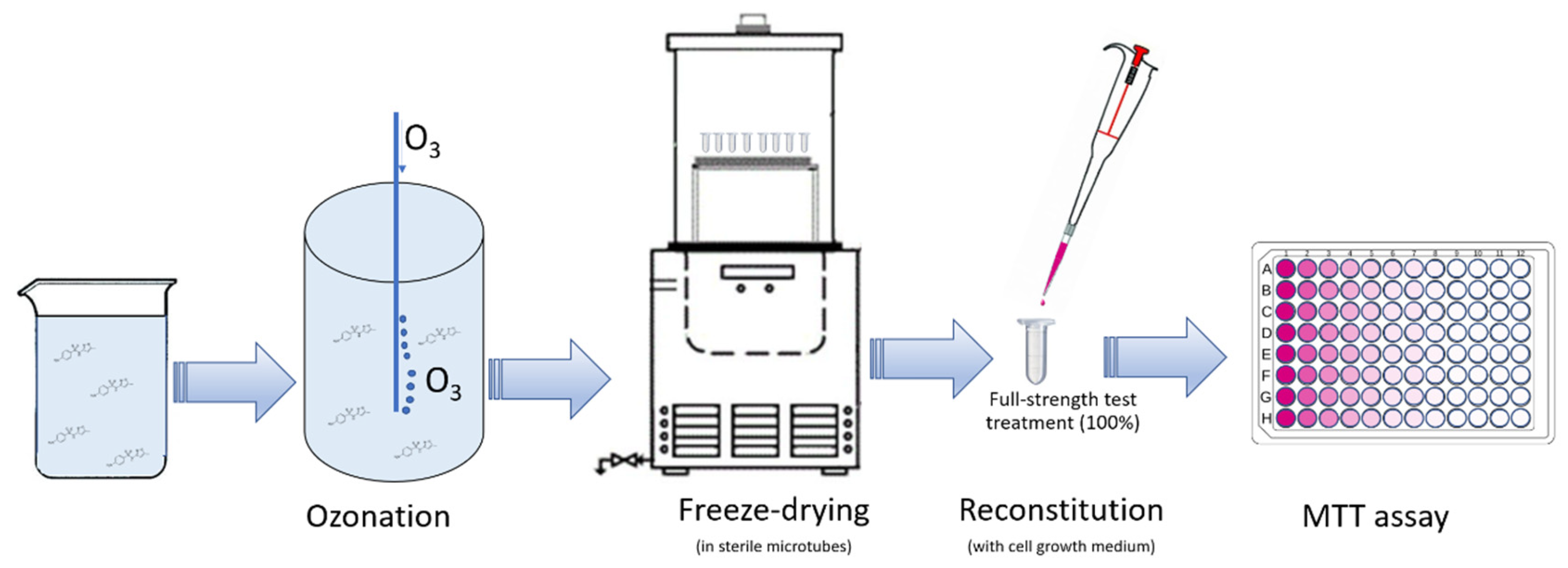
2.1. Samples Preparation

Methylparaben (MP), propylparaben (PP), paracetamol (PCT), sulfamethoxazole (SMX), and carbamazepine (CBZ) were dissolved in ultrapure water (1 mg/L of each compound) to create solutions with individual PPCPs and mixtures of two to five PPCPs (Table 1). These solutions were then treated by ozonation as described in detail by Jesus et al. [18]. Briefly, ozonation of each effluent was performed in a 2 L glass reactor at a temperature of 25 ± 1 °C. The duration of ozonation was set by the time required for the total degradation of the initial compounds (reaction time; Table 1). Ozone was produced by an ozone generator (802N, BMT, Berlin, Germany) from pure oxygen (99.9%). The ozone concentrations at the entrance and exit of the reactor were quantified by gas analyzers (Ozone analyzer 963 and 964, BMT, Berlin, Germany, respectively) and used to determine the transferred ozone dose (TOD; Table 1). All the materials used in the preparation process were sterilized beforehand. The degradation of the PPCPs in the solutions following ozonation was followed by high-performance liquid chromatography (HPLC) as also described in detail by Jesus et al. [18].
Considering the requirements of the toxicity tests applied in this study (see below), namely the need to ensure that cells are supplied with the necessary nutrients to grow and the effects noticed relate exclusively to the treatments, samples (2 mL) of ozone-treated and untreated solutions were freeze-dried (ScanVac, CoolSafe Basic & Pro, LaboGene ApS, Lillerød, Denmark) for 36 h at −100 °C in sterile microtubes, which were then stored at 4 °C in the dark until shortly before the onset of each corresponding toxicity test. As necessary to set up the test, lyophilized samples were reconstituted by adding 2 mL of cell growth medium (see below). This allowed establishing the full-strength test treatment (100%), which was then diluted with cell growth medium to obtain the remaining test treatments (12.5, 25 and 50% of sample).

2.2. Cell Culture and Testing

Human skin keratinocytes (HaCaT cell line—Cell Lines Service, Eppelheim, Germany) were cultured in 25 cm2 flasks in Dulbecco’s modified Eagle’s medium (DMEM) supplemented with 10% (v/v) fetal bovine serum, 1% fungizone, 1% glutamine, and 1% penicillin–streptomycin, incubated at 37 °C and 5% CO2. The cell culture was observed daily under a microscope to check morphology, confluence, and possible contamination.
The cytotoxicity of the treated and untreated solutions was assessed based on cell viability determined by the 3-(4,5-dimethyl-2-thiazolyl)-2,5-diphenyl tetrazolium bromide (MTT) colorimetric assay. The assay is based on the reduction of MTT, by mitochondrial dehydrogenases, to formazan crystals. Previously cultured cells were seeded in 96-well plates with supplemented DMEM at a density of 60,000 cells/mL, 40,000 cells/mL, or 20,000 cells/mL, for assays lasting 24, 48, and 72 h, respectively. Then, the plates were incubated for 20–24 h at 37 °C and 5% CO2 for cell adhesion. Afterwards, the medium was replaced with supplemented DMEM (negative control) or supplemented DMEM with each reconstituted sample (100%) and its dilutions (12.5; 25; 50%) and test plates were incubated at 37 °C and 5% CO2 for the desired exposure time. Three independent assays were run for each treated and untreated PPCP solution, each including six technical replicates of each test treatment. After 24, 48, or 72 h of exposure, the MTT reagent in PBS (1 mg/mL) was added to each well and left to incubate for four hours at 37 °C and 5% CO2 in the dark. The culture medium with the MTT reagent was then removed and dimethyl sulfoxide (DMSO) was added to each well for crystal solubilization. The plates were then shaken on an orbital shaker for two hours. The optical density of the reduced MTT was measured at 570 nm in a microtiter plate reader (Synergy HT, BioTeK Instruments Inc., Winooski, VT, USA). The values obtained allowed the determination of the percentage of viable cells, according to Equation (1).
Cell   viability   % = Optical   density test Optical   density ( blank ) Optical   density ( control ) Optical   density ( blank ) 100
The schematic outline of the experimental workflow from sample preparation to cytotoxicity testing is presented in Figure 1.

2.3. Data Analysis

The results were expressed and represented in graphs using the mean ± standard deviation of the three independent assays. The effects of each sample (untreated and ozone-treated PPCP solutions) were analyzed using one-way Analysis of Variance (ANOVA). For heteroscedastic data as confirmed by the Brown–Forsythe test, the non-parametric equivalent of the F-test (Kruskal–Wallis test) was used. To statistically distinguish the treated groups that differed from the negative control, Dunnett’s multiple comparisons test was used. An alpha level of 0.05 was used in all statistical tests.

3. Results and Discussion

3.1. Ozonation Treatment Efficacy

Ozonation efficacy analysis was carried out within the scope of the work by Jesus et al. [18]. Complete degradation of the initial compounds was confirmed in all samples. However, several degradation by-products of these same compounds were detected [18,22]: 2,4-dihydroxybenzoic acid (2,4-DHBA) and 3,4-dihydroxybenzoic acid (3,4-DHBA) for the MP effluent; 4-hydroxybenzoic acid (4-HBA) and 3,4-DMBA for the PP effluent; Oxalic acid (AO) and Hydroquinone (HQ) for the PCT effluent; 3-amino-5-methylisoxazole (AMI) for the SMX effluent; 1-(2-benzaldehyde)-4-hydro-(1H,3H)-quinazolin-2-one (BQM) for the CBZ effluent; and 4-HBA, 2,4-DHBA, 3,4-DHBA, and 3,4-DMBA for Mix 2. As the quoted authors discuss, this list represents the main by-products, and it may be incomplete since there are by-products that could not be identified in this analysis due to the low concentration and/or method limitations.

3.2. Potential Cytotoxicity Associated with the Use of Ozonation to Degrade PPCPs

The present study is, to our best knowledge, a pioneer in addressing the effects of municipal wastewater treatment (using synthetic effluents as a proxy) in human cell models, whereas there are studies focused on the efficiency of treatment of metal-loaded electroplating industrial wastewaters [25,26] and on cytotoxicity assessment of phosphogypsum transport water from waste ponds of a fertilizer factory [27]. This is somewhat surprising given the likelihood of human exposure, particularly of skin cells, during the use of reclaimed water. For example, the use of reclaimed water in pools leads to direct exposure of pool-users skin cells; also, its use for irrigation may imply occupational exposure. Technical issues may have been the reason for this scarcity of studies. Indeed, the cell testing set up has high requirements in terms of sterilization of all elements involved, and the testing of effluents poses challenges concerning the preparation of test solutions in such a way that the assessment over full-strength solutions can be made without constraining the nutrient supply to the cells. The methods applied herein were successful in both preventing contaminations (prioritization of sterile conditions while preparing the test solutions) and allowing testing of full-strength effluents (lyophilization and resuspension in cell culture medium), thus, the overcoming of these technical difficulties will hopefully stimulate the investigation in the field. Considering the above, although some studies examine the cytotoxicity of various PPCPs used here in different cell lines, it should be noted that the results are not fully comparable with those of the present study, yet some parallels can be made, feeding several discussion items in the following sections.
The cytotoxicity associated with the application of ozonation for effluent treatment was assessed by recording the mitochondrial metabolic rate (MTT) following HaCaT cell exposure to the synthetic effluents before and after ozonation treatment. In this context, three different exposure times were tested (24, 48, and 72 h) to empirically understand whether the elongation of the exposure period could modulate the toxic response. In general, extending the exposure period led to an increase in toxicity from 24 to 72 h (compare Figure 2 and Figure 3 with Figures S1 and S2), providing a larger breadth for interpretation of effects and thus driving most of the discussion below.

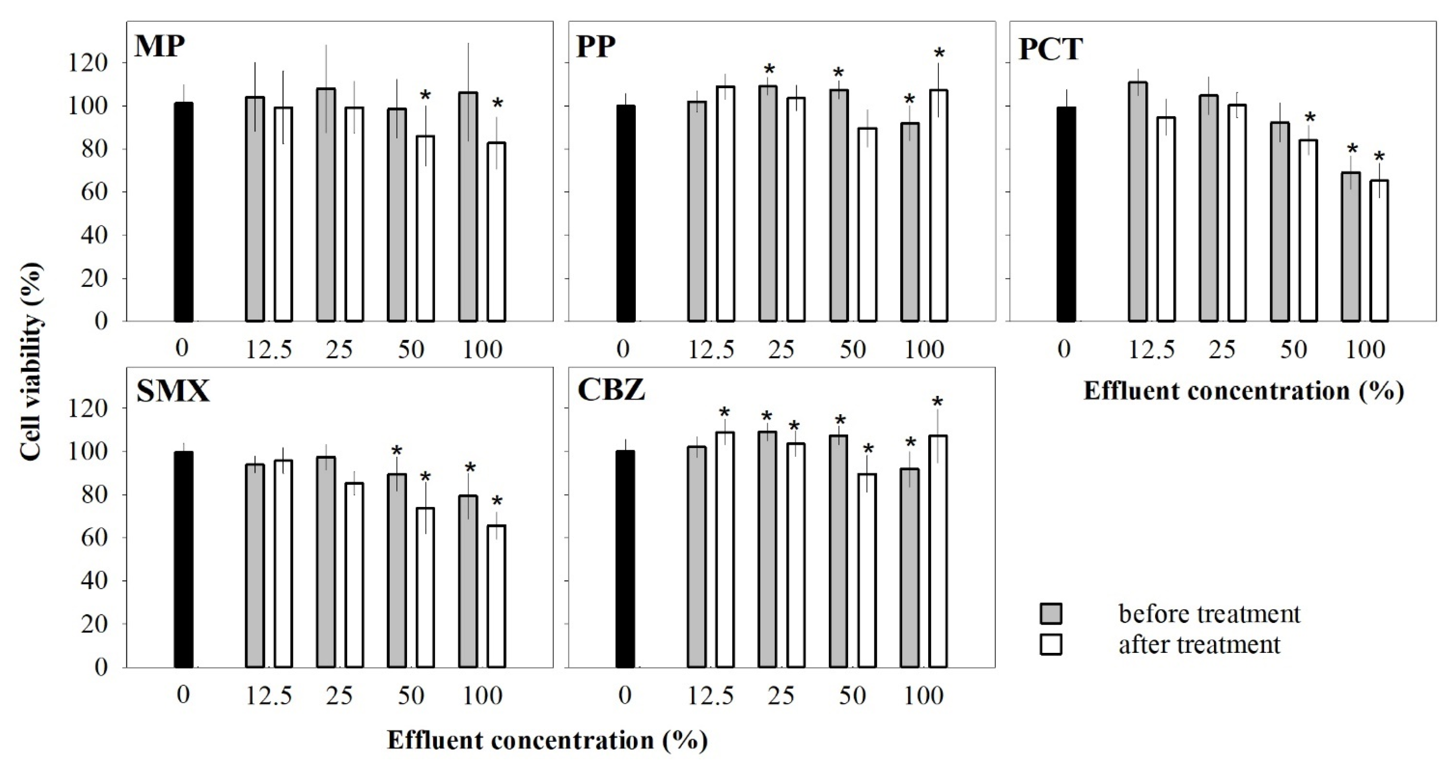
3.2.1. Effects of Single-Chemical Effluents on Cell Viability

In general, exposure to the single-chemical effluent samples resulted in toxic effects on HaCaT cells, significantly decreasing cell viability, mostly when the full-strength effluent (100%) or its half dilution (50%) were tested (Figure 2; Table S1).
The untreated MP effluent was ineffective towards cell viability after 24 h (Figure S1) and 72 h of exposure (Figure 2), while significantly decreased cell viability was found after 48 h of exposure to 25–100% effluent strength (Figure S1). This apparent inconsistency in toxicity records at 48 h is likely a result of the lower variability within treatment, exposing a potential negative effect of MP in cell viability. MP has already been reported to accelerate the cellular aging of human normal primary epidermal keratinocytes (NHEK) at concentrations ranging within 7.6 × 10⁻⁴–0.152 mg/L [28], which is below the ranges expected for MP concentration in effluent concentrations of 25–100%. A possible explanation for the toxicity of parabens considers their mode of action at the cellular level: alteration of the mitochondrial permeability transition pore, causing an increase in intracellular calcium and an inhibition in electron transport, which may induce apoptosis [29]. On the contrary, the treated MP effluent at 50 and 100% significantly decreased HaCaT viability after 72 h of exposure (after 24 h no cytotoxicity was found, and after 48 h, the same pattern arose as after 72 h; Figure S1), suggesting that the formed by-products, 2,4-DHBA and 3,4-DHBA (see Section 3.1), should be cytotoxic upon prolonged exposure. In fact, 3,4-DHBA decreased the viability of breast cancer (MCF-7), lung cancer (A549), liver cancer (HepG2), cervical cancer (HeLa), and prostate cancer (LNCaP) cell lines [30], while 2,4-DHBA decreased viability in MCF-7 cell line and breast cancer cells (MDA-MB-231) [31]. There is some controversy about the mechanism of toxicity of these by-products, specifically concerning their interaction with reactive oxygen species (ROS). The antioxidant efficiency of hydroxybenzoic acids (the class to which some of these degradation products belong) is argued to depend on several factors, namely the number and position of the hydroxyl group attached to the aromatic ring [32], but overall, they would not be expected to present toxicity as observed herein (Figure 2). Although by-product concentrations could not be determined, the results obtained are consistent with the quoted works, showing toxicity by 2,4-DHBA and 3,4-DHBA to HaCaT cells rather than any protection derived from their claimed primary function as antioxidants, depending on molecular structure, but rather due to mechanisms not yet unveiled.
The untreated PP effluent significantly decreased cell viability in HaCaT only at full-strength (1 mg/L PP), regardless of the exposure period (Figure 2 and Figure S1). This pattern is in general agreement with previous studies with other cell lines, such as MCF-7 tested with PP concentrations above 3.604 × 10⁻³ mg/L [33], human breast carcinoma cells (MDA-kb2) at more than 0.0225 mg/L PP [34], and HepG2 at 4 × 10⁻⁴ to 4.6 × 10⁻³ mg/L PP [35]. However, Fouyet et al. [36] reported that 0.0180 mg/L of PP slightly reduced cell viability, but in a non-cytotoxic manner, in human placenta (JEG-Tox) and HaCaT cell lines. These data are not fully comparable with our results since the concentrations of the untreated PP effluent affecting cell viability in the present work are higher (0.125 to 1 mg/L), with negative effects being noticed only at a concentration equivalent of 1 mg/L regardless of the exposure period (Figure 2 compared to Figure S1). The noticed negative effects could result from the increased production of peroxide anions, which causes a reduction in mitochondrial membrane potential and induces cytotoxicity [35]. The treated PP effluent showed significant toxicity only when tested at full-strength (after 48 h and 72 h). For 4-HBA, one of the identified by-products of the PP oxidation, no decrease in cell viability was reported in the immortalized human colorectal adenocarcinoma cell line (Caco-2) at 1, 3.5, 18, and 36 mg/L [37], or in HaCaT cells at 25, 50, and 100 mg/L [38]. Also, 3,4-DMBA, the other by-product identified, was not proved to induce cytotoxicity in this latter cell line at 10, 50, and 100 mg/L [39]. These data seem inconsistent with the results obtained herein after exposure to the treated PP effluent (Figure 2) after 72 h of exposure, unless unidentified by-products other than 4-HBA and 3,4-DMBA (Section 3.1) are present that induce cytotoxic effects.
Behrends et al. [40] described that exposure to PCT at concentrations of 0.302 to 2.267 mg/L led to cell death in HepG2, probably due to damage to mitochondrial proteins, potentially reducing respiration and inducing oxidative stress, although the exact mechanism is not yet understood. This evidence is consistent with the results obtained herein, as cytotoxicity was identified at 1 mg/L of PCT in the untreated effluent after 72 h of exposure (Figure 2). In the treated PCT sample, the initial compound was degraded, so the toxicity evidenced following exposure to 50 and 100% treated effluent (Figure 2) should originate from the by-products produced, namely AO and HQ (Section 3.1). Guo et al. [41] describe that AO exposure (≥2 mg/L) causes cell death in human proximal tubule cells (HPT) and exhibits cytotoxicity in porcine renal tubule cells (LLC-PK1) and canine Madin-Darby kidney cells (MDCK). These toxic effects may reflect the accumulation of hydroxyl radicals [42]. Pereira et al. [43] showed that HQ decreased the cell viability of colon cancer cells (HCT116) and primary human fibroblasts. According to some authors, HQ may induce DNA damage by a combination of mitotic spindle damage, type II topoisomerase inhibition, and breaks in DNA chains through ROS generation [43].
There are not many studies evaluating the cytotoxicity associated with SMX as an isolated compound. However, Monteiro [44] demonstrated that SMX (1 to 800 mg/L) significantly reduced cell viability in breast tumor (4T1), primary osteosarcoma (Saos-2), normal osteoblast (MC3T3), and fibroblast (3T3-L1) cell lines, like our results obtained following a 72-h exposure to 0.5 and 1 mg/L SMX in the untreated effluent for HaCaT (Figure 2). After ozonation, SMX was degraded, originating AMI as the main by-product (Section 3.1), and the cytotoxicity of the effluent appreciably increased: significantly decreased cell viability was found for treated effluents, undiluted and diluted by 50% and 75% (Figure 2). Although no studies concerning AMI cytotoxicity were found that can provide confirmation, it seems reasonable to postulate the link between the by-product formed and the observed cell viability decrease.
Finally, toxicity was observed with exposure for 72 h to 0.25–1 mg/L of CBZ in the untreated effluent (Figure 2), with similar patterns found for shorter exposures (Figure S1). This evidence is apparently contrary to what would be expected from data available in the literature. Al-Musawi et al. [45] reported no toxicity of 0.402 mg/L CBZ to HaCaT cells, despite cytotoxicity to rat brain microvascular endothelial cells (rCMECs) being found after exposure to 0.236 mg/L CBZ and reduced viability of oral keratinocyte cells (OKF6-TERT1) being observed for CBZ at 0.402 mg/L. The cytotoxicity was not alleviated by the ozonation treatment (Figure 2), although CBZ had been fully degraded [18]. The treated CBZ effluent contains BQM as highlighted in Section 3.1, which was reported to induce cytotoxicity in Chinese Hamster ovary fibroblast cells (CHO-K1), presumably due to the quinazoline within the compound molecular structure [46].
Overall, the cytotoxicity of the untreated and ozone-treated effluents is similar within each concentration. After a 72-h exposure period, the differences in the cell viability between untreated and ozone-treated samples were null or minor: for full-strength samples, the most pronounced difference was found for MP (24% improvement on cell viability). These data suggest that, despite yielding the full removal of the target compounds, ozonation was not remarkably effective on removing cytotoxicity to the HaCaT cells, even causing, in some cases, a slight increase in cytotoxicity (see, e.g., the data for the full-strength PP sample in Figure 2, where an 8% decrease in cell viability was observed).

3.2.2. Effects of Effluents with Mixed PPCPs on Cell Viability

There was a progressive and significant decrease in cell viability with increasing concentration of the effluents (both untreated and treated), at least from 25 to 100%, with, in many cases, a significant decrease being noticed also at 12.5% (Figure 3 for 72 h and Figure S2 for 24 and 48 h; Table S2 for statistical summaries). This decrease was more pronounced for the mixture of parabens (Mix 2), with average cell viability reaching values as low as 48.69% and 42.66% (untreated and ozone-treated effluent, respectively) upon exposure to the 100% effluent concentration (72 h exposure; Figure 3).
As observed for single PPCPs, treated effluents yielded effects in cells that are similar across concentrations to their untreated counterparts, with differences in the cell viability between untreated and ozone-treated samples in the range 0.78–8.46%. Remarkably, the cell viability of the treated samples was commonly lower than that of the untreated samples. The narrower range observed for the mixtures suggests that ozonation is less efficient (concerning toxicity removal) towards mixtures than towards single PPCP contaminants. For instance, Mix 2 caused higher cytotoxicity (Figure 3) than each of the components alone (Figure 2), once cell viability inhibition higher than 20% was found from 12.5% effluent onwards, while these records were found only at full strength for single paraben effluents. Moreover, taking the treated 100% effluent concentration as an example, viability inhibition was ≤20% for MP and PP, individually, but reached 60% for the mixture. The same pattern was observed for untreated effluents. This suggests that the parabens or their by-products exhibit more-than-additivity regarding cytotoxicity to human keratinocyte cells. Interestingly, a synergistic interaction among the components of an ozone-treated mixture of MP and PP has also been observed for aquatic species, namely Daphnia magna and Raphidocelis subcapitata [18], suggesting that the increased toxicity of this mixture following ozonation might be transversal to humans and aquatic species. Mixture 3, adding parabens and PCT, followed a similar trend, despite being less cytotoxic than Mix 2. The untreated and treated Mix 4 and 5 effluents presented lower cytotoxicity than Mix 2 and Mix 3 (Figure 3), as interpreted from the lower magnitude of the effects promoted (slightly more than 20% cell viability inhibition following exposure to full-strength effluents) compared to the effects noted in simpler mixtures (40–60% cell viability inhibition following exposure to full-strength effluents).
This apparent decrease in relative toxicity with the increase in effluent mixture complexity, which is common to both the untreated and ozone-treated effluents, suggests that as one adds more compounds, there is a trend for reversing the interactive behavior of either the parent compounds or the by-products generated after treatment. Regarding the treated effluents, this might be partially explained by the increased reaction time (and TOD) with increasing the mixture complexity (Table 1). In fact, the increase in the ozonation time could enhance the by-products oxidation (e.g., [18]), which can result in less cytotoxic solutions. However, the similarity of the cell viability between untreated and ozone-treated samples suggests that this effect might be independent of the reaction time, being mainly determined by the number and type of contaminants in the mixture.
Assessing the toxicity of treated mixtures of PPCPs to human cell lines is crucial to reliably conclude on the safety of reclaimed water. In the present study, it was shown that ozonation of synthetic effluents containing parabens, PCT, SMX, and CBZ had no pronounced beneficial effect on their cytotoxicity. Interestingly, these results agree, in general, with the conclusions of a previous study addressing the toxicity of these samples to aquatic species [18]. It is then evident that monitoring the concentration of parent compounds and by-products is not sufficient to safely decide on the potential use of reclaimed water. Hence, it is recommended to optimize the ozonation processes towards safe use of reclaimed water, e.g., by increasing the ozone doses, increasing pH to produce more hydroxyl radicals, or even using catalysts to favor the ozone decomposition, which will increase the oxidation potential [47]. It should be kept in mind that for low concentrations of single contaminants (such as the dilution 12.5%), which are closer to environmental concentrations [23], commonly the observed cytotoxicity of both untreated and ozone-treated samples was negligible compared to the control treatment. However, regarding mixtures, cytotoxicity was observed for all ozone-treated samples, including the dilution of 12.5%, which suggests that ozonation of samples containing mixtures of contaminants might cause higher cytotoxicity than ozonation of individual contaminants. Future studies should extend the mixture complexity to real wastewater samples, and should consider other advanced oxidation processes and other cell lines. For instance, intestinal epithelial cells as well as lung epithelial cells might be targeted when handling reclaimed water.

4. Conclusions

Starting from synthetic effluents contaminated with single or mixed PPCPs (MP, PP, PCT, SMX, CBZ), ozonation fully degraded the initial compounds, either if they were occurring singly or in mixtures. Exposure to ozone-treated effluents induced adverse effects on HaCaT cells, comparable to those following exposure to parent compounds in counterpart effluents. The cytotoxicity of treated effluents, formerly contaminated with single PPCPs or their mixtures, is likely related to by-products formation during ozonation. The cytotoxicity of untreated and treated effluents tended to decrease with increasing complexity (number of contaminants) of the effluents, with the mixture of parabens showing the highest toxicity. For this mixture, there was also an evidently increased toxicity compared to single PPCPs, suggesting a synergistic interaction among the produced by-products. Our results suggest, for the first time, that ozonation is poorly efficient in rendering water contaminated with PPCPs non-toxic for human cells, flagging the need for optimization of the treatment processes towards a safe use of reclaimed water. However, the scarcity of comparable studies in the literature for validation of the present conclusions should be noted, claiming for further investment in understanding the potential human health toxicity of treated effluents beyond the successful degradation of parent contaminants. This naturally includes the follow-up on highly controlled studies with synthetic effluents such as ours by addressing more complex (real) matrices contaminated with PPCPs, individually or in mixture, and the by-products formed during ozonation and other advanced oxidation processes. Lastly, the experimental procedure developed in the present study should be highlighted, as it was proven effective to appropriately meet the sterilization conditions required and to test full-strength solutions. This is a new asset that promotes the much-needed knowledge enrichment concerning the safety of reclaimed water to human health.

Supplementary Materials

The following supporting information can be downloaded at: https://www.mdpi.com/article/10.3390/app15147784/s1, Table S1: Summary of the one-way ANOVA (F-test) or non-parametric equivalent (Kruskall-Wallis test; H) applied to the data obtained in the cell viability tests with the untreated and ozonated effluents under study, considering exposures of 24 h, 48 h and 72 h (all independent tests); Table S2: Summary of the one-way ANOVA (F-test) or non-parametric equivalent (Kruskall-Wallis test; H) applied to the data obtained in the cell viability tests with the untreated and ozonated effluents under study, considering exposures of 24 h, 48 h and 72 h (all independent tests); Figure S1: Cell viability recorded in MTT assays with the HaCaT cell line exposed for 24 and 48 h to a range of concentrations of the untreated and ozone-treated MP, PP, PCT, SMX and CBZ effluents. Bars represent the mean of three independent assays and error bars represent the corresponding standard deviation. Asterisks indicate the groups that differ significantly from the control (Dunnett’s test or non-parametric equivalent; p < 0.05); Figure S2. Cell viability recorded in MTT assays with the HaCaT cell line exposed for 24 and 48 h to a range of concentrations of the untreated and ozone-treated Mix 2 (MP + PP), Mix 3 (MP + PP + PCT), Mix 4 (MP + PP + PCT + SMX) and Mix 5 (MP + PP + PCT + SMX + CBZ) effluents. Bars represent the mean of three independent assays and error bars represent the corresponding standard deviation. Asterisks indicate the groups that differ significantly from the control (Dunnett’s test or the non-parametric equivalent; p < 0.05).

Author Contributions

Conceptualization, A.T.R., H.O., J.G. and J.L.P.; investigation, A.T.R. and F.J.; resources, H.O., J.G. and J.L.P.; supervision, H.O., J.G. and J.L.P.; writing—original draft, A.T.R.; writing—review and editing, F.J., H.O., J.G. and J.L.P. All authors have read and agreed to the published version of the manuscript.

Funding

This research was funded by national funds through FCT within the scope of the project/support to CESAM (UID/50006 + LA/P/0094/2020), as well as CERES (UIDB/00102/2020). JG and HO acknowledge FCT/MCTES for the research contracts under the Scientific Employment Stimulus CEECIND/01207/2018 (doi: 10.54499/CEECIND/01207/2018/CP1585/CT0003) and CEECIND/04050/2017 (doi: 10.54499/CEECIND/04050/2017/CP1459/CT0023), respectively.

Institutional Review Board Statement

Not applicable.

Informed Consent Statement

Not applicable.

Data Availability Statement

The original contributions presented in this study are included in the article and Supplementary Materials. Further inquiries can be directed to the corresponding authors.

Conflicts of Interest

The authors declare no conflicts of interest.

References

  1. Santos, A.F.; Alvarenga, P.; Gando-Ferreira, L.M.; Quina, M.J. Urban Wastewater as a Source of Reclaimed Water for Irrigation: Barriers and Future Possibilities. Environments 2023, 10, 17. [Google Scholar] [CrossRef]
  2. Maryam, B.; Büyükgüngör, H. Wastewater Reclamation and Reuse Trends in Turkey: Opportunities and Challenges. J. Water Process Eng. 2019, 30, 100501. [Google Scholar] [CrossRef]
  3. Garrido-Cardenas, J.A.; Esteban-García, B.; Agüera, A.; Sánchez-Pérez, J.A.; Manzano-Agugliaro, F. Wastewater Treatment by Advanced Oxidation Process and Their Worldwide Research Trends. Int. J. Environ. Res. Public Health 2020, 17, 170. [Google Scholar] [CrossRef] [PubMed]
  4. Shahriar, A.; Tan, J.; Sharma, P.; Hanigan, D.; Verburg, P.; Pagilla, K.; Yang, Y. Modeling the Fate and Human Health Impacts of Pharmaceuticals and Personal Care Products in Reclaimed Wastewater Irrigation for Agriculture. Environ. Pollut. 2021, 276, 116532. [Google Scholar] [CrossRef]
  5. Kumar, S.; Talan, A.; Boyle, K.; Ormeci, B.; Drogui, P.; Tyagi, R.D. Water Recycling: Economic and Environmental Benefits. In Biomass, Biofuels, Biochemicals. Circular Bioeconomy—Current Developments and Future Outlook; Elsevier: Amsterdam, The Netherlands, 2021; pp. 91–120. [Google Scholar] [CrossRef]
  6. Deng, S.; Yan, X.; Zhu, Q.; Liao, C. The Utilization of Reclaimed Water: Possible Risks Arising from Waterborne Contaminants. Environ. Pollut. 2019, 254, 113020. [Google Scholar] [CrossRef]
  7. Niemi, L.; Taggart, M.; Boyd, K.; Zhang, Z.; Gaffney, P.P.J.; Pfleger, S.; Gibb, S. Assessing Hospital Impact on Pharmaceutical Levels in a Rural ‘Source-to-Sink’ Water System. Sci. Total Environ. 2020, 737, 139618. [Google Scholar] [CrossRef]
  8. Silva, C.; Cachada, A.; Gonçalves, F.J.M.; Nannou, C.; Lambropoulou, D.; Patinha, C.; Abrantes, N.; Pereira, J.L. Chemical Characterization of Riverine Sediments Affected by Wastewater Treatment Plant Effluent Discharge. Sci. Total Environ. 2022, 839, 156305. [Google Scholar] [CrossRef]
  9. Wu, Y.; Song, S.; Chen, X.; Shi, Y.; Cui, H.; Liu, Y.; Yang, S. Source-Specific Ecological Risks and Critical Source Identification of PPCPs in Surface Water: Comparing Urban and Rural Areas. Sci. Total Environ. 2023, 854, 158792. [Google Scholar] [CrossRef]
  10. Ebele, A.J.; Abou-Elwafa Abdallah, M.; Harrad, S. Pharmaceuticals and Personal Care Products (PPCPs) in the Freshwater Aquatic Environment. Emerg. Contam. 2017, 3, 1–16. [Google Scholar] [CrossRef]
  11. Kujawska, A.; Kiełkowska, U.; Atisha, A.; Yanful, E.; Kujawski, W. Comparative Analysis of Separation Methods Used for the Elimination of Pharmaceuticals and Personal Care Products (PPCPs) from Water—A Critical Review. Sep. Purif. Technol. 2022, 290, 120797. [Google Scholar] [CrossRef]
  12. Narayanan, M.; El-sheekh, M.; Ma, Y.; Pugazhendhi, A.; Natarajan, D.; Kandasamy, G.; Raja, R.; Saravana Kumar, R.M.; Kumarasamy, S.; Sathiyan, G.; et al. Current Status of Microbes Involved in the Degradation of Pharmaceutical and Personal Care Products (PPCPs) Pollutants in the Aquatic Ecosystem. Environ. Pollut. 2022, 300, 118922. [Google Scholar] [CrossRef] [PubMed]
  13. Mellah, A.; Fernandes, S.P.S.; Rodríguez, R.; Otero, J.; Paz, J.; Cruces, J.; Medina, D.D.; Djamila, H.; Espiña, B.; Salonen, L.M. Adsorption of Pharmaceutical Pollutants from Water Using Covalent Organic Frameworks. Chem. A Eur. J. 2018, 24, 10601–10605. [Google Scholar] [CrossRef] [PubMed]
  14. Wang, J.; Chen, H. Catalytic Ozonation for Water and Wastewater Treatment: Recent Advances and Perspective. Sci. Total Environ. 2020, 704, 135249. [Google Scholar] [CrossRef] [PubMed]
  15. Gomes, J.; Costa, R.; Quinta-Ferreira, R.M.; Martins, R.C. Application of Ozonation for Pharmaceuticals and Personal Care Products Removal from Water. Sci. Total Environ. 2017, 586, 265–283. [Google Scholar] [CrossRef]
  16. Kanakaraju, D.; Glass, B.D.; Oelgemöller, M. Advanced Oxidation Process-Mediated Removal of Pharmaceuticals from Water: A Review. J. Environ. Manag. 2018, 219, 189–207. [Google Scholar] [CrossRef]
  17. Potivejkul, S.; Pimdee, P.; Phimolsathien, T. Perceptions on Ozonation Water Treatment Use: An Alternative Idea for Asean Water Resource Sustainability. J. Sustain. Sci. Manag. 2017, 12, 49–59. [Google Scholar]
  18. Jesus, F.; Bernardo, C.; Martins, R.C.; Gomes, J.; Pereira, J.L. Ecotoxicological Consequences of the Abatement of Contaminants of Emerging Concern by Ozonation—Does Mixture Complexity Matter? Water 2022, 14, 1801. [Google Scholar] [CrossRef]
  19. Gomes, J.; Frasson, D.; Pereira, J.L.; Gonçalves, F.J.M.; Castro, L.M.; Quinta-Ferreira, R.M.; Martins, R.C. Ecotoxicity Variation through Parabens Degradation by Single and Catalytic Ozonation Using Volcanic Rock. Chem. Eng. J. 2019, 360, 30–37. [Google Scholar] [CrossRef]
  20. Gomes, J.; Jesus, F.; Domingues, E.; Gonçalves, F.; Pereira, J.L.; Martins, R.C. Photocatalytic Oxidation of Pharmaceutical Contaminants of Emerging Concern Using Sunlight and Visible Radiation: Mechanism and Ecotoxicological Evaluation. J. Water Process Eng. 2021, 43, 102204. [Google Scholar] [CrossRef]
  21. Pereira, A.; Silva, L.; Lino, C.; Meisel, L.; Pena, A. Assessing Environmental Risk of Pharmaceuticals in Portugal: An Approach for the Selection of the Portuguese Monitoring Stations in Line with Directive 2013/39/EU. Chemosphere 2016, 144, 2507–2515. [Google Scholar] [CrossRef]
  22. Gomes, J.; Bernardo, C.; Jesus, F.; Pereira, J.L.; Martins, R.C. Ozone Kinetic Studies Assessment for the PPCPs Abatement: Mixtures Relevance. Chem. Eng. 2022, 6, 20. [Google Scholar] [CrossRef]
  23. Khasawneh, O.F.S.; Palaniandy, P. Occurrence and removal of pharmaceuticals in wastewater treatment plants. Process Saf. Environ. Prot. 2021, 150, 532–556. [Google Scholar] [CrossRef]
  24. Saeid, S.; Kråkström, M.; Tolvanen, P.; Kumar, N.; Eränen, K.; Mikkola, J.-P.; Kronberg, L.; Eklund, P.; Peurla, M.; Aho, A.; et al. Advanced Oxidation Process for Degradation of Carbamazepine from Aqueous Solution: Influence of Metal Modified Microporous, Mesoporous Catalysts on the Ozonation Process. Catalysts 2020, 10, 90. [Google Scholar] [CrossRef]
  25. Durgo, K.; Oreščanin, V.; Horvat, T.; Oreščanin, V.; Mikelić, L.; Čolić, J.F.; Lulić, S. Cytotoxicity and Mutagenicity Study of Waste and Purified Water Samples from Electroplating Industries Prepared by Use of Ferrous Sulfate and Wood Fly Ash. J. Environ. Sci. Health Part A 2005, 40, 949–957. [Google Scholar] [CrossRef]
  26. Orescanin, V.; Kopjar, N.; Durgo, K.; Elez, L.; Gustek, S.F.; Colic, J.F. Citotoxicity Status of Electroplating Wastewater Prior/after Neutralization/Purification with Alkaline Solid Residue of Electric Arc Furnace Dust. J. Environ. Sci. Health Part. A 2009, 44, 273–278. [Google Scholar] [CrossRef]
  27. Durgo, K.; Oreščanin, V.; Lulić, S.; Kopjar, N.; elježić, D.Ž.; Čolić, J.F. The Assessment of Genotoxic Effects of Wastewater from a Fertilizer Factory. J. Appl. Toxicol. 2009, 29, 42–51. [Google Scholar] [CrossRef]
  28. Kizhedath, A.; Wilkinson, S.; Glassey, J. Assessment of Hepatotoxicity and Dermal Toxicity of Butyl Paraben and Methyl Paraben Using HepG2 and HDFn In Vitro Models. Toxicol. Vitr. 2019, 55, 108–115. [Google Scholar] [CrossRef]
  29. Nakagawa, Y.; Moldéus, P. Mechanism of P-Hydroxybenzoate Ester-Induced Mitochondrial Dysfunction and Cytotoxicity in Isolated Rat Hepatocytes. Biochem. Pharmacol. 1998, 55, 1907–1914. [Google Scholar] [CrossRef]
  30. Yin, M.-C.; Lin, C.-C.; Wu, H.-C.; Tsao, S.-M.; Hsu, C.-K. Apoptotic Effects of Protocatechuic Acid in Human Breast, Lung, Liver, Cervix, and Prostate Cancer Cells: Potential Mechanisms of Action. J. Agric. Food Chem. 2009, 57, 6468–6473. [Google Scholar] [CrossRef]
  31. Yip, E.C.H.; Chan, A.S.L.; Pang, H.; Tam, Y.K.; Wong, Y.H. Protocatechuic Acid Induces Cell Death in HepG2 Hepatocellular Carcinoma Cells through a C-Jun N-Terminal Kinase-Dependent Mechanism. Cell Biol. Toxicol. 2006, 22, 293–302. [Google Scholar] [CrossRef]
  32. Kalinowska, M.; Gołębiewska, E.; Świderski, G.; Męczyńska-Wielgosz, S.; Lewandowska, H.; Pietryczuk, A.; Cudowski, A.; Astel, A.; Świsłocka, R.; Samsonowicz, M.; et al. Plant-derived and dietary hydroxybenzoic acids—A comprehensive study of structural, anti-/pro-oxidant, lipophilic, antimicrobial, and cytotoxic activity in mda-mb-231 and mcf-7 cell lines. Nutrients 2021, 13, 3107. [Google Scholar] [CrossRef] [PubMed]
  33. Wróbel, A.; Gregoraszczuk, E.L. Effects of Single and Repeated In Vitro Exposure of Three Forms of Parabens, Methyl-, Butyl- and Propylparabens on the Proliferation and Estradiol Secretion in MCF-7 and MCF-10A Cells. Pharmacol. Rep. 2013, 65, 484–493. [Google Scholar] [CrossRef] [PubMed]
  34. Klopčič, I.; Kolšek, K.; Dolenc, M.S. Glucocorticoid-like Activity of Propylparaben, Butylparaben, Diethylhexyl Phthalate and Tetramethrin Mixtures Studied in the MDA-Kb2 Cell Line. Toxicol. Lett. 2015, 232, 376–383. [Google Scholar] [CrossRef] [PubMed]
  35. Szelag, S.; Zabłocka, A.; Trzeciak, K.; Drozd, A.; Baranowska-Bosiacka, I.; Kolasa, A.; Goschorska, M.; Chlubek, D.; Gutowska, I. Propylparaben-Induced Disruption of Energy Metabolism in Human HepG2 Cell Line Leads to Increased Synthesis of Superoxide Anions and Apoptosis. Toxicol. Vitr. 2016, 31, 30–34. [Google Scholar] [CrossRef]
  36. Fouyet, S.; Olivier, E.; Leproux, P.; Dutot, M.; Rat, P. Pregnant Women and Endocrine Disruptors: Role of P2X7 Receptor and Mitochondrial Alterations in Placental Cell Disorders. Cells 2022, 11, 495. [Google Scholar] [CrossRef]
  37. Ottaviano, E.; Baron, G.; Fumagalli, L.; Leite, J.; Colombo, E.A.; Artasensi, A.; Aldini, G.; Borghi, E. Candida Albicans Biofilm Inhibition by Two Vaccinium Macrocarpon (Cranberry) Urinary Metabolites: 5-(3′,4′-DihydroxyPhenyl)-γ-Valerolactone and 4-Hydroxybenzoic Acid. Microorganisms 2021, 9, 1492. [Google Scholar] [CrossRef]
  38. Mittraphab, Y.; Amen, Y.; Nagata, M.; Matsumoto, M.; Wang, D.; Shimizu, K. Anti-Phototoxicity Effect of Phenolic Compounds from Acetone Extract of Entada Phaseoloides Leaves via Activation of COX-2 and INOS in Human Epidermal Keratinocytes. Molecules 2022, 27, 440. [Google Scholar] [CrossRef]
  39. Shin, S.W.; Jung, E.; Kim, S.; Lee, K.-E.; Youm, J.-K.; Park, D. Antagonist Effects of Veratric Acid against UVB-Induced Cell Damages. Molecules 2013, 18, 5405–5419. [Google Scholar] [CrossRef]
  40. Behrends, V.; Giskeødegård, G.F.; Bravo-Santano, N.; Letek, M.; Keun, H.C. Acetaminophen Cytotoxicity in HepG2 Cells Is Associated with a Decoupling of Glycolysis from the TCA Cycle, Loss of NADPH Production, and Suppression of Anabolism. Arch. Toxicol. 2019, 93, 341–353. [Google Scholar] [CrossRef]
  41. Guo, C.; McMartin, K.E. The Cytotoxicity of Oxalate, Metabolite of Ethylene Glycol, Is Due to Calcium Oxalate Monohydrate Formation. Toxicology 2005, 208, 347–355. [Google Scholar] [CrossRef]
  42. Schepers, M.S.J.; Van Ballegooijen, E.S.; Bangma, C.H.; Verkoelen, C.F. Oxalate Is Toxic to Renal Tubular Cells Only at Supraphysiologic Concentrations. Kidney Int. 2005, 68, 1660–1669. [Google Scholar] [CrossRef] [PubMed]
  43. Pereira, P.; Enguita, F.J.; Ferreira, J.; Leitão, A.L. DNA Damage Induced by Hydroquinone Can Be Prevented by Fungal Detoxification. Toxicol. Rep. 2014, 1, 1096–1105. [Google Scholar] [CrossRef] [PubMed]
  44. Monteiro, A. Preparação e Caracterização de Nanofibras Magnéticas Para Liberação Controlada de Fármacos e Hipertemia. Ph.D Thesis, Universidade Federal de Minas Gerais, Belo Horizonte, Brazil, 2018. [Google Scholar]
  45. Al-Musawi, M.; Durham, J.; Whitworth, J.M.; Stone, S.J.; Nixdorf, D.R.; Valentine, R.A. Effect of Topical Neuromodulatory Medications on Oral and Skin Keratinocytes. J. Oral. Pathol. Med. 2017, 46, 134–141. [Google Scholar] [CrossRef] [PubMed]
  46. Han, Y.; Ma, M.; Li, N.; Hou, R.; Huang, C.; Oda, Y.; Wang, Z. Chlorination, Chloramination and Ozonation of Carbamazepine Enhance Cytotoxicity and Genotoxicity: Multi-Endpoint Evaluation and Identification of Its Genotoxic Transformation Products. J. Hazard. Mater. 2018, 342, 679–688. [Google Scholar] [CrossRef]
  47. Gomes, J.F.; Lopes, A.; Gmurek, M.; Quinta-Ferreira, R.M.; Martins, R.C. Study of the influence of the matrix characteristics over the photocatalytic ozonation of parabens using Ag-TiO2. Sci. Total Environ. 2019, 646, 1468–1477. [Google Scholar] [CrossRef]
Figure 1. Schematic outline of the experimental workflow.
Applsci 15 07784 g001
Figure 2. Cell viability recorded in MTT assays with the HaCaT cell line exposed for 72 h to a range of concentrations of the untreated and ozone-treated MP, PP, PCT, SMX, and CBZ effluents. Bars represent the mean of three independent assays and error bars represent the corresponding standard deviation. Asterisks indicate the groups that differ significantly from the control (Dunnett’s test; p < 0.05). The detailed statistical summaries are available in Supplementary Materials (Table S1).
Applsci 15 07784 g002
Figure 3. Cell viability recorded in MTT assays with the HaCaT cell line exposed for 72 h to a range of concentrations of the untreated and ozone-treated Mix 2 (MP + PP), Mix 3 (MP + PP + PCT), Mix 4 (MP + PP + PCT + SMX), and Mix 5 (MP + PP + PCT + SMX + CBZ) effluents. Bars represent the mean of three independent assays and error bars represent the corresponding standard deviation. Asterisks indicate the groups that differ significantly from the control (Dunnett’s test; p < 0.05). The detailed statistical summaries are available in Supplementary Materials (Table S2).
Applsci 15 07784 g003
Table 1. Chemical composition of each solution under study (1 mg/L of each compound), and ozonation treatment parameters allowing the full degradation of the compounds as confirmed by Jesus et al. [18]. MP: methylparaben; PP: propylparaben; PCT: paracetamol; SMX: sulfamethoxazole; CBZ: carbamazepine; TOD: transferred ozone dose.
IDSolution CompositionReaction Time (min)TOD (mg O3/L)
MPMP107.56
PPPP810.85
PCTPCT2016.70
SMXSMX66.53
CBZCBZ1.52.10
Mix 2MP + PP1212.39
Mix 3MP + PP + PCT3014.17
Mix 4MP + PP + PCT + SMX4020.85
Mix 5MP + PP + PCT + SMX + CBZ6025.09
Disclaimer/Publisher’s Note: The statements, opinions and data contained in all publications are solely those of the individual author(s) and contributor(s) and not of MDPI and/or the editor(s). MDPI and/or the editor(s) disclaim responsibility for any injury to people or property resulting from any ideas, methods, instructions or products referred to in the content.

