Review

A Narrative Review on Functionalized Nanoparticles for the Treatment and Early Detection of Hepatocellular Carcinoma

1
Department of Physiology, “Iuliu Hatieganu” University of Medicine and Pharmacy, 400006 Cluj-Napoca, Romania
2
Nanomedicine Department, Regional Institute of Gastroenterology and Hepatology, 400162 Cluj-Napoca, Romania
3
3rd Surgery and Medical Clinic, “Iuliu Hatieganu” University of Medicine and Pharmacy, 400162 Cluj-Napoca, Romania
*
Author to whom correspondence should be addressed.
Appl. Sci. 2025, 15(14), 7649; https://doi.org/10.3390/app15147649
Submission received: 27 May 2025 / Revised: 1 July 2025 / Accepted: 6 July 2025 / Published: 8 July 2025

Abstract

(1) Background: Hepatocellular carcinoma (HCC) is a major global health issue, ranking among the most frequently diagnosed cancers and one of the leading causes of cancer-related mortality. (2) Methods: To identify studies that focus on nanotechnology-mediated treatment and early diagnosis in hepatocellular carcinoma, our group conducted a thorough literature search across major scientific databases. (3) Results: In this narrative review, we demonstrated that nanotechnology—particularly the use of nanoparticles—holds significant potential for both the treatment and early detection of hepatocellular carcinoma. Nanoparticles act as carriers for the targeted delivery of drugs to cancer cells, greatly enhancing treatment efficacy while minimizing adverse effects on healthy tissues. Due to their physicochemical properties, these nanoparticles can also carry contrast agents, enabling precise identification of tumor cells and contributing to the early diagnosis of hepatocellular carcinoma. (4) Conclusions: While significant progress has been made, challenges such as toxicity, cost, and regulatory hurdles remain.

1. Introduction

Hepatocellular carcinoma is a major global health issue, ranking among the most frequently diagnosed cancers and one of the leading causes of cancer-related mortality [1].
According to the most recent data from the Global Cancer Observatory, the global incidence of primary liver cancer is estimated at 866,136 new cases diagnosed annually. This places liver cancer as the sixth most common malignancy worldwide. Alarmingly, the disease also accounts for approximately 758,725 deaths each year, ranking third among the leading causes of cancer-related mortality. Hepatocellular carcinoma, the most prevalent form of primary liver cancer, accounts for the majority of these cases. A pronounced sex disparity is evident in the incidence of liver cancer, with men being disproportionately affected compared to women [2].
Hepatocellular carcinoma typically develops in the setting of chronic liver injury, with cirrhosis serving as a common pathological background across diverse etiologies. Globally, chronic infections with the hepatitis B virus (HBV) and hepatitis C virus (HCV) remain major contributors to HCC, accounting for approximately 21% to 55% of cases. Despite notable progress in reducing the burden of viral hepatitis through vaccination programs and antiviral therapies, HCC continues to represent a significant public health concern. This persistent incidence highlights the growing impact of non-viral risk factors. Long-term alcohol consumption, aflatoxin exposure, obesity, and type 2 diabetes [1] are known to drive hepatocarcinogenesis.
From an etiopathogenic perspective, HCC arises gradually in the context of chronic inflammation, progressing through various stages of hepatic transformation—from repeated injury and hepatocyte necrosis to regeneration, fibrosis, cirrhosis, and ultimately, malignant transformation [3]. Metabolically associated steatohepatitis, the progressive form of steatotic liver disease, now drives the fastest increase in HCC cases and shifts the disease burden from viral to metabolic causes.
Hepatocellular carcinoma often reaches an advanced stage before diagnosis because early development typically lacks symptoms. In its initial phases, the disease progresses silently and remains undetected without dedicated screening programs, delaying early intervention. Early detection can raise the five-year survival rate to over 70%. In contrast, patients with symptomatic, advanced-stage HCC face a much poorer prognosis, with a median survival of only 1 to 1.5 years. This sharp difference highlights the urgent need to implement effective surveillance strategies for high-risk individuals and to develop advanced early diagnostic tools that can detect the disease before symptoms appear [4].
Therapeutic approaches for hepatocellular carcinoma vary significantly depending on the stage of the disease, residual liver function, and specific tumor characteristics. Curative interventions, such as surgical resection, are feasible for fewer than 20% of patients. In eligible cases, other treatment options, including liver transplantation, percutaneous ablation, and radiotherapy, are also considered [5].
The characteristics of the tumor microenvironment (TME) add further complexity to treatment. Although tumor-infiltrating T lymphocytes (TILs) are often present, the TME in HCC is profoundly immunosuppressive. This state results from the recruitment and activation of immunosuppressive cells such as regulatory T cells (Tregs), myeloid-derived suppressor cells (MDSCs), and tumor-associated macrophages (TAMs), all of which contribute to immune evasion. Another defining feature of HCC is its intense angiogenesis, mainly driven by pro-angiogenic factors, especially vascular endothelial growth factor (VEGF) [3].
For many patients, chemotherapy remains a central therapeutic strategy, despite its limited impact on survival and considerable toxicity. However, their clinical benefits remain modest, with a median overall survival of around 3 months. Their clinical impact is further limited by significant toxicities (e.g., hypertension, hand-foot skin reactions, gastrointestinal disturbances), rapid immune clearance reducing effective tumor targeting, heterogeneous patient responses, and the emergence of drug resistance. All these factors underscore the urgent need for more effective and personalized treatment strategies in advanced HCC.
In the second-line setting, regorafenib and cabozantinib have demonstrated survival benefits in patients who tolerated prior sorafenib therapy. However, their broader clinical application is constrained by the significant adverse events associated with their use. Ramucirumab provides benefits only for patients with elevated AFP (Alpha-fetoprotein) levels, while immunotherapy with nivolumab and pembrolizumab, although promising, has yet to demonstrate clear superiority over sorafenib in phase III trials. The majority of therapies recently investigated in phase II trials have failed to deliver outcomes superior to standard treatments [6].
This highlights the urgent need for innovative methods to enhance drug delivery to tumors while minimizing harm to healthy tissues. Recent advances in nanotechnology offer a promising path forward. Nanoparticles are emerging as valuable tools for precise drug delivery, cancer imaging, and diagnosis. They enable earlier detection of cancer, more accurate diagnostics, and the simultaneous treatment of disease, presenting a transformative potential for improving chemotherapy outcomes [7].

2. Materials and Methods

2.1. Aim of the Study

This review aimed to consolidate comprehensive and up-to-date information on various types of nanoparticles functionalized with different agents, with the goal of enhancing the targeted treatment of liver cancer and exploring innovative therapeutic modalities. Additionally, we sought to highlight the potential role of nanotechnology in the early diagnosis of hepatocellular carcinoma, which guided our extensive literature search across major scientific databases.
Beyond targeted drug delivery, our focus encompassed a wide range of nanotechnology-mediated strategies, including photothermal and photodynamic therapies, gene-based approaches such as RNA interference and CRISPR/Cas9, and immunotherapeutic applications—particularly nanoparticle-based vaccines and immune checkpoint blockade. We also reviewed theranostic platforms that integrate imaging and therapy within a single nanosystem. Finally, we addressed key translational aspects, including scalability, cost implications, and regulatory approval, which are essential for the future clinical application of these technologies.

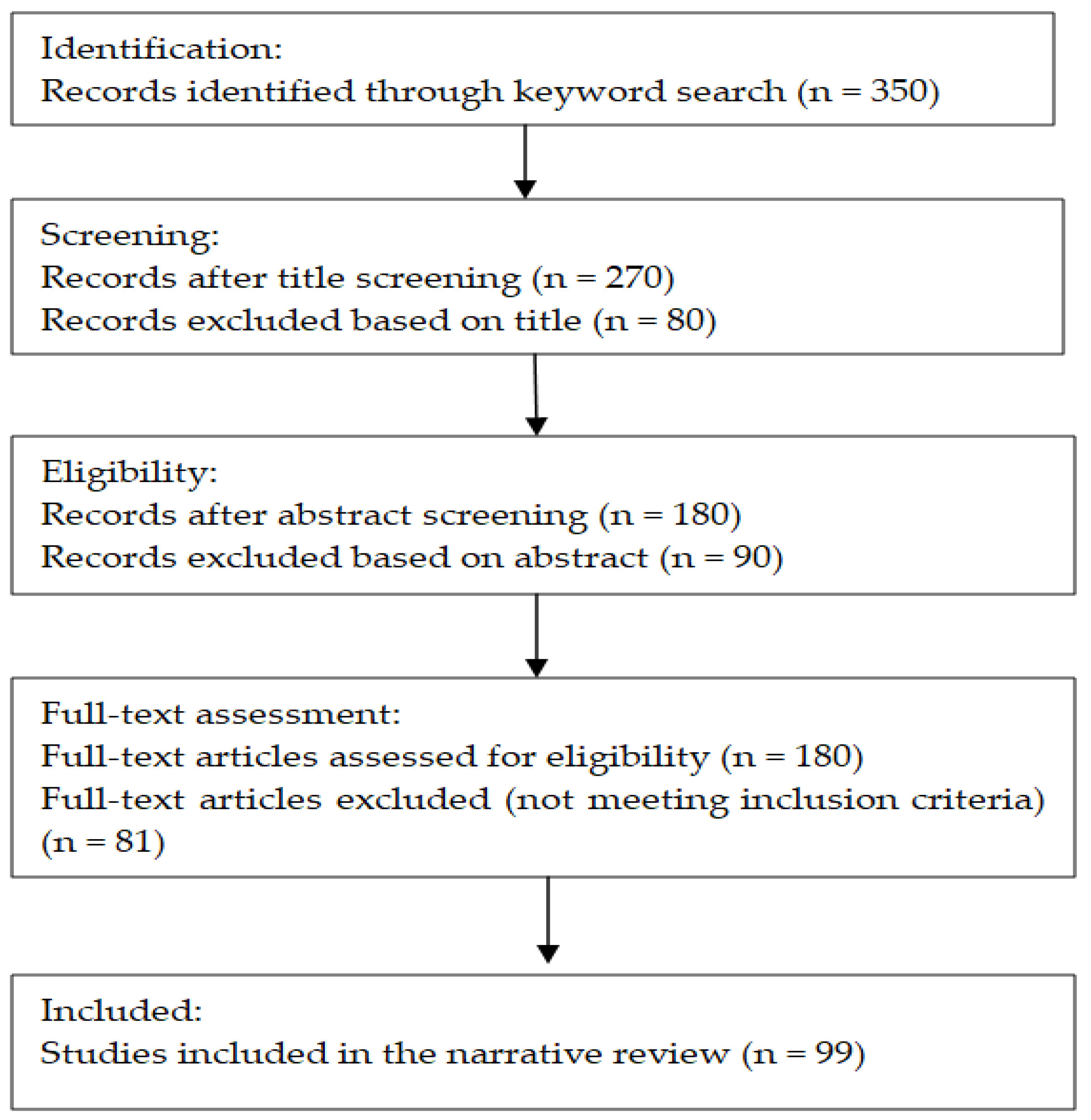
2.2. Search Strategy

This narrative review was conducted over a five-month period with the aim of synthesizing the most relevant and recent studies on the use of biofunctionalized nanoparticles in the diagnosis and treatment of hepatocellular carcinoma. The literature search was carried out using major scientific databases, including PubMed, Web of Science, Scopus, and Google Scholar. No strict temporal limitations were imposed, though particular emphasis was placed on the most recently published studies.
The initial screening process was performed manually by evaluating titles and abstracts. Articles considered potentially relevant were then reviewed in full.

2.3. Inclusion Criteria

Both original research articles and review papers were included, provided they met the following criteria:
  • Investigated the use of biofunctionalized nanoparticles in the targeted treatment of HCC;
  • Addressed nanoparticle-based strategies involving either active targeting mechanisms (e.g., ligand–receptor interactions) or passive targeting (e.g., the Enhanced Permeability and Retention [EPR] effect);
  • Explored gene therapy or immunotherapy approaches facilitated by nanotechnology platforms;
  • Examined innovative or emerging therapeutic modalities such as photothermal therapy (PTT) or photodynamic therapy (PDT);
  • Focused on the early detection of HCC using nanoparticle-based or nanotechnology-enhanced diagnostic techniques;
  • Were published recently (preferably within the last 5–7 years), to ensure the inclusion of the most up-to-date advancements in the field.

2.4. Exclusion Criteria

We excluded the following:
  • Studies primarily focused on cancers other than hepatocellular carcinoma;
  • Articles that lacked a clear relevance to nanotechnology or nanoparticle-mediated therapeutic or diagnostic approaches;
  • Experimental studies that were outdated or had been superseded by more recent and comprehensive findings;
  • Review articles that did not offer novel perspectives or substantially overlapped with previously published content;
  • Publications in languages other than English.
Figure 1 provides a visual representation of the steps taken, from the initial identification of the literature to the final inclusion of the articles.

3. Results

3.1. Multifunctional Nanoparticles for Therapy and Imaging

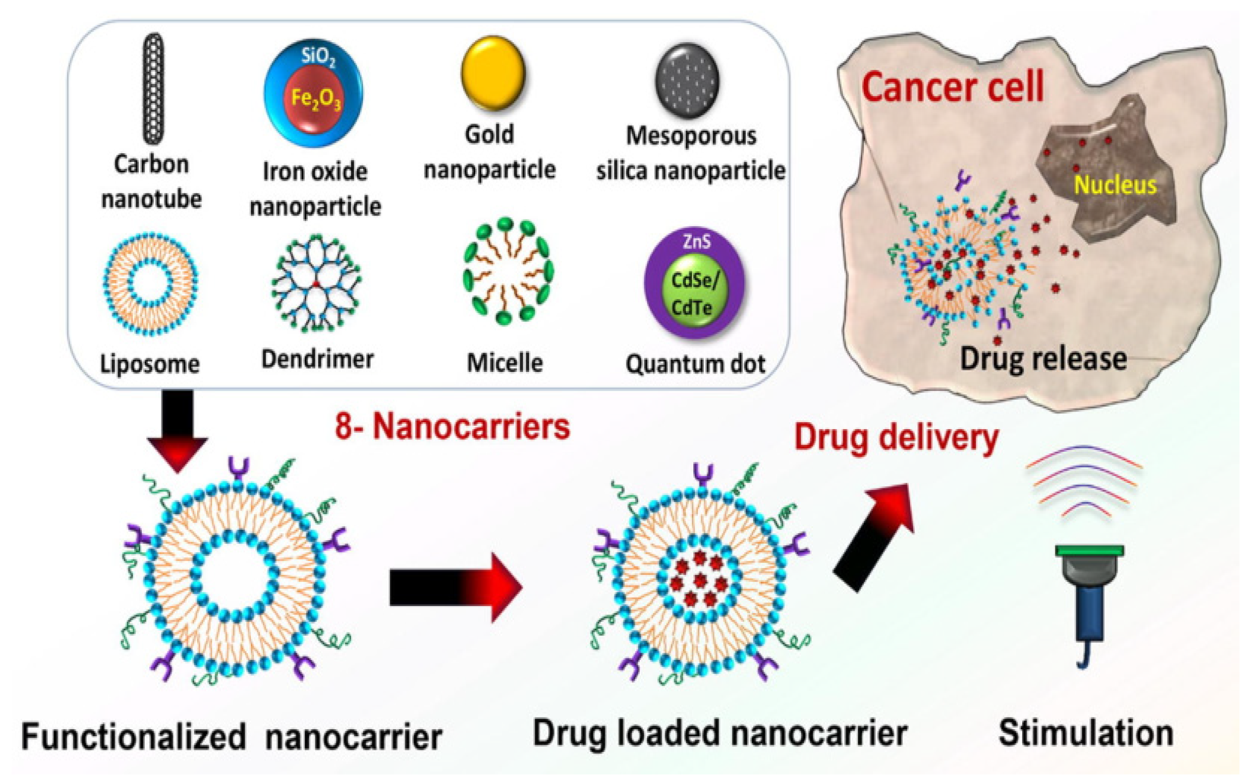
3.1.1. Nanoparticle Drug Carriers

Nanoparticles (NPs) play a crucial role in improving the precision and effectiveness of drug delivery systems, and their design is optimized in terms of coating, size, and shape to enhance biodistribution and pharmacokinetics, and to allow controlled drug release [8]. They possess the unique capability to deliver drugs directly to targeted cells or tissues while minimizing side effects, making them ideal for delivering various therapeutic agents to different regions of the body. Nanocarriers encapsulate and deliver therapeutic agents effectively, improving drug solubility, stability, and bioavailability, while also protecting them from premature degradation and clearance. When designing nanoparticle-based drug delivery carriers, it is essential to take into account both drug release and polymer biodegradation [9]. Several factors influence the rate of drug release, including: diffusion of the drug through the nanoparticle matrix, the drug’s solubility, a combination of erosion and diffusion processes, desorption of the surface-bound or adsorbed drug, and erosion or degradation of the nanoparticle matrix [10].
Multifunctional nanoparticles are under active investigation, paving the way for personalized and improved therapies [8]. In an effort to overcome the limitations of current liver cancer therapies, several authors have synthesized a variety of nanostructured materials employed as drug delivery systems. Figure 2 shows a schematic illustration of the different types of nanocarriers used in the treatment of HCC [11].
  • Solid Lipid Nanocarriers
Solid lipid nanocarriers (SLNc) represent a modern and efficient drug delivery platform, with significant potential in oncological treatments due to their favorable physicochemical properties. They are colloidal carriers with diameters typically ranging between 50 and 100 nm, which are optimal for avoiding rapid renal clearance and uptake by the reticuloendothelial system [12]. SNLc are composed of a lipid matrix that remains solid at both room temperature and physiological temperature (37 °C), stabilized by surfactants or cosurfactants. This solid-state behavior is due to the use of lipids with melting points well above body temperature. The solid lipid core provides high physical stability, enables efficient encapsulation of hydrophobic drugs, and supports a sustained and controlled drug release profile [13].
SLNc are capable of improving the bioavailability of active substances by prolonging their half-life, controlling their release, and reducing premature elimination from the body. Through these mechanisms, increased drug concentration at the target tissues is achieved, along with a reduction in systemic toxicity. They protect active compounds from unfavorable physiological conditions, can reduce the frequency of administration, and are versatile in terms of administration routes (intravenous, oral, transdermal). However, their use may be limited by certain challenges, such as premature leakage of the active substance during systemic circulation, insufficient colloidal stability in complex biological environments, and undesirable accumulation in healthy tissues [14].
  • Dendrimers
Dendrimers have demonstrated enhanced functionalities compared to other frequently used nanomaterials. They are synthetic macromolecules with well-defined, spherical, three-dimensional hyperbranched structures, enabling them to exhibit great biocompatibility and unique physicochemical properties.
Structurally, dendrimers are composed of a multifunctional core and branched dendrons, with internal cavities capable of encapsulating hydrophobic drugs. This structure allows for improved water solubility, enhanced bioavailability, and increased therapeutic efficacy. Various classes of dendrimers have been developed to date, the most commonly used being polypropylenimine (PPI), polyamidoamine (PAMAM), carbosilane dendrimers, and polyester-based dendrimers [15].
Due to their exceptional water solubility, dendrimers are highly suitable for biological and medical applications. Their nanometric size allows effective penetration into tumor tissues while remaining large enough to avoid rapid renal clearance, thereby extending their circulation time in the body [16].
They are considered non-toxic, offer high drug-loading capacity, and exhibit strong chemotherapeutic and photothermal properties, making them particularly promising candidates for hepatocellular carcinoma therapy [17].
  • Liposomes
Liposomes possess a distinct structure consisting of a lipid bilayer membrane surrounding an aqueous core. This structure allows them to encapsulate both hydrophobic and hydrophilic substances without requiring chemical modification. The lipid bilayer of liposomes can fuse with other bilayers, such as the cell membrane, thereby facilitating the release of their contents. This property makes them highly valuable for drug delivery applications. Additionally, their surface can be modified with ‘stealth’ materials to enhance stability in the body or with targeting ligands for more selective delivery. Liposomes can range in size from 15 nanometers to several micrometers and may consist of either a single phospholipid layer (unilamellar) or multiple bilayer membranes (multilamellar) [10].
Multivesicular liposomes (MVLs) have a distinct structure compared to unilamellar and multilamellar vesicles. MVLs have gained attention due to their high stability and prolonged drug release duration, ranging from days to several weeks. These systems feature a complex internal structure of densely packed, non-aligned aqueous chambers, each enclosed by interconnected lipid membranes. Triolein, a neutral triglyceride composed of glycerol and three oleic acid chains, gives MVLs this distinctive structure. Traditional liposomes do not contain triolein, but MVLs require it to modify drug release profiles. Triolein occupies the corners and junctions of the lipid bilayers within MVLs, ensuring that only breaches in the outer membranes allow drug release while the inner vesicles retain the drug. This feature enables controlled and sustained drug release, making triolein essential for the depot-like properties of MVL-based delivery systems [18].
With their unique lipid bilayer structure surrounding an aqueous core, liposomes are one of the most promising drug delivery platforms [10].
  • Chitosan
Chitosan is a polycationic polymer, carrying a positive charge in acidic conditions, which enables it to interact with negatively charged biological surfaces, such as cell membranes. Structurally, it is a linear aminopolysaccharide that is biodegradable, biocompatible, exhibits low toxicity, and possesses mucoadhesive properties [19]. The size of chitosan nanoparticles typically ranges between 10 and 500 nm, allowing them to pass through the fenestrated blood vessels of tumors and accumulate in the tumor mass.
In addition, they are capable of controlled drug release, enhancing the stability and solubility of the loaded compounds while reducing systemic toxicity through targeted delivery. In the context of hepatocellular carcinoma treatment, chitosan nanoparticles have proven effective both due to their targeted delivery capabilities and their potential intrinsic antitumor activity. Preclinical studies on cell lines and animal models have demonstrated tumor growth inhibition, induction of apoptosis, and reduced angiogenesis. Antioxidant and anti-inflammatory effects have also been observed [20].
  • Micelles
Polymeric micelles (PMs) have been utilized as a drug delivery platform for liver cancer due to their core–shell structure, small and uniform size distribution, and high drug delivery efficiency [21]. These nanostructured systems are formed through the spontaneous self-assembly of amphiphilic polymers in aqueous environments, resulting in a hydrophobic core capable of encapsulating lipophilic drugs and a hydrophilic shell that provides colloidal stability and prolonged blood circulation time.
Their ability to co-encapsulate multiple therapeutic agents within a single carrier offers the potential for synergistic therapeutic effects, which is particularly valuable in oncological treatments. Moreover, polymeric micelles—especially those based on prodrug strategies—are considered a promising platform for combination drug delivery, offering enhanced anticancer efficacy and reduced systemic toxicity [22].
Polymeric micellar drug delivery technology is actively studied as a promising nanocarrier for multi-drug delivery.
  • Carbon Nanotubes
Carbon nanotubes (CNTs) are notable for their unique characteristics that distinguish them from other potential drug carriers. They consist of cylindrical tubes formed by rolled graphene sheets, made entirely of carbon, featuring high aspect ratios in both length and width. Depending on the number of concentric walls, CNTs exhibit exceptional mechanical strength, high electrical conductivity, and remarkable chemical stability. Based on their layered structure and diameter, researchers classify CNTs into three types: single-walled nanotubes (SWCNTs), double-walled nanotubes (DWCNTs), and multi-walled nanotubes (MWCNTs) [23].
Due to their distinct chemical composition, shape, ultra-small size, and targeting capabilities, carbon nanotubes can function effectively at the biomolecular level. They can carry various therapeutic agents, enabling site-specific delivery and prolonged accumulation in targeted tumor tissues, thereby reducing potential systemic toxicities.
However, the toxicity of CNTs remains a concern, as factors such as concentration, size, degree of functionalization, and the presence of catalytic residues can influence it. Recent studies indicate that CNTs can induce oxidative stress or exhibit asbestos-like behavior under certain conditions. Therefore, researchers need to conduct further studies to fully assess their biocompatibility and safety profiles [24].
  • Gold Nanoparticles
Gold nanoparticles (AuNPs) have been widely utilized in both therapeutic applications and diagnostics. Due to their small size, they have a large surface-area-to-volume ratio, which gives them unique physicochemical and optoelectronic properties, including controllable sizes below 100 nm, high chemical stability, strong absorption in the near-infrared (NIR) spectrum, and the presence of localized surface plasmons. These features make them excellent candidates for drug delivery and cancer therapy [25].
Technological advances have allowed scientists to synthesize AuNPs in various shapes and sizes—spheres, rods, nanocages, and nanocavities. Surface functionalization with ligands such as polyethylene glycol (PEG), peptides, nucleic acids, or antibodies enhances their biocompatibility and cellular specificity, while simultaneously reducing phagocytosis and degradation during systemic circulation.
Their favorable interaction with biological systems, combined with the ease of tailoring their surfaces and targeting therapeutic agents to specific sites, positions them as valuable platforms for precise and controlled cancer drug delivery [26].
Although AuNPs are generally considered biocompatible, their unmodified forms may accumulate in reticuloendothelial organs such as the liver and spleen. This raises concerns regarding potential toxicity, particularly with repeated administrations. Consequently, further studies are warranted to comprehensively validate their safety profile [27].
A comparative overview of key findings from recent studies using different nanoparticle-based drug delivery systems in HCC therapy is presented in Table 1.

3.1.2. Engineered Nanoparticles for Enhanced Diagnostic Imaging

  • Magnetic Nanoparticles
In the realm of tumor diagnostics, MRI is widely utilized; however, its specificity and sensitivity remain suboptimal. Additionally, adverse reactions to contrast agents in sensitive patients make this method unsuitable for certain groups. To overcome these limitations, researchers have focused on using magnetic nanoparticles in nuclear magnetic resonance, offering potential advancements in imaging precision and safety. These nanoparticles act as contrast agents, improving the resolution and clarity of MRI scans. They can also be functionalized with targeting ligands to allow for more precise imaging of specific tissues, facilitating early detection of diseases such as cancer [39].
Magnetic nanoparticles, particularly those based on superparamagnetic iron oxide (SPIONs), attract significant interest due to their tunable physicochemical properties. Their morphology—typically spherical or ellipsoidal—influences circulation time, cellular internalization, and biodistribution. For clinical applications, ensuring biocompatibility remains essential. Surface functionalization with polymers such as dextran or polyvinyl alcohol improves stability and reduces cytotoxicity.
SPION toxicity depends on both dose and particle size. Concentrations below 100 μg/mL are generally considered safe in vitro, although cellular responses may vary depending on the cell type and route of administration. When administered intravenously, SPIONs primarily accumulate in the liver, spleen, and kidneys. In normal hepatic tissue, Kupffer cells selectively uptake these particles, while malignant lesions show minimal or no uptake. This selective distribution enhances lesion-to-liver contrast in MRI.
In hepatocellular carcinoma, the absence or depletion of Kupffer cells leads to minimal SPION retention within tumor tissue, producing marked signal differences on T2-weighted MRI. This contrast enhancement improves visualization and supports early detection of HCC, particularly in cases where conventional imaging methods are limited or contraindicated [40].
  • Quantum Dots
Quantum dots (QDs) are semiconductor nanoparticles with exceptional optical properties, including intense, stable, and size-tunable fluorescence, broad absorption spectra, and narrow emission bands. These characteristics make them ideal for advanced biomedical imaging applications, particularly in cancer diagnostics [41].
Thanks to their photostability and strong signal intensity, QDs enable long-term monitoring of cellular processes and the acquisition of high-contrast images, thereby surpassing the limitations of traditional fluorophores. Their well-defined emission also enables the multiplex detection of multiple biomarkers simultaneously, contributing to early cancer diagnosis and the differentiation of tumor-associated molecular patterns [42].
Functionalizing QD surfaces with recognition molecules such as antibodies, aptamers, or peptides allows the specific identification of tumor cells by targeting biomarkers such as AFP, EpCAM, or Her2. This molecular specificity significantly improves diagnostic accuracy by allowing precise tumor localization and facilitating the capture of cancer cells from biological samples such as circulating tumor cells (CTCs) [43].
Researchers have successfully applied QDs in both in vitro and in vivo imaging, even under high autofluorescence conditions, due to their distinct emission signals. Their integration into advanced optical systems generates detailed spectral tumor maps, enabling a clear distinction between healthy and pathological tissue, even at early disease stages [42].
Although earlier generations of QDs raised toxicity concerns, advances in synthesis and functionalization have produced more biocompatible variants—such as carbon-, graphene-, or phosphorus-based QDs—which significantly reduce toxic potential without compromising diagnostic performance [43].
Therefore, QDs represent a powerful platform for cancer diagnosis, offering high sensitivity and specificity while remaining compatible with modern imaging and cellular analysis techniques. Their multiplexing capacity, spectral clarity, and chemical adaptability position them among the most promising fluorescent contrast agents in precision medicine.

3.1.3. Theranostic Nanoparticles

Theranostic nanoparticles are multifunctional platforms that combine therapeutic and diagnostic capabilities within a single system [44]. These nanoparticles deliver therapeutic agents while simultaneously providing imaging feedback on treatment efficacy.
The theranostic delivery-based strategy has emerged as a promising approach for managing liver cancer. This method integrates therapeutic agents—such as chemotherapy drugs, genetic materials, peptides, and proteins—into specialized nanocarriers. Alongside these therapeutic components, diagnostic elements are incorporated to enhance imaging and detection. Commonly used diagnostic agents include gadolinium, radionuclides, QDs, fluorescent dyes, superparamagnetic iron oxides, and dense elements like iodine. These agents support various imaging modalities, including optical imaging, MRI, and CT, offering a comprehensive solution for both treatment and diagnosis [7]. NPs loaded with both a chemotherapeutic drug and a contrast agent enable real-time monitoring of drug release and tumor response.
A schematic overview of the major types of theranostic nanoplatforms, their imaging modalities, and ligand-based targeting strategies is presented in Figure 3, highlighting the integration of therapeutic and diagnostic components within a single nanoparticle system [44].
This integration of diagnosis and therapy holds great promise for personalized medicine, where treatment can be tailored based on individual patient responses.
Table 2 provides a summary of various nanoparticle classifications used in diagnostic or theranostic applications, highlighting the diagnostic agents incorporated, experimental models utilized, techniques employed for detection or monitoring, and reported outcomes in terms of efficacy.

3.2. Targeted Drug Delivery

3.2.1. Active Targeting

Active targeting involves functionalizing the surface of nanoparticles with specific ligands, which is particularly advantageous in cancer treatment where overexpression of certain receptors is common. Various ligands, including antibodies, peptides, aptamers, and small molecules, have been explored for their ability to recognize and bind to specific receptors on target cells. In hepatocellular carcinoma, receptors such as asialoglycoprotein, transferrin, folate, CD44 antigen, and integrins bind to complementary ligands on nanocarriers [51]. These targeting ligands promote receptor-mediated endocytosis of the drugs into liver tumor cells, enhancing the selective delivery of chemotherapeutics to the tumor, thus improving treatment efficacy while minimizing damage to healthy tissues [7].
The CD44 receptor, a transmembrane glycoprotein, serves as the primary receptor for hyaluronan. Hyaluronan is a polysaccharide and a key component of the extracellular matrix, alongside collagen. The interaction between CD44 and hyaluronan influences cancer cell phenotypic behaviors, such as tumor progression, metastasis, and cell proliferation. The literature also indicates that the tumor microenvironment should be considered in disease management, as it significantly influences the development of hepatocellular carcinoma.
Based on this interaction, liposomes functionalized with hyaluronic acid (HA) and/or PEG have demonstrated promising therapeutic potential in hepatocellular carcinoma. These nanocarriers exhibit favorable physicochemical properties and are efficiently internalized by cells that overexpress CD44, such as Huh7 cells. Additionally, PEG can modulate interactions with immune cells, particularly tumor-associated macrophages, while reducing off-target uptake by tumor cells with low CD44 expression. Thus, dual-targeting strategies addressing both tumor cells and the immunological components of the tumor microenvironment are emerging as promising approaches to enhance therapeutic efficacy in the treatment of hepatocellular carcinoma, as demonstrated in the study conducted by Cannito et al. [52].
The asialoglycoprotein receptor (ASGPR), also known as the galactose receptor, has drawn attention due to its highly specific expression on hepatocytes and near its absence in other tissues. ASGPR mediates nanoparticle internalization through clathrin-mediated endocytosis when nanoparticles are surface-decorated with galactose or N-acetylgalactosamine [53].
Among the delivery systems exploiting this mechanism, galactosylated chitosan (Gal-CS) nanoparticles—produced by grafting lactobionic acid onto chitosan—have demonstrated considerable potential in HCC therapy. Researchers have successfully loaded these carriers with various therapeutic agents, including doxorubicin, 5-fluorouracil, gemcitabine, and triptolide, for targeted treatment of liver cancer [54].
In their study, Sun et al. developed several types of self-assembled chitosan nanoparticles loaded with DOX to selectively target HCC via ASGPR. Chitosan was first chemically modified with glycidol to enhance its solubility, then further functionalized with hydrophobic moieties such as vitamin E succinate (VE) or deoxycholic acid (DCA) to impart amphiphilic properties, and finally galactosylated by conjugation with lactobionic acid to enable ASGPR targeting. Doxorubicin was subsequently loaded into the resulting nanoparticles using the dialysis method. In vitro drug release studies confirmed that DOX release from all nanoparticle types was pH-dependent, with faster release under acidic conditions (pH 5), which mimic the tumor microenvironment. Internalization tests conducted on HepG2 cells demonstrated superior uptake for the galactosylated nanoparticles, supporting the involvement of ASGPR in the cellular uptake mechanism. Among the variants, galactosylated nanoparticles showed superior in vitro performance and enhanced in vivo anticancer efficacy. Moreover, all tested formulations exhibited good biocompatibility, with no significant histological damage observed.
However, the study also highlighted certain limitations. The exact molecular mechanisms of the interaction between the nanoparticles and ASGPR were not explored in detail. Another aspect that requires further investigation is the behavior of these nanoparticles in systemic circulation over the long term, as well as potential accumulation in other organs that weakly express ASGPR. Lastly, detailed pharmacokinetic and biodistribution studies are essential before any potential clinical applications [54].
Based on the fact that folic acid receptors are overexpressed on liver tumor cells, Ghalekhondabi et al. employed nanomicelles functionalized with silibinin (SLB) to enhance treatment efficiency. Silibinin, a polyphenol derived from Silybum marianum, is known for its anti-inflammatory, anticancer, and hepatoprotective effects, with minimal side effects. However, its poor water solubility limits clinical use [55].
To address this, the researchers synthesized FA-functionalized Pluronic F127 copolymer (F127/FA) using the Steglich esterification method and encapsulated SLB into its hydrophobic core via thin-film hydration. The resulting nanomicelles had a spherical morphology, an average size of 17.7 nm, and demonstrated 2.36% drug loading and 79.43% encapsulation efficiency. In vitro release studies showed that SLB was released more slowly and sustainably from the FA-targeted nanomicelles than in its free form. Cytotoxicity assays in HepG2 cells revealed significantly greater anticancer activity for SLB/F127/FA compared to SLB/F127 or free SLB. In summary, these findings highlight SLB/F127/FA nanomicelles as a promising targeted delivery system for liver cancer therapy, especially in tumors with overexpressed folate receptors.
While the study demonstrates promising in vitro results, its translational potential remains limited due to the absence of in vivo validation. Additionally, the relatively low drug loading capacity could restrict the amount of silibinin delivered per dose, necessitating larger quantities of the carrier to achieve therapeutic effects. The formulation’s long-term stability also remains uncertain, as no data were provided on potential changes in particle size, aggregation, or drug retention under physiological conditions [56].

3.2.2. Passive Targeting: The Enhanced Permeability and Retention Effect

Passive targeting exploits the unique physiological properties of the tumor vasculature through the Enhanced Permeability and Retention effect, first described by Prof. Maeda’s group in 1986 [57]. The EPR effect is central to the passive targeting of nanoparticles in cancer therapy, as it allows for the preferential accumulation of drug carriers in the tumor [58], without the need for specific targeting ligands. The EPR effect arises from the distinct differences in vascular structure between normal and cancerous tissues. Tumor blood vessels are often disorganized and dilated, with larger and irregular fenestrations, allowing for easier penetration and retention of nanoparticles within the tumor. Additionally, impaired lymphatic drainage in tumor tissues helps retain these particles longer, resulting in a higher local concentration of therapeutic agents compared to normal tissues. This can be particularly advantageous for hepatocellular carcinoma, where systemic treatments often have limited efficacy and high toxicity [59]. Furthermore, high interstitial fluid pressure is common in HCC tumors, influencing drug distribution within the tumor tissue and contributing to the retention of larger molecules. This effect, while beneficial in retaining drugs within the tumor, can also present a barrier to uniform drug distribution, which is being addressed through innovative drug delivery systems.
The literature has demonstrated that several physicochemical properties of nanocarriers—such as particle size, molecular weight, surface charge, hydrophobicity, and hydrophilicity—are critical for effective passive targeting in drug delivery systems. These features influence how nanocarriers interact with biological environments, enabling improved retention and accumulation in target tissues. The optimal size range for these nanostructures is generally considered to be between 50 and 200 nm, which helps avoid rapid renal clearance (as occurs with smaller particles) and uptake by the reticuloendothelial system (common with larger particles). For instance, stealth liposomes with sizes around 100–150 nm have shown efficient retention in tumor tissues due to this favorable size window [60].
Moreover, nanoparticles with surface modifications such as PEGylation have shown improved drug delivery performance in hepatocellular carcinoma. PEGylation, in particular, increases circulation time and enhances uptake within the liver tumor microenvironment. As a result of the EPR effect, these modified nanoparticles accumulate more effectively in HCC tissues and have demonstrated better therapeutic outcomes in animal models [61].
In addition to surface modifications, the nature of the nanocarrier itself plays a key role in passive targeting efficiency. pH-sensitive liposomes, polymeric micelles, and solid lipid nanoparticles have each shown specific advantages, such as enhanced stability, controlled drug release, and responsiveness to the acidic tumor environment. Studies indicate that dioleoylphosphatidylethanolamine (DOPE)- and cholesteryl hemisuccinate (CHEMS)-based formulations, modified with polyethylene glycol, exhibit superior pharmacokinetics and significantly greater tumor accumulation than free drug formulations.
Morphology also influences targeting performance. Spherical structures are most commonly used due to their stability and efficient extravasation. However, other shapes—such as nanorods or flexible particles—may offer additional benefits in certain tumor types by improving retention or prolonging circulation time [60].
Studies have shown that nanoparticles that rely on the EPR effect can enhance the concentration of chemotherapeutic agents within tumors, improving therapeutic outcomes compared to free drugs [62]. However, the efficiency of the EPR effect can vary significantly between patients and even within different regions of the same tumor, due to factors such as tumor size, type, and location. Some tumors exhibit limited vascular permeability, reducing the effectiveness of passive targeting. Moreover, the dense extracellular matrix within tumors can impede the diffusion of nanoparticles, limiting their penetration into the deeper tumor tissues. Researchers are actively exploring strategies to enhance the EPR effect, such as using vasodilators to increase tumor blood vessel permeability or designing nanoparticles that respond to the tumor microenvironment by releasing their payload in response to specific stimuli like pH or enzymes.
By using vascular normalization agents, such as anti-angiogenic drugs, it is possible to temporarily stabilize the abnormal blood vessels in HCC tumors. This enhances the penetration of nanoparticles into the tumor, thus improving the efficacy of EPR-based drug delivery, as shown by Subhan et al. in their study [63].
A study published by Kong et al. explored the use of polymeric micelles loaded with sorafenib, an HCC therapeutic, to harness the EPR effect. The study revealed that these micelles accumulated within the tumor, improved therapeutic efficacy, and lowered toxicity in liver tissues. It also highlighted that modifying the micelles with pH-sensitive coatings enhanced drug release at the tumor site, further improving targeting in the HCC microenvironment [64].
The study by Li et al. showed that passive targeting can depend on pH, as well as surface changes produced on tumor cells. They successfully created galactose- and PEG-decorated disulfide-linked core–shell mesoporous silica nanoparticles (GPDC-MSNs), designed for selective tumor accumulation and controlled drug release within HCC cells [65].
Despite promising preclinical findings, translating the EPR effect into clinical efficacy in HCC patients has been challenging. Heterogeneity in vascularization, patient liver function, and tumor size all contribute to variability in the EPR effect’s efficacy. This suggests a need for personalized approaches, where patients’ vascular profiles might be used to determine the suitability of EPR-based therapies.

3.3. Gene Therapy and RNA Interference (RNAi)

3.3.1. siRNA-Loaded Nanoparticles

RNA interference is a powerful gene-silencing technique that uses small interfering RNA (siRNA) molecules to suppress the expression of specific genes. However, free siRNA is unstable in the bloodstream due to rapid degradation by nucleases and elimination via glomerular filtration, resulting in low bioavailability and limited therapeutic potential [66]. To address this, siRNA-loaded nanoparticles have emerged as a promising delivery platform, protecting siRNA and enhancing its accumulation in target tissues.
Among these, lipid-based nanoparticles are particularly effective, as they can fuse with cellular membranes and facilitate the release of siRNA directly into the cytoplasm.
The progression of hepatocellular carcinoma is largely driven by angiogenesis, which results from the excessive secretion of VEGF by tumor cells within the tumor microenvironment [67]. Thus, reducing VEGF expression through siRNA could form the basis for a promising anti-HCC therapy.
Huang et al. developed a system that overcomes these barriers by encapsulating siRNA in a pH-sensitive calcium phosphate core coated with a lipid membrane. This structure protects the siRNA from enzymatic degradation and enables the release of the nucleic acid directly into the cytoplasm of tumor cells. To achieve efficient siRNA delivery and targeting within HCC cells, given that galactoside derivatives possess strong targeting capabilities toward ASGPR on tumor cells, the team synthesized various galactoside derivatives and encapsulated siRNA/VEGF into lipid/calcium/phosphate (LCP) nanoparticles specifically targeted to ASGPR. Their findings illustrate that LCP nanoparticles decorated with phenyl β-D-galactoside (L4-LCPNPs) enabled highly efficient siRNA delivery to HCC cells, with significantly less uptake in normal hepatocytes.
In vitro and in vivo, VEGF siRNA released by L4-LCPNPs successfully downregulated VEGF expression in HCC, producing a marked antiangiogenic effect within the tumor microenvironment. This led to substantial tumor regression and suggests that phenyl galactoside could serve as a promising HCC-targeting ligand for therapeutic siRNA delivery in the treatment of liver cancer. Nonetheless, limitations remain, including limited biodistribution analysis and lack of long-term safety data, which warrant further investigation before clinical translation [68].
Previous research has indicated that AFP, a common HCC marker, plays a crucial role in the proliferation and apoptosis of hepatocellular carcinoma cells. Elevated levels of AFP are associated with increased cell growth and survival, highlighting its potential as a therapeutic target in HCC management. Studies suggest that AFP may influence various signaling pathways that regulate these processes, thus contributing to cancer progression [69].
This relationship highlights the potential of siRNA in targeting and modulating the pathways involved in HCC progression, offering a novel approach to treatment.
Research on polymeric nanoparticles loaded with siRNA against AFP has demonstrated significant anticancer effects in HCC cells. Epigallocatechin-3-gallate (EGCG), a potent polyphenol found in green tea, has shown notable anticancer properties by blocking tumor progression through mechanisms such as cell cycle arrest and induction of apoptosis in cancer cells.
In this context, co-delivery systems that combine biodegradable PLGA nanoparticles with AFP-targeting siRNA and EGCG have shown enhanced cytotoxicity against liver cancer cells compared to either treatment alone, suggesting a strong synergistic effect. However, these strategies have only shown efficacy in vitro, lacking in vivo validation, and their underlying molecular mechanisms remain only partially understood [69].
Among siRNA delivery strategies for HCC, selenium nanoparticles (SeNPs) functionalized with HA and coated with polyethylenimine (PEI) have gained attention due to their favorable targeting and delivery properties. In the literature, such nanoparticle systems are associated with enhanced cellular uptake and gene-silencing efficiency, owing to the ability of HA to mediate active targeting through CD44 receptors—commonly overexpressed on HCC cells—and the cationic nature of PEI, which facilitates electrostatic binding of siRNA and cellular internalization.
Furthermore, reports suggest that these systems can achieve notable antitumor efficacy both in vitro and in vivo, while maintaining a favorable safety profile, with minimal systemic toxicity observed under tested conditions. However, it is important to note that treatment with such nanoparticles induced apoptosis in only approximately 32.7% of tumor cells, indicating that a significant proportion of cancer cells survived, and that the therapeutic efficacy may require further optimization or combination with other treatment strategies [70].

3.3.2. CRISPR-Cas9

In recent years, CRISPR-Cas9 technology has transformed the field of genetic engineering, enabling precise and efficient modification of DNA sequences across a wide array of organisms. Discovered as part of the adaptive immune system in bacteria and archaea, CRISPR-Cas9 functions by introducing site-specific breaks in DNA, allowing for targeted gene knockout, repair, or insertion. This makes it a powerful approach for studying and potentially treating HCC [71]. Recent studies have shown that CRISPR-Cas9 can inhibit the expression of oncogenes in HCC cells, disrupt HBV replication by targeting viral DNA, and enhance the susceptibility of cancer cells to existing treatments [72].
In one of their studies, Song et al. investigated whether the CRISPR/Cas9 gene-editing system could be used to knock out the hepatitis B surface antigen (HBsAg) in HCC cells infected with HBV. Given that chronic HBV infection is a major contributor to liver cancer, the researchers aimed to determine whether eliminating HBsAg could reduce the cancerous behavior of these cells—specifically, their ability to proliferate and form tumors. Using CRISPR/Cas9 constructs designed to target the preS1/preS2/S open reading frame of the HBV genome, they successfully disabled the gene in HBV-positive HCC cell lines.
The impact of the knockout was assessed through a series of in vitro assays, such as CCK-8-based proliferation tests and cell counting, which revealed a clear reduction in cell proliferation following HBsAg disruption. To evaluate tumorigenic potential, the team conducted both xenograft experiments in mice and immunohistochemical analyses. In both settings, cells lacking HBsAg showed a significantly reduced ability to form tumors. Overall, the study demonstrated that targeting HBsAg with CRISPR/Cas9 not only suppresses the growth of HBV-positive liver cancer cells but also diminishes their tumor-forming potential. These results suggest that this gene-editing approach could be a promising therapeutic strategy for HBV-related HCC.
However, the study also presents some important limitations. Potential adverse effects on normal (non-tumor) hepatocytes were not discussed, limiting the safety assessment of the intervention. Additionally, the effect on cccDNA (covalently closed circular DNA)—the persistent form of HBV in hepatocytes—was not evaluated. This viral reservoir is notoriously difficult to eliminate and remains a major barrier to curing chronic HBV infection [73].
In another study, Zhang et al. explored the role of NSD1, a nuclear receptor-binding SET domain protein, in HCC progression. They used CRISPR/Cas9 to knock out NSD1 in HCC cell lines, aiming to disrupt tumor-related signaling—particularly the NSD1–H3–Wnt10b axis.
After editing the NSD1 gene with specific guide RNAs, the team measured histone H3 methylation changes. The knockout increased H3K27 trimethylation and reduced H3K36 dimethylation, two modifications known to suppress gene transcription. These epigenetic changes blocked Wnt10b signaling, a key pathway in tumor growth.
To evaluate the biological impact, the researchers performed cell proliferation assays and xenograft experiments in mice. NSD1-deficient cells showed slower proliferation, reduced tumor formation, and lower metastatic potential. These results indicate that NSD1 actively promotes HCC through both epigenetic regulation and oncogenic signaling, positioning it as a strong candidate for targeted liver cancer therapy.
Although the results of this study are promising, it presents certain limitations, such as the use of a relatively small clinical sample size, the lack of functional validation of other pathways regulated by NSD1, and the absence of advanced in vivo models. Moreover, the risk of off-target effects associated with the use of CRISPR/Cas9 was not thoroughly evaluated, which could affect the accuracy of the conclusions [74].
The application of CRISPR-Cas9 in HCC therapy remains complex due to challenges such as off-target effects and the delivery of gene-editing components to liver cells. Despite these obstacles, ongoing advancements in CRISPR-Cas9 technology and delivery methods hold promise for developing highly specific and minimally invasive treatments for liver cancer, potentially paving the way for CRISPR-based therapies in clinical practice [75].

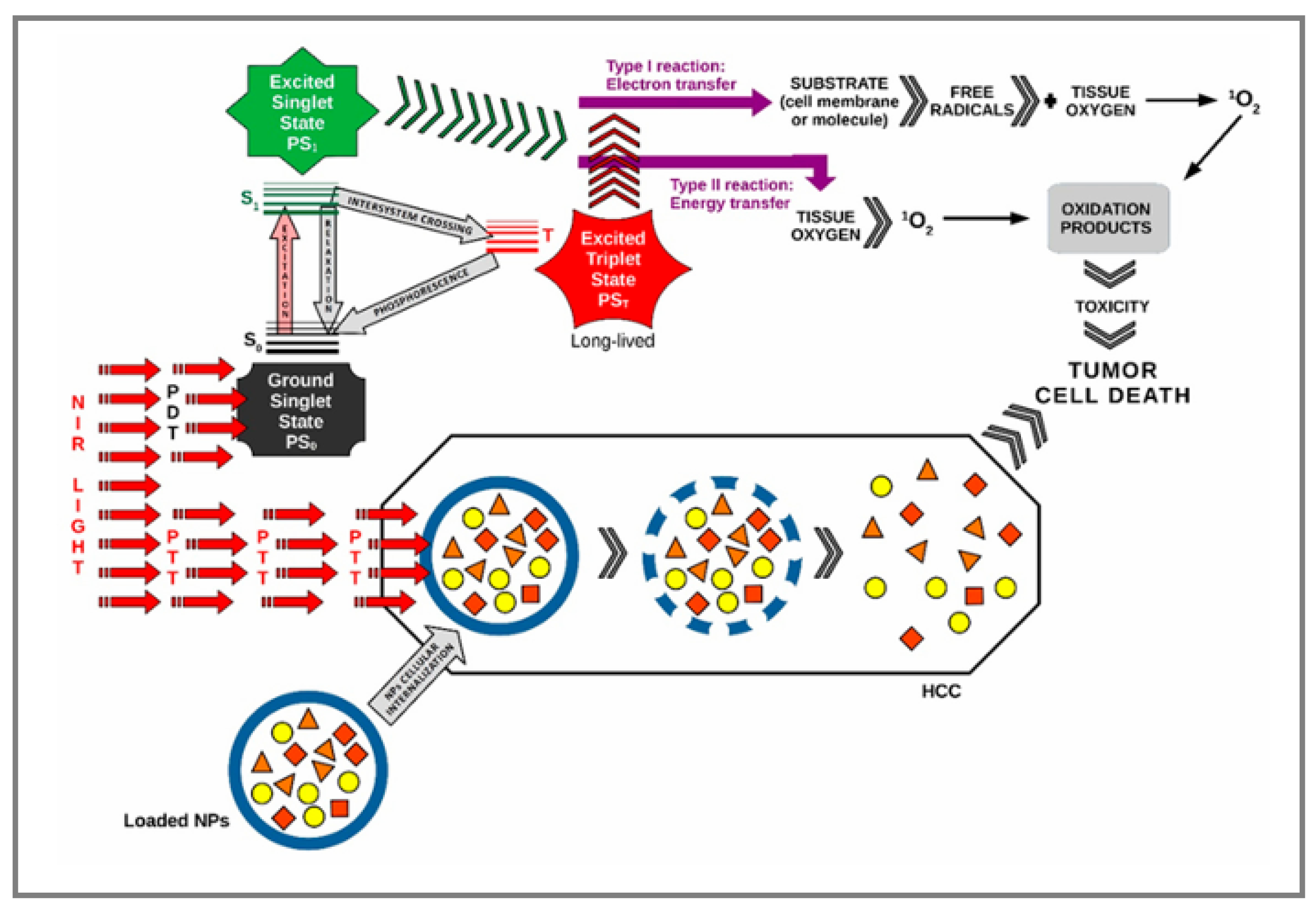
3.4. Photothermal and Photodynamic Therapy

Among nanotechnology-based approaches, photothermal and photodynamic therapy have shown significant potential for liver cancer treatment due to their non-invasive nature and high specificity [76]. PTT employs light-absorbing agents to convert NIR light into heat, selectively destroying cancer cells, while PDT utilizes light-activated photosensitizers to generate reactive oxygen species that induce cell death [77]. Recent developments have led to the combination of PTT and PDT into multifunctional nanoprobes, which simultaneously exploit these complementary mechanisms to enhance therapeutic efficacy and overcome the limitations of each individual approach.
Innovative nanoplatforms, such as targeted multifunctional nanoparticle systems, offer promising avenues for integrating PTT and PDT with imaging capabilities. This integration enables real-time tumor visualization, guided therapy, and precise targeting of HCC tumors [74]. These advances underscore the potential of multifunctional nanotechnology to enable a robust, image-guided, dual-modal therapeutic strategy for HCC, improving treatment outcomes. A schematic representation of the synergistic application of PTT and PDT in hepatocarcinoma is shown in Figure 4, illustrating their mechanism of action and potential therapeutic integration [78].

3.4.1. Photothermal Therapy

The use of light-to-heat energy conversion for tumor treatment has gained significant attention among researchers. Metal nanoparticles are particularly valuable in this area, playing a key role in treating tumors. Their strong absorption of near-infrared light is fundamental to their effectiveness in photothermal therapy. Unlike traditional treatments, metal nanoparticles offer high selectivity and efficiency with minimal invasiveness. Among these, gold-based nanomaterials are most commonly used due to their tunable surface plasmon resonance and excellent photothermal conversion efficiency [76].
In this context, recent studies have investigated gold nanoparticles functionalized with albumin (Alb-AuNPs), designed to specifically bind to gp60 receptors, which are overexpressed on hepatocellular carcinoma cells. This approach enables precise thermal energy delivery upon exposure to near-infrared light. Following internalization by HepG2 cells, the nanoparticles generate localized heat, disrupting the Golgi apparatus and endoplasmic reticulum (ER), leading to caspase-3 activation and the initiation of apoptosis. This selective interaction triggered strong pro-apoptotic effects in tumor cells, with minimal impact on normal cells. Experimental evidence suggests the effectiveness of this strategy in reducing tumor mass; however, in vivo validation remains limited, and efficient nanoparticle accumulation may be influenced by vascular heterogeneity, the tumor microenvironment, and variability in target receptor expression [79].
In parallel, combined strategies have been proposed, integrating PTT with the controlled release of chemotherapeutic agents such as sorafenib, using copper selenide nanoparticles. These facilitate thermally activated drug release at the tumor site, increasing local drug concentration and antitumor efficacy. Both in vitro and in vivo experiments have demonstrated superior apoptotic induction compared to treatments applied separately. In animal models, this therapy significantly inhibited tumor growth while showing reduced toxicity to surrounding healthy tissues. However, the use of subcutaneous tumor models limits their translational relevance, and the absence of long-term pharmacokinetic data and systemic toxicity studies calls for caution when extrapolating these findings [80].
Despite these limitations, PTT remains a promising and minimally invasive therapeutic option, especially when integrated into targeted drug delivery platforms, with real clinical application potential for hepatocellular carcinoma treatment.

3.4.2. Photodynamic Therapy
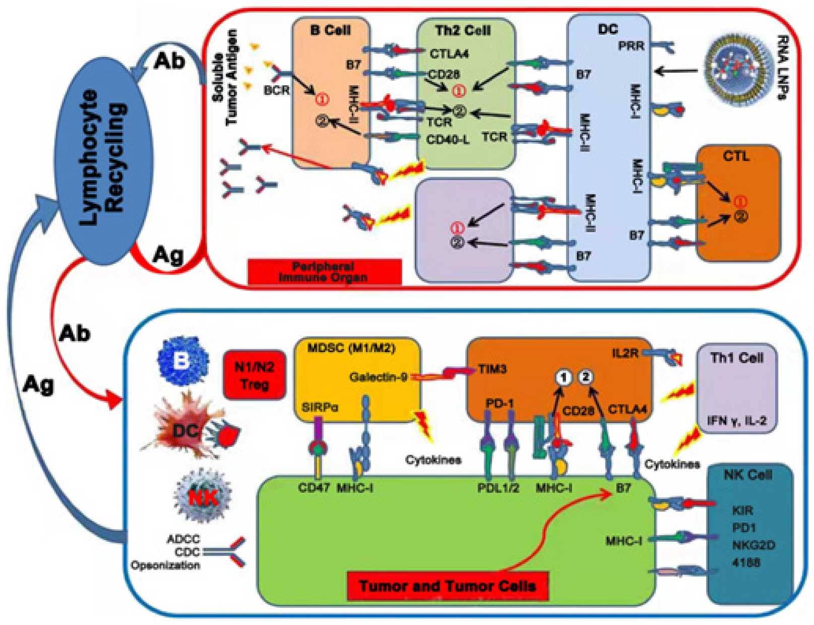
Photodynamic therapy involves administering a light-sensitive dye (photosensitizer), locally or systemically, to allow accumulation at the target site. When exposed to light of a specific wavelength, the photosensitizer becomes activated and transfers energy to cellular oxygen, generating reactive oxygen species (ROS). These ROS selectively damage tumor cells by inducing cell death, disrupting tumor vasculature, and stimulating the innate immune system [76,81].
In a pioneering study, Ogbodu et al. evaluated the use of tetra-triethyleneoxysulfonyl zinc phthalocyanine (ZnPc) as a novel photosensitizer for photodynamic therapy in hepatocellular carcinoma. ZnPc was chosen for its favorable optical properties and ability to generate ROS upon light activation. The compound demonstrated strong uptake by liver cancer cells (HepG2 and Huh-7), and when combined with light exposure, it induced substantial oxidative stress, leading to apoptosis and inhibition of cell proliferation. The treatment proved highly effective both in vitro and in vivo. In cell cultures, PDT with ZnPc resulted in a dose- and time-dependent reduction in cancer cell viability, associated with increased ROS generation and caspase-3 activation. Protein expression analysis further revealed modulation of apoptosis-related markers, indicating mitochondria-mediated apoptosis. In vivo validation using the chorioallantoic membrane (CAM) assay showed significant tumor size reduction, without observable harm to the developing embryo. These results underscore ZnPc’s potential as a selective and powerful agent for photodynamic treatment of HCC, offering both efficacy and biocompatibility.
Although the results obtained are promising, the study presents certain limitations. The in vivo validation was restricted to the CAM model of the chicken embryo, which does not allow for a complete assessment of pharmacokinetics, immunogenicity, or potential systemic toxic effects. Additionally, selectivity toward normal hepatic cells was not investigated, and the molecular mechanisms involved in cell death were only partially explored [82]. These aspects highlight the need for further research in preclinical and clinical studies.

3.5. Immunotherapy

Immunotherapy has emerged as a promising treatment for hepatocellular carcinoma, with immune checkpoint inhibitors (ICIs) like anti-PD-1/PD-L1 antibodies showing efficacy in clinical trials. However, resistance mechanisms and the immunosuppressive microenvironment challenge its success, necessitating combinatory strategies to enhance therapeutic outcomes [83].

3.5.1. Nanoparticle-Based Vaccines

Nanoparticle-based vaccines represent a promising approach in the treatment of hepatocellular carcinoma, leveraging their ability to enhance immune responses and effectively target tumor cells. These vaccines utilize nanoparticles as delivery systems to protect antigens, enhance cellular uptake, and promote immune activation against tumor-specific antigens [84].
Advances in mRNA technology have introduced nanoparticle-based mRNA vaccines as a versatile platform for liver cancer. These vaccines encode tumor antigens, ensuring their expression in targeted cells, and utilize LNPs to protect and deliver the mRNA effectively.
In an innovative study, Zhang et al. investigated the use of lipid nanoparticles to deliver tumor-derived RNA as a therapeutic vaccine against hepatocellular carcinoma. This strategy aimed to stimulate dendritic cells (DCs), promoting their maturation and enhancing the activation of cytotoxic T lymphocytes (CTLs) capable of recognizing and eliminating tumor cells. The RNA was extracted from Hepa1-6 liver cancer cells and encapsulated into LNPs formulated with DOTAP and cholesterol to ensure both structural stability and efficient cellular uptake.
A schematic overview of this immune activation process, highlighting the interplay between tumor antigens, immune cells, and checkpoint molecules, is illustrated in Figure 5 [85].
The RNA-LNP vaccine was tested both in vitro and in vivo, where it demonstrated strong immunostimulatory effects. Dendritic cells exposed to the vaccine showed enhanced maturation, while cytotoxic T lymphocyte activation was significantly improved. In mouse models, the treatment led to a marked reduction in tumor growth, confirming the vaccine’s therapeutic potential. Altogether, the findings support RNA-loaded LNPs as a promising and well-tolerated platform for antigen-specific immunotherapy in liver cancer. However, the authors emphasized that tumor heterogeneity poses a challenge in identifying universally expressed tumor antigens, and although total RNA expands antigenic diversity, it does not guarantee immunogenicity or tumor specificity. Furthermore, systemic and local immunosuppression may reduce vaccine efficacy, particularly in patients with a compromised immune response [85].
Lin et al. explored a next-generation vaccine delivery strategy for hepatocellular carcinoma that enhances immune activation through targeted antigen transport. They designed spike-shaped silicon nanoparticles capable of co-delivering tumor-specific neoantigens and immune adjuvants directly into the cytoplasm of antigen-presenting cells. By leveraging caveolin-mediated uptake, this system improved antigen presentation and minimized degradation, thereby promoting the activation of cytotoxic CD8+ T cells. The study demonstrated that this platform enhanced immune cell responses in vitro and led to significant tumor suppression and prolonged survival in mouse models. When used alongside checkpoint inhibitors, the therapeutic effect was even greater, suggesting a promising synergy with established immunotherapies. These results point to the potential of such nanoparticle systems to support personalized vaccination strategies for HCC. However, limitations of the study include the need to explore the durability of the immune response in long-term models, potential variability in antigen uptake across different tumor environments, and the requirement to optimize dosing and delivery strategies before clinical translation [86].

3.5.2. Nanoparticles Carrying Checkpoint Inhibitors

Checkpoint inhibitors, such as those targeting PD-1/PD-L1, have revolutionized cancer immunotherapy by reactivating anti-tumor T-cell responses. However, their efficacy in HCC has been limited due to the immunosuppressive tumor microenvironment. Nanoparticle-based delivery systems are being developed to improve the delivery and efficacy of these inhibitors in HCC [87].
In their work, Liu et al. introduced a dual-functional nanobubble platform incorporating both a PD-L1 antibody and the sonosensitizer Chlorin E6 (Ce6) for the treatment of hepatocellular carcinoma. By integrating immune checkpoint blockade with ultrasound-triggered sonodynamic therapy (SDT), this approach aimed to enhance tumor-specific cytotoxicity and immune activation. The PD-L1 Ab/Ce6-NBs increased ROS production upon ultrasound exposure, leading to immunogenic cell death and the release of tumor-associated antigens. This, in turn, activated natural killer cells and CD8+ cytotoxic T lymphocytes, alongside elevated expression of immune-stimulatory markers such as calreticulin (CRT), interferon-γ (IFN-γ), interleukin-2 (IL-2), CD80, and CD86. Pro-apoptotic signaling was also reinforced. Importantly, enhanced CD8+ T cell tumor infiltration and suppression of PD-L1 expression were observed, underscoring the synergistic potential of combining SDT with immune checkpoint inhibition for improved anti-HCC efficacy.
Despite the promising results, the study has several limitations: systemic toxicity and biodistribution were not extensively evaluated, leaving potential off-target effects uncharacterized; the therapeutic efficacy was not compared with standard-of-care treatments; and the long-term immunological memory or relapse prevention remains unexplored [88].
In a recent study, Da et al. developed a nanoplatform that combines targeted therapy with immunotherapy for treating hepatocellular carcinoma. Their approach involved hollow mesoporous silica nanoparticles loaded with sorafenib and coated with platelet membranes. These biomimetic particles were further functionalized with anti-PD-1 antibodies. The coated nanoparticles showed superior uptake in liver cancer cells and demonstrated strong stability in biological environments. In vivo experiments confirmed that this system not only improved the delivery and efficacy of sorafenib, but also promoted robust infiltration of CD8+ and CD4+ T cells into the tumor microenvironment. This immune stimulation was accompanied by reduced expression of VEGF-A and Treg cells, indicating an enhanced anti-tumor immune response. Importantly, the combination of aPD-1 and sorafenib in a single delivery system led to a more significant suppression of tumor growth than either treatment alone. The study highlights the potential of platelet-mimicking nanocarriers to simultaneously target tumor cells and modulate immune responses, paving the way for more effective and personalized therapeutic strategies in HCC. Despite its innovative approach, the study presents several important limitations. Long-term safety, including the assessment of immune-related adverse effects, was not evaluated. Moreover, the platelet-based nanoparticle system may encounter scalability and translational challenges due to the complexity of its preparation [87].

3.6. Challenges and Future Directions

3.6.1. Toxicity, Biocompatibility, and Immune Clearance

Despite significant progress in nanomedicine, nanoparticle toxicity and biocompatibility remain key concerns. Due to their high surface area and energy, NPs are thermodynamically unstable and prone to aggregation or dissolution in biological environments. These changes—driven by factors such as protein concentration, ionic strength, and pH—can alter their physicochemical properties and biological interactions [89].
Gold nanoparticles exhibit favorable biocompatibility, largely due to their chemical inertness and stability in physiological media. In contrast, silver nanoparticles, though valued for their antimicrobial properties, may cause oxidative stress and cellular damage at elevated concentrations, resulting in non-specific cytotoxicity [90].
NP toxicity is influenced by their ability to interact with cell membranes, generate reactive oxygen species (ROS), and penetrate biological barriers like the blood–brain barrier. Titanium dioxide and zinc oxide NPs, for instance, have been associated with inflammation and DNA damage under certain conditions. Systemic toxicity also arises from NP accumulation in organs such as the liver, kidneys, and lungs, with severity depending on plasma half-life, administration route, and biodegradability [91].
Surface modifications can mitigate toxicity. Coating NPs with polyethylene glycol or bioactive ligands reduces immune recognition and enhances biocompatibility. PEGylation improves colloidal stability and prolongs circulation by evading phagocytosis via the reticuloendothelial system (RES) [90].
Nevertheless, rapid immune clearance remains a major hurdle. Unmodified NPs are often eliminated by macrophages, particularly Kupffer cells in the liver, through opsonin-mediated phagocytosis. These cells evaluate size, surface charge, and protein corona composition, favoring uptake of larger, negatively charged, or hydrophobic particles. Liver sinusoidal endothelial cells also contribute via clathrin-mediated endocytosis [92,93].
Hepatic architecture and blood flow significantly influence NP sequestration. Upon entering the sinusoidal space, NPs experience a marked reduction in velocity, which increases their interaction time with hepatic cells. Studies show that Kupffer cells, hepatic B cells, and LSECs readily internalize NPs, while hepatocyte uptake is minimal. For example, over 80% of Kupffer and B cells internalized quantum dots within 12 h of injection, with uptake varying by lobular location and protein adsorption [94].
Some NPs, particularly metallic ones like silver or carbon nanotubes, also exhibit immunotoxicity. They can induce oxidative stress, inflammation, and cell injury [92], and they may activate inflammasomes such as NLRP3, promoting cytokine release and pyroptosis in phagocytes.
To reduce hepatic clearance, researchers are optimizing NP design—adjusting size, surface charge, and employing PEGylation or decoy particles to block Kupffer cell receptors [93]. However, modifying NP characteristics alone is not always sufficient. Altering the biological environment can also be effective. For instance, increasing hepatic blood flow in microfluidic models reduced Kupffer cell uptake by over 80%, while immunomodulation with cytokine cocktails decreased gold NP internalization by about 37% [94].
Still, clinical translation is hindered by individual variability in immune responses. A deeper understanding of immune recognition mechanisms, protein corona dynamics, and long-term tissue interactions remains essential for ensuring the safe and effective application of NPs in medicine [92].

3.6.2. Cost and Scalability

Cost considerations play a decisive role in the selection of production methods for nanomedicines. Advanced technologies such as supercritical fluid processing or microfluidization offer significant technical advantages in controlling particle size and homogeneity. However, these methods require substantial investments in equipment, consume high energy, and involve the use of large volumes of CO2, which limits their industrial applicability.
In contrast, techniques such as nanoprecipitation or emulsion–diffusion are more easily adaptable to large-scale production and significantly reduce processing time. For example, studies have shown that nanoprecipitation can shorten processing time by approximately one-third compared to emulsion-based methods, thus contributing to reduced operational costs [95].
Methods based on sonication or milling introduce additional challenges, as they may cause contamination due to equipment wear. This contamination increases expenditures related to quality control. Moreover, losses of active pharmaceutical ingredients and the need for extensive washing steps negatively affect economic efficiency [96].
The absence of standardized quality control methods, coupled with difficulties in establishing in vitro–in vivo correlations, leads to additional costs during development and regulatory approval phases. Manufacturers are therefore required to constantly balance technological performance, economic feasibility, and market demands [97].
Batch-to-batch reproducibility continues to represent a major hurdle in the clinical application of nanotechnology-based therapies for hepatocellular carcinoma. Maintaining therapeutic efficacy requires strict control over essential parameters such as particle size, surface charge, drug distribution, and physicochemical stability.
Modern technologies, such as microfluidics or soft lithography-based templating, allow precise control over nanoparticle size, composition, and properties, thus enhancing batch consistency [98]. Nevertheless, both laboratory-scale and industrial-scale synthesis processes frequently generate significant variability. This variability affects crucial characteristics—such as size, morphology, porosity, crystallinity, colloidal stability, and surface chemistry—which directly influence the interaction of nanoparticles with biological systems.
Even when researchers use large batches of the same material, dispersion instability may result in property degradation over a few months, whereas in vivo studies may span several years. Furthermore, a single batch does not necessarily reflect the full complexity and variability of the entire manufacturing process, thereby limiting the accuracy and clinical relevance of the data.
To address these limitations, it has been argued that batch-to-batch reproducibility must be actively integrated into the synthesis process, rather than treated as a secondary outcome. Proposed solutions include continuous flow synthesis, the implementation of stringent quality control measures, and the adoption of standardized characterization protocols using complementary analytical techniques [96]. Without such measures, batch variability risks compromising the interpretation of biological data and undermining trust in the safety assessments of nanomaterials.
To make nanotechnology-based therapies accessible to a broader population, manufacturing processes must be optimized to ensure consistency, reproducibility, and economic efficiency. Therefore, the development of standardized protocols for nanoparticle synthesis and characterization is essential to overcoming these challenges [95].

3.6.3. Regulatory Approval

The regulatory landscape for nanoparticle-based therapies remains complex, as these systems frequently fall under both pharmaceutical and medical device categories. Their nanoscale-specific properties may generate distinct toxicological risks compared to conventional materials, necessitating a tailored regulatory approach. Approval requires rigorous preclinical and clinical testing to demonstrate safety and efficacy—often a lengthy and costly process [97].
To support the standardization of such procedures, international organizations such as the OECD and ISO have developed guidelines for the testing, characterization, and safety evaluation of nanomaterials.
At the national level, the United States regulates nanomaterials through agencies such as the Environmental Protection Agency (EPA) and the Food and Drug Administration (FDA), which mandate detailed notifications and safety evaluations before product commercialization. The European Union applies REACH and CLP regulations, requiring mandatory registration and labeling of products containing nanomaterials. Countries such as Japan, South Korea, and Canada have adopted their own policies or aligned with international models to effectively manage associated risks.
In parallel, the industry has introduced voluntary self-regulation initiatives such as the Responsible Nano Code and the Nano Risk Framework, aimed at promoting transparency and safety throughout the entire product lifecycle [99].
Within this evolving framework, companies must carefully navigate a dynamic legislative environment. Collaborative efforts among researchers, manufacturers, and regulatory authorities are essential to facilitate the safe and timely approval of nanoparticle-based therapies, while ensuring patient protection and supporting innovation in nanomedicine [97,99].

4. Discussion

The results synthesized in this review highlight the remarkable progress made in the field of nanomedicine regarding the treatment and diagnosis of hepatocellular carcinoma. The analyzed studies clearly demonstrate that the use of biofunctionalized nanoparticles enables targeted and efficient delivery of anticancer drugs, with reduced side effects on healthy tissues. Additionally, these nanostructures have been shown to improve diagnostic accuracy by transporting contrast agents directly to tumor cells, thus facilitating early disease detection.
The diversity of materials used—from lipid-based nanoparticles, dendrimers, and liposomes to carbon nanotubes, chitosan, and gold nanoparticles—reflects the impressive adaptability of nanostructures to meet various therapeutic needs. The studies included in this analysis have demonstrated the efficacy of combination therapies (e.g., DOX/Cur, SF/Gd), as well as the advantages of co-delivery strategies and the synergistic effects of combining chemotherapy with photodynamic or photothermal therapies.
Furthermore, active delivery systems functionalized with specific ligands have shown an increased capacity for accumulation in HCC cells, emphasizing their potential for personalized treatment approaches. Gene therapy and RNAi were also extensively covered, highlighting the ability of these strategies to downregulate oncogene expression or specific HCC markers.
Regarding diagnosis, the integration of nanoparticles into advanced imaging methods such as MRI or fluorescence with quantum dots, allows for earlier and more accurate detection. Theranostic approaches, which combine diagnosis and therapy into a single platform, hold significant promise for the advancement of personalized medicine in liver cancer management.
Nevertheless, although significant progress has been made in nanomedicine for the treatment and diagnosis of hepatocellular carcinoma, the clinical translation of these therapies faces substantial challenges. Batch-to-batch reproducibility remains a key issue, as variations in nanoparticle size and stability can affect treatment efficacy and safety. Rapid immune clearance reduces the circulation time of nanoparticles and their accumulation in tumor tissue, thereby limiting therapeutic efficiency. Additionally, high costs and the difficulty of scaling up manufacturing processes constrain industrial applicability. Regulatory approvals also involve complex safety evaluations, requiring collaboration between researchers, industry, and authorities. Overcoming these obstacles is essential to transform the potential demonstrated in preclinical studies into clinical reality for patients with hepatocellular carcinoma.

5. Future Research Trends

Combinatorial technologies that integrate immunotherapy, nanoparticle delivery, photothermal therapy, and/or siRNA could open new opportunities for optimized HCC treatment. These combinations may enhance immune checkpoint blockade, enable mRNA-based cancer vaccines with lipid nanocarrier delivery, improve tumor penetration, and support gene silencing using mRNA. Such approaches hold great promise for imaging and simultaneous multi-stage drug delivery; therefore, further research should focus on these combined strategies.

6. Conclusions

Nanotechnology has revolutionized the field of targeted drug delivery, gene therapy, phototherapy, and immunotherapy, as well as imaging, offering promising solutions for various diseases, especially cancer. While significant progress has been achieved, challenges including toxicity, cost, and regulatory hurdles remain. Addressing these issues will be crucial for translating nanomedicine from the lab to the clinic, paving the way for more effective and personalized therapeutic strategies in the future.

Author Contributions

Conceptualization, M.C. and T.M.; methodology, T.M.; investigation, T.P., O.M. and L.I.S.; resources, O.M.; writing—original draft preparation, M.C.; writing—review and editing, M.C.; visualization, L.M.; supervision, T.M.; project administration, L.M. All authors have read and agreed to the published version of the manuscript.

Funding

This research was funded by “Iuliu Hatieganu” University of Medicine and Pharmacy, Cluj-Napoca, Romania, grant contract no. 1680/54/19.01.2018.

Data Availability Statement

No new data were created or analyzed in this study.

Conflicts of Interest

The authors declare no conflicts of interest.

References

  1. Khemlina, G.; Ikeda, S.; Kurzrock, R. The biology of Hepatocellular carcinoma: Implications for genomic and immune therapies. Mol. Cancer 2017, 16, 149. [Google Scholar] [CrossRef]
  2. International Agency for Research on Cancer. Global Cancer Observatory: Cancer Today. World Health Organization. Available online: https://gco.iarc.fr/today (accessed on 20 May 2025).
  3. Bloom, M.; Podder, S.; Dang, H.; Lin, D. Advances in Immunotherapy in Hepatocellular Carcinoma. Int. J. Mol. Sci. 2025, 26, 1936. [Google Scholar] [CrossRef]
  4. Sequeira, L.M.; Ozturk, N.B.; Sierra, L.; Gurakar, M.; Toruner, M.D.; Zheng, M.; Simsek, C.; Gurakar, A.; Kim, A.K. Hepatocellular Carcinoma and the Role of Liver Transplantation: An Update and Review. J. Clin. Transl. Hepatol. 2025, 13, 327–338. [Google Scholar] [CrossRef]
  5. Yuan, Y.; Sun, W.; Xie, J.; Zhang, Z.; Luo, J.; Han, X.; Xiong, Y.; Yang, Y.; Zhang, Y. RNA nanotherapeutics for hepatocellular carcinoma treatment. Theranostics 2025, 15, 965–992. [Google Scholar] [CrossRef]
  6. Chen, Z.; Xie, H.; Hu, M.; Huang, T.; Hu, Y.; Sang, N.; Zhao, Y. Recent progress in treatment of hepatocellular carcinoma. Am. J. Cancer Res. 2020, 10, 2993–3036. [Google Scholar]
  7. Ruman, U.; Fakurazi, S.; Masarudin, M.J.; Hussein, M.Z. Nanocarrier-Based Therapeutics and Theranostics Drug Delivery Systems for Next Generation of Liver Cancer Nanodrug Modalities. Int. J. Nanomed. 2020, 15, 1437–1456. [Google Scholar] [CrossRef]
  8. Dadwal, A.; Baldi, A.; Kumar Narang, R. Nanoparticles as carriers for drug delivery in cancer. Artif. Cells Nanomed. Biotechnol. 2018, 46, 295–305. [Google Scholar] [CrossRef]
  9. Yu, X.; Trase, I.; Ren, M.; Duval, K.; Guo, X.; Chen, Z. Design of Nanoparticle-Based Carriers for Targeted Drug Delivery. J. Nanomater. 2016, 2016, 1087250. [Google Scholar] [CrossRef]
  10. Mudshinge, S.R.; Deore, A.B.; Patil, S.; Bhalgat, C.M. Nanoparticles: Emerging carriers for drug delivery. Saudi Pharm. J. 2011, 19, 129–141. [Google Scholar] [CrossRef]
  11. Hossen, S.; Hossain, M.K.; Basher, M.K.; Mia, M.N.H.; Rahman, M.T.; Uddin, M.J. Smart nanocarrier-based drug delivery systems for cancer therapy and toxicity studies: A review. J. Adv. Res. 2019, 15, 1–18. [Google Scholar] [CrossRef]
  12. Xu, Z.; Chen, L.; Gu, W.; Gao, Y.; Lin, L.; Zhang, Z.; Xi, Y.; Li, Y. The performance of docetaxel-loaded solid lipid nanoparticles targeted to hepatocellular carcinoma. Biomaterials 2009, 30, 226–232. [Google Scholar] [CrossRef]
  13. Zhao, X.; Chen, Q.; Li, Y.; Tang, H.; Liu, W.; Yang, X. Doxorubicin and curcumin co-delivery by lipid nanoparticles for enhanced treatment of diethylnitrosamine-induced hepatocellular carcinoma in mice. Eur. J. Pharm. Biopharm. 2015, 93, 27–36. [Google Scholar] [CrossRef]
  14. Xu, Y.; Wang, M.; Wu, J.; Zou, M.; Wu, D.; Gong, J.; Wang, P.; Yan, H.; Xia, X. Targeted treatment of hepatocellular carcinoma with aptamer-guided solid lipid nanoparticles loaded with norcantharidin. Drug Deliv. 2025, 32, 2519470. [Google Scholar] [CrossRef]
  15. Fu, F.; Wu, Y.; Zhu, J.; Wen, S.; Shen, M.; Shi, X. Multifunctional Lactobionic Acid-Modified Dendrimers for Targeted Drug Delivery to Liver Cancer Cells: Investigating the Role Played by PEG Spacer. ACS Appl. Mater. Interfaces 2014, 6, 16416–16425. [Google Scholar] [CrossRef]
  16. Iacobazzi, R.M.; Porcelli, L.; Lopedota, A.A.; Laquintana, V.; Lopalco, A.; Cutrignelli, A.; Altamura, E.; Di Fonte, R.; Azzariti, A.; Franco, M.; et al. Targeting human liver cancer cells with lactobionic acid-G(4)-PAMAM-FITC sorafenib loaded dendrimers. Int. J. Pharm. 2017, 528, 485–497. [Google Scholar] [CrossRef]
  17. Jędrzak, A.; Grześkowiak, B.F.; Coy, E.; Wojnarowicz, J.; Szutkowski, K.; Jurga, S.; Jesionowski, T.; Mrówczyński, R. Dendrimer based theranostic nanostructures for combined chemo- and photothermal therapy of liver cancer cells in vitro. Colloids Surf. B Biointerfaces 2019, 173, 698–708. [Google Scholar] [CrossRef]
  18. Zhong, Z.; Liu, Z.; Zhang, X.; Huang, J.; Yu, X.; Li, J.; Xiong, D.; Sun, X.; Luo, Y. Effect of a controlled-release drug delivery system made of oleanolic acid formulated into multivesicular liposomes on hepatocellular carcinoma in vitro and in vivo. Int. J. Nanomed. 2016, 11, 3111–3129. [Google Scholar] [CrossRef]
  19. Qi, L.; Xu, Z.; Chen, M. In vitro and in vivo suppression of hepatocellular carcinoma growth by chitosan nanoparticles. Eur. J. Cancer 2007, 43, 184–193. [Google Scholar] [CrossRef]
  20. Bonferoni, M.C.; Gavini, E.; Rassu, G.; Maestri, M.; Giunchedi, P. Chitosan Nanoparticles for Therapy and Theranostics of Hepatocellular Carcinoma (HCC) and Liver-Targeting. Nanomaterials 2020, 10, 870. [Google Scholar] [CrossRef]
  21. Huang, W.; Wang, W.; Wang, P.; Tian, Q.; Zhang, C.; Wang, C.; Yuan, Z.; Liu, M.; Wan, H.; Tang, H. Glycyrrhetinic acid-modified poly(ethylene glycol)–b-poly(γ-benzyl l-glutamate) micelles for liver targeting therapy. Acta Biomater. 2010, 6, 3927–3935. [Google Scholar] [CrossRef]
  22. Yang, T.; Lan, Y.; Cao, M.; Ma, X.; Cao, A.; Sun, Y.; Yang, J.; Li, L.; Liu, Y. Glycyrrhetinic acid-conjugated polymeric prodrug micelles co-delivered with doxorubicin as combination therapy treatment for liver cancer. Colloids Surf. B Biointerfaces 2019, 175, 106–115. [Google Scholar] [CrossRef] [PubMed]
  23. Rastogi, V.; Yadav, P.; Bhattacharya, S.S.; Mishra, A.K.; Verma, N.; Verma, A.; Pandit, J.K. Carbon Nanotubes: An Emerging Drug Carrier for Targeting Cancer Cells. J. Drug Deliv. 2014, 2014, 670815. [Google Scholar] [CrossRef] [PubMed]
  24. Singh, B.G.P.; Baburao, C.; Pispati, V.; Pathipati, H.; Muthy, N.; Prassana, S.R.V.; Rathode, B.G. Carbon nanotubes. A novel drug delivery system. Int. J. Res. Pharm. Chem. 2012, 2, 523–532. [Google Scholar]
  25. Salem, D.S.; Sliem, M.A.; El-Sesy, M.; Shouman, S.A.; Badr, Y. Improved chemo-photothermal therapy of hepatocellular carcinoma using chitosan-coated gold nanoparticles. J. Photochem. Photobiol. B Biol. 2018, 182, 92–99. [Google Scholar] [CrossRef]
  26. Siddique, S.; Chow, J.C.L. Gold Nanoparticles for Drug Delivery and Cancer Therapy. Appl. Sci. 2020, 10, 3824. [Google Scholar] [CrossRef]
  27. Ji, M.; Qiu, X.; Hou, L.; Huang, S.; Li, Y.; Liu, Y.; Duan, S.; Hu, Y. Construction and application of a liver cancer-targeting drug delivery system based on core–shell gold nanocages. Int. J. Nanomed. 2018, 13, 1773–1789. [Google Scholar] [CrossRef]
  28. Grześkowiak, B.F.; Maziukiewicz, D.; Kozłowska, A.; Kertmen, A.; Coy, E.; Mrówczyński, R. Polyamidoamine Dendrimers Decorated Multifunctional Polydopamine Nanoparticles for Targeted Chemo- and Photothermal Therapy of Liver Cancer Model. Int. J. Mol. Sci. 2021, 22, 738. [Google Scholar] [CrossRef]
  29. Shen, Z.; Li, B.; Liu, Y.; Zheng, G.; Guo, Y.; Zhao, R.; Jiang, K.; Fan, L.; Shao, J. A self-assembly nanodrug delivery system based on amphiphilic low generations of PAMAM dendrimers-ursolic acid conjugate modified by lactobionic acid for HCC targeting therapy. Nanomed. Nanotechnol. Biol. Med. 2018, 14, 227–236. [Google Scholar] [CrossRef]
  30. Xu, Y.; Guo, X.; Tu, L.; Zou, Q.; Li, Q.; Tang, C.; Chen, B.; Wu, C.; Wei, M. Lactoferrin-modified PEGylated liposomes loaded with doxorubicin for targeting delivery to hepatocellular carcinoma. Int. J. Nanomed. 2015, 5123. [Google Scholar] [CrossRef]
  31. Cheng, Y.; Zhao, P.; Wu, S.; Yang, T.; Chen, Y.; Zhang, X.; He, C.; Zheng, C.; Li, K.; Ma, X.; et al. Cisplatin and curcumin co-loaded nano-liposomes for the treatment of hepatocellular carcinoma. Int. J. Pharm. 2018, 545, 261–273. [Google Scholar] [CrossRef]
  32. Hefnawy, A.; Khalil, I.H.; Arafa, K.; Emara, M.; El-Sherbiny, I.M. Dual-Ligand Functionalized Core-Shell Chitosan-Based Nanocarrier for Hepatocellular Carcinoma-Targeted Drug Delivery. Int. J. Nanomed. 2020, 15, 821–837. [Google Scholar] [CrossRef]
  33. Li, W.; Tan, G.; Zhang, H.; Wang, Z.; Jin, Y. Folate chitosan conjugated doxorubicin and pyropheophorbide acid nanoparticles (FCDP–NPs) for enhance photodynamic therapy. RSC Adv. 2017, 7, 44426–44437. [Google Scholar] [CrossRef]
  34. Li, H.; Li, X.; Zhang, C.; Sun, Q.; Yi, W.; Wang, X.; Cheng, D.; Chen, S.; Liang, B.; Shuai, X. Regulated pH-Responsive Polymeric Micelles for Doxorubicin Delivery to the Nucleus of Liver Cancer Cells. J. Biomed. Nanotechnol. 2016, 12, 1258–1269. [Google Scholar] [CrossRef] [PubMed]
  35. Zhou, Y.; Vinothini, K.; Dou, F.; Jing, Y.; Chuturgoon, A.A.; Arumugam, T.; Rajan, M. Hyper-branched multifunctional carbon nanotubes carrier for targeted liver cancer therapy. Arab. J. Chem. 2022, 15, 103649. [Google Scholar] [CrossRef]
  36. Meng, L.; Zhang, X.; Lu, Q.; Fei, Z.; Dyson, P.J. Single walled carbon nanotubes as drug delivery vehicles: Targeting doxorubicin to tumors. Biomaterials 2012, 33, 1689–1698. [Google Scholar] [CrossRef] [PubMed]
  37. Tomuleasa, C.; Soritau, O.; Orza, A.; Dudea, M.; Petrushev, B.; Mosteanu, O.; Susman, S.; Florea, A.; Pall, E.; Aldea, M.; et al. Gold nanoparticles conjugated with cisplatin/doxorubicin/capecitabine lower the chemoresistance of hepatocellular carcinoma-derived cancer cells. J. Gastrointestin Liver Dis. 2012, 21, 187–196. [Google Scholar] [PubMed]
  38. Wang, J.; Zhang, Y.; Liu, L.; Cui, Z.; Liu, X.; Wang, L.; Li, Y.; Li, Q. Combined chemo/photothermal therapy based on mesoporous silica-Au core-shell nanoparticles for hepatocellular carcinoma treatment. Drug Dev. Ind. Pharm. 2019, 45, 1487–1495. [Google Scholar] [CrossRef]
  39. Farinha, P.; Coelho, J.M.P.; Reis, C.P.; Gaspar, M.M. A Comprehensive Updated Review on Magnetic Nanoparticles in Diagnostics. Nanomaterials 2021, 11, 3432. [Google Scholar] [CrossRef]
  40. Ungureanu, B.S.; Teodorescu, C.-M.; Săftoiu, A. Magnetic Nanoparticles for Hepatocellular Carcinoma Diagnosis and Therapy. J. Gastrointest. Liver Dis. 2016, 25, 375–383. [Google Scholar] [CrossRef]
  41. Guo, W.; Song, X.; Liu, J.; Liu, W.; Chu, X.; Lei, Z. Quantum Dots as a Potential Multifunctional Material for the Enhancement of Clinical Diagnosis Strategies and Cancer Treatments. Nanomaterials 2024, 14, 1088. [Google Scholar] [CrossRef]
  42. Yu, X.; Chen, L.; Li, K.; Li, Y.; Xiao, S.; Luo, X.; Liu, J.; Zhou, L.; Deng, Y.; Pang, D.; et al. Immunofluorescence detection with quantum dot bioconjugates for hepatoma in vivo. J. Biomed. Opt. 2007, 12, 014008. [Google Scholar] [CrossRef] [PubMed]
  43. Wang, F.-B.; Rong, Y.; Fang, M.; Yuan, J.-P.; Peng, C.-W.; Liu, S.-P.; Li, Y. Recognition and capture of metastatic hepatocellular carcinoma cells using aptamer-conjugated quantum dots and magnetic particles. Biomaterials 2013, 34, 3816–3827. [Google Scholar] [CrossRef]
  44. Sharmiladevi, P.; Girigoswami, K.; Haribabu, V.; Girigoswami, A. Nano-enabled theranostics for cancer. Mater. Adv. 2021, 2, 2876–2891. [Google Scholar] [CrossRef]
  45. Liang, J.; Zhang, X.; Miao, Y.; Li, J.; Gan, Y. Lipid-coated iron oxide nanoparticles for dual-modal imaging of hepatocellular carcinoma. Int. J. Nanomed. 2017, 12, 2033–2044. [Google Scholar] [CrossRef]
  46. Zhao, H.Y.; Liu, S.; He, J.; Pan, C.C.; Li, H.; Zhou, Z.Y.; Ding, Y.; Huo, D.; Hu, Y. Synthesis and application of strawberry-like Fe3O4-Au nanoparticles as CT-MR dual-modality contrast agents in accurate detection of the progressive liver disease. Biomaterials 2015, 51, 194–207. [Google Scholar] [CrossRef]
  47. Karahaliloglu, Z.; Kilicay, E.; Hazer, B. PLinaS-g-PEG coated magnetic nanoparticles as a contrast agent for hepatocellular carcinoma diagnosis. J. Biomater. Sci. Polym. Ed. 2020, 31, 1580–1603. [Google Scholar] [CrossRef] [PubMed]
  48. Vaughan, H.J.; Zamboni, C.G.; Hassan, L.F.; Radant, N.P.; Jacob, D.; Mease, R.C.; Minn, I.; Tzeng, S.Y.; Gabrielson, K.L.; Bhardwaj, P.; et al. Polymeric nanoparticles for dual-targeted theranostic gene delivery to hepatocellular carcinoma. Sci. Adv. 2022, 8, eabo6406. [Google Scholar] [CrossRef]
  49. Kozenkova, E.; Levada, K.; Efremova, M.V.; Omelyanchik, A.; Nalench, Y.A.; Garanina, A.S.; Pshenichnikov, S.; Zhukov, D.G.; Lunov, O.; Lunova, M.; et al. Multifunctional Fe3O4-Au Nanoparticles for the MRI Diagnosis and Potential Treatment of Liver Cancer. Nanomaterials 2020, 10, 1646. [Google Scholar] [CrossRef]
  50. Chen, X.; Song, L.; Li, X.; Zhang, L.; Li, L.; Zhang, X.; Wang, C. Co-delivery of hydrophilic/hydrophobic drugs by multifunctional yolk-shell nanoparticles for hepatocellular carcinoma theranostics. Chem. Eng. J. 2020, 389, 124416. [Google Scholar] [CrossRef]
  51. Yoo, J.; Park, C.; Yi, G.; Lee, D.; Koo, H. Active Targeting Strategies Using Biological Ligands for Nanoparticle Drug Delivery Systems. Cancers 2019, 11, 640. [Google Scholar] [CrossRef]
  52. Cannito, S.; Bincoletto, V.; Turato, C.; Pontisso, P.; Scupoli, M.T.; Ailuno, G.; Andreana, I.; Stella, B.; Arpicco, S.; Bocca, C. Hyaluronated and PEGylated Liposomes as a Potential Drug-Delivery Strategy to Specifically Target Liver Cancer and Inflammatory Cells. Molecules 2022, 27, 1062. [Google Scholar] [CrossRef] [PubMed]
  53. Huang, Y.; Hu, L.; Huang, S.; Xu, W.; Wan, J.; Wang, D.; Zheng, G.; Xia, Z. Curcumin-loaded galactosylated BSA nanoparticles as targeted drug delivery carriers inhibit hepatocellular carcinoma cell proliferation and migration. Int. J. Nanomed. 2018, 13, 8309–8323. [Google Scholar] [CrossRef]
  54. Sun, R.; Fang, L.; Lv, X.; Fang, J.; Wang, Y.; Chen, D.; Wang, L.; Chen, J.; Qi, Y.; Tang, Z.; et al. In vitro and in vivo evaluation of self-assembled chitosan nanoparticles selectively overcoming hepatocellular carcinoma via asialoglycoprotein receptor. Drug Deliv. 2021, 28, 2071–2084. [Google Scholar] [CrossRef] [PubMed]
  55. Takke, A.; Shende, P. Nanotherapeutic silibinin: An insight of phytomedicine in healthcare reformation. Nanomed. Nanotechnol. Biol. Med. 2019, 21, 102057. [Google Scholar] [CrossRef]
  56. Ghalehkhondabi, V.; Soleymani, M.; Fazlali, A. Folate-targeted nanomicelles containing silibinin as an active drug delivery system for liver cancer therapy. J. Drug Deliv. Sci. Technol. 2021, 61, 102157. [Google Scholar] [CrossRef]
  57. Wu, J. The Enhanced Permeability and Retention (EPR) Effect: The Significance of the Concept and Methods to Enhance Its Application. J. Pers. Med. 2021, 11, 771. [Google Scholar] [CrossRef] [PubMed]
  58. Xiao, K.; Li, Y.; Luo, J.; Lee, J.S.; Xiao, W.; Gonik, A.M.; Agarwal, R.G.; Lam, K.S. The effect of surface charge on in vivo biodistribution of PEG-oligocholic acid based micellar nanoparticles. Biomaterials 2011, 32, 3435–3446. [Google Scholar] [CrossRef]
  59. Onzi, G.; Guterres, S.S.; Pohlmann, A.R.; Frank, L.A. Passive Targeting and the Enhanced Permeability and Retention (EPR) Effect. In The ADME Encyclopedia; Springer International Publishing: Cham, Switzerland, 2021; pp. 1–13. [Google Scholar] [CrossRef]
  60. Ferreira, D.D.S.; Lopes, S.C.D.A.; Franco, M.S.; Oliveira, M.C. pH-sensitive Liposomes for Drug Delivery in Cancer Treatment. Ther. Deliv. 2013, 4, 1099–1123. [Google Scholar] [CrossRef]
  61. Partikel, K.; Korte, R.; Stein, N.C.; Mulac, D.; Herrmann, F.C.; Humpf, H.-U.; Langer, K. Effect of nanoparticle size and PEGylation on the protein corona of PLGA nanoparticles. Eur. J. Pharm. Biopharm. 2019, 141, 70–80. [Google Scholar] [CrossRef]
  62. Jain, R.K. Transport of molecules in the tumor interstitium: A review. Cancer Res. 1987, 47, 3039–3051. [Google Scholar]
  63. Subhan, M.A.; Parveen, F.; Filipczak, N.; Yalamarty, S.S.K.; Torchilin, V.P. Approaches to Improve EPR-Based Drug Delivery for Cancer Therapy and Diagnosis. J. Pers. Med. 2023, 13, 389. [Google Scholar] [CrossRef]
  64. Kong, F.-H.; Ye, Q.-F.; Miao, X.-Y.; Liu, X.; Huang, S.-Q.; Xiong, L.; Wen, Y.; Zhang, Z.-J. Current status of sorafenib nanoparticle delivery systems in the treatment of hepatocellular carcinoma. Theranostics 2021, 11, 5464–5490. [Google Scholar] [CrossRef] [PubMed]
  65. Li, Y.; Miao, Y.; Chen, M.; Chen, X.; Li, F.; Zhang, X.; Gan, Y. Stepwise targeting and responsive lipid-coated nanoparticles for enhanced tumor cell sensitivity and hepatocellular carcinoma therapy. Theranostics 2020, 10, 3722–3736. [Google Scholar] [CrossRef] [PubMed]
  66. Gavrilov, K.; Saltzman, W.M. Therapeutic siRNA: Principles, challenges, and strategies. Yale J. Biol. Med. 2012, 85, 187–200. [Google Scholar]
  67. Hanahan, D.; Folkman, J. Patterns and Emerging Mechanisms of the Angiogenic Switch during Tumorigenesis. Cell 1996, 86, 353–364. [Google Scholar] [CrossRef] [PubMed]
  68. Huang, K.-W.; Lai, Y.-T.; Chern, G.-J.; Huang, S.-F.; Tsai, C.-L.; Sung, Y.-C.; Chiang, C.-C.; Hwang, P.-B.; Ho, T.-L.; Huang, R.-L.; et al. Galactose Derivative-Modified Nanoparticles for Efficient siRNA Delivery to Hepatocellular Carcinoma. Biomacromolecules 2018, 19, 2330–2339. [Google Scholar] [CrossRef]
  69. Rodponthukwaji, K.; Pingrajai, P.; Jantana, S.; Taya, S.; Duangchan, K.; Nguyen, K.T.; Srisawat, C.; Punnakitikashem, P. Epigallocatechin Gallate Potentiates the Anticancer Effect of AFP-siRNA-Loaded Polymeric Nanoparticles on Hepatocellular Carcinoma Cells. Nanomaterials 2023, 14, 47. [Google Scholar] [CrossRef]
  70. Xia, Y.; Guo, M.; Xu, T.; Li, Y.; Wang, C.; Lin, Z.; Zhao, M.; Zhu, B. siRNA-loaded selenium nanoparticle modified with hyaluronic acid for enhanced hepatocellular carcinoma therapy. Int. J. Nanomed. 2018, 13, 1539–1552. [Google Scholar] [CrossRef]
  71. Jinek, M.; Chylinski, K.; Fonfara, I.; Hauer, M.; Doudna, J.A.; Charpentier, E. A Programmable Dual-RNA–Guided DNA Endonuclease in Adaptive Bacterial Immunity. Science 2012, 337, 816–821. [Google Scholar] [CrossRef]
  72. Seeger, C.; Sohn, J.A. Targeting Hepatitis B Virus With CRISPR/Cas9. Mol. Ther. Nucleic Acids 2014, 3, e216. [Google Scholar] [CrossRef]
  73. Song, J.; Zhang, X.; Ge, Q.; Yuan, C.; Chu, L.; Liang, H.; Liao, Z.; Liu, Q.; Zhang, Z.; Zhang, B. CRISPR/Cas9-mediated knockout of HBsAg inhibits proliferation and tumorigenicity of HBV-positive hepatocellular carcinoma cells. J. Cell. Biochem. 2018, 119, 8419–8431. [Google Scholar] [CrossRef] [PubMed]
  74. Zhang, S.; Zhang, F.; Chen, Q.; Wan, C.; Xiong, J.; Xu, J. CRISPR/Cas9-mediated knockout of NSD1 suppresses the hepatocellular carcinoma development via the NSD1/H3/Wnt10b signaling pathway. J. Exp. Clin. Cancer Res. 2019, 38, 467. [Google Scholar] [CrossRef] [PubMed]
  75. Goh, Z.Y.; Ren, E.C.; Ko, H.L. Intracellular interferon signalling pathways as potential regulators of covalently closed circular DNA in the treatment of chronic hepatitis B. World J. Gastroenterol. 2021, 27, 1369–1391. [Google Scholar] [CrossRef] [PubMed]
  76. Fan, Z.; Zhuang, C.; Wang, S.; Zhang, Y. Photodynamic and Photothermal Therapy of Hepatocellular Carcinoma. Front. Oncol. 2021, 11, 787780. [Google Scholar] [CrossRef]
  77. Li, B.; Niu, X.; Xie, M.; Luo, F.; Huang, X.; You, Z. Tumor-Targeting Multifunctional Nanoprobe for Enhanced Photothermal/Photodynamic Therapy of Liver Cancer. Langmuir 2021, 37, 8064–8072. [Google Scholar] [CrossRef]
  78. Ailioaie, L.M.; Ailioaie, C.; Litscher, G. Synergistic Nanomedicine: Photodynamic, Photothermal and Photoimmune Therapy in Hepatocellular Carcinoma: Fulfilling the Myth of Prometheus? Int. J. Mol. Sci. 2023, 24, 8308. [Google Scholar] [CrossRef]
  79. Iancu, C.; Mocan, T.; Mocan, L.; Matea, C.; Tabaran, F.; Mosteanu, O.; Pop, T. Photothermal treatment of liver cancer with albumin-conjugated gold nanoparticles initiates Golgi Apparatus–ER dysfunction and caspase-3 apoptotic pathway activation by selective targeting of Gp60 receptor. Int. J. Nanomed. 2015, 10, 5435–5445. [Google Scholar] [CrossRef]
  80. Huang, A.-T.; Du, J.; Liu, Z.-Y.; Zhang, G.-C.; Abuduwaili, W.; Yan, J.-Y.; Sun, J.-L.; Xu, R.-C.; Liu, T.-T.; Shen, X.-Z.; et al. Sorafenib-Loaded Cu2−xSe Nanoparticles Boost Photothermal–Synergistic Targeted Therapy against Hepatocellular Carcinoma. Nanomaterials 2022, 12, 3191. [Google Scholar] [CrossRef]
  81. Kumar, A.; Moralès, O.; Mordon, S.; Delhem, N.; Boleslawski, E. Could Photodynamic Therapy Be a Promising Therapeutic Modality in Hepatocellular Carcinoma Patients? A Critical Review of Experimental and Clinical Studies. Cancers 2021, 13, 5176. [Google Scholar] [CrossRef]
  82. Ogbodu, R.O.; Nitzsche, B.; Ma, A.; Atilla, D.; Gürek, A.G.; Höpfner, M. Photodynamic therapy of hepatocellular carcinoma using tetra-triethyleneoxysulfonyl zinc phthalocyanine as photosensitizer. J. Photochem. Photobiol. B Biol. 2020, 208, 111915. [Google Scholar] [CrossRef]
  83. Buonaguro, L.; Mauriello, A.; Cavalluzzo, B.; Petrizzo, A.; Tagliamonte, M. Immunotherapy in hepatocellular carcinoma. Ann. Hepatol. 2019, 18, 291–297. [Google Scholar] [CrossRef]
  84. Chung, S.; Lee, C.M.; Zhang, M. Advances in nanoparticle-based mRNA delivery for liver cancer and liver-associated infectious diseases. Nanoscale Horiz. 2023, 8, 10–28. [Google Scholar] [CrossRef] [PubMed]
  85. Zhang, Y.; Xie, F.; Yin, Y.; Zhang, Q.; Jin, H.; Wu, Y.; Pang, L.; Li, J.; Gao, J. Immunotherapy of Tumor RNA-Loaded Lipid Nanoparticles Against Hepatocellular Carcinoma. Int. J. Nanomed. 2021, 16, 1553–1564. [Google Scholar] [CrossRef] [PubMed]
  86. Lin, Z.; Jiang, C.; Wang, P.; Chen, Q.; Wang, B.; Fu, X.; Liang, Y.; Zhang, D.; Zeng, Y.; Liu, X. Caveolin-mediated cytosolic delivery of spike nanoparticle enhances antitumor immunity of neoantigen vaccine for hepatocellular carcinoma. Theranostics 2023, 13, 4166–4181. [Google Scholar] [CrossRef] [PubMed]
  87. Da, X.; Cao, B.; Mo, J.; Xiang, Y.; Hu, H.; Qiu, C.; Zhang, C.; Lv, B.; Zhang, H.; He, C.; et al. Inhibition of growth of hepatocellular carcinoma by co-delivery of anti-PD-1 antibody and sorafenib using biomimetic nano-platelets. BMC Cancer 2024, 24, 273. [Google Scholar] [CrossRef]
  88. Liu, Y.; Yang, S.; Zhou, Q.; Zhou, J.; Li, J.; Ma, Y.; Hu, B.; Liu, C.; Zhao, Y. Nanobubble-based anti-hepatocellular carcinoma therapy combining immune check inhibitors and sonodynamic therapy. Nanoscale Adv. 2022, 4, 4847–4862. [Google Scholar] [CrossRef]
  89. Fiorito, S.; Serafino, A.; Andreola, F.; Togna, A.; Togna, G. Toxicity and Biocompatibility of Carbon Nanoparticles. J. Nanosci. Nanotechnol. 2006, 6, 591–599. [Google Scholar] [CrossRef]
  90. Li, X.; Wang, L.; Fan, Y.; Feng, Q.; Cui, F. Biocompatibility and Toxicity of Nanoparticles and Nanotubes. J. Nanomater. 2012, 2012, 548389. [Google Scholar] [CrossRef]
  91. Liu, X.; Bai, Y.; Zhou, B.; Yao, W.; Song, S.; Liu, J.; Zheng, C. Recent advances in hepatocellular carcinoma-targeted nanoparticles. Biomed. Mater. 2024, 19, 042004. [Google Scholar] [CrossRef]
  92. Baig, B.; Halim, S.A.; Farrukh, A.; Greish, Y.; Amin, A. Current status of nanomaterial-based treatment for hepatocellular carcinoma. Biomed. Pharmacother. 2019, 116, 108852. [Google Scholar] [CrossRef]
  93. Li, J.; Chen, C.; Xia, T. Understanding Nanomaterial–Liver Interactions to Facilitate the Development of Safer Nanoapplications. Adv. Mater. 2022, 34, 2106456. [Google Scholar] [CrossRef] [PubMed]
  94. Tsoi, K.M.; MacParland, S.A.; Ma, X.-Z.; Spetzler, V.N.; Echeverri, J.; Ouyang, B.; Fadel, S.M.; Sykes, E.A.; Goldaracena, N.; Kaths, J.M.; et al. Mechanism of hard-nanomaterial clearance by the liver. Nature Mater. 2016, 15, 1212–1221. [Google Scholar] [CrossRef] [PubMed]
  95. Paliwal, R.; Babu, R.J.; Palakurthi, S. Nanomedicine Scale-up Technologies: Feasibilities and Challenges. Aaps Pharmscitech 2014, 15, 1527–1534. [Google Scholar] [CrossRef]
  96. Mülhopt, S.; Diabaté, S.; Dilger, M.; Adelhelm, C.; Anderlohr, C.; Bergfeldt, T.; Gómez De La Torre, J.; Jiang, Y.; Valsami-Jones, E.; Langevin, D.; et al. Characterization of Nanoparticle Batch-To-Batch Variability. Nanomaterials 2018, 8, 311. [Google Scholar] [CrossRef] [PubMed]
  97. Hussain, S.M.; Warheit, D.B.; Ng, S.P.; Comfort, K.K.; Grabinski, C.M.; Braydich-Stolle, L.K. At the Crossroads of Nanotoxicology in vitro: Past Achievements and Current Challenges. Toxicol. Sci. 2015, 147, 5–16. [Google Scholar] [CrossRef]
  98. Yu, X.; Zhang, Q.; Wang, L.; Zhang, Y.; Zhu, L. Engineered nanoparticles for imaging and targeted drug delivery in hepatocellular carcinoma. Exp. Hematol. Oncol. 2025, 14, 62. [Google Scholar] [CrossRef]
  99. Malik, R.; Patil, S. Nanotechnology: Regulatory Outlook on Nanomaterials and Nanomedicines in United States, Europe and India. Appl. Clin. Res. Clin. Trials Regul. Aff. 2020, 7, 225–236. [Google Scholar] [CrossRef]
Figure 1. Flow diagram of the literature selection process.
Applsci 15 07649 g001
Figure 2. Examples of nanocarriers for targeted drug delivery in liver cancer. Reproduced with permission from Hossen et al. [11].
Applsci 15 07649 g002
Figure 3. Theranostic applications of nanomaterials. Reproduced with permission from Sharmiladevi et al. [44].
Applsci 15 07649 g003
Figure 4. Schematic representation of the synergistic application of photothermal therapy and photodynamic therapy in hepatocellular carcinoma. The process illustrates how nanoparticle-loaded photosensitizers, upon light irradiation, generate heat and reactive oxygen species, leading to tumor cell death through both thermal ablation and oxidative stress. Reproduced with permission from Ailioaie et al. [78].
Applsci 15 07649 g004
Figure 5. Schematic representation of tumor vaccine and immunotherapy strategy using lipid nanoparticles for RNA delivery. The image illustrates antigen presentation by dendritic cells, activation of helper and cytotoxic T cells, immune checkpoints, and mechanisms of immune evasion by tumor cells. Reproduced with permission from Zhang et al. [85].
Applsci 15 07649 g005
Table 1. Summary of recent studies on various types of nanoparticles used as drug carriers in the treatment of hepatocellular carcinoma.
TypePayloadTargeting LigandTarget ModelEfficacy OutcomesToxicity Outcomes and LimitationsReference
SLNsDocetaxelGalactoseBEL7402 cells;
tumor-bearing nude mice
(Preclinical study)
Reduced systemic toxicity vs. free docetaxelNo liver damage; long-term toxicity and immunogenicity not evaluated; limited pharmacokinetics (PK) profile.[12]
SLNsDoxorubicin (DOX) + Curcumin-BEL-7402, BEL-7402/5-FU cells; DEN-induced HCC in mice
(Preclinical study)
High drug loading; sustained release; enhanced efficacy in drug-resistant cellsReduced liver toxicity; no long-term toxicity/accumulation
data
[13]
Poly-
dopamine
core NPs decorated with PAMAM
DoxorubicinFolic AcidHepG2 cells
(Preclinical study)
High drug loading; synergistic chemo-photothermal effectLow toxicity to normal liver cells; PAMAM increases cytotoxicity at high concentrations[28]
Dendrimer
based self-assem-
bling nanodrug
Ursolic acid (UA)Lactobionic acidASGPR-overexpressing SMMC7721 cells; H22
tumor-bearing mice
(Preclinical study)
Enhanced cytotoxicity to SMMC7721 cells; significant tumor suppression in vivoReduced cytotoxicity in ASGPR-negative HeLa cells; better safety profile than free UA; limited data on long-term toxicity[29]
MVLsOleanolic Acid (OA) HepG2 cells (in vitro); H22 murine hepatoma (in vivo)
(Preclinical study)
Enhanced cellular uptake; apoptosis induction; high tumor inhibition at high dose; prolonged survivalMild hepatic and pulmonary toxicity at high dose; no renal or hematologic effects; not suitable for IV use due to poor solubility.[18]
PEGylated
liposome (PLS)
DoxorubicinLactoferrin (Lf)ASGPR-positive HCC cells (HepG2, BEL7402, SMMC7721); BALB/c nude mice with HepG2 xenografts
(Preclinical study)
Improved therapeutic effect vs. non-targeted formulationsNo significant body weight loss; low systemic toxicity; no PK or biodistribution data[30]
LiposomesCisplatin (CDDP) + Curcumin (CUR)-HepG2 xenografts in BALB/c nude mice; H22 tumors in Kunming mice
(Preclinical study)
Enhanced tumor inhibition; synergistic effect; reduced tumor volume; prolonged survivalReduced nephrotoxicity and hepatotoxicity vs. free CDDP; maintained body weight; normal liver/kidney markers[31]
Core–shell
chitosan-
based NPs
DoxorubicinLactobionic acid, Glycyrrhetinic acidHepG2 cells; Wistar rats with induced HCC
(Preclinical study)
Enhanced cellular uptake; apoptosis induction; and tissue regeneration observedReduced nephrotoxicity and cardiotoxicity vs. free DOX; less liver/kidney damage histologically[32]
Chitosan
NPs
Doxorubicin Folic AcidHepG2 cells
(Preclinical study)
Enhanced anti-tumor effectNo major toxic effects observed in vitro[33]
Polymeric micellesDoxorubicinGlycyrrhetinic Acid (also therapeutic)HepG2 cells; HepG2 xenograft in nude mice
(Preclinical study)
Synergistic tumor growth inhibition; enhanced apoptosis; improved cytotoxicity
prolonged survival
Reduced cardiac accumulation of DOX; no significant body weight loss[22]
Polymeric micelles (PEG-PGA(DIP))Doxorubicin-Bel-7402 cell line
(Preclinical study)
Increased delivery of DOX into the nucleus;improved therapeutic perfor-
mance vs. controls
Reduced systemic toxicity due to controlled release in acidic environment; possible instability in bloodstream[34]
MWCNT-
based nanocarrier
Doxorubicin Folic AcidHepG2 cells; HEK293 cells
(Preclinical study)
High DOX loading; pH-sensitive release; high cytotoxicity toward HepG2 cellsMinimal toxicity in HEK293 cells; good biocompatibility; long-term toxicity not assessed[35]
SWCNT-
based nanocarrier
Doxorubicin Folic acidSMMC-7721 cells; SMMC-7721 xenograft in BALB/c nude mice
(Preclinical study)
Superior in vitro cytotoxicity vs. free DOX; enhanced tumor suppression in vivoNo significant weight loss; lower AST/PLT elevation vs. free DOX; minimal renal/liver histological damage[36]
AuNPsCisplatin, Doxorubicin, Capecitabine-HepG2 cells; CSC (chemotherapy-resistant HCC cells); LIV (normal liver cells)
(Preclinical study)
Reduced viability in CSC and HepG2 cells vs. free drugs; enhanced apoptosis in CSCSlightly increased toxicity in LIV cells vs. free drugs; possible false-positives from sample handling[37]
Gold-coated mesoporous silica nanoparticles (Au-MSNs)Sorafenib
(SO)
-Huh-7, SMMC-7721, HepG2; L-02 as control
(Preclinical study)
Reduced viability in HCC cells, enhanced under NIR; improved uptake and cytotoxicity vs. SO-MSNs or Au-MSNs aloneModerate toxicity without NIR; increased toxicity with NIR due to hyperthermia[38]
Table 2. Summary of nanoparticle-based platforms with diagnostic and theranostic applications.
Nanoparticle ClassificationApplication TypeDiagnostic AgentModelTechniqueEfficacy OutcomesReference
Lipid-coated iron oxide
nanoparticles (GPC@IR783-Fe3O4)
DiagnosticIR-783 (NIR fluorescent dye) + Fe3O4 (superpara-
magnetic)
In vitro (Huh-7 cells); In vivo (Huh-7 tumor-bearing
nude mice)
(Preclinical study)
NIR fluorescence imaging; T2-weighted MRIHigher tumor uptake; improved MRI contrast; enhanced tumor signal[45]
Hybrid magnetic-metallic nanoparticle (Fe3O4-Au)DiagnosticFe3O4 (T2 MRI contrast) + Au clusters (CT enhancement)In vitro: HepG2 cells; In vivo: rats (normal, fatty
liver, cirrhosis, HCC)
(Preclinical study)
3T MRI; 64-slice CTStrong T2 and CT contrast;
Clear lesion visualization across liver disease stages; good biocompatibility;
low cytotoxicity
[46]
Poly(linoleic acid)-grafted PEG-coated SPIONsDiagnosticFe3O4 (T2 MRI contrast)In vitro: HepG2 and L929 fibroblasts; MRI phantom model
(Preclinical study)
T2-MRI, TEM, DLS, cytotoxicity assay, confocal microscopyHigh T2 relaxivity; selective uptake by cancer cells; minimal toxicity in normal cells[47]
Core–shell quantum dots (CdSe/ZnS)DiagnosticFluorescent anti-AFP immunoprobeIn vitro: HCCLM6 cells; In vivo: nude mice with subcutaneous hepatoma
(Preclinical study)
Fluorescence imaging (confocal and spectral)High specificity for hepatoma cells;
strong tumor-site
fluorescence; active targeting;
demonstrated
biocompatibility
[42]
Quantum dots + magnetic particlesDetection and capture of metastatic HCC cellsAptamer LY-1HCCLM9 and MHCC97-L cells; mouse xenograft with lung
metastasis;
(Preclinical study)
clinical HCC tissues; spiked
human blood
(Clinical study)
Cell-SELEX, flow cytometry, fluorescence microscopy, magnetic captureHigh specificity and affinity for HCCLM9 cells; detected metastatic cells in tissues; enabled magnetic capture[43]
Poly(beta-amino-ester)Theranostic18F-FHBG (PET radiotracer)Orthotopic xenograft in NU/J mice (Hep3b HCC cells)
(Preclinical study)
PET, IR fluorescence imaging, MRI PET signal specific to AFP+ HCC;
MRI confirmed targeting and size
reduction;
no liver toxicity; selective in vitro cancer cell killing
[48]
Fe3O4-Au hybrid nanoparticlesTheranosticFe3O4
(magnetite)
Huh7, PLC/PRF/5-
Alexander, HepG2
(Preclinical study)
MRI relaxometry, magnetic hyperthermia, cytotoxicity assaysEffective MRI contrast; potential for heat-based therapy; reduced toxicity in liver cells[49]
Upconversion
based on yolk-shell nanoparticles
TheranosticYb3+-doped upconversion coreIn vitro (HepG2); In vivo (HCC-bearing mice)
(Preclinical study)
CT, MRI, UCL imaging, PTT, confocal microscopyStrong CT/MRI contrast (Yb3+); high UCL signal; pH/NIR-triggered dual drug release (DOX, HCPT); tumor localization; high photothermal efficiency; significant tumor inhibition; minimal systemic toxicity[50]
Abbreviations: MRI—Magnetic Resonance Imaging; CT—Computed Tomography; TEM—Transmission Electron Microscopy; DLS—Dynamic Light Scattering; UCL—Upconversion Luminescence; PET—Positron Emission Tomography; AST—Aspartate Aminotransferase; PLT—Platelets.
Disclaimer/Publisher’s Note: The statements, opinions and data contained in all publications are solely those of the individual author(s) and contributor(s) and not of MDPI and/or the editor(s). MDPI and/or the editor(s) disclaim responsibility for any injury to people or property resulting from any ideas, methods, instructions or products referred to in the content.

