Article

Effect of the Functional Appliances on Skeletal, Dentoalveolar, and Facial Soft Tissue Characteristics

by
Doris Šimac Pavičić
1,2,
Anđelo Svirčić
3,
Boris Gašparović
4,
Luka Šimunović
3,
Sara Crnković
3 and
Višnja Katić
1,3,*
1
Clinical Hospital Center Rijeka, 51000 Rijeka, Croatia
2
Faculty of Medicine, University of Rijeka, 51000 Rijeka, Croatia
3
Faculty of Dental Medicine, University of Rijeka, 51000 Rijeka, Croatia
4
Faculty of Engineering, University of Rijeka, 51000 Rijeka, Croatia
*
Author to whom correspondence should be addressed.
Appl. Sci. 2025, 15(13), 7529; https://doi.org/10.3390/app15137529
Submission received: 24 May 2025 / Revised: 24 June 2025 / Accepted: 3 July 2025 / Published: 4 July 2025

Abstract

This study aimed to evaluate the impact of Twin Block appliance therapy on skeletal, dentoalveolar, and facial soft tissue characteristics. The study included 18 participants with Class II skeletal malocclusion who were treated with the Twin Block appliance. Lateral cephalograms and 3D face scans were analyzed before and after therapy for each participant. Dependent t-test results showed a significant increase in the protrusion of the lower incisors (p < 0.001), proclination of the lower incisors (p = 0.021), SNB (p = 0.005), Ls:E (p = 0.040), mandibular length (p < 0.001), and soft tissue mandible length (p < 0.001) and a significant decrease in the ANB (p = 0.003), Wits (p = 0.001), ANPG (p = 0.001), overbite (p = 0.001), and the retrusion of upper incisors (p = 0.002). Twin Block therapy caused changes in skeletal and soft tissue characteristics. The increase in the SNB angle and mandibular length, accompanied by the decrease in the ANB and Wits values reduced the skeletal discrepancy. The reduction in the ANPG indicated an improvement in the skeletal profile. Additionally, the increase in the soft tissue mandible length and distance between the upper lip and E-line contributed to improved esthetic soft tissue profile characteristics.

1. Introduction

Skeletal Class II can result from maxillary prognathism, mandibular retrognathism, or their combination. Dental characteristics often include the relationship of the upper first permanent molars and canines with their antagonists in Class II, the protrusion of the upper incisors, a mainly decreased overbite, and a narrow maxilla with a high palate, which can result in a bilateral crossbite; in Class II, division 2, upper incisors are retruded. Additionally, the presence of an open or deep bite, adenoid vegetations, tonsillar hypertrophy, and functional disorders such as mouth breathing and infantile swallowing may be observed [1,2,3]. Functional appliances are frequently used during the growth phase to encourage the forward movement of the lower jaw. They have shown short-term success in correcting Class II malocclusions by decreasing overjet mostly due to tipping of the lower front teeth [4]. Several appliances play a crucial role in modifying mandibular growth. Traditional appliances such as Twin Block and Herbst and the various activators are commonly used [5]. The Herbst appliance uses a telescopic rod to advance the mandible while also preventing the maxilla from moving forward, while the Twin Block appliance uses upper and lower plates to reposition the mandible forward. The activator-type appliances apply masticatory muscles’ forces to move the mandible forward. Additionally, advancements in orthodontics have led to the development of functional clear aligners which can include mandibular advancement elements like custom attachments and elastics [6]. These studies mainly measured effects on hard and soft tissues using lateral cephs, and not soft tissue scans. Our study focuses on therapy with the functional Twin Block appliance and the impact of this therapy on soft tissue, with traditional cephalometric characteristics used to demonstrate effects of Twin Block therapy. The goal of the therapy, depending on the dental and skeletal characteristics of the patient, is to create more harmonious skeletal and soft tissue relations. The skeletal effect of the Twin Block appliance is the increase in the total length of the mandible, the increase in the height of the ascending ramus, and the posterior direction of the condylar growth [7]. Dentoalveolar changes that can be observed include the retrusion of the upper incisors and protrusion of the lower incisors, and reduction in overbite. Additionally, there is extrusion of the lower molars, which increases the vertical dimension of the occlusion [8]. Soft tissue changes are reflected in an increase in the height of the lower third of the face, and the forward movement of the lower lip and chin, which ultimately results in an improved facial profile appearance [8,9]. Research has also confirmed a significant reduction in facial convexity, lip incompetence, and protrusion of the upper lip, and the inclination of the columellar line [9]. Three-dimensional (3D) facial scanning has emerged as a reliable and efficient method for assessing changes in facial soft tissues. Combined with artificial intelligence (AI)-based analysis, facial scanning is emerging as a powerful approach for early disease detection [10]. Beyond internal structural and functional abnormalities, many diseases are associated with distinctive craniofacial features. These facial phenotypes can serve as clinically relevant biomarkers, particularly in the diagnosis of genetic syndromes, metabolic disorders, and neuromuscular conditions [10]. Compared to conventional imaging techniques, 3D facial scans capture the entire face without radiation exposure or angular distortion, enabling the precise quantification of linear and angular distances, area superimposition, and volumetric analysis [11,12]. A high degree of precision and accuracy is making 3D facial scans suitable for monitoring in orthodontic practice. This method enables the evaluation of facial morphological changes in pediatric patients, the analysis of craniofacial pathologies and asymmetries, and the detailed assessment of soft tissue structures in individuals undergoing oral surgery [11,13]. The Fränkel maneuver (FM) is a diagnostic procedure that simulates Class I occlusal relationship in patients with Class II malocclusion. If a soft tissue profile changes from a convex toward a less convex or straight profile, which is more esthetically pleasing, functional orthodontic appliances can be utilized to encourage forward mandibular growth in peak pubertal growth. Recording the initial soft tissue profile and comparing it with the end result after treatment facilitates a more accurate evaluation of post-treatment outcomes [13]. The effects of the Twin Block appliance are based on numerous studies, which showed that a longer and relatively constant change in the position of the lower jaw, through the use of the appliance and function, triggers a neuromuscular proprioceptive response. This response stimulates bone remodeling to establish and maintain a new balance and a stabilizing position of the teeth [5,7,14]. Additionally, it was early proven in experimental animals that the skeletal effect exists exclusively in growing and developing animals, while in adolescent and adult animals, the effect is purely dental due to the resistance and stability of the temporomandibular joint [14]. This is supported by more recent research on functional appliances, which show a significantly greater effect on mandibular growth when applied during the peak of pubertal growth [15,16,17].
The hypothesis proposed was that Twin Block appliance therapy will improve skeletal, dentoalveolar, and soft tissue characteristics in patients with Class II malocclusion, leading to reduced skeletal discrepancy between the maxilla and mandible, improved facial profile, and enhanced esthetic outcomes.

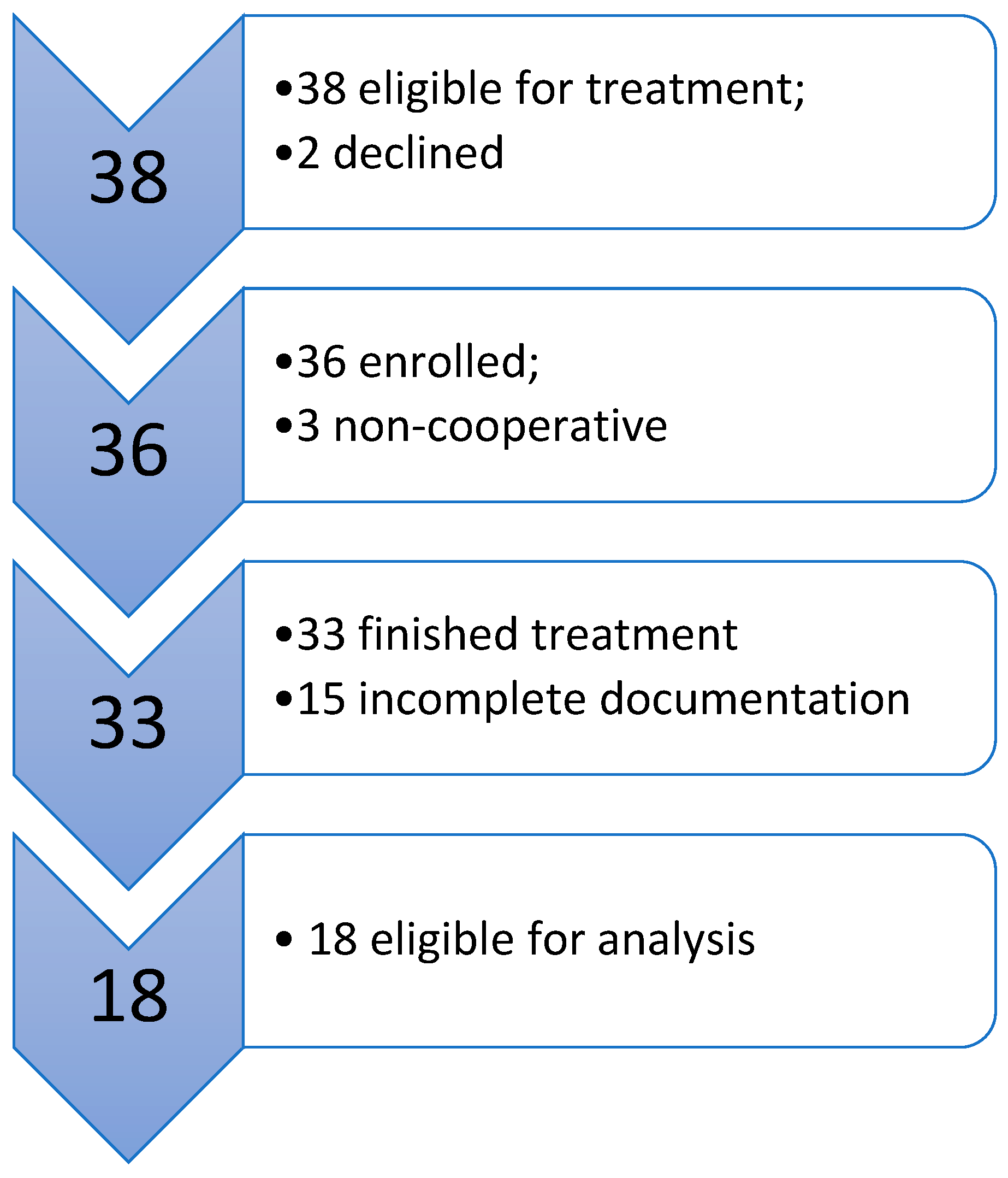
2. Materials and Methods

At the beginning of this retrospective study, it was planned to include 38 participants in the peak puberty growth phase or shortly after (CVS3 and CVS4 stages) who were receiving treatment with a Twin Block appliance during the period from 2023 to 2024. Two participants declined to participate in the study, a further three were non-cooperative (discontinued treatment), and the remaining 33 finished the treatment. Unfortunately, it was established that a further 15 participants had either missed one of the facial scans, or they were not suitable for the analysis, even after inspection upon scanning. Therefore, the final sample consisted of 18 participants (Figure 1).
All parents/guardians of the patients signed an informed consent form for the therapy, thereby giving their consent for the use of medical documentation for research purposes.
Finally, this study included 18 participants (7 girls and 11 boys) who had all the necessary documentation before and after the treatment. Participants were receiving treatment with functional appliances at the clinical unit for orthodontics, which is part of the Department of Dental Medicine at the Clinical Hospital Center Rijeka. The median age of the participants at the start of therapy was 12.4 years, and at the end of therapy it was 13.6 years. The therapy lasted an average of 14.1 months. The Twin Block appliance consists of upper and lower plates with occlusal ramps and hooks that retain the appliance on the dental arch. The registration of the constructional bite is the most important step in the design and final fabrication of the functional appliance. It determines the new sagittal, vertical, and horizontal relationships of the lower jaw to the upper jaw. The activation of the lower jaw must sufficiently stretch the masticatory muscles and allow a positive proprioceptive response, while also staying within the physiological limits of the muscles and joints [14]. The quality of 2D images was also considered, as well as the possibility for their most precise analysis. The sample size was calculated with a power of 90% and a significance level of 0.05, using an online calculator and previous published studies. It was calculated that a minimum of 14 participants was needed [12].
The inclusion criteria for participants in the study were as follows: individuals of both genders with the presence of skeletal Class II with a retrognathic mandible, overjet > 6 mm, ANB > 2.5°, Wits > 2.5 mm, Class II on the first permanent molars and canines, and patients who were undergoing orthodontic treatment for the first time. Additionally, participants included in the study had to have lateral cephalograms taken before and after therapy. All subjects were Class II, division 1; six of them had a horizontal growth pattern (SN:GoGn ≤ 26°), ten had a neutral growth pattern (SN:GoGn > 26° < 37°), and two exhibited a vertical growth pattern (SN:GoGn > 37°). The treatment choice for the functional appliance was made upon performing the Fränkel maneuver.
The exclusion criteria for the study were as follows: the presence of hypodontia, temporomandibular joint disorders, obstructive airway disorders, craniofacial syndromes, congenital malformations, severe facial asymmetries, or systemic diseases that could affect facial morphology.

2.1. Lateral Cephalogram Analysis

Lateral cephalograms and 3D scans of patients were analyzed before and after treatment with the Twin Block appliance. Each lateral cephalogram analyzed in the study was taken on the same device at the Department of Dental Medicine, Clinical Hospital Center Rijeka. The radiographic images were analyzed by one researcher on a personal computer with the AudaxCeph software program, version 6.6.12.4731 (Audax, Ljubljana, Slovenia). Calibration was performed before each measurement. The reference points of the cephalometric analysis can be seen in Figure 2; the defined parameters used are listed and explained in Table 1.

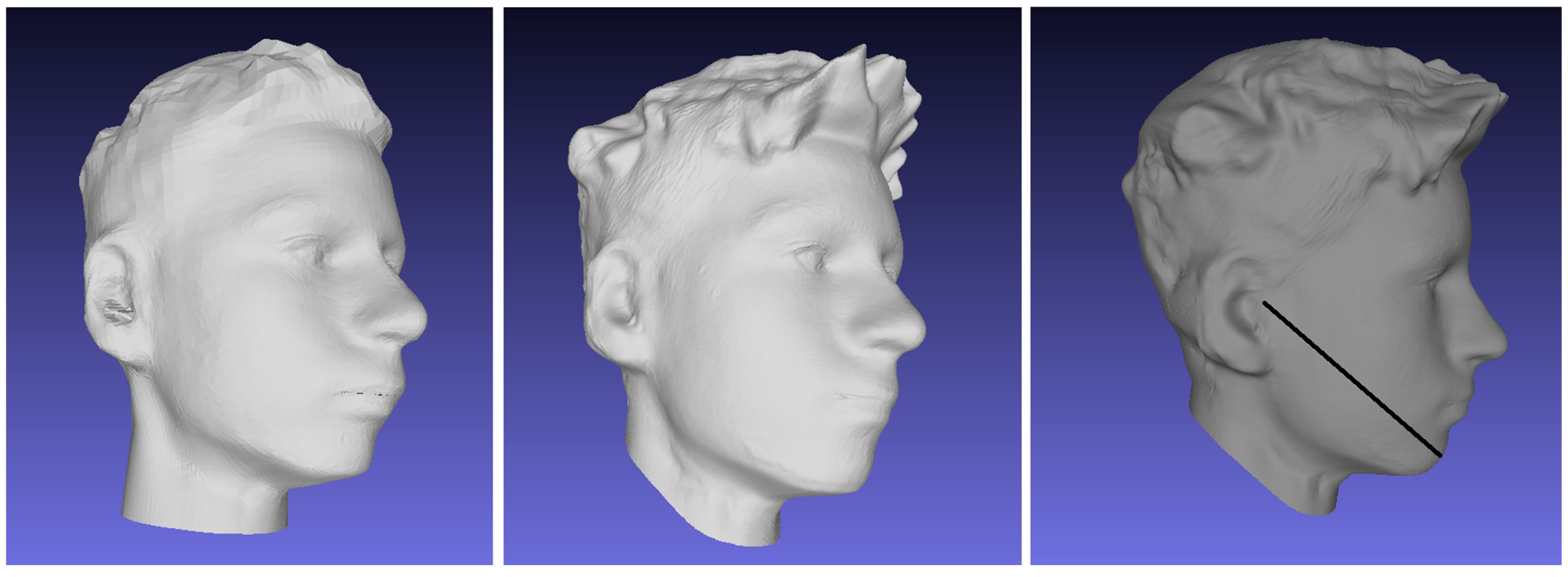
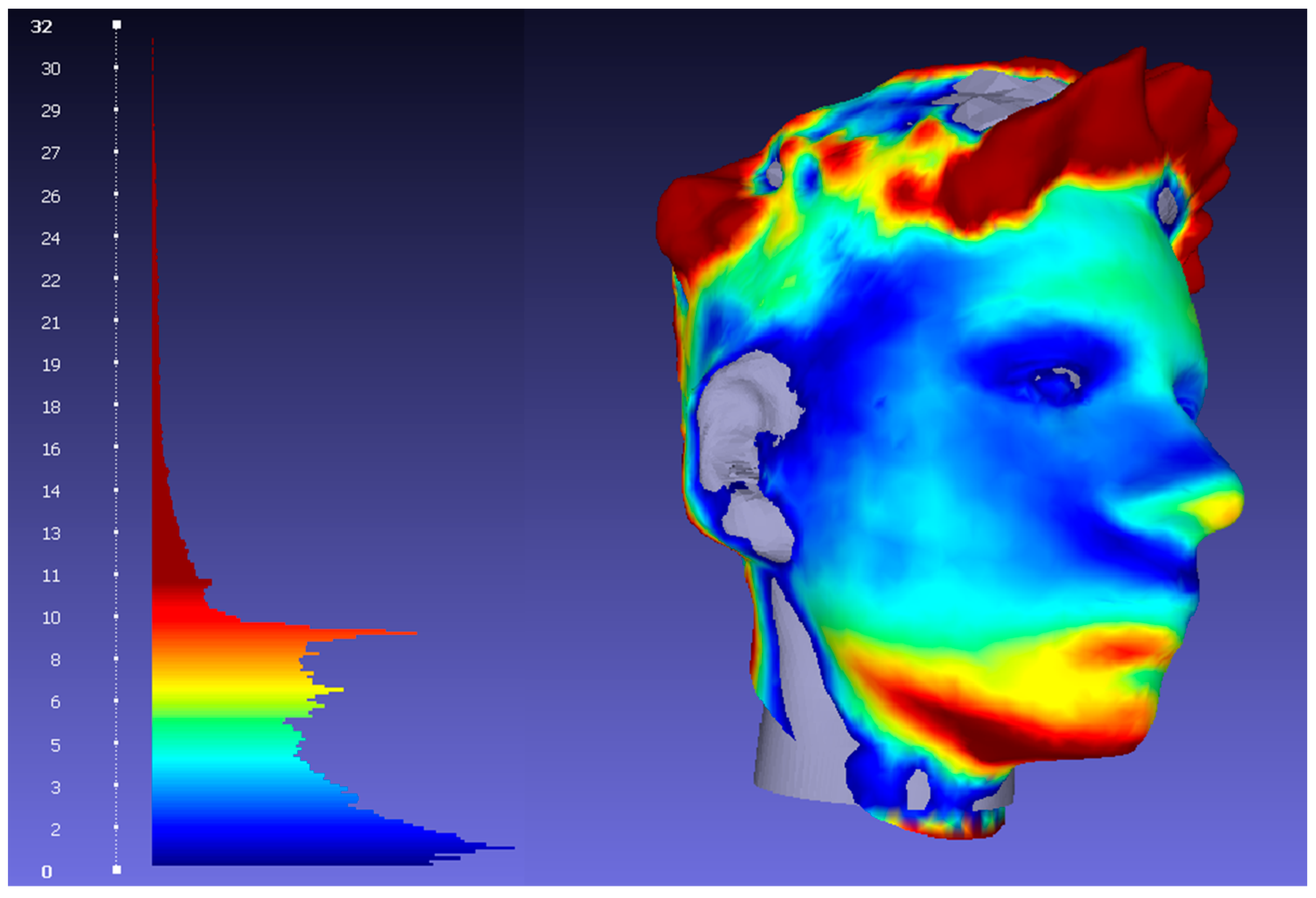
2.2. Three-Dimensional Facial Scans Analysis

Three-dimensional facial scans were captured using the Arc scanner (Bellus3D, Bellus3D, Inc., Campbell, CA, USA) along with the Bellus3D software, version 1.6.2. The scanning procedure involved four distinct head movements: rotating 90° to the left and right, and tilting approximately 45° upward and downward. Facial scanning was performed under illumination provided by a ring light with a correlated color temperature (CCT) of 3000 K. Natural head position (NHP) was used to ensure consistency in image capture as it is proven to be reproducible [18,19]. Participants were seated on a height-adjustable stool and instructed to align their eyes with the level of the front-facing camera. Seating height was modified as needed to facilitate proper NHP alignment. Prior to scanning, subjects were asked to maintain a relaxed jaw posture with their mouths gently closed. Scans were repeated in cases where any irregularities or deviations from the required positioning were observed.
Measurements were taken and recorded in millimeters, specifically the distance between the tragus and the pogonion, to assess soft tissue changes related to the length of the mandible. Mandibular skeletal measurements are typically assessed using the linear distance between condylion (Co) and gnathion (Gn), which represents the effective mandibular length [17]. In soft tissue, the tragus is the anatomical landmark most analogous to the condylion. Therefore, the distance between the tragus and pogonion is commonly used to evaluate soft tissue changes in the lower jaw region [18].
The discrete geodesic problem refers to the shortest path connecting a source and a target across a spherical surface. In the current study, we used the “single source, all destinations” technique proposed by Mitchell and Mount [20]. Their method is based on a specialized variant of Dijkstra’s algorithm, known as the “continuous Dijkstra,” which computes the shortest paths along the edges of a subdivided mesh. For our implementation on triangular mesh structures, we utilized Kirsanov’s geodesic algorithm [21], accessed through a Cython wrapper for the original C++ code.
The overlapping of pre-treatment and post-treatment 3D facial models was performed to analyze soft tissue changes resulting from Twin Block appliance therapy. The 3D scans were obtained separately before and after treatment, comprising approximately a period of one year, and then processed for comparative analysis.
Generating detailed and accurate visualizations is crucial for detecting differences between the reconstructed 3D models. However, this requires the precise alignment of the models. The initial alignment process, where two 3D models are brought into a roughly corresponding spatial configuration without relying on prior position data, is known as global registration [22].
This procedure is a key step in shape matching and 3D model alignment. Global registration does not depend on known starting positions; it tends to produce less accurate results and is generally used to initialize more precise, local alignment techniques. The Iterative Closest Point (ICP) algorithm is one of the most commonly used [22]. The ICP algorithm refines alignment by reducing the squared distances between comparable points in two point clouds, known as the target and reference models. To ensure accuracy, it is essential to register these scans using anatomically stable regions. Anatomically stable regions include portions of the forehead, nasal bridge and infraorbital region. Although centrally located, this region lies within the upper third of the face and demonstrates minimal morphological variation due to growth. Its stability makes it particularly suitable for assessing changes in the middle and lower facial thirds, which are more susceptible to developmental and treatment-induced modifications [12,23,24]. Once these key landmarks are identified for overlap, the alignment process becomes more straightforward and helps minimize fitting errors specifically at those locations. Points outside of these chosen areas are excluded from the alignment function and therefore do not influence the minimization of differences.
The scans were then matched with the ICP algorithm to generate a unified coordinate space in which spatial differences between the models could be measured. This approach facilitated the assessment of morphological changes, particularly in regions influenced by the Twin Block appliance, such as the chin, jawline, and mandible. As this process was conducted, visual and metric inspection was applied to validate the registration accuracy prior to measurement extraction.
A distance map was generated by computing point-to-point distances across the facial surface, which were then visualized using a color-coded heat map. This visualization technique assigns colors to specific ranges of distances, facilitating the interpretation of areas where soft tissue protrusion or retrusion occurred. For instance, regions with minimal displacement were displayed in cooler tones (blue/gray), while regions with greater soft tissue movement—such as the mandible, lower lip, chinand hair—were shown in red tones. Changes in hair were significant mainly because of a change in haircut.
For distance measurements we used Hausdorff distance which is a commonly used metric for comparing two 3D surfaces or point clouds [25]. It measures the greatest of all the shortest distances from a point on one surface to the closest point on the other surface, capturing the maximum deviation between the two shapes. This makes it particularly useful in evaluating geometric accuracy and detecting local misalignments in applications such as model registration, surface reconstruction, and shape comparison. A lower Hausdorff distance indicates better alignment and greater similarity between the compared models.
These visual results were not only useful for confirming the quantitative measurements but also provided a valuable visual tool for communicating changes. The combination of model superimposition and color-based analysis offers an accessible and effective way to interpret complex 3D facial changes resulting from orthodontic treatment.
A statistical analysis was conducted using the SPSS software package (version 26.0; IBM Corp., Armonk, NY, USA). Changes pre- and post-treatment were evaluated using paired t-tests, with statistical significance defined as p < 0.05. Descriptive statistics, including means and standard deviations, were calculated to summarize sample characteristics. Measurement reliability was verified through the Intraclass Correlation Coefficient (ICC), ensuring precision and reproducibility. The Kolmogorov–Smirnov test was employed to assess the normality of variable distributions.

3. Results

The Kolmogorov–Smirnov test for normality indicated that all measured variables followed a normal distribution (p > 0.05), with the exception of the intermaxillary angle and the position of the upper incisors relative to the apical base of the maxilla. For these two variables, the test demonstrated statistically significant results, which compromised the assumption of normality of distribution required for conducting a paired samples t-test. Consequently, the Wilcoxon test was applied only for these two variables.
Using the Intraclass Correlation Coefficient (ICC), high reliability was determined for all measured variables (ICC was greater than 0.9). The measurement reliability test was performed by repeating the measurements of all variables on 10 randomly selected lateral cephalograms from the main sample, before and after therapy, with the measurements being repeated 14 days after the initial measurements by the same examiner.

3.1. Cephalometric Analysis

Descriptive data of cephalometric measurements before and after treatment are shown in Table 2.
Paired t-test results (Table 3) showed that there is a statistically significant difference towards an increase between the first and the second measurement of the SNB angle (p = 0.005), the CoGo (p < 0.001), the Ls:E (p = 0.040), and the proclination and protrusion of the lower incisors (p = 0.021 and p < 0.001, respectively). The decrease was observed for the ANB angle (p = 0.003), Wits (p < 0.001), ANPG (p < 0.001) and overbite (p < 0.001) values and the protrusion of the upper incisors (p = 0.022) (Table 3 and Table 4). The results of the paired t-test are shown in Table 3, and the results of the Wilcoxon test for the protrusion of upper incisors to the apical base of maxilla are shown in Table 4.
Figure 3 shows a typical example of a case before and after treatment with Twin Block from our study. Superimposed analyses before and after treatment show the downwards and forwards displacement of the lower facial third, but no change in vertical relations in terms of the change in the mandibular plane angle (p = 0.283, visible in results from Table 3).

3.2. Soft Tissue Analysis

Measurements from the point tragus to point pogonion in millimeters for every participant were taken on 3D face scans before and after the treatment. The measurements are visible in Table 5. Figure 4 shows a typical head scan before (left) and after treatment (center), and the measurement of the tragus–pogonion distance (right).
Kolmogorov–Smirnov test for normality showed normal distribution of all variables visible in Figure 5.
There is a high statistically significant difference in the results of the paired t-test for the measurements of the soft tissue changes in the length of the mandible. On average, for the first measurement (T0) the mean is 148.71 and the standard deviation is 26.43. For the second measurement (T1), on average, the mean is 153.16 and the standard deviation is 19.86, p < 0.001. After the superimposition of 3D face scans before and after the treatment for every patient, the greatest recorded distances in the chin area are shown in Table 6.
The color-coded heat map indicated that the most significant soft tissue changes following treatment were observed in the protrusion of the chin and the elongation of the mandible. Typical changes are visible in Figure 6.

4. Discussion

In this study, the effects of Twin Block appliance therapy on cephalometric variables and 3D face scans in patients with Class II malocclusion were analyzed. The results of the paired t-test showed a significant increase in the values of SNB, mandibular length, LsE, and soft tissue mandible length and the protrusion and inclination of lower incisors, while the values of ANB, Wits, ANPG, and overbite and the protrusion of upper incisors to the apical base of the maxilla showed a significant decrease. Other variables did not show significant changes, including maxillary and mandibular plane angles, and intermaxillary angle, indicating no change in vertical dimensions.
An increase in the lower facial third is a favorable response to therapy in Class II patients, and it is mostly attributed to the vertical growth in the condyle and mandibular ramus, without an opening of the mandibular plane angle [7]. These results suggest that Twin Block appliance therapy effectively influenced certain skeletal and soft tissue parameters. Previous studies have indicated that patients with a condylar angle (Co-Go-Me) less than 125.5° before therapy are likely to exhibit favorable responses to treatment with functional appliances. In the present study, a condylar angle of approximately 119°, both pre- and post-treatment, demonstrated that the use of the Twin Block functional appliance was favorable for treatment outcomes [26]. Comparing the results obtained with previous studies, certain similarities and differences can be observed. The increase in the SNB angle after Twin Block appliance therapy is consistent with findings from other studies, such as those by Jena et al. (2013), Ghodke et al. (2014), and Çoban and Camci (2023), indicating sagittal advancement of the mandible and a reduction in the skeletal discrepancy between the mandible and maxilla [27,28,29]. Vertical cephalometric parameters did not change significantly after therapy, and as such, did not contribute to masking or emphasizing the sagittal changes. The distance between the reference point of the upper lip (Ls) and Ricketts’ esthetic line (Ls:E) increased significantly, contributing to a more optimal facial profile and reduced protrusion of the upper lip. Most of the other related studies support the statistically significant increase in the Ls:E distance, such as those by Varlik et al. (2008), Khoja et al. (2016), and Baysal and Uysal (2013) [30,31,32]. All of these studies also included a control group, which further strengthens their findings. Although the present study did not include a control group, previous research incorporating a control cohort found no statistically significant differences in skeletal discrepancy or overjet which could be contributed to the growth alone in control subjects [15]. Additionally, another study reported an average mandibular length increase of only 0.3 mm due to natural growth, an amount that was not statistically significant, particularly when compared to the treatment group managed with the Twin Block appliance, which exhibited a statistically significant increase in mandibular length [16], similarly to our findings. Furthermore, the study of maxillomandibular growth in untreated Class II showed that mandibular deficiency will not self-correct during pubertal peak growth [33]. The systematic literature review by Flores et Major (2006) concluded that two-dimensional measurement methods, such as those based on lateral cephalograms, are not the most optimal approach for evaluating soft tissues, which may explain the contradictory results [34]. The statistically significant decrease in the skeletal class angle (ANB) and Wits as a linear measure for class contributes to the reduction in the discrepancy between the upper and lower jaws, bringing their relationship closer to Class I. The results suggest that the increase in the SNB angle, accompanied by the increase in the effective mandibular length, is the main cause of the reduction in the skeletal class angle. The mean value of the maxillary prognathism angle (SNA) did not change significantly after Twin Block therapy, indicating that there was no inhibition of maxillary growth, also seen in other studies [27,28,29,31,32]. Similar results can be seen in the study by Lombardo et al. (2024), which also reported statistically significant changes, including an increase in the SNB angle and a decrease in the ANB angle [35]. However, after therapy, the average value of the SNA decreased, albeit without statistical significance, similarly to our data, and without significant change in the control groups of the cited studies [16,33,34,35]. In a study by Çoban and Camci (2023), in addition to the decrease in the ANB angle and the increase in the SNB angle, there was also a statistically significant decrease in the SNA, which may indicate the minimal, but statistically significant, inhibition of maxillary growth [28]. The statistically significant decrease in the Wits value correlates with the results from other studies [34,35]. A statistically significant decrease can also be observed in the angle of skeletal convexity (ANPG), which supports the reduction in the convexity of the bony facial profile. A review of the literature revealed contradictions in the results, with some studies indicating a significant decrease in the angle of skeletal convexity, while others report an increase [6,34]. The results did show statistically significant changes in the inclination and protrusion of lower incisors relative to the mandibular base and protrusion of upper incisors to the apical base of the maxilla. This suggests that the Twin Block appliance also affects the dentoalveolar level, leading to protrusion of lower incisors and retrusion of upper incisors. Most studies in this literature review confirm statistically significant changes in the dental segment, similarly to our data [16,17,34,35].
Limitations of this study include the absence of a control group, which prevents our drawing conclusions about the extent to which the changes in cephalometric and soft tissue parameters are solely due to the effects of the functional appliance, rather than the interaction between growth, development, and the appliance. Also, it would be interesting to see in future research whether the increase in the lower facial third would contribute to more harmonious relations, as compared to lower facial heights before therapy.

5. Conclusions

The Twin Block appliance in this study reduced the skeletal discrepancy between the maxilla and mandible. The statistically significant increase in the mandibular prognathism angle and mandibular length allowed the mandible to achieve a more favorable anterior position relative to the maxilla. The angular and linear values that define skeletal class (ANB and Wits) led to a significant reduction in Class II malocclusion. No statistically significant impact on the upper jaw was recorded. The significant increase in the distance between the upper lip and Ricketts’ esthetic line (Ls:E) and in soft tissue mandible length is evidence of the appliance’s effect on the soft tissues, contributing to a more harmonious facial appearance.

Author Contributions

V.K. and B.G. were responsible for the conceptualization, methodology, software, and project administration. V.K. handled resources and funding acquisition. Validation was performed by V.K. and D.Š.P. The formal analysis was performed by V.K., D.Š.P. and B.G., while the investigation was conducted by S.C. and A.S. Data curation was managed by V.K. and L.Š.; D.Š.P. wrote the original draft, while A.S. contributed to writing—review and editing and handled visualization. Supervision was provided by V.K. All authors have read and agreed to the published version of the manuscript.

Funding

The study was supported by the University of Rijeka via grant number uniri-iskusni-biomed-23-36, titled “Impact of functional appliances on 3D craniodentofacial characteristics and reported sleep related breathing disorders in children”.

Institutional Review Board Statement

The study was conducted in accordance with the Declaration of Helsinki and approved by the Ethics Committee of the Faculty of dental medicine, University of Rijeka (protocol code 12-23, 19 June 2023), and the Ethics Committee of Clinical Hospital center Rijeka (22 December 2022) for studies involving humans.

Informed Consent Statement

Informed consent was obtained from all subjects involved in the study.

Data Availability Statement

The datasets presented in this article are not readily available because the data are part of an ongoing study and contain identifiable details. Requests to access the datasets should be directed to visnja.katic@fdmri.uniri.hr.

Conflicts of Interest

The authors declare no conflicts of interest. The funders had no role in the design of the study; in the collection, analyses, or interpretation of data; in the writing of the manuscript; or in the decision to publish the results.

References

  1. Guo, R.; Tian, Y.; Li, X.; Li, W.; He, D.; Sun, Y. Facial profile evaluation and prediction of skeletal class II patients during camouflage extraction treatment: A pilot study. Head Face Med. 2023, 19, 51. [Google Scholar] [CrossRef]
  2. Lone, I.M.; Zohud, O.; Midlej, K.; Proff, P.; Watted, N.; Iraqi, F.A. Skeletal Class II Malocclusion: From Clinical Treatment Strategies to the Roadmap in Identifying the Genetic Bases of Development in Humans with the Support of the Collaborative Cross Mouse Population. J. Clin. Med. 2023, 12, 5148. [Google Scholar] [CrossRef]
  3. Bittencourt Neto, A.C.; Saga, A.Y.; Pacheco, A.A.R.; Tanaka, O. Therapeutic approach to Class II, Division 1 malocclusion with maxillary functional orthopedics. Dental Press. J. Orthod. 2015, 20, 99–125. [Google Scholar] [CrossRef]
  4. Santana, L.G.; Avelar, K.; Flores-Mir, C.; Marques, L.S. Incremental or maximal mandibular advancement in the treatment of class II malocclusion through functional appliances: A systematic review with meta-analysis. Orthod. Craniofac. Res. 2020, 23, 371–384. [Google Scholar] [CrossRef]
  5. Ivorra-Carbonell, L.; Montiel-Company, J.M.; Almerich-Silla, J.M.; Paredes-Gallardo, V.; Bellot-Arcís, C. Impact of functional mandibular advancement appliances on the temporomandibular joint—A systematic review. Med. Oral. Patol. Oral. Cir. Bucal. 2016, 21, e565–e572. [Google Scholar] [CrossRef]
  6. Ghorbani, M.; Mousavi, S.A.; Bardideh, E.; Saeedi, P.; Shahnaseri, S.; Shafaee, H.; Akyalcin, S. The Effectiveness of Functional Clear Aligners for Class II Correction in Growing Patients: A Systematic Review and Meta-Analysis. Orthod. Craniofac. Res. 2025. [Google Scholar] [CrossRef]
  7. Souki, B.Q.; Vilefort, P.L.C.; Oliveira, D.D.; Andrade, I., Jr.; Ruellas, A.C.; Yatabe, M.S.; Nguyen, T.; Franchi, L.; McNamara, J.A., Jr.; Cevidanes, L.H.S. Three-dimensional skeletal mandibular changes associated with Herbst appliance treatment. Orthod. Craniofac. Res. 2017, 20, 111–118. [Google Scholar] [CrossRef]
  8. Kim, J.E.; Mah, S.J.; Kim, T.W.; Kim, S.J.; Park, K.H.; Kang, Y.G. Predictors of favorable soft tissue profile outcomes following Class II Twin-block treatment. Korean J. Orthod. 2018, 48, 11–22. [Google Scholar] [CrossRef]
  9. Shahamfar, M.; Atashi, M.H.A.; Azima, N. Soft tissue esthetic changes following a modified twin block appliance therapy: A prospective study. Int. J. Clin. Pediatr. Dent. 2020, 13, 255–260. [Google Scholar] [CrossRef]
  10. Qiang, J.; Wu, D.; Du, H.; Zhu, H.; Chen, S.; Pan, H. Review on Facial-Recognition-Based Applications in Disease Diagnosis. Bioengineering 2022, 9, 273. [Google Scholar] [CrossRef]
  11. Pellitteri, F.; Scisciola, F.; Cremonini, F.; Baciliero, M.; Lombardo, L. Accuracy of 3D facial scans: A comparison of three different scanning system in an in vivo study. Prog. Orthod. 2023, 24, 44. [Google Scholar] [CrossRef]
  12. Pellitteri, F.; Albertini, P.; Brucculeri, L.; Cremonini, F.; Guiducci, D.; Falconi, V.; Lombardo, L. Soft tissue changes during orthopedic therapy: An in vivo 3-dimensional facial scan study. Am. J. Orthod. Dentofac. Orthop. 2025, 167, 154–165. [Google Scholar] [CrossRef]
  13. Gašparović, B.; Morelato, L.; Lenac, K.; Mauša, G.; Zhurov, A.; Katić, V. Comparing Direct Measurements and Three-Dimensional (3D) Scans for Evaluating Facial Soft Tissue. Sensors 2023, 23, 2412. [Google Scholar] [CrossRef]
  14. McNamara, J.A. Neuromuscular and skeletal adaptations to altered function in the orofacial region. Am. J. Orthod. 1973, 64, 578–606. [Google Scholar] [CrossRef]
  15. Cozza, P.; Baccetti, T.; Franchi, L.; De Toffol, L.; McNamara, J.A. Mandibular changes produced by functional appliances in Class II malocclusion: A systematic review. Am. J. Orthod. Dentofac. Orthop. 2006, 129, 599.e1–599.e12. [Google Scholar] [CrossRef]
  16. Baysal, A.; Uysal, T. Dentoskeletal effects of Twin Block and Herbst appliances in patients with Class II division 1 mandibular retrognathy. Eur. J. Orthod. 2014, 36, 164–172. [Google Scholar] [CrossRef]
  17. Khan, M.I.; Neela, P.K.; Unnisa, N.; Jaiswal, A.K.; Ahmed, N.; Purkayastha, A. Dentoskeletal effects of Twin Block appliance in patients with Class II malocclusion. Med. Pharm. Rep. 2022, 95, 191–196. [Google Scholar] [CrossRef]
  18. Kaplan, V.; Ciğerim, L.; Ciğerim, S.Ç.; Bazyel, Z.D.; Dinç, G. Comparison of Various Measurement Methods in the Evaluation of Swelling After Third Molar Surgery. Van. Med. J. 2021, 28, 412–420. [Google Scholar] [CrossRef]
  19. Chiu, C.S.; Clark, R.K. Reproducibility of natural head position. J. Dent. 1991, 19, 130–131. [Google Scholar] [CrossRef]
  20. Mitchell, J.S.B.; Mount, D.M.; Papadimitriou, C.H. The Discrete Geodesic Problem. SIAM J. Comput. 1987, 16, 647–668. [Google Scholar] [CrossRef]
  21. Kirsanov, D. Exact Geodesic for Triangular Meshes. 2022. Available online: https://www.mathworks.com/matlabcentral/fileexchange/18168-exact-487geodesic-for-triangular-meshes (accessed on 15 April 2025).
  22. Besl, P.; McKay, N.D. A method for registration of 3-D shapes. IEEE Trans. Pattern Anal. Mach. Intell. 1992, 14, 239–256. [Google Scholar] [CrossRef]
  23. Farkas, L.G. Anthropometry of the Head and Face; Lippincott Williams & Wilkins: Philadelphia, PA, USA, 1994. [Google Scholar]
  24. Häner, S.T.; Kanavakis, G.; Matthey, F.; Gkantidis, N. Valid 3D surface superimposition references to assess facial changes during growth. Sci. Rep. 2021, 11, 16456. [Google Scholar] [CrossRef]
  25. Aspert, N.; Santa-Cruz, D.; Ebrahimi, T. MESH: Measuring errors between surfaces using the Hausdorff distance. In Proceedings of the Proceedings. IEEE International Conference on Multimedia and Expo, Lausanne, Switzerland, 26–29 August 2002; Volume 1, pp. 705–708. [Google Scholar] [CrossRef]
  26. Franchi, L.; Baccetti, T. Prediction of individual mandibular changes induced by functional jaw orthopedics followed by fixed appliances in Class II patients. Angle Orthod. 2006, 76, 950–954. [Google Scholar] [CrossRef]
  27. Jena, A.K.; Singh, S.P.; Utreja, A.K. Effectiveness of twin-block and Mandibular Protraction Appliance-IV in the improvement of pharyngeal airway passage dimensions in Class II malocclusion subjects with a retrognathic mandible. Angle Orthod. 2013, 83, 728–734. [Google Scholar] [CrossRef]
  28. Çoban Büyükbayraktar, Z.; Camcı, H. Dentoalveolar, skeletal, pharyngeal airway, cervical posture, hyoid bone position, and soft palate changes with Myobrace and Twin-block: A retrospective study. BMC Oral Health 2023, 23, 53. [Google Scholar] [CrossRef]
  29. Ghodke, S.; Utreja, A.K.; Singh, S.P.; Jena, A.K. Effects of twin-block appliance on the anatomy of pharyngeal airway passage (PAP) in class II malocclusion subjects. Prog. Orthod. 2014, 15, 68. [Google Scholar] [CrossRef]
  30. Varlik, S.K.; Gultan, A.; Tumer, N. Comparison of the effects of Twin Block and activator treatment on the soft tissue profile. Eur. J. Orthod. 2008, 30, 128–134. [Google Scholar] [CrossRef]
  31. Khoja, A.; Fida, M.; Shaikh, A. Cephalometric evaluation of the effects of the twin block appliance in subjects with class II, division 1 malocclusion amongst different cervical vertebral maturation stages. Dent. Press. J. Orthod. 2016, 21, 73–84. [Google Scholar] [CrossRef]
  32. Baysal, A.; Uysal, T. Soft tissue effects of twin block and herbst appliances in patients with class II division 1 mandibular retrognathy. Eur. J. Orthod. 2013, 35, 71–81. [Google Scholar] [CrossRef]
  33. Perinetti, G.; Sbardella, V.; Bertolami, V.; Contardo, L.; Primozic, J. Circumpubertal maxillomandibular growth in untreated subjects with skeletal Class II relationship: A controlled longitudinal study according to the third finger middle phalanx maturation. Am. J. Orthod. Dentofac. Orthop. 2022, 162, 937–946. [Google Scholar] [CrossRef]
  34. Flores-Mir, C.; Major, P.W. Cephalometric facial soft tissue changes with the twin block appliance in class II division 1 malocclusion patients: A systematic review. Angle Orthod. 2006, 76, 876–881. [Google Scholar]
  35. Lombardo, E.C.; Lione, R.; Franchi, L.; Gaffuri, F.; Maspero, C.; Cozza, P.; Pavoni, C. Dentoskeletal effects of clear aligner vs twin block-a short-term study of functional appliances. J. Orofac. Orthop. 2024, 85, 317–326. [Google Scholar] [CrossRef]
Figure 1. Study enrolment flow chart.
Applsci 15 07529 g001
Figure 2. The reference cephalometric points marked on latero-lateral cephalogram of an example of a case before treatment.
Applsci 15 07529 g002
Figure 3. The reference cephalometric points marked on latero-lateral cephalogram of an example of a case before treatment (left) and after the Twin Block treatment (center); on the (right): pre- and post-treatment lateral cephalograms superimposed at SN line (center at S) with drawn lines representing maxilla and mandible, upper and lower frontal teeth, and first permanent molars (red—before treatment; green—after treatment).
Applsci 15 07529 g003
Figure 4. Examples of a typical head scan before (left) and after treatment (center), and measurement of the tragus–pogonion distance shown with black line (right).
Applsci 15 07529 g004
Figure 5. Distribution of data normality for the soft tissue tragus–pogonion measurements.
Applsci 15 07529 g005
Figure 6. Color-coded heat map showing significant changes in the chin, mandible, and lower lip areas.
Applsci 15 07529 g006
Table 1. Variables measured in the cephalometric analysis.
VariablesDescription
SNASagittal relationship of the maxilla to the anterior cranial base
SNBSagittal relationship of the mandible to the anterior cranial base
ANBSagittal relationship of the maxilla to the mandible
WitsLinear measurement of skeletal class
ANPGAngle of convexity of the facial bony structures
SN:NLMaxillary plane angle
MaxMandAngleIntermaxillary angle
N-S-ArCranial base angle
SArGoArticular angle
MeGoArMandibular angle
NSGnY-axis angle
SN:GoGnMandibular plane angle
CoGoMeCondylar angle
U1:MaxProclination of the upper incisors to the maxillary base
L1:MandProclination of the lower incisors to the mandibular base
U1:L1Interincisal angle
U1MxPlaneProtrusion of the upper incisors to the apical base of the maxilla
L1MdPlaneProtrusion of the lower incisors to the apical base of the mandible
OJOverjet
OBOverbite
Ls:E1Distance of the upper lip from the Ricketts’ esthetic line (Prn-Pgn)
Li:E1Distance of the lower lip from the Ricketts’ esthetic line (Prn-Pgn)
CoGnMandibular length
Table 2. Descriptive statistics of the measured variables before (T0) and after (T1) therapy (N = 18).
T0T1
MSDMSD
SNA (°)82.174.3081.994.30
SNB (°)76.973.5478.193.97
ANB (°)5.212.113.811.67
Wits (mm)3.283.190.792.49
ANPG (°)8.355.655.354.80
SN:NL (°)8.433.108.123.16
MaxMandAngle (°)20.775.9319.986.53
NSAr (°)124.334.07124.304.53
SArGo (°)142.665.97142.526.08
MeGoAr (°)122.187.20121.285.59
NSGn (°)65.684.8965.625.12
SN:GoGn (°)26.777.3026.006.86
CoGoMe (°)119.576.92119.796.27
U1:Max (°)113.778.90111.334.44
L1:Mand (°)97.187.0399.655.81
U1:L1 (°)128.2910.38129.055.79
U1MxPlane (mm)6.232.835.281.81
L1MdPlane (mm)4.391.935.162.10
OJ (mm)3.873.493.572.84
OB (mm)7.583.793.322.80
Ls:E line (mm)1.671.372.902.00
Li:E line (mm)2.091.802.631.84
CoGn (mm)104.704.58109.945.06
M—mean; SD—standard deviation.
Table 3. Paired samples t-test.
Paired DifferencestdfSig. (2-Tailed)
MeanStd. Deviation
OB (mm)−4.2620914.688027−3.857170.001
OJ (mm)−0.3065365.041305−0.258170.800
L1MdPlane (mm)0.7673390.5547965.868170.000
U1:L1 (°)0.7620897.2457530.446170.661
L1:Mand (°)2.4674544.1315992.534170.021
U1:Max (°)−2.4407426.227762−1.663170.115
CoGoMe (°)0.2266505.4349270.177170.862
SN:GoGn (°)−0.7633752.922641−1.108170.283
NSGn (°)−0.0546931.372553−0.169170.868
MeGoAr (°)−0.8929035.603276−0.676170.508
SArGo (°)−0.1681773.400253−0.210170.836
NSAr (°)−0.0305322.387120−0.054170.957
SN:NL (°)−0.3028032.333445−0.551170.589
ANPG (°)−2.9940333.262767−3.893170.001
Wits (mm)−2.4885792.678087−3.942170.001
ANB (°)−1.4020671.684264−3.532170.003
SNB (°)1.2237921.5997293.246170.005
SNA (°)−0.1782721.153377−0.656170.521
Li:E line (mm)0.5408462.0827171.102170.286
Ls:E line (mm)1.2235232.3321832.226170.040
CoGn (mm)5.240563.457936.430170.001
Table 4. Wilcoxon test for protrusion of upper incisors to the apical base of the maxilla.
Related-Samples Wilcoxon Signed Rank Test Summary
Total N18
Test Statistic33.000
Standard Error22.962
Standardized Test Statistic−2.286
Asymptotic Sig. (2-sided test)0.022
Table 5. Measurements of the soft tissue changes in the length of the mandible.
SampleTragus–Pogonion (mm)
T0
Tragus–Pogonion (mm)
T1
1145.05151.70
2144.23147.67
3146.54149.04
4148.99158.62
5151.77154.38
6156.46159.83
7148.40151.74
8147.28149.53
9145.29148.67
10151.80153.94
11149.02158.71
12150.79152.76
13154.15156.97
14137.61148.88
15150.27152.18
16157.93162.24
17150.78153.09
18140.39147.90
T0—before treatment; T1—after treatment.
Table 6. Hausdorff distance from reference mesh.
SampleMin (mm)Max (mm)
10.00032521.22
20.00056219.89
30.00018720.47
4018.12
50.00096922.65
6023.41
7037.59
80.0004546.96
90.0001128.39
100.00088812.45
110.00057426.74
1209.45
130.00048316.96
140.00028812.41
150.00072916.58
16034.12
17017.68
180.00034214.29
Disclaimer/Publisher’s Note: The statements, opinions and data contained in all publications are solely those of the individual author(s) and contributor(s) and not of MDPI and/or the editor(s). MDPI and/or the editor(s) disclaim responsibility for any injury to people or property resulting from any ideas, methods, instructions or products referred to in the content.

