Article

Clinical Characterization of Patients with Syncope of Unclear Cause Using Unsupervised Machine-Learning Tools: A Pilot Study

by
María-José Muñoz-Martínez
1,2,3,*,
Manuel Casal-Guisande
2,4,5,*,
María Torres-Durán
1,2,4,
Bernardo Sopeña
3,6 and
Alberto Fernández-Villar
1,2,4,7
1
Pulmonary Department, Hospital Universitario Álvaro Cunqueiro, 36312 Vigo, Spain
2
NeumoVigo I+i Research Group, Galicia Sur Health Research Institute (IIS Galicia Sur), SERGAS-UVIGO, 36312 Vigo, Spain
3
Faculty of Medicine and Dentistry, University of Santiago de Compostela, 15782 Santiago de Compostela, Spain
4
Centro de Investigación Biomédica en Red, CIBERES ISCIII, 28029 Madrid, Spain
5
Fundación Pública Galega de Investigación Biomédica Galicia Sur, Hospital Álvaro Cunqueiro, 36312 Vigo, Spain
6
Internal Medicine Department, Hospital Clínico Universitario de Santiago de Compostela, 15782 Santiago de Compostela, Spain
7
School of Industrial Engineering, University of Vigo, 36310 Vigo, Spain
*
Authors to whom correspondence should be addressed.
Appl. Sci. 2025, 15(13), 7176; https://doi.org/10.3390/app15137176
Submission received: 10 June 2025 / Revised: 20 June 2025 / Accepted: 24 June 2025 / Published: 26 June 2025

Abstract

Syncope of unclear cause (SUC) presents a significant diagnostic challenge, with a considerable proportion of patients remaining without a definitive diagnosis despite comprehensive clinical evaluation. This study aims to explore the potential of unsupervised machine learning (ML), specifically clustering algorithms, to identify clinically meaningful subgroups within a cohort of 123 patients with SUC. Patients were prospectively recruited from the cardiology, neurology, and emergency departments, and clustering was performed using the k-prototypes algorithm, which is suitable for mixed-type data. The number of clusters was determined through cost function analysis and silhouette index, and visual validation was performed using UMAP. Five distinct patient clusters were identified, each exhibiting unique profiles in terms of age, comorbidities, and symptomatology. After clustering, nocturnal cardiorespiratory polygraphy and heart rate variability (HRV) parameters were analyzed across groups to uncover potential physiological differences. The results suggest distinct autonomic and respiratory patterns in specific clusters, pointing toward possible links among sympathetic dysregulation, sleep-related disturbances, and syncope. While the sample size imposes limitations on generalizability, this pilot study demonstrates the feasibility of applying unsupervised ML to complex clinical syndromes. The integration of clinical, autonomic, and sleep-related data may provide a foundation for future, larger-scale studies aiming to improve diagnostic precision and guide personalized management strategies in patients with SUC.

1. Introduction

Syncope is a clinical condition characterized by a sudden and transient loss of consciousness caused by a temporary decrease in cerebral blood flow. Its onset is rapid, its duration is brief, and recovery is usually complete and spontaneous [1]. It is estimated that between 20% and 37% of patients with syncope experience recurrences within the first three years, a factor that has been associated with a higher risk of mortality and serious cardiovascular events [2,3,4]. Additionally, syncope carries a significant risk of trauma [5] and is associated with an all-cause mortality rate ranging from 4% to 6% [4]. It also negatively impacts quality of life and represents a considerable burden on healthcare systems [6]. The incidence of syncope shows two peaks: between the ages of 15 and 20, and around the age of 60, with the latter group exhibiting the highest morbidity and mortality [7].
From a pathophysiological perspective, syncope can be classified as reflex or neurally mediated, cardiogenic, or orthostatic [1]. Identifying the etiology is essential for guiding treatment and establishing a prognosis. However, the initial evaluation is often inconclusive, necessitating additional studies in many cases [8]. Despite these complementary evaluations, up to 48% of cases remain without an etiological diagnosis after 2.5 years of follow-up [9]. Furthermore, there are discrepancies among the recommendations of different scientific societies regarding the management of syncope [10]. The European Society of Cardiology, for example, suggests the early use of implantable loop recorders in patients with syncope of unclear cause (SUC) [1].
The clinical management of syncope remains complex, especially when the cause is uncertain, which complicates diagnosis and increases healthcare costs. Clinical decision rules have been developed to predict outcomes and stratify risk, but recent evidence suggests that most of these scales have limited predictive accuracy, restricting their utility in clinical practice [11].
In this context, artificial intelligence (AI) techniques, particularly machine learning (ML), have shown promising results in various areas of medicine [12,13,14,15,16,17,18]. However, their application in the field of syncope has been limited. Most previous studies on AI in syncope have used supervised ML methods for risk stratification, yielding only modest results, comparable to traditional approaches [19]. AI techniques have also been applied to improve diagnosis and risk prediction [20], but these studies have primarily focused on Holter and tilt-table data, which are not typically available in the early stages of diagnosis, when they could be most useful.
Unsupervised ML techniques, particularly clustering methods, hold great potential in medicine, as they enable the identification of homogeneous patient groups within the clinical heterogeneity of a disease. This approach could facilitate better stratification and personalization of diagnostic and therapeutic strategies. In other conditions, such as COPD [21], clustering has proven useful for identifying clinical subtypes with prognostic and therapeutic implications. However, to date, few published studies have applied clustering methods in the context of SUC.
The possibility of identifying syncope patient clusters based on clinical and demographic data—commonly available in the initial diagnostic phases—could allow for more precise analyses. This would facilitate the identification of patients at higher risk and promote more personalized and comprehensive care, potentially avoiding the need for invasive procedures such as implantable loop recorders.
The aim of this study is to evaluate the utility of unsupervised ML techniques in identifying clusters among patients with SUC, characterized using clinical and demographic information. Unlike other studies, this work also includes data obtained through overnight cardiorespiratory polygraphy, based on the hypothesis that respiratory events and nocturnal heart rate variability may be related to syncope of uncertain etiology [22,23]. However, polygraphic data will not be used for the initial clustering but will instead be integrated later to assess key differences among clusters in polygraphic terms, which could support clinical decision-making by identifying groups that may benefit from polygraphic studies.

2. Materials and Methods

2.1. Study Cohort

The cohort used in this study included 179 patients recruited from the Cardiology (mainly those on the waiting list for an implantable loop recorder), Neurology, or Emergency Departments between January 2019 and May 2024 at Álvaro Cunqueiro Hospital (Vigo, Spain). The study was approved by the Galicia Ethics and Research Committee under protocol number 2019/048. This work is part of the SINCOSAS study [24], an initiative aimed at exploring the relationship between SUC and sleep apnea (SA), a connection that has been little studied to date. All participants were informed about the nature and objectives of the study, and written informed consent was obtained from all subjects involved.
Patients over 18 years of age with a diagnosis of syncope of unknown cause after neurological and cardiological evaluation in accordance with clinical guidelines [1] were included. Patients with a diagnosis of SA, epilepsy, or illicit drug use were excluded.
The first column of Table 1 and Table 2 presents a summary of the collected variables. Anthropometric variables were recorded [age, sex, body mass index (BMI)], known cardiovascular diseases [ischemic or valvular heart disease, arterial hypertension, stroke, arrhythmias], respiratory diseases (COPD, asthma), dyslipidemia, and diabetes. Smoking habits (status and pack-years), symptoms of SA (daytime sleepiness assessed using the Epworth Sleepiness Scale, awakenings, non-restorative sleep, daytime fatigue), total number of syncopal episodes, and number of episodes in the previous year were also recorded. In addition, the use of negative inotropic (cardiodepressive) drugs capable of reducing heart rate was documented.
All patients underwent overnight home respiratory polygraphy using the Embletta® MPR model (Natus Medical Inc., Middleton, WI, USA), a type III portable system that allows synchronized recording of electrocardiographic signals and automated analysis of heart rate variability (HRV). The device is equipped with sensors to measure nasal airflow (via pressure transducer and thermistor), thoracic and abdominal respiratory effort (inductive bands), and oxygen saturation and heart rate via a finger pulse oximeter, as well as continuous electrocardiographic monitoring. All electrocardiographic signals were obtained with a sampling rate of 200 Hz.
Recordings were conducted between 00:00 and 07:00. Respiratory events were initially detected through automated analysis and subsequently reviewed and manually corrected by trained personnel. HRV analysis was performed automatically.
The definition and classification of apneas and hypopneas followed the clinical guidelines of the Spanish Society of Pulmonology and Thoracic Surgery (SEPAR) [25]. The apnea–hypopnea index (AHI) was calculated as the total number of apneas and hypopneas divided by the total recording time. The respiratory events included in the AHI are apneas and hypopneas, defined as follows: obstructive apnea: ≥90% reduction in airflow lasting at least 10 s, with continued respiratory effort; central apnea: ≥90% reduction in airflow for at least 10 s, without respiratory effort; mixed apnea: initially presents without respiratory effort (as a central apnea), followed by resumption of effort; and hypopnea: ≥30% reduction in airflow for at least 10 s, associated with a ≥3% oxygen desaturation. AHI was used to classify SA severity as follows: normal: AHI < 5 events/h; mild: 5 ≤ AHI < 15 events/h; moderate: 15 ≤ AHI < 30 events/h; and severe: AHI ≥ 30 events/h.
Although in this study HRV was obtained from the electrocardiogram (ECG) channel of the polygraph device itself, in future methodological applications, it could be derived from ambulatory 24 h Holter recordings, without the need to conduct respiratory studies.
Time-domain HRV measures were included, such as mean RR interval, SDNN (standard deviation of RR intervals), SDANN (standard deviation of the average RR intervals in all 1 min segments of the full recording), RMSSD (root mean square of successive differences between adjacent RR intervals), SDNN index (mean of the standard deviations of all RR intervals for all 1 min segments), NN50 (number of successive RR intervals differing by more than 50 ms), pNN50 (NN50 count divided by the total number of RR intervals, expressed as a percentage), and the triangular index (total number of RR intervals divided by the height of the RR interval histogram). Frequency-domain parameters were also analyzed, including total power (variance of all RR intervals), high-frequency (HF) power, low-frequency (LF) power, and very-low-frequency (VLF) power.
Respiratory parameters derived from the polygraphy included the AHI, time with oxygen saturation below 90% (TC90), the ≥3% desaturation index (ID3), as well as the number of obstructive, central, and mixed apneas and the number of hypopneas.

2.2. Conceptual Design of the Study

Figure 1 presents a conceptual diagram outlining the study methodology.
In Stage 1, the clustering of the cohort was conducted. Prior to this, a thorough data preprocessing was performed. The distribution of numerical variables was assessed using the skewness coefficient, calculated according to the moment-based Fisher–Pearson method [26]. Several variables with moderate or high skewness were identified (e.g., total number of syncopal episodes, SDNN, SDANN), which could distort the distance calculations used in clustering techniques. Since skewed distributions may bias clustering results, the Box–Cox transformation [27] was applied to those numerical variables with skewness greater than 0.5, after adding one unit to each value to ensure positivity. Subsequently, all numerical variables were rescaled using min–max normalization to bring them into a common range [0, 1]. This sequence of transformations helped reduce skewness and prevent certain numerical variables from dominating the distance calculations.
Categorical variables were included as categorical inputs, allowing the algorithm to handle them using specific dissimilarity measures based on category matching, without requiring additional transformation.
To avoid biases introduced by artificial imputations in an unsupervised approach, patients with missing values in any of the selected variables were excluded from the analysis. As a result, out of the 179 patients initially available in the cohort, a total of 123 were ultimately included in the clustering analysis.
Since the dataset includes both continuous and categorical variables [28,29], the k-prototypes [30] algorithm was selected, as it is specifically suited for mixed-type data. This algorithm combines Euclidean distance for numerical variables with a dissimilarity measure based on mode matching for categorical variables, thus allowing a unified computation of the total distance. To determine the optimal number of clusters, the elbow method was used—a graphical heuristic approach to identify the optimal number of clusters [31]. Clustering was performed using the variables listed in Table 1. Following the analysis, five clusters were identified.
To explore different clustering solutions, values of k ranging from 2 to 10 were evaluated. In each case, the algorithm was initialized using the Cao method [32], and multiple random restarts were performed to enhance the stability of the solution. As previously mentioned, the selection of the optimal number of clusters was based on the elbow method [31] applied to the model’s cost function and complemented by the calculation of the silhouette index [33] using the Gower distance matrix [34]. This metric allowed for a quantitative assessment of the cohesion and separation among the generated groups. The five-cluster solution was ultimately selected as it offered a suitable balance among internal consistency, clinical relevance, and interpretability.
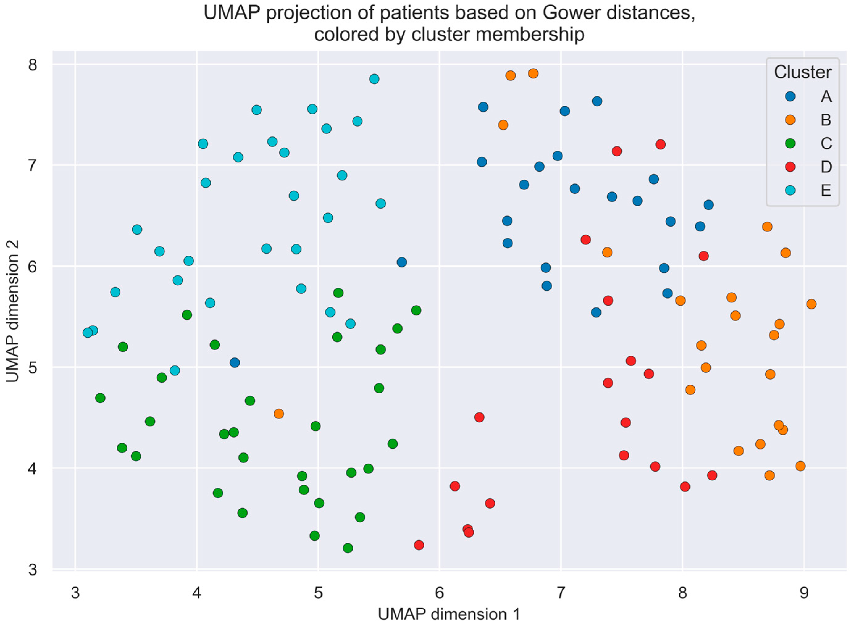
As an additional visual validation, the UMAP (Uniform Manifold Approximation and Projection for Dimension Reduction) technique [35] was applied, also using the Gower distance matrix, to graphically represent the distribution of patients and the separation among clusters in a two-dimensional space. This unsupervised technique preserves both the local and global structure of the original dataset. For its implementation, the number of neighbors was set to 30 and the minimum distance between points to 0.3—values commonly used in exploratory visualizations. This projection was employed exclusively for illustrative purposes, providing visual support for the cluster structure identified through k-prototypes.
Once the clusters were defined, Stage 2 involved a statistical analysis to assess differences among them. At this stage, both the variables shown in Table 1 and those in Table 2 were included. For continuous variables, the Mann–Whitney U test [36] was used, while for categorical variables, the chi-square test [36] was applied. A significance level of 5% was established.
The study was conducted on a computer equipped with an Intel Core i9 processor, an NVIDIA RTX 4080 GPU, and 32 GB of RAM, using Python (version 3.10.13). The clustering algorithm and statistical analyses were implemented using the kmodes library (version 0.12.2), scipy (version 1.11.4), pandas (version 2.2.2), and scikit-learn (version 1.4.2) for internal validation metrics. Dissimilarity matrices suitable for mixed-type data were computed using the Gower library (version 0.1.2). Dimensionality reduction with UMAP-learn (version 0.5.7) was applied solely for visualization purposes. Data visualization was performed using Matplotlib (version 3.10.3). and Seaborn (version 0.13.2).

3. Results

3.1. Study Population

Of the 179 patients initially enrolled in the study, those without missing data were included, resulting in a total of 123 patients. Among them, 55.28% were male, with a mean age of 62 ± 14.38 years.
Clustering was performed using the variables listed in Table 1, resulting in five clusters: Cluster A (n = 23), Cluster B (n = 23), Cluster C (n = 30), Cluster D (n = 19), and Cluster E (n = 28).
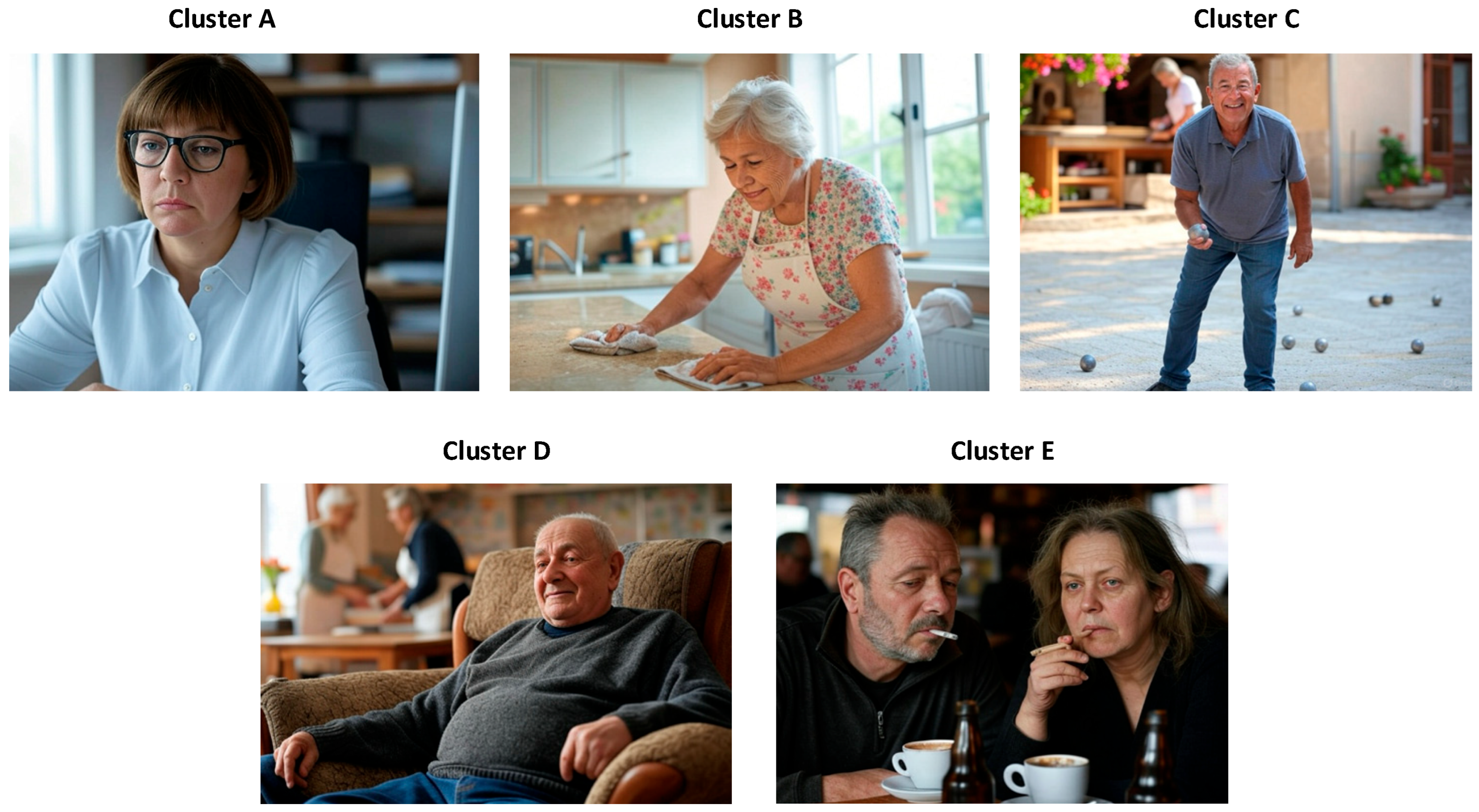
To facilitate the interpretation of the groups, Figure 2 provides a graphical representation of the clusters, generated using Grok 2 (xAI, San Fracisco, CA, USA).

3.2. Cluster Analysis

A detailed description of each cluster is presented below, integrating demographic, clinical, and autonomic features based on the data summarized in Table 1:
  • Cluster A—Young women with daytime fatigue and sleepiness, who are overweight, without cardiovascular risk factors: This group represents 18.7% of the sample and is composed predominantly of women (60.9%). It is the youngest cluster (51.5 ± 14.2 years), followed by Cluster E, with an intermediate BMI (28.2 ± 4.9 kg/m2), no history of diabetes, who are non-smokers and without cardiorespiratory disease. It presents the highest level of daytime sleepiness (Epworth score: 11.9 ± 6.2). Regarding HRV, this cluster shows low SDNN (100.57 ± 29.84 ms), RMSSD (58.22 ± 24.68 ms), and SDANN (78.0 ± 37.7 ms) values, indicating predominant nocturnal sympathetic activation.
  • Cluster B—Elderly women with no daytime symptoms, who are overweight, with cardiovascular risk factors: This group also accounts for 18.7% of the patients and includes the oldest individuals (75.4 ± 5.8 years). It is characterized by a predominance of women (60.9%) with multiple cardiovascular risk factors, such as hypertension (91.6%) and dyslipidemia (66.6%), but low daytime sleepiness (Epworth score: 5.8 ± 5.4). Atrial fibrillation is present in 25% of patients. None are smokers. HRV is also diminished (SDNN: 80.52 ± 26.79 ms; RMSSD: 62.17 ± 32.23 ms), again suggesting enhanced nocturnal sympathetic activity.
  • Cluster C—Older, but not elderly, asymptomatic men without cardiovascular risk factors: Representing 24.4% of the sample, this cluster consists mainly of men (73.3%) with intermediate age (62.0 ± 9.9 years) and the lowest BMI (24.2 kg/m2). Patients report no significant fatigue or excessive sleepiness (Epworth score: 4.2 ± 3.9) and have the lowest number of syncopal episodes in the past 12 months. HRV parameters (SDNN: 140.93 ± 166.27 ms; RMSSD: 88.17 ± 68.11 ms) are consistent with a preserved parasympathetic tone.
  • Cluster D—Elderly obese men with cardiovascular risk factors: This group represents 15.4% of the sample, with a predominance of men (63.2%), and is the only cluster characterized by obesity (BMI: 30.0 ± 4.8 kg/m2). Patients present with hypertension (73.7%), dyslipidemia (68%), and the highest prevalence of diabetes (36.8%). Nearly half have atrial fibrillation (47.4%), and 26.3% have ischemic heart disease. While most are ex-smokers, they show the highest cumulative tobacco exposure. Together with Cluster B, they report the highest total number of syncopal episodes (10.7 ± 13.0). HRV parameters (SDNN: 266.37 ± 206.98 ms; RMSSD: 296.89 ± 70.73 ms) reflect parasympathetic predominance, though with wide variability.
  • Cluster E—Young overweight male or female smokers with dyslipidemia, daytime fatigue, and frequent nocturnal awakenings: This is the largest cluster (28.8%) and the second youngest (52.6 ± 11.2 years), with a balanced male–female distribution. A high proportion are current smokers (67.9%), while 32.1% are former smokers. This group shows the highest prevalence of asthma (17.9%), and although they do not present with hypertension or diabetes, 39.3% have dyslipidemia. They report the highest number of syncopal episodes in the last 12 months (5.5 ± 7.5). Daytime sleepiness is comparable to Cluster A (Epworth score: 11.7 ± 6.1), and sleep quality is the poorest, with more awakenings and marked non-restorative sleep. HRV indicates marked nocturnal sympathetic activity (SDNN: 102.64 ± 50.21 ms; RMSSD: 90.11 ± 96.48 ms), with the lowest mean RR interval (893.89 ± 136.89 ms) and the highest LF/HF ratio (3.8 ± 3.0).
To assess the robustness and clinical relevance of the clustering solution, both quantitative metrics and visual evaluation were applied. These assessments aimed to determine the internal consistency of the five-cluster model and its potential to reflect meaningful patient stratification. The average silhouette index value for the five-cluster solution was 0.15, calculated from the Gower distance matrix. While this value indicates modest separation among groups, it is consistent with what is typically observed in real-world clinical datasets, which are characterized by high heterogeneity and mixed-type variables. In such contexts, clusters often partially overlap and lack sharp boundaries, which limits the ability of geometric metrics to fully capture their clinical validity. Nonetheless, the value obtained suggests the presence of an underlying structure in the data, consistent with clinically meaningful differences among patient subgroups.
The two-dimensional UMAP projection, applied to the Gower distance matrix, enabled visual inspection of patient distribution. This representation shows a coherent grouping of individuals according to cluster assignment, with some overlap, as expected. The layout supports the internal validity of the model and aligns with the modest silhouette score. Figure 3 illustrates this distribution, with color and letter codes (A–E) for clarity.

3.3. Association of Clusters with Respiratory Polygraphy Variables

It is important to note that the parameters obtained through overnight cardiorespiratory polygraphy were intentionally excluded from the initial clustering process. This decision reflects the reality of routine clinical practice, where such information is typically unavailable in the early stages of diagnosing SUC. However, the subsequent inclusion of polygraphic data to analyze inter-cluster differences enabled the identification of underlying pathophysiological patterns and their association with the clinical characteristics of each group. This approach adds valuable insight that could help guide clinical decisions and optimize diagnostic and therapeutic strategies in patients with syncope.
It is worth clarifying that HRV values do not originate from polygraphy but are instead derived from the nocturnal segment of 24 h Holter monitoring.
Table 2 presents a comparison of the polygraphy parameters across the five identified clusters. Patients in Cluster A exhibited mild sleep apnea (AHI: 9.4 ± 8.1 events/hour), accompanied by fatigue and significant daytime sleepiness. In Cluster B, sleep apnea was moderate (AHI: 21.4 ± 19.4 events/hour), with greater oxygen desaturation and poor sleep quality (ID3: 22.3 ± 19.3; TC90: 14.0 ± 22.3), although without subjective daytime symptoms. Cluster C also showed mild sleep apnea (AHI: 13.2 ± 13.8 events/hour), but without associated clinical manifestations. Cluster D was characterized by severe sleep apnea (AHI: 30.8 ± 13.7 events/hour), with a notable presence of central apneas (24.2 ± 46.3 events/hour) and oxygen desaturation (ID3: 28.6 ± 14.5; TC90: 13.1 ± 23.1), although symptom perception was limited. Finally, Cluster E showed mild sleep apnea (AHI: 14.8 ± 16.7 events/hour), with the highest burden of both daytime and nocturnal symptoms, including fatigue, awakenings, and non-restorative sleep.

4. Discussion

This study employs a novel and exploratory methodology based on unsupervised ML tools, which has proven highly useful for grouping patients with SUC. This clustering ability is especially relevant given that, despite advances in clinical guidelines and complementary testing, a high percentage of patients remain without a clear etiological diagnosis of syncope [9]. This underscores the need for innovative and efficient strategies to improve the management of these patients.
In this context, AI represents a promising opportunity to optimize risk stratification, diagnosis, and clinical decision-making in patients with syncope [37,38]. Previous studies have explored the use of AI algorithms to address the limitations of traditional risk stratification tools such as the Canadian Syncope Risk Score [19]. While these approaches have demonstrated performance comparable to conventional methods, integrating AI with broader clinical, demographic, and pathophysiological data could improve diagnostic accuracy and risk assessment in the future.
More recent studies have applied AI to classify and stratify risk in patients with syncope. For example, a recent systematic review evaluated ML algorithms for syncope classification using hemodynamic parameters from the Head-Up Tilt Test (HUTT), reporting a sensitivity of 88.8%, specificity of 81.5%, and accuracy of 85.8% [20]. Additionally, AI has been used to predict prognosis and hospital admission [39], as well as to identify structural abnormalities in ECG [40].
Although AI has been used in syncope diagnosis [20], only two studies have applied clustering techniques. Moloney et al. [41] identified eight clusters using clinical data, but with limited translational impact. Guftar et al. [42] combined clinical and HUTT data for a more refined characterization, although the findings did not translate into changes in patient management. Unlike these prior studies, the present work innovatively incorporates autonomic parameters (such as heart rate variability) and respiratory data obtained via polygraphy. Notably, these respiratory variables were not used in the generation of clusters but were analyzed post hoc to explore pathophysiological differences among the predefined subgroups. This strategy enables a multidimensional phenotyping approach, suggesting that the interaction between autonomic dysfunction and nocturnal respiratory disturbances—particularly nocturnal sympathetic hyperactivity—may represent a relevant pathophysiological axis. This perspective complements the traditional clinical typology (reflex, cardiac, orthostatic) and opens new avenues for more precise and personalized stratification.
Unlike these previous studies, our approach allows not only for a more robust clinical characterization but also for a pathophysiological perspective based on objective parameters. A key advantage of the unsupervised approach is its ability to identify latent subgroups without the need for predefined clinical labels, which is particularly useful in the context of SUC, where many patients remain undiagnosed despite standard testing.
Thus, this model facilitates the detection of shared clinical patterns and may help generate pathophysiological hypotheses (e.g., autonomic dysfunction or significant sleep apnea) that could inform future studies aimed at developing more personalized stratification strategies. These hypotheses might eventually help prioritize diagnostic and therapeutic resources, pending further validation.
In addition, in our model, HRV parameters were included directly as input variables in the clustering algorithm, along with clinical and demographic data. This decision is based on the fact that nocturnal HRV can be easily obtained from 24 h Holter recordings, making it an accessible and reproducible tool in clinical practice. It also enhances the model’s ability to detect relevant autonomic patterns early in the diagnostic process.
Our study applies unsupervised ML approaches to identify clusters among patients with SUC, enabling a preliminary, data-driven stratification that could potentially inform early-stage clinical decision-making, prior to the use of costly or invasive diagnostic tools. Five clusters with distinct demographic and clinical characteristics were identified, revealing specific profiles that may require personalized management strategies.
A particular strength of this patient cohort is that all participants underwent overnight cardiorespiratory polygraphy with ECG recording and nocturnal HRV analysis, conducted within the scope of the SINCOSAS study [43], aimed at exploring autonomic and respiratory profiles in patients with SUC. This enabled the integration of polygraphic parameters in addition to demographic and clinical variables. Identifying clusters that combine clinical, demographic, and nocturnal respiratory event data and HRV may help determine which patients with syncope and sleep apnea could benefit from treatment for sleep-disordered breathing, with the goal of reducing syncope incidence.
Since 24 h Holter monitoring is often part of the diagnostic work-up for patients with unexplained syncope, nocturnal HRV can be readily obtained by selecting sleep-time intervals from Holter data. HRV follows a circadian rhythm, characterized by parasympathetic predominance at night and sympathetic predominance during the day [44]. Disruptions to this circadian pattern can be observed in 24 h Holter recordings, which are routinely used in the assessment of syncope of unclear origin. SA [45], restless leg syndrome [46], insomnia [47], and other sleep disorders [43] may impact this circadian HRV cycle. Managing nocturnal autonomic nervous system (ANS) activity may contribute to greater stability of daytime ANS function.
Cluster analysis identified five patient groups with distinct clinical and pathophysiological profiles. Importantly, respiratory polygraphy variables were not included in the initial clustering algorithm. This was a deliberate methodological decision, grounded in the clinical reality that cardiorespiratory sleep studies are not routinely performed as part of the initial evaluation of patients with syncope of uncertain cause. By excluding polygraphy data from the clustering inputs, we aimed to simulate a first-line, data-driven stratification process based on variables that are typically available during the early diagnostic workup—such as clinical history, demographics, and nocturnal HRV derived from Holter recordings. The goal was to identify latent patient profiles that might benefit from further testing, including respiratory polygraphy. This design avoids biasing the clustering process with post hoc diagnostic data and reflects a more pragmatic, stepwise clinical strategy. Nonetheless, we acknowledge that excluding polygraphy data from the unsupervised model may have limited the identification of certain pathophysiological patterns.
The five identified clusters showed distinct demographic, clinical, autonomic, and respiratory profiles. Although this analysis does not support causal inference, the observed patterns suggest potentially relevant clinical groups that may inform future stratification strategies. Below, we provide exploratory interpretations for each cluster based on current clinical knowledge and pathophysiological plausibility. These hypotheses aim to generate future research directions and should not be construed as prescriptive recommendations.
Cluster A represents young, overweight, non-smoking women with prominent daytime fatigue and sleepiness, no or mild sleep apnea, and no nocturnal oxygen desaturation. They present the lowest SDNN and RMSSD values, suggesting elevated nocturnal sympathetic activity. This cluster reflects a typical profile of hypersomnia or non-respiratory sleep disorders, in which full polysomnography may be more appropriate than simple respiratory polygraphy. The absence of apneas, combined with marked nocturnal sympathetic activation and low HRV values, suggests functional dysautonomia, possibly associated with conditions such as chronic insomnia, restless legs syndrome, or even undiagnosed anxiety. Treatment aimed at improving sleep quality and regulating nocturnal autonomic activity could be hypothesized to help reduce morning hemodynamic instability and, consequently, syncopal events.
Cluster B includes elderly, overweight, hypertensive women with few symptoms, moderate sleep apnea, and nocturnal desaturation. HRV shows decreased values in SDNN, RMSSD, NN50, and SDANN. The presence of cardiovascular risk factors in this group suggests a clinical profile in which syncope may be related to nocturnal bradyarrhythmic events or unrecognized intermittent hypoxia. Despite the limited daytime symptomatology, the altered HRV and nocturnal desaturation support a proactive strategy that could include treatment of sleep apnea, prolonged electrocardiographic monitoring, and potential evaluation for pacemaker implantation if an arrhythmic cause of syncope is confirmed.
Cluster C represents older men, mostly former smokers, who are asymptomatic, with mild sleep apnea. HRV does not reveal significant alterations. This group represents a low-risk phenotype, in which the likelihood of syncope related to sleep apnea or dysautonomia is minimal. In this context, expectant clinical follow-up and education on warning signs may be sufficient, reserving invasive or costly complementary tests only in the event of recurrence.
Cluster D is characterized by obese men with hypertension and dyslipidemia, presenting with severe sleep apnea and a central component. They exhibit abnormal HRV patterns, likely influenced by the use of negative inotropic agents [48]. This group may benefit from intensive intervention, which should include treatment of sleep apnea with CPAP, strict control of cardiovascular risk factors, and electrophysiological evaluation. Nocturnal autonomic dysfunction and sustained hypoxia may act as triggers for bradyarrhythmias or sinus pauses, making the combination of polygraphy and overnight Holter monitoring a potentially valuable strategy to identify these mechanisms. The presence of symptomatic SA supports the indication for treatment and subsequent clinical follow-up to assess the potential reduction in syncope.
Cluster E includes young men and women who are active smokers, with poor sleep quality, fatigue, daytime sleepiness, mild or absent apnea, and moderate desaturation. High nocturnal sympathetic activity may represent an early or subclinical form of dysautonomia related to poor sleep hygiene and harmful habits. Despite presenting only mild apnea, the HRV pattern, along with both daytime and nighttime symptoms, points to nocturnal autonomic nervous system dysfunction with potential impact on clinical stability. This group could be considered for non-pharmacological interventions focused on improving sleep hygiene, smoking cessation, and stress management. Nevertheless, high nocturnal sympathetic activity suggests that addressing nocturnal events may help improve HRV and reduce the frequency of syncope episodes.
Taken together, the identification of these five clinical profiles not only highlights the heterogeneity of SUC but also allows, for the first time, the proposal of differentiated management strategies based on routine clinical data and non-invasive studies, although these proposals remain exploratory and require further validation. Thus, the use of artificial intelligence tools, combined with polygraphy and HRV, could be integrated into clinical decision-making algorithms to guide the rational use of diagnostic resources, prioritize treatments, and reduce diagnostic uncertainty in this complex group of patients.
The finding of increased nocturnal sympathetic activity in patients with sleep apnea [49] supports the hypothesis that treatment with CPAP may improve nocturnal HRV [50] and thus stabilize daytime ANS activity. However, to date, no studies have assessed whether patients with syncope and sleep apnea exhibiting elevated nocturnal sympathetic activity improve after CPAP treatment, nor whether such improvement correlates with a reduction in syncopal events. To confirm this relationship, studies with 24 h Holter monitoring before and after CPAP therapy are needed. Evaluating the circadian HRV cycle in these patients could help establish a causal link among apnea, syncope, and circadian rhythm disturbances in HRV.
From an ethical standpoint, the adoption of AI in medicine requires transparency, explainability, and human oversight to maintain the trust of both patients and healthcare professionals [51,52,53,54]. Morley et al. [51] propose governance frameworks that ensure accountability and fairness, while Grote and Berens [52] highlight the need to assess algorithmic biases and their distributive impact. Studies on public perception show that patients are receptive to the use of AI for supportive tasks but prefer that critical clinical decisions remain under the physician’s control [53,54]. Our clustering model, designed to generate hypotheses and support early stratification, is applied without automating individual decision-making; its future implementation will require ongoing validation, clear communication, and alignment with bioethical principles to ensure its responsible and ethical integration into clinical practice.
This study has some limitations. The most significant is the absence of standardized reference values for daytime and nocturnal HRV analysis. Home respiratory polygraphy, used instead of polysomnography (the gold standard), may have introduced interpretation errors. External factors such as stress, insomnia, or ambient temperature could also have affected the results. Moreover, a larger sample size might have revealed statistically significant differences in more HRV parameters.
In clustering analysis involving mixed clinical data, traditional cohesion metrics such as the within-cluster sum of squares are not applicable when using algorithms like k-prototypes, which rely on hybrid dissimilarity functions combining categorical and numerical variables. For this reason, we used the silhouette index calculated from the Gower distance matrix—a recognized metric for internal validation in mixed-type datasets. The average silhouette score obtained (0.15) may appear modest, but it aligns with expectations for real-world clinical data, where phenotypic boundaries are often diffuse. To better contextualize this metric, a table was added in Appendix A (Table A1) showing silhouette scores by cluster, which indicate greater internal cohesion within more clinically defined subgroups. Additionally, future strategies should include complementary internal validation metrics, such as the Davies–Bouldin or Calinski–Harabasz indices, as well as stability analyses via bootstrapping to evaluate the reproducibility of the clustering results more robustly. Moreover, the clustering structure was supported by a UMAP projection, which showed a coherent distribution of patients in the reduced space. Furthermore, since this is an unsupervised analysis, no reference labels (ground truth) exist for direct external validation of the identified clusters. This is a widely recognized limitation in clustering studies involving clinical data, where well-defined phenotypic classifications are often lacking. Both elements reinforce the interpretation of the results and support their potential clinical relevance.
Nonetheless, it is important to acknowledge that potentially confounding factors—such as patients’ lifestyle (e.g., physical activity, substance use, dietary habits) and mental health status (e.g., anxiety, depression, stress)—were not assessed in this study. These factors are known to modulate autonomic nervous system function and may therefore influence both the incidence of syncope and HRV parameters, potentially affecting the interpretation of clustering results. Their absence may have limited the identification of clinically meaningful subgroups by omitting relevant sources of phenotypic variability. Furthermore, psychological and behavioral factors could interact with sleep-disordered breathing in complex ways that remain unexplored in the present analysis. This limitation underscores the need for future studies to incorporate detailed and standardized assessments of these dimensions using validated questionnaires, alongside statistical models capable of adjusting for their confounding influence. Doing so will enhance the precision and clinical interpretability of unsupervised clustering approaches, enabling a more comprehensive phenotypic characterization of patients with syncope and sleep-disordered breathing.
A relevant methodological limitation of this study concerns the handling of missing data. We chose to exclude cases with incomplete values (56 out of the initial 179 patients) to avoid introducing bias through unsupervised imputations, which could distort the latent structure of the data. This decision was based on methodological recommendations that discourage the use of multiple imputation in unsupervised analyses without adequate control of imputation uncertainty, especially when no “ground truth” exists to validate cluster stability [55,56]. Several studies have noted that certain imputation methods can lead to artificial or unstable clusters if not accompanied by rigorous validation procedures, such as sensitivity analyses, consensus techniques, or probabilistic models [57,58]. Although excluding cases may introduce some selection bias, it was deemed the most appropriate option given the exploratory nature of this study and the limited sample size. Future research involving larger, multicenter cohorts should incorporate advanced approaches to handle missing data, allowing for a more precise assessment of cluster robustness.
Finally, it should be noted that this work is presented as an exploratory pilot study, and the available sample size imposes certain constraints on the generalizability of the findings, particularly in the cluster comparisons. This limitation is partly due to the inherent difficulty in recruiting patients with syncope of unclear cause—a relatively rare and diagnostically challenging condition. Nevertheless, appropriate statistical tools were applied for this type of sample, and the results should be understood as hypothesis-generating. Future studies with larger, multicenter samples and longitudinal follow-up will be needed to validate and expand these findings, as well as to explore their clinical applicability in routine practice.
Despite these limitations, the study has important strengths. It is the first to characterize clinical, demographic, and nocturnal cardiorespiratory variables in syncope patients using unsupervised ML. The combination of daytime and nocturnal analysis offers an integrated view that may help develop personalized management strategies based on AI. Characterizing syncope patients through AI by integrating clinical, demographic, and nocturnal cardiorespiratory data could represent a promising non-invasive strategy to better understand unexplained syncope. Further research is needed to determine its role in improving long-term clinical outcomes.

Author Contributions

Conceptualization, M.-J.M.-M., A.F.-V., M.C.-G. and B.S.; methodology, M.-J.M.-M., A.F.-V., M.C.-G., M.T.-D. and B.S.; validation, M.-J.M.-M., A.F.-V. and M.C.-G.; formal analysis, M.-J.M.-M., A.F.-V. and M.C.-G.; investigation, M.-J.M.-M. and M.T.-D.; data curation, M.-J.M.-M. and M.T.-D.; writing—original draft preparation, M.-J.M.-M.; writing—review and editing, M.C.-G.; supervision, B.S. and A.F.-V.; project administration, M.-J.M.-M.; funding acquisition, M.-J.M.-M. All authors have read and agreed to the published version of the manuscript.

Funding

This research was funded by the Galician Society of Respiratory Pathology (Sociedade Galega de Patoloxía Respiratoria, SOGAPAR). The grant (EUR 18,000) was used to acquire a home respiratory polygraph and to support data management. No other external funding was received.

Institutional Review Board Statement

The study was conducted in accordance with the Declaration of Helsinki and approved by the Clinical Research Ethics Committee of Galicia, protocol code 2019/048, on 14 February 2019.

Informed Consent Statement

Informed consent was obtained from all subjects involved in the study.

Data Availability Statement

The dataset is available on request from the authors.

Acknowledgments

The authors would like to express their sincere gratitude to the Galician Society of Respiratory Pathology (SOGAPAR) for the grant awarded to support this project. We also wish to acknowledge the Spanish Sleep Network and the Integrated Sleep Research Program of SEPAR for their valuable contributions to the development and subsequent analysis of the study. This paper is part of the research conducted in fulfillment of the requirements for the Ph.D. degree of María-José Muñoz-Martínez.

Conflicts of Interest

The authors declare no conflicts of interest.

Appendix A

Table A1. Silhouette score per cluster.
ClusterSilhouette Score Per Cluster
A0.24
B0.17
C0.14
D0.14
E0.08

References

  1. Brignole, M.; Moya, A.; De Lange, F.J.; Deharo, J.C.; Elliott, P.M.; Fanciulli, A.; Fedorowski, A.; Furlan, R.; Kenny, R.A.; Martín, A.; et al. 2018 ESC Guidelines for the Diagnosis and Management of Syncope. Eur. Heart J. 2018, 39, 1883–1948. [Google Scholar] [CrossRef] [PubMed]
  2. Zimmermann, T.; du Fay de Lavallaz, J.; Nestelberger, T.; Gualandro, D.M.; Strebel, I.; Badertscher, P.; Lopez-Ayala, P.; Widmer, V.; Freese, M.; Miró, Ò.; et al. Incidence, Characteristics, Determinants, and Prognostic Impact of Recurrent Syncope. Europace 2020, 22, 1885–1895. [Google Scholar] [CrossRef] [PubMed]
  3. Barón-Esquivias, G.; Quintanilla, M.; Díaz-Martín, A.J.; Barón-Solís, C.; Almeida-González, C.V.; García-Romero, C.; Paneque, I.; Rubio-Guerrero, C.; Rodríguez-Corredor, R.; Valle-Racero, J.I.; et al. Long-Term Recurrences and Mortality in Patients with Noncardiac Syncope. Rev. Española Cardiol. 2022, 75, 568–575. [Google Scholar] [CrossRef]
  4. Galron, E.; Kehat, O.; Weiss-Meilik, A.; Furlan, R.; Jacob, G. Diagnostic Approaches to Syncope in Internal Medicine Departments and Their Effect on Mortality. Eur. J. Intern. Med. 2022, 102, 97–103. [Google Scholar] [CrossRef]
  5. Johansson, M.; Rogmark, C.; Sutton, R.; Fedorowski, A.; Hamrefors, V. Risk of Incident Fractures in Individuals Hospitalised Due to Unexplained Syncope and Orthostatic Hypotension. BMC Med. 2021, 19, 188. [Google Scholar] [CrossRef]
  6. Sun, B.C. Quality-of-Life, Health Service Use, and Costs Associated with Syncope. Prog. Cardiovasc. Dis. 2013, 55, 370–375. [Google Scholar] [CrossRef]
  7. Kenny, R.A.; Bhangu, J.; King-Kallimanis, B.L. Epidemiology of Syncope/Collapse in Younger and Older Western Patient Populations. Prog. Cardiovasc. Dis. 2013, 55, 357–363. [Google Scholar] [CrossRef]
  8. Bennett, M.T.; Leader, N.; Krahn, A.D. Recurrent Syncope: Differential Diagnosis and Management. Heart 2015, 101, 1591–1599. [Google Scholar] [CrossRef]
  9. Ruwald, M.H.; Hansen, M.L.; Lamberts, M.; Vinther, M.; Torp-Pedersen, C.; Hansen, J.; Gislason, G.H. Unexplained Syncope and Diagnostic Yield of Tests in Syncope According to the ICD-10 Discharge Diagnosis. J. Clin. Med. Res. 2013, 5, 441. [Google Scholar] [CrossRef]
  10. Goldberger, Z.D.; Petek, B.J.; Brignole, M.; Shen, W.K.; Sheldon, R.S.; Solbiati, M.; Deharo, J.C.; Moya, A.; Hamdan, M.H. ACC/AHA/HRS Versus ESC Guidelines for the Diagnosis and Management of Syncope: JACC Guideline Comparison. J. Am. Coll. Cardiol. 2019, 74, 2410–2423. [Google Scholar] [CrossRef]
  11. Wakai, A.; Sinert, R.; Zehtabchi, S.; de Souza, I.S.; Benabbas, R.; Allen, R.; Dunne, E.; Richards, R.; Ardilouze, A.; Rovic, I. Risk-Stratification Tools for Emergency Department Patients with Syncope: A Systematic Review and Meta-Analysis of Direct Evidence for SAEM GRACE. Acad. Emerg. Med. 2024, 32, 72–86. [Google Scholar] [CrossRef] [PubMed]
  12. Wang, L.; Chen, X.; Zhang, L.; Li, L.; Huang, Y.; Sun, Y.; Yuan, X. Artificial Intelligence in Clinical Decision Support Systems for Oncology. Int. J. Med. Sci. 2023, 20, 79–86. [Google Scholar] [CrossRef] [PubMed]
  13. Ramgopal, S.; Sanchez-Pinto, L.N.; Horvat, C.M.; Carroll, M.S.; Luo, Y.; Florin, T.A. Artificial Intelligence-Based Clinical Decision Support in Pediatrics. Pediatr. Res. 2022, 93, 334–341. [Google Scholar] [CrossRef]
  14. Casal-Guisande, M.; Cerqueiro-Pequeño, J.; Comesaña-Campos, A.; Bouza-Rodríguez, J.B. Proposal of a Methodology Based on Expert Systems for the Treatment of Diabetic Foot Condition. In Proceedings of the Eighth International Conference on Technological Ecosystems for Enhancing Multiculturality, Association for Computing Machinery. Salamanca, Spain, 21 October 2020; pp. 491–495. [Google Scholar]
  15. Casal-Guisande, M.; Comesaña-Campos, A.; Núñez-Fernández, M.; Torres-Durán, M.; Fernández-Villar, A. Proposal and Definition of an Intelligent Clinical Decision Support System Applied to the Prediction of Dyspnea after 12 Months of an Acute Episode of COVID-19. Biomedicines 2024, 12, 854. [Google Scholar] [CrossRef]
  16. Corbacho-Abelaira, D.; Casal-Guisande, M.; Corbacho-Abelaira, F.; Arnaiz-Fernandez, M.; Trinidad-Lopez, C.; Delgado Sanchez-Gracian, C.; Sanchez-Montanes, M.; Ruano-Ravina, A.; Fernandez-Villar, A. Proposal and Definition of an Intelligent Decision- Support System Based on Deep Learning Techniques for the Management of Possible COVID-19 Cases in Patients Attending Emergency Departments. IEEE Access 2024, 12, 95035–95046. [Google Scholar] [CrossRef]
  17. López-Canay, J.; Casal-Guisande, M.; Pinheira, A.; Golpe, R.; Comesaña-Campos, A.; Fernández-García, A.; Represas-Represas, C.; Fernández-Villar, A. Predicting COPD Readmission: An Intelligent Clinical Decision Support System. Diagnostics 2025, 15, 318. [Google Scholar] [CrossRef]
  18. Casal-Guisande, M.; Fernández-Villar, A.; Mosteiro-Añón, M.; Comesaña-Campos, A.; Cerqueiro-Pequeño, J.; Torres-Durán, M. Integrating Tabular Data through Image Conversion for Enhanced Diagnosis: A Novel Intelligent Decision Support System for Stratifying Obstructive Sleep Apnoea Patients Using Convolutional Neural Networks. Digital Health 2024, 10, 20552076241272632. [Google Scholar] [CrossRef]
  19. Grant, L.; Joo, P.; Nemnom, M.J.; Thiruganasambandamoorthy, V. Machine Learning versus Traditional Methods for the Development of Risk Stratification Scores: A Case Study Using Original Canadian Syncope Risk Score Data. Intern. Emerg. Med. 2022, 17, 1145–1153. [Google Scholar] [CrossRef]
  20. Goh, C.H.; Ferdowsi, M.; Gan, M.H.; Kwan, B.H.; Lim, W.Y.; Tee, Y.K.; Rosli, R.; Tan, M.P. Assessing the Efficacy of Machine Learning Algorithms for Syncope Classification: A Systematic Review. MethodsX 2023, 12, 102508. [Google Scholar] [CrossRef]
  21. Casal-Guisande, M.; Represas-Represas, C.; Golpe, R.; Fernández-García, A.; González-Montaos, A.; Comesaña-Campos, A.; Ruano-Raviña, A.; Fernández-Villar, A. Clinical and Social Characterization of Patients Hospitalized for COPD Exacerbation Using Machine Learning Tools. Arch. Bronconeumol. 2024, 61, 264–273. [Google Scholar] [CrossRef]
  22. Puel, V.; Pepin, J.L.; Gosse, P. Sleep Related Breathing Disorders and Vasovagal Syncope, a Possible Causal Link? Int. J. Cardiol. 2013, 168, 1666–1667. [Google Scholar] [CrossRef] [PubMed]
  23. Puel, V.; Godard, I.; Papaioannou, G.; Gosse, P.; Pepin, J.L.; Thoin, F.; Deharo, J.C.; Roche, F.; Zarqane, N.; Gagnadoux, F.; et al. Management of Sleep Apnoea Syndrome (SAS) in Patients with Vasovagal Syncope (VVS): A Protocol for the VVS-SAS Cohort Study. BMJ Open 2020, 10, e038791. [Google Scholar] [CrossRef] [PubMed]
  24. Muñoz-Martínez, M.J.; Fernández-Villar, A.; Casal-Guisande, M.; García-Campo, E.; Corbacho-Abelaira, D.; Souto-Alonso, A.; Sopeña, B. Prevalence of Sleep Apnea in Patients with Syncope of Unclear Cause: SINCOSAS Study. Medicina 2025, 61, 887. [Google Scholar] [CrossRef]
  25. Mediano, O.; González Mangado, N.; Montserrat, J.M.; Alonso-Álvarez, M.L.; Almendros, I.; Alonso-Fernández, A.; Barbé, F.; Borsini, E.; Caballero-Eraso, C.; Cano-Pumarega, I.; et al. International Consensus Document on Obstructive Sleep Apnea. Arch. Bronconeumol. 2022, 58, 52–68. [Google Scholar] [CrossRef]
  26. Doane, D.P.; Seward, L.E. Measuring Skewness: A Forgotten Statistic? J. Stat. Educ. 2011, 19. [Google Scholar] [CrossRef]
  27. Osborne, J.W. Improving Your Data Transformations: Applying the Box-Cox Transformation. Pract. Assess. Res. Eval. 2010, 15, 12. [Google Scholar] [CrossRef]
  28. Agresti, A. Categorical Data Analysis; John Wiley & Sons, Inc.: Hoboken, NJ, USA, 2002; ISBN 0471360937. [Google Scholar]
  29. Powers, D.; Xie, Y. Statistical Methods for Categorical Data Analysis; Emerald Group Publishing: Bradford, UK, 2008. [Google Scholar]
  30. Huang, Z.X. Clustering Large Datasets with Mixed Numeric and Categorical Values. In Proceedings of the First Pacific-Asia Knowledge Discovery and Data Mining Conference, Singapore, 23–24 February 1997; pp. 21–34. [Google Scholar]
  31. Han, J.; Kamber, M.; Pei, J. Data Mining: Concepts and Techniques, 3rd ed.; Morgan Kaufmann: Waltham, MA, USA, 2012. [Google Scholar]
  32. Cao, F.; Liang, J.; Bai, L. A New Initialization Method for Categorical Data Clustering. Expert. Syst. Appl. 2009, 36, 10223–10228. [Google Scholar] [CrossRef]
  33. Shahapure, K.R.; Nicholas, C. Cluster Quality Analysis Using Silhouette Score. In Proceedings of the 2020 IEEE 7th International Conference on Data Science and Advanced Analytics, DSAA, Sydney, Australia, 6–9 October 2020; pp. 747–748. [Google Scholar] [CrossRef]
  34. Gower, J.C. A General Coefficient of Similarity and Some of Its Properties. Biometrics 1971, 27, 857. [Google Scholar] [CrossRef]
  35. McInnes, L.; Healy, J.; Melville, J. UMAP: Uniform Manifold Approximation and Projection for Dimension Reduction. arXiv 2018, arXiv:1802.03426. [Google Scholar]
  36. Samuels, M.L.; Witmer, J.A.; Schaffner, A.A. Statistics for the Life Sciences, 5th ed.; Pearson Education: London, UK, 2016. [Google Scholar]
  37. Statz, G.M.; Evans, A.Z.; Johnston, S.L.; Adhaduk, M.; Mudireddy, A.R.; Sonka, M.; Lee, S.; Barsotti, E.J.; Ricci, F.; Dipaola, F.; et al. Can Artificial Intelligence Enhance Syncope Management?: A JACC: Advances Multidisciplinary Collaborative Statement. JACC Adv. 2023, 2, 100323. [Google Scholar] [CrossRef]
  38. Aamir, A.; Jamil, Y.; Bilal, M.; Diwan, M.; Nashwan, A.J.; Ullah, I. Artificial Intelligence in Enhancing Syncope Management—An Update. Curr. Probl. Cardiol. 2024, 49, 102079. [Google Scholar] [CrossRef] [PubMed]
  39. Dipaola, F.; Gebska, M.A.; Gatti, M.; Levra, A.G.; Parker, W.H.; Menè, R.; Lee, S.; Costantino, G.; Barsotti, E.J.; Shiffer, D.; et al. Will Artificial Intelligence Be “Better” Than Humans in the Management of Syncope? JACC Adv. 2024, 3, 101072. [Google Scholar] [CrossRef] [PubMed]
  40. Muzammil, M.A.; Javid, S.; Afridi, A.K.; Siddineni, R.; Shahabi, M.; Haseeb, M.; Fariha, F.N.U.; Kumar, S.; Zaveri, S.; Nashwan, A.J. Artificial Intelligence-Enhanced Electrocardiography for Accurate Diagnosis and Management of Cardiovascular Diseases. J. Electrocardiol. 2024, 83, 30–40. [Google Scholar] [CrossRef] [PubMed]
  41. Moloney, D.; O’Connor, J.; Newman, L.; Scarlett, S.; Hernandez, B.; Kenny, R.A.; Romero-Ortuno, R. Clinical Clustering of Eight Orthostatic Haemodynamic Patterns in The Irish Longitudinal Study on Ageing (TILDA). Age Ageing 2021, 50, 854–860. [Google Scholar] [CrossRef]
  42. Guftar, M.; Ali, S.H.; Raja, A.A.; Qamar, U. A Novel Framework for Classification of Syncope Disease Using K-Means Clustering Algorithm. In Proceedings of the 2015 SAI Intelligent Systems Conference (IntelliSys), London, UK, 10–11 November 2015; pp. 127–132. [Google Scholar] [CrossRef]
  43. Stein, P.K.; Pu, Y. Heart Rate Variability, Sleep and Sleep Disorders. Sleep. Med. Rev. 2012, 16, 47–66. [Google Scholar] [CrossRef]
  44. Baharav, A.; Kotagal, S.; Gibbons, V.; Rubin, B.K.; Pratt, G.; Karin, J.; Akselrod, S. Fluctuations in Autonomic Nervous Activity during Sleep Displayed by Power Spectrum Analysis of Heart Rate Variability. Neurology 1995, 45, 1183–1187. [Google Scholar] [CrossRef]
  45. Noda, A.; Yasuma, F.; Okada, T.; Yokota, M. Circadian Rhythm of Autonomic Activity in Patients with Obstructive Sleep Apnea Syndrome. Clin. Cardiol. 1998, 21, 271–276. [Google Scholar] [CrossRef]
  46. Lin, C.Y.; Tsai, S.J.; Peng, C.K.; Yang, A.C. Sleep State Instabilities in Patients with Periodic Limb Movements in Sleep—Detection and Quantification with Heart Rate Variability. Psychiatry Res. 2020, 293, 113454. [Google Scholar] [CrossRef]
  47. Zhao, W.; Jiang, B. Heart Rate Variability in Patients with Insomnia Disorder: A Systematic Review and Meta-Analysis. Sleep. Breath. 2023, 27, 1309–1313. [Google Scholar] [CrossRef]
  48. Weber, F.; Schneider, H.; Von Arnim, T.; Urbaszek, W. Heart Rate Variability and Ischaemia in Patients with Coronary Heart Disease and Stable Angina Pectoris; Influence of Drug Therapy and Prognostic Value. TIBBS Investigators Group. Total Ischemic Burden Bisoprolol Study. Eur. Heart J. 1999, 20, 38–50. [Google Scholar] [CrossRef]
  49. Wang, Z.; Jiang, F.; Xiao, J.; Chen, L.; Zhang, Y.; Li, J.; Yi, Y.; Min, W.; Su, L.; Liu, X.; et al. Heart Rate Variability Changes in Patients with Obstructive Sleep Apnea: A Systematic Review and Meta-Analysis. J. Sleep. Res. 2023, 32, e13708. [Google Scholar] [CrossRef] [PubMed]
  50. Guo, W.; Lv, T.; She, F.; Miao, G.; Liu, Y.; He, R.; Xue, Y.; Nu, N.K.; Yang, J.; Li, K.; et al. The Impact of Continuous Positive Airway Pressure on Heart Rate Variability in Obstructive Sleep Apnea Patients during Sleep: A Meta-Analysis. Heart Lung 2018, 47, 516–524. [Google Scholar] [CrossRef] [PubMed]
  51. Morley, J.; Machado, C.C.V.; Burr, C.; Cowls, J.; Joshi, I.; Taddeo, M.; Floridi, L. The Ethics of AI in Health Care: A Mapping Review. Soc. Sci. Med. 2020, 260, 113172. [Google Scholar] [CrossRef] [PubMed]
  52. Grote, T.; Berens, P. On the Ethics of Algorithmic Decision-Making in Healthcare. J. Med. Ethics 2020, 46, 205–211. [Google Scholar] [CrossRef]
  53. Vayena, E.; Blasimme, A.; Cohen, I.G. Machine Learning in Medicine: Addressing Ethical Challenges. PLoS Med. 2018, 15, e1002689. [Google Scholar] [CrossRef]
  54. Sovrano, F.; Palmirani, M.; Vitali, F. Combining Shallow and Deep Learning Approaches against Data Scarcity in Legal Domains. Gov. Inf. Q. 2022, 39, 101715. [Google Scholar] [CrossRef]
  55. Juan, W.; Ahn, K.W.; Chen, Y.G.; Lin, C.W. CCI: A Consensus Clustering-Based Imputation Method for Addressing Dropout Events in ScRNA-Seq Data. Bioengineering 2025, 12, 31. [Google Scholar] [CrossRef]
  56. Harder, A.A.; Olbricht, G.R.; Ekuma, G.; Hier, D.B.; Obafemi-Ajayi, T. Multiple Imputation for Robust Cluster Analysis to Address Missingness in Medical Data. IEEE Access 2024, 12, 42974–42991. [Google Scholar] [CrossRef]
  57. Hao, K.A.; Vasilopoulos, T.; Elwell, J.; Roche, C.P.; Hones, K.M.; Wright, J.O.; King, J.J.; Wright, T.W.; Simovitch, R.W.; Schoch, B.S. Missing Data in Orthopaedic Clinical Outcomes Research: A Sensitivity Analysis of Imputation Techniques Utilizing a Large Multicenter Total Shoulder Arthroplasty Database. J. Clin. Med. 2025, 14, 3829. [Google Scholar] [CrossRef]
  58. Zhou, H.; Chen, J.; Zhang, X. BMDD: A Probabilistic Framework for Accurate Imputation of Zero-Inflated Microbiome Sequencing Data. bioRxiv 2025. bioRxiv:2025.05.08.652808. [Google Scholar] [CrossRef]
Figure 1. Study methodology. The process consists of two stages. Before Stage 1, the data is preprocessed. In Stage 1, the clustering of the cohort is performed, resulting in five clusters. Subsequently, in Stage 2, a comparative analysis of differences among groups is conducted, including those related to cardiorespiratory polygraphy.
Applsci 15 07176 g001
Figure 2. Visualization of patients within each cluster. Generated using Grok 2 (xAI).
Applsci 15 07176 g002
Figure 3. Two-dimensional visualization of the clusters identified using the k-prototypes algorithm, based on UMAP projection applied to the Gower distance matrix. Each point represents a patient, color- and letter-coded (A–E) according to the assigned cluster. This graphical representation illustrates the relative distribution of the groups in the feature space, complementing the quantitative validation performed with the silhouette index.
Applsci 15 07176 g003
Table 1. Cohort and cluster characteristics.
VariablePopulation
Summary
Cluster A
(n = 23)
Cluster B
(n = 23)
Cluster C
(n = 30)
Cluster D
(n = 19)
Cluster E
(n = 28)
Significant Comparisons
Age62.0 ± 14.451.5 ± 14.275.4 ± 5.862.0 ± 9.972.5 ± 12.252.6 ± 11.2A ≠ B: p < 0.001, A ≠ C: p = 0.005, A ≠ D: p < 0.001, B ≠ C: p < 0.001, B ≠ E: p < 0.001, C ≠ D: p < 0.001, C ≠ E: p = 0.002, D ≠ E: p < 0.001
Man (%)55.3% (43.5, 67.1)39.1% (19.2, 59.1)39.1% (19.2, 59.1)73.3% (57.5, 89.2)63.2% (41.5, 84.9)57.1% (38.8, 75.5)A ≠ C: p = 0.026, B ≠ C: p = 0.026
BMI (5)27.8 ± 5.328.2 ± 4.927.6 ± 3.226.9 ± 5.330.0 ± 4.827.1 ± 6.8C ≠ D: p = 0.024, D ≠ E: p = 0.035
Diabetes: No (%)86.2% (79.6, 92.8)100.0% (100.0, 100.0)73.9% (56.0, 91.9)90.0% (79.3, 100.0)63.2% (41.5, 84.9)96.4% (89.6, 100.0)A ≠ B: p = 0.029, A ≠ D: p = 0.006, D ≠ E: p = 0.010
Diabetes: Yes, no insulin (%)11.4% (0.0, 28.0)0.0% (0.0, 0.0)26.1% (8.1, 44.0)10.0% (0.0, 20.7)26.3% (6.5, 46.1)0.0% (0.0, 0.0)A ≠ B: p = 0.029, A ≠ D: p = 0.032, B ≠ E: p = 0.015, D ≠ E: p = 0.017
Diabetes: Yes, with insulin (%)2.4% (0.0, 19.9)0.0% (0.0, 0.0)0.0% (0.0, 0.0)0.00% (0.00, 0.00)10.5% (0.0, 24.3)3.6% (0.0, 10.5)-
Dyslipidemia (%)41.5% (27.9, 55.0)17.4% (1.9, 32.9)69.6% (50.8, 88.4)23.3% (8.2, 38.5)68.% (47.5, 89.3)39.3% (21.2, 57.4)A ≠ B: p = 0.001, A ≠ D: p = 0.002, B ≠ C: p = 0.002, C ≠ D: p = 0.005
Hypertension (%)43.1% (29.8, 56.4)13.0% (0.0, 26.8)87.0% (73.2, 100.0)30.0% (13.6, 46.4)73.7% (53.9, 93.5)25.0% (9.0, 41.0)A ≠ B: p < 0.001, A ≠ D: p < 0.001, B ≠ C: p < 0.001, B ≠ E: p < 0.001, C ≠ D: p = 0.007, D ≠ E: p = 0.003
Never smoker (%)45.5% (32.5, 58.6)91.3% (79.8, 100.0)95.7% (87.3, 100.0)0.0% (0.0, 0.0)68.4% (47.5, 89.3)0.0% (0.0, 0.0)A ≠ C: p < 0.001, A ≠ E: p < 0.001, B ≠ C: p < 0.001, B ≠ E: p < 0.001, C ≠ D: p < 0.001, D ≠ E: p < 0.001
Smoker (%)21.95% (6.3, 37.6)4.4% (0.0, 12.7)0.0% (0.0, 0.0)20.0% (5.7, 34.3)5.3% (0.0, 15.3)67.9% (50.6, 85.2)A ≠ E: p < 0.001, B ≠ E: p < 0.001, C ≠ E: p < 0.001, D ≠ E: p < 0.001
Former smoker (%)32.52% (18.0, 47.0)4.4% (0.0, 12.7)4.4% (0.0, 12.7)80.0% (65.7, 94.3)26.3% (6.5, 46.1)32.1% (14.8, 49.4)A ≠ C: p < 0.001, A ≠ E: p = 0.033, B ≠ C: p < 0.001, B ≠ E: p = 0.033, C ≠ D: p < 0.001, C ≠ E: p < 0.001
Pack-year16.5 ± 24.80.4 ± 1.20.3 ± 1.530.1 ± 26.518.0 ± 33.327.3 ± 22.5A ≠ C: p < 0.001, A ≠ D: p = 0.041, A ≠ E: p < 0.001, B ≠ C: p < 0.001, B ≠ D: p = 0.016, B ≠ E: p < 0.001, C ≠ D: p = 0.002, D ≠ E: p = 0.003
Asthma (%)8.9% (0.0, 25.8)8.7% (0.0, 20.2)8.7% (0.0, 20.2)6.7% (0.0, 15.6)0.0% (0.0, 0.0)17.9% (3.7, 32.0)-
COPD (%)5.7% (0.0, 22.9)4.4% (0.0, 12.7)0.0% (0.0, 0.0)10.0% (0.0, 20.7)10.5% (0.0, 24.3)3.6% (0.0, 10.5)-
Ischemic heart disease (%)13.01% (0.0, 29.5)0.0% (0.0, 0.0)17.4% (1.9, 32.9)16.7% (3.3, 30.0)26.3% (6.5, 46.1)7.1% (0.0, 16.7)A ≠ D: p = 0.032
Atrial fibrillation (%)15.5% (0.0, 31.7)0.0% (0.0, 0.0)26.1% (8.1, 44.0)13.3% (1.2, 25.5)47.4% (24.9, 69.8)0.0% (0.0, 0.0)A ≠ B: p = 0.029, A ≠ D: p < 0.001, B ≠ E: p = 0.015, C ≠ D: p = 0.022, D ≠ E: p < 0.001
Stroke (%)1.6% (0.0, 19.2)0.0% (0.0, 0.0)4.4% (0.0, 12.7)0.0% (0.0, 0.0)0.0% (0.0, 0.0)3.6% (0.0, 10.5)-
Syncope last 12 months (%)3.7 ± 5.34.1 ± 6.73.8 ± 4.51.7 ± 1.63.89 ± 3.165.5 ± 7.5B ≠ C: p = 0.045, C ≠ D: p = 0.006
Total number of syncopes8.6 ± 11.79.7 ± 11.810.5 ± 15.74.6 ± 4.110.7 ± 13.08.9 ± 12.0-
Injuries last 12 months1.0 ± 1.70.5 ± 0.81.3 ± 2.20.9 ± 1.91.6 ± 2.50.9 ± 0.9-
Daytime Tiredness48.8% (36.1, 61.4)60.9% (40.9, 80.8)34.8% (15.3, 54.3)20.0% (5.7, 34.3)42.1% (19.9, 64.3)85.7% (72.8, 98.7)A ≠ C: p = 0.006, B ≠ E: p < 0.001, C ≠ E: p < 0.001, D ≠ E: p = 0.005
Nocturnal awakenings52.0% (39.8, 64.3)47.8% (27.4, 68.2)69.6% (50.8, 88.4)40.0% (22.5, 57.5)42.1% (19.9, 64.3)60.7% (42.6, 78.8)-
Lack of concentration35.0 (20.7, 49.2)26.1% (8.1, 44.0)26.1% (8.1, 44.0)16.7% (3.3, 30.0)36.8% (15.2, 58.5)67.9% (50.6, 85.2)A ≠ E: p = 0.007, B ≠ E: p = 0.007, C ≠ E: p < 0.001
Witnessed apneas19.5% (3.7, 35.4)17.4% (1.9, 32.9)13.0% (0.0, 26.8)6.7% (0.0, 15.6)15.8% (0.0, 32.2)42.9% (24.5, 61.2)B ≠ E: p = 0.044, C ≠ E: p = 0.004
Asphyxia episodes8.9% (0.0, 25.8)21.7% (4.9, 38.6)8.7% (0.0, 20.2)3.3% (0.0, 9.8)0.0% (0.0, 0.0)10.7% (0.0, 22.2)-
Non-restorative sleep50.4% (38.0, 62.9)69.6% (50.8, 88.4)30.4% (11.6, 49.2)33.3% (16.5, 50.2)36.8% (15.2, 58.5)78.6% (63.4, 93.8)A ≠ B: p = 0.018, A ≠ C: p = 0.019, B ≠ E: p = 0.002, C ≠ E: p = 0.001, D ≠ E: p = 0.010
Epworth8.5 ± 6.511.9 ± 6.25.8 ± 5.44.2 ± 3.99.5 ± 6.911.7 ± 6.1A ≠ B: p = 0.001, A ≠ C: p < 0.001, B ≠ E: p = 0.001, C ≠ D: p = 0.009, C ≠ E: p < 0.001
Average RR967.44 ± 152.86994.04 ± 150.97953.13 ± 149.781034.73 ± 157.73954.68 ± 132.73893.89 ± 136.89A ≠ E: p = 0.017, C ≠ E: p < 0.001
SDNN132.75 ± 132.12100.57 ± 29.8480.52 ± 26.79140.93 ± 166.27266.37 ± 206.98102.64 ± 50.21A ≠ B: p = 0.011, A ≠ D: p < 0.001, B ≠ C: p < 0.001, B ≠ D: p < 0.001, C ≠ D: p < 0.001, D ≠ E: p < 0.001
SDNN index93.44 ± 64.5468.96 ± 22.3954.87 ± 21.8284.33 ± 40.26214.37 ± 47.7772.93 ± 46.53A ≠ B: p = 0.036, A ≠ D: p < 0.001, B ≠ C: p = 0.001, B ≠ D: p < 0.001, C ≠ D: p < 0.001, D ≠ E: p < 0.001
RMSSD110.39 ± 103.8458.22 ± 24.6862.17 ± 32.2388.17 ± 68.11296.89 ± 70.7390.11 ± 96.48A ≠ D: p < 0.001, B ≠ D: p < 0.001, C ≠ D: p < 0.001, D ≠ E: p < 0.001
NN504774.1 ± 6303.04038.3 ± 3105.71787.2 ± 2229.53683.5 ± 3385.715,350.6 ± 8937.21823.7 ± 2216.5A ≠ B: p = 0.007, A ≠ D: p < 0.001, A ≠ E: p = 0.004, B ≠ C: p = 0.042, B ≠ D: p < 0.001, C ≠ D: p < 0.001, C ≠ E: p = 0.024, D ≠ E: p < 0.001
pNN5022.2 ± 34.616.5 ± 13.45.4 ± 5.816.7 ± 15.578.9 ± 56.48.1 ± 8.9A ≠ B: p = 0.003, A ≠ D: p < 0.001, A ≠ E: p = 0.017, B ≠ C: p = 0.009, B ≠ D: p < 0.001, C ≠ D: p < 0.001, C ≠ E: p = 0.042, D ≠ E: p < 0.001
SDANN184.7 ± 531.478.0 ± 37.790.3 ± 122.4131.3 ± 158.3481.1 ± 1226.0206.0 ± 388.6B ≠ C: p = 0.040
Total power27,605.2 ± 18,804.338,752.5 ± 15,615.915,682.9 ± 10,137.836,500.8 ± 24,360.412,323.3 ± 4680.329,080.5 ± 13,485.7A ≠ B: p < 0.001, A ≠ D: p < 0.001, A ≠ E: p = 0.027, B ≠ C: p < 0.001, B ≠ E: p < 0.001, C ≠ D: p < 0.001, D ≠ E: p < 0.001
VLF power12,664.6 ± 11,167.018,494.0 ± 9711.67203.7 ± 5403.418,113.1 ± 14,762.21471.6 ± 1724.414,119.4 ± 7006.8A ≠ B: p < 0.001, A ≠ D: p < 0.001, B ≠ C: p = 0.006, B ≠ D: p < 0.001, B ≠ E: p < 0.001, C ≠ D: p < 0.001, D ≠ E: p < 0.001
LF10,033.1 ± 8326.813,331.7 ± 6290.25489.4 ± 4888.514,095.4 ± 11,352.63708.0 ± 2158.910,995.3 ± 6763.4A ≠ B: p < 0.001, A ≠ D: p < 0.001, B ≠ C: p < 0.001, B ≠ E: p < 0.001, C ≠ D: p < 0.001, D ≠ E: p < 0.001
HF3814.0 ± 2249.06106.6 ± 2785.32267.0 ± 1491.94113.1 ± 1721.43568.3 ± 1728.83048.0 ± 1509.1A ≠ B: p < 0.001, A ≠ C: p = 0.007, A ≠ D: p = 0.006, A ≠ E: p < 0.001, B ≠ C: p < 0.001, B ≠ D: p = 0.029, C ≠ E: p = 0.016
Triangular index17.0 ± 8.716.6 ± 4.710.3 ± 6.17.7 ± 5.926.8 ± 13.115.4 ± 5.6A ≠ B: p < 0.001, A ≠ D: p = 0.005, B ≠ C: p < 0.001, B ≠ D: p < 0.001, B ≠ E: p = 0.003, C ≠ D: p = 0.016, D ≠ E: p < 0.001
LF/HF2.4 ± 2.52.2 ± 2.02.0 ± 1.62.8 ± 2.90.5 ± 0.73.8 ± 3.0A ≠ D: p < 0.001, A ≠ E: p = 0.028, B ≠ D: p = 0.001, B ≠ E: p = 0.027, C ≠ D: p < 0.001, D ≠ E: p < 0.001
Table 2. Comparison of polygraphy parameters across the five identified clusters.
VariablePopulation
Summary
Cluster A
(n = 23)
Cluster B
(n = 23)
Cluster C
(n = 30)
Cluster D
(n = 19)
Cluster E (n = 28)Significant Comparisons
AHI17.1 ± 16.29.4 ± 8.121.4 ± 19.413.2 ± 13.830.8 ± 13.714.8 ± 16.7A ≠ B: p = 0.041, A ≠ D: p < 0.001, C ≠ D: p < 0.001, D ≠ E: p < 0.001
ID316.7 ± 16.18.8 ± 9.122.3 ± 19.314.0 ± 13.828.6 ± 14.513.5 ± 16.0A ≠ B: p = 0.007, A ≠ D: p < 0.001, C ≠ D: p < 0.001, D ≠ E: p < 0.001
TC909.5 ± 17.70.9 ± 2.314.0 ± 22.39.5 ± 18.213.1 ± 23.110.6 ± 14.0A ≠ B: p = 0.001, A ≠ C: p = 0.004, A ≠ D: p < 0.001, A ≠ E: p < 0.001
Number of obstructive apneas24.7 ± 52.08.5 ± 21.939.3 ± 79.516.7 ± 38.453.8 ± 63.415.0 ± 35.6A ≠ B: p = 0.011, A ≠ D: p = 0.008, C ≠ D: p = 0.033
Number of central apneas6.9 ± 20.93.9 ± 7.74.2 ± 8.24.8 ± 12.824.2 ± 46.32.0 ± 3.8B ≠ D: p = 0.020, C ≠ D: p = 0.007, D ≠ E: p = 0.006
Number of hypopneas79.5 ± 74.851.0 ± 42.795.4 ± 93.966.3 ± 57.9120.1 ± 65.276.3 ± 88.8A ≠ D: p < 0.001, C ≠ D: p = 0.004, D ≠ E: p = 0.010
Disclaimer/Publisher’s Note: The statements, opinions and data contained in all publications are solely those of the individual author(s) and contributor(s) and not of MDPI and/or the editor(s). MDPI and/or the editor(s) disclaim responsibility for any injury to people or property resulting from any ideas, methods, instructions or products referred to in the content.