Review

The Key Role of Porphyromonas gingivalis in the Pathogenesis of Periodontitis Linked with Systemic Diseases

1
Unit of Periodontology, Department of General Surgery and Medical-Surgical Specialities, University of Catania, 95124 Catania, Italy
2
University Department of Head, Neck and Sense Organs, Università Cattolica del Sacro Cuore, Largo F. Vito 1, 00168 Rome, Italy
3
UOC Dental Clinic, Department of Neuroscience, Sense Organs and Thorax, Fondazione Policlinico Universitario A. Gemelli IRCCS, 00168 Rome, Italy
4
Department of Neuroscience, Reproductive Science and Oral Science, University of Naples Federico II, 80138 Naples, Italy
*
Author to whom correspondence should be addressed.
Appl. Sci. 2025, 15(12), 6847; https://doi.org/10.3390/app15126847
Submission received: 29 April 2025 / Revised: 8 June 2025 / Accepted: 16 June 2025 / Published: 18 June 2025
(This article belongs to the Section Applied Biosciences and Bioengineering)

Abstract

New technologies and tools are emerging in periodontology and oral health. Periodontitis is a chronic inflammatory condition that destroys the supporting tissues of the teeth and ultimately leads to tooth loss. As one of the most prevalent oral conditions, periodontitis endangers the oral health of 70% of people worldwide, and has been increasingly linked to various systemic diseases. In this regard, Porphyromonas gingivalis (P. gingivalis) is a key pathogen in the oral microbiome and a Gram-negative oral anaerobic bacterium that plays a key role in the pathogenesis of periodontitis. Porphyromonas gingivalis can express various virulence factors to evade innate and adaptive immunities, which causes P. gingivalis to survive and propagate in the host, destroy periodontal tissues, and contribute to systemic diseases. This narrative review aims to summarize the current knowledge on the impact of P. gingivalis in oral microbiome formation and its mechanistic links to systemic diseases such as cardiovascular disease, diabetes mellitus, rheumatoid arthritis, and Alzheimer’s disease. This review will explore the pathogenic mechanisms employed by P. gingivalis, including immune evasion, dissemination, and molecular mimicry, and evaluate the clinical and epidemiological evidence linking periodontitis with systemic health outcomes. By consolidating these insights, this review seeks to highlight the importance of periodontal health in preventing systemic diseases and propose potential therapeutic interventions targeting P. gingivalis. These findings highlight that early diagnosis and effective treatment of periodontitis, particularly targeting P. gingivalis, are essential not only to preserving oral health but also to reducing the risk and progression of systemic diseases.

1. Introduction of Periodontitis

1.1. Overview of Periodontitis and Its Prevalence

Periodontitis is an inflammatory condition of the periodontal tissues characterized by the progressive destruction of the tooth-supporting structures. The primary features include the loss of periodontal tissue support, which is evidenced by clinical attachment loss (CAL) and radiographically assessed alveolar bone loss, as well as the presence of periodontal pocketing and gingival bleeding. Periodontitis is a major public health problem due to its high prevalence, as well as because it may lead to tooth loss and disease, negatively affect chewing function and aesthetics, be a source of social inequality, and impair quality of life [1]. According to the latest international classification scheme, periodontitis is further subdivided as follows: periodontal health and gingival diseases and conditions; periodontitis; periodontal manifestations of systemic diseases and acquired conditions; and peri-implant diseases and conditions [2]. The classification of periodontitis according to the EFP (European Federation of Periodontology) includes four stages (I to IV) and three grades (A to C), which assess the severity, complexity, and rate of disease progression. The grades (A, B, C) reflect the risk of progression, influenced by factors such as smoking and diabetes. The stages (I–IV) classify the extent of periodontal tissue loss, including clinical attachment loss and bone loss [3]. These are multifactorial conditions primarily driven by bacterial etiologies. The development and progression of these diseases are influenced by three key factors [4]:
  • Host Susceptibility: Genetic and immune system factors that predispose individuals to periodontal disease;
  • Environmental Factors: External influences, such as environmental pollutants, diet, and microbiome composition, that may exacerbate or mitigate the progression of periodontal disease;
  • Behavioral Factors: Lifestyle choices, including oral hygiene practices, smoking, and dietary habits, which significantly impact the onset and severity of periodontal conditions.
A comprehensive understanding of the complex interactions between these cofactors is essential for developing effective strategies for the prevention, diagnosis, and management of both periodontal and systemic health conditions.
Periodontitis is the most common chronic inflammatory non-communicable disease in humans. According to the Global Burden of Disease 2010 study, the global age-standardized prevalence of severe periodontitis (1990–2010) was 11.2%, making it the sixth-most prevalent condition worldwide. In the Global Burden of Disease 2015 study, the prevalence of severe periodontitis was estimated at 7.4%. Periodontal diseases are a major global public health issue, with prevalence rates reaching up to 50% worldwide [5].
Patients with periodontal diseases are at increased risk of tooth loss, edentulism, and masticatory dysfunction, all of which negatively impact their nutrition, quality of life, and self-esteem. Importantly, periodontitis is a chronic condition that cannot be cured but can only be managed through long-term treatment and maintenance therapy [6]. Furthermore, periodontal infections are associated with a range of systemic diseases, contributing to premature mortality. These include diabetes mellitus, cardiovascular diseases, rheumatoid arthritis, Alzheimer’s disease, and adverse pregnancy outcomes [7].

1.2. Importance of Oral Microbiome in Periodontal Disease

The oral microbiome represents one of the most intricate and diverse ecosystems, arising from successive colonization by more than 600 bacterial species. The development of the microbiome begins with the attachment of early colonizers, such as Actinomyces species and oral streptococci, to the acquired pellicle and enamel surface. These initial colonizers not only adhere to the tooth surface but also engage in reciprocal interactions that facilitate the subsequent attachment of bridging species such as Fusobacterium nucleatum, followed by late colonizers, including the red complex pathogens—Porphyromonas gingivalis, Tannerella forsythia, and Treponema denticola—which are strongly implicated in the pathogenesis of periodontal disease [8].
As the biofilm transitions from supragingival to subgingival sites, a shift in the local environment occurs, progressing from an aerobic to an anaerobic milieu. This transition promotes the proliferation of primarily Gram-negative obligate anaerobes, while simultaneously restricting the growth of the early colonizers, which are predominantly Gram-positive facultative aerobes. Microorganisms present at the supragingival level are primarily associated with gingivitis and root caries, whereas subgingival species play a pivotal role in the destruction of periodontal tissues, contributing to the development of periodontitis.
The subgingival biofilm within periodontal pockets comprises over 300 different species. In this context, P. gingivalis is considered to be a late colonizer, often co-aggregating at the top layer with initial and secondary colonizers. While most oral microbes are seen as commensal, some, including P. gingivalis, are recognized as opportunistic or keystone pathogens [9].
Alterations in the oral microenvironment enhance the presence of pathogenic species, often at the expense of those beneficial microorganisms typically associated with oral health. This dysbiosis fosters the upregulation of virulence factors within these pathogenic organisms. Furthermore, this dynamic is influenced by the interaction between microbial communities and the host’s inflammatory response. During periodontal infection, an increase in gingival crevicular fluid initiates early modifications in the bacterial composition of the supragingival biofilm, with these changes becoming more pronounced as the microbiome matures. This process is further perpetuated by autogenic microbial succession and regulated interactions among the microbiota, which contribute to the dynamic equilibrium within the oral ecosystem. Such interactions promote the persistence of dysbiotic communities that evade host immune responses, leading to chronic inflammation and tissue destruction. Moreover, these microbial consortia produce virulence factors and metabolic byproducts that exacerbate local and systemic pathological processes, thereby reinforcing the bidirectional relationship between periodontal disease and systemic conditions [10].

1.3. Introduction to P. gingivalis and Its Role as a Keystone Pathogen

The oral microbiota comprises hundreds of bacterial species that typically maintain a commensal relationship with the host. In the gingival compartment, epithelial cells are the first line of contact with colonizing microorganisms, including P. gingivalis. Gingival epithelial cells (GECs) form a key interface capable of sensing and responding to bacterial stimuli. P. gingivalis, a Gram-negative anaerobe, plays a central role in the pathogenesis of periodontitis—especially in its severe clinical forms—and is recognized as a prominent colonizer of the oral epithelium and a major etiological agent of chronic periodontal inflammation [11].
More than 500 oral bacterial species have been implicated in the progression of advanced periodontal lesions [12]. Over the past two decades, extensive phylogenetic and proteomic research has significantly advanced the understanding of P. gingivalis as a dominant periodontopathogen [13]. Notably, P. gingivalis may surpass other anaerobic pathogens, such as Bacteroides fragilis and Bacteroides thetaiotaomicron, in its relevance to oral and periodontal disease [14]. Recent evidence suggests that heme-binding proteins expressed by B. fragilis may enhance the capacity of P. gingivalis to promote dysbiosis in the gut microbiome. Furthermore, the translocation of P. gingivalis to the gastrointestinal tract has been associated with the onset and progression of gastrointestinal disorders, including colitis and inflammatory bowel disease [15]. The present study investigates this microbe–host interaction, focusing on the subsequent secretion of innate immune effectors—such as inflammatory cytokines and antimicrobial peptides—which are critical for host defense [16,17,18,19].

2. Materials and Methods

Inclusion and Exclusion Criteria

An open and time-unrestricted literature search was conducted using major electronic databases, including PubMed, Scopus, Google Scholar, and Web of Science. This narrative review was developed based on articles meeting the following inclusion criteria: (1) written in English and (2) study designs including in vivo and in vitro studies, prospective and retrospective studies, cross-sectional studies, narrative reviews, systematic reviews, and meta-analyses. Articles were excluded if they (1) were not available in English; (2) were not related to P. gingivalis; or (3) consisted of opinion pieces or conference abstracts.

3. Pathogenic Mechanisms of P. gingivalis

3.1. Microbiome Formation and Microbial Synergy

Biofilm formation is a critical step in the pathogenesis of periodontal disease. P. gingivalis expresses several virulence factors that facilitate its survival, modulate interactions with other species within the microbiome, and influence the inflammatory response of colonized host tissue. P. gingivalis expresses several virulence factors, including fimbriae, LPS, and its cysteine proteases, namely, gingipains. These include the arginine-specific proteinases RgpA and RgpB and the lysine-specific proteinase Kgp [20]. Gingipains can utilize free amino acids as a carbon and nitrogen source, which allows them to evade host defenses by degrading antibacterial peptides, such as neutrophil-derived α-defensins, complement factors, such as C3 and C4, and T-cell receptors, such as CD4 and CD8. Unlike other bacteria, P. gingivalis does not produce siderophores to sequester and transport iron, but its gingipains mediate iron uptake from hemoglobin, heme proteins, and ferritin [21].
This may disrupt the crosstalk between C5a receptor and Toll-like receptor signaling to prevent bacterial clearance and cause dysbiosis, resulting in periodontal bone loss [22]. Despite the importance of gingipains in the pathogenicity of P. gingivalis, limited research has been conducted on their role in biofilm formation. However, it has been shown that P. gingivalis initially attaches to the substrate as a primary colonizer, subsequently co-aggregating with Treponema denticola to form a mixed microbiome. These biological interactions are essential to supporting the virulence of the microbial community [23].

3.2. Immune Evasion Strategies

To establish chronic infection in the gingival pocket, P. gingivalis employs strategies to evade or subvert host immune defenses. Its pathogenicity is linked to several virulence factors—such as gingipains, hemagglutinins, lipopolysaccharides (LPSs), and fimbriae that aid in colonization and proliferation within the gingival crevice [24]. P. gingivalis can manipulate innate immunity, particularly Toll-like receptors (TLRs) and the complement system, thereby suppressing host immune responses and influencing adaptive immunity [25]. A key mechanism involves TLR4: P. gingivalis inhibits antimicrobial peptide (β-defensin) expression and releases LPS-containing vesicles that antagonize TLR4, weakening antimicrobial defense within the polymicrobial biofilm [26]. Unlike TLR4, TLR2 is not antagonized directly; instead, P. gingivalis promotes disruptive crosstalk between TLR2 and other innate receptors to attenuate host responses. These immune evasion mechanisms are summarized in Table 1 [27].

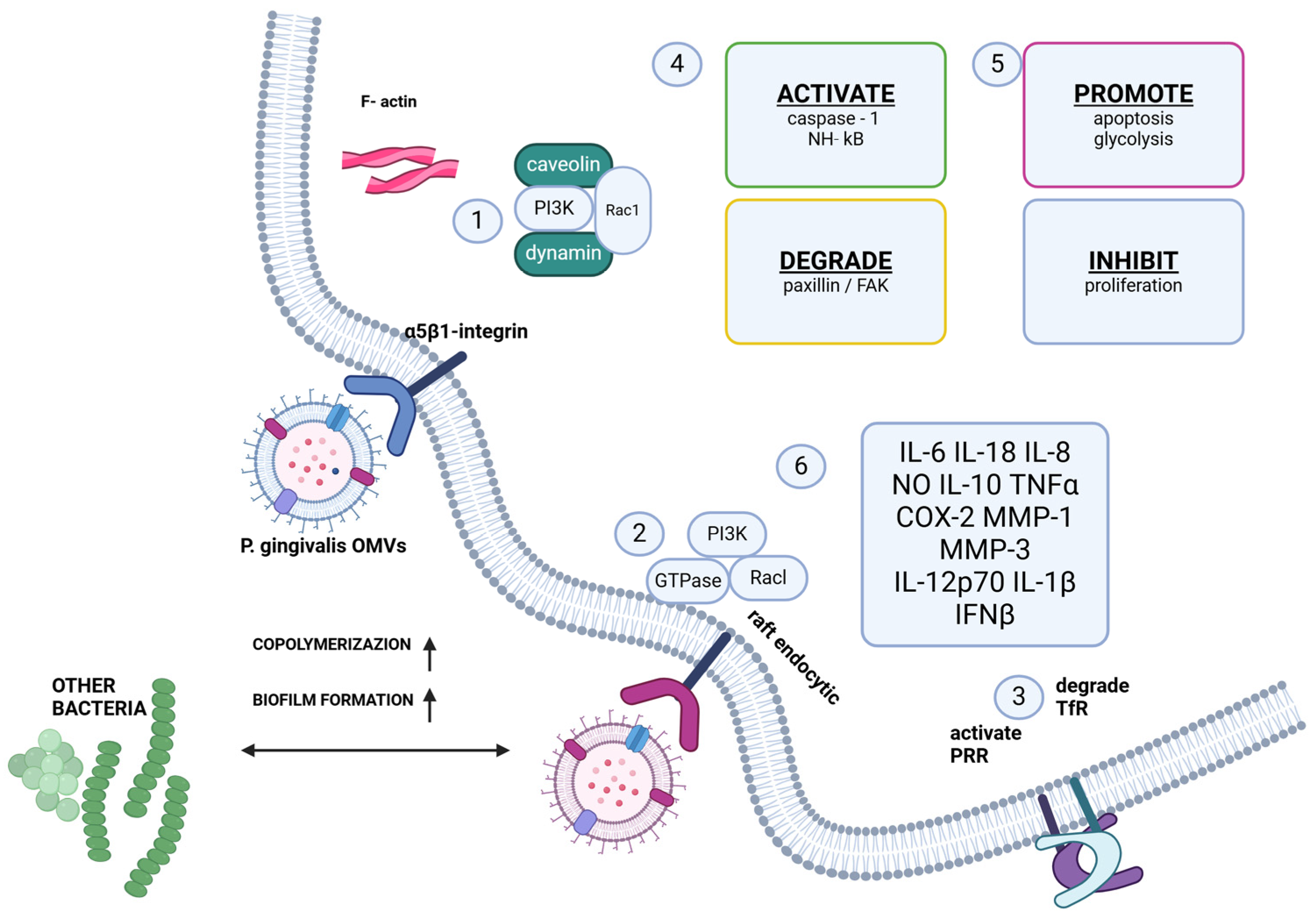
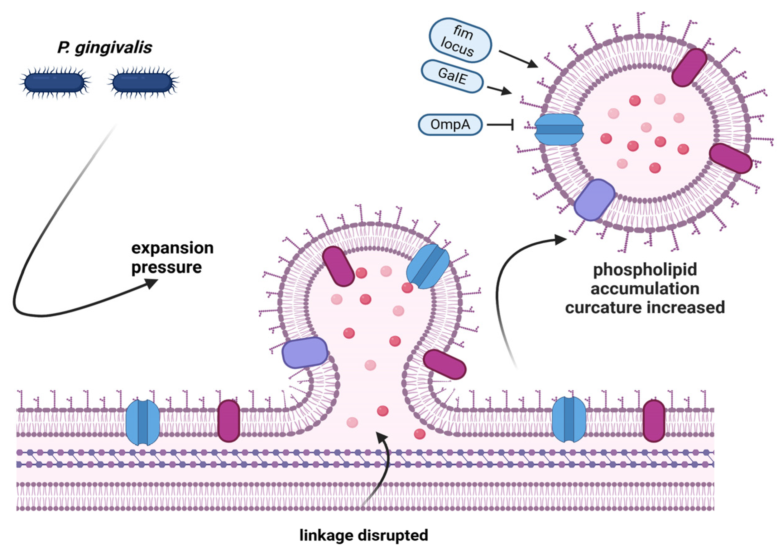
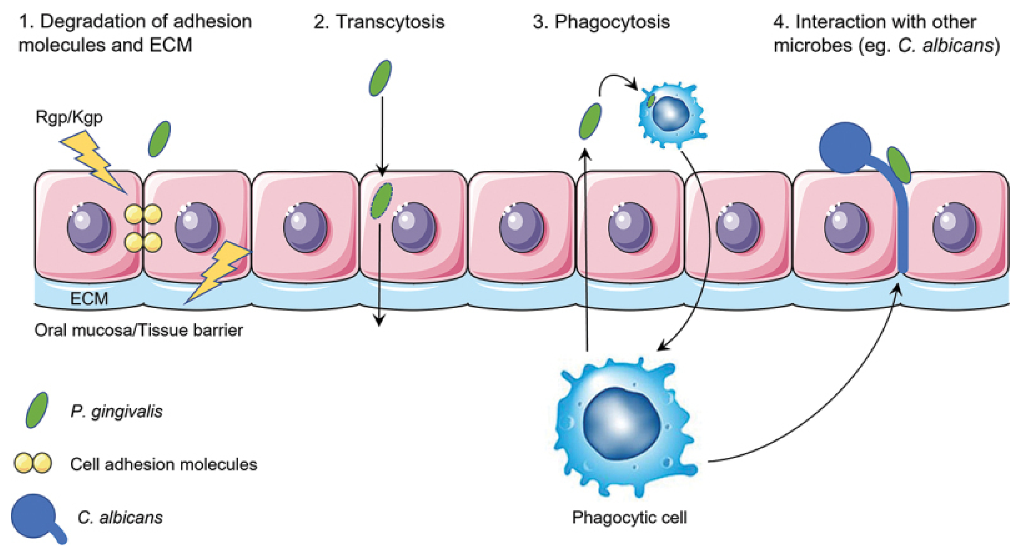
3.3. Virulence Factors and Tissue Invasion

P. gingivalis evades host defense mechanisms primarily through the disruption of epithelial barrier integrity, an early event that triggers inflammatory and immune responses ultimately leading to tissue destruction [28].
This evasion is mediated by a range of virulence factors that dysregulate innate immune and inflammatory pathways [29].
Upon adherence to host cells, P. gingivalis is internalized via lipid rafts and incorporated into early phagosomes, where it engages the autophagic machinery to establish a replicative niche while simultaneously inhibiting apoptosis. Autophagy provides essential host-derived proteins that support intracellular survival, whereas its inhibition—via agents such as 3-methyladenine or wortmannin—leads to bacterial trafficking to the phagolysosome and subsequent degradation [30].
The persistence of P. gingivalis within the periodontal environment is further enhanced by its ability to withstand oxidative stress and by the activity of dipeptidyl peptidase IV (DPPIV), which facilitates nutrient acquisition through dipeptides [31].
Additionally, the bacterium exploits gingival crevicular fluid, an inflammatory exudate rich in peptides and iron (primarily in the form of hemin), as a critical nutrient source [13]. Iron plays a central regulatory role in both bacterial growth and the expression of virulence factors [32].
Hemin is essential to fulfilling the organism’s iron requirements, and P. gingivalis can store endogenous hemin within its membrane, conferring a nutritional advantage under limiting conditions and promoting long-term survival within the periodontal pocket [33].

3.4. Molecular Mimicry and Systemic Dissemination

P. gingivalis is considered a key bacterial pathogen in periodontal disease largely due to its ability to produce outer membrane vesicles (OMVs), which are released via a molecular mimicry mechanism and play a central role in its pathogenesis [34]. OMVs are spherical, bilayered structures (50–250 nm in diameter) shed from the bacterial surface during growth without compromising membrane integrity (Figure 1 and Figure 2). These vesicles participate in essential processes such as stress adaptation, nutrient acquisition, and intercellular communication with both host tissues and microbial communities [35].
Compared to free bacteria, OMVs are more stable, highly adhesive, and protease-resistant, enabling them to concentrate and deliver virulence factors efficiently into the surrounding environment, thereby enhancing bacterial pathogenicity. Importantly, P. gingivalis-derived OMVs can enter the bloodstream and reach distant tissues, potentially contributing to systemic diseases associated with the pathogen.

4. P. gingivalis and Systemic Diseases

4.1. Cardiovascular Diseases

According to the World Health Organization (WHO), cardiovascular diseases (CVDs) remain the leading cause of death worldwide [36]. These diseases encompass a range of cardiac and vascular conditions, including ischemia, atherosclerosis, peripheral arterial disease, infective endocarditis, and acute myocardial infarction. Numerous studies have highlighted a correlation between CVDs and periodontal disease, as both conditions share common risk factors and are multifactorial in nature.
These studies [37] demonstrated that individuals with periodontitis are at a significantly higher risk of developing coronary heart disease compared to those without periodontitis. A 2023 meta-analysis confirmed a consistent association between periodontal disease and adverse cardiovascular outcomes, such as myocardial infarction and stroke, across different populations and genders [38].
Similarly, Geerts et al. [39] reported that activities such as tooth brushing, debridement, or scaling could facilitate the entry of oral pathogens and their virulence factors into the bloodstream. It has also been shown that P. gingivalis can be detected in atherosclerotic plaques [40,41], and more recent studies indicate that the presence of periodontal bacteria in the bloodstream or in situ within vascular lesions is associated with an increased risk of aneurysmal disease [42]. Recent studies have shown that periodontal disease is linked to a significantly increased risk of developing coronary artery disease, stroke, and heart failure, with this association observed consistently in both males and females [38].
The literature identifies two main mechanisms through which periodontitis may influence the development of cardiovascular diseases: direct and indirect mechanisms. The direct mechanism involves the bacterial invasion of blood vessels, whereas the indirect mechanism operates through the activation of excessive inflammatory responses in the host, which in turn may influence the onset of systemic diseases [43].
The interaction between bacterial antigens and host cells can provoke both local and systemic immune responses. P. gingivalis has been shown to invade endothelial cells, induce dysfunction, and trigger pro-atherogenic responses, promoting the development and progression of atherosclerotic lesions [44].

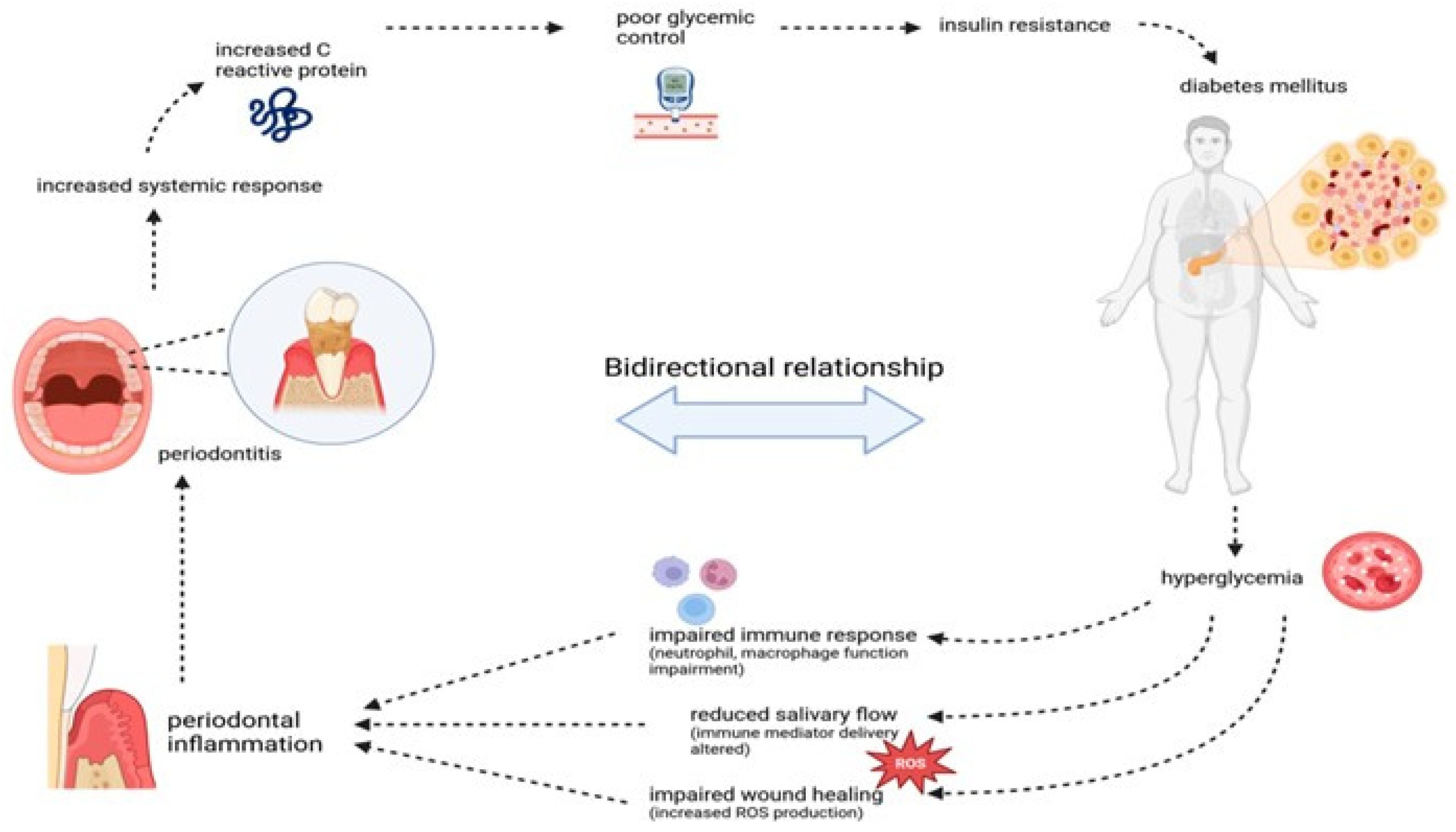
4.2. Diabetes Mellitus

Diabetes mellitus is a metabolic disorder characterized by hyperglycemia resulting from defects in insulin secretion, insulin action, or both. The biological relationship between diabetes and periodontal disease is well documented, with periodontitis having been shown to influence the pathogenesis of several systemic conditions, including diabetes.
Periodontitis and diabetes exhibit a bidirectional interactive relationship: diabetes promotes the onset and progression of periodontitis, while periodontitis, in turn, impairs glycemic control and contributes to the development of diabetic complications. Diabetic patients generally present with more severe clinical manifestations of periodontal inflammation than non-diabetic individuals, and the severity of periodontal destruction is related to the presence of systemic complications, the degree of glycemic control, and the type of diabetes [45].
Microbial activity in periodontal infections is one of the main sources of the bidirectional connection between periodontitis and diabetes, because the hyperinflammatory state associated with this disorder could impose a significant increase in reactive oxygen species (ROS) (Figure 3 from [46]). A complex interplay exists between oxidative stress and advanced glycation end-products (AGEs)—a heterogeneous group of compounds that accumulate under hyperglycemic and oxidative conditions. AGEs intensify oxidative stress and inflammation, further impairing periodontal and systemic health [47]. Infections induced by periodontal bacteria trigger the production of lipopolysaccharides (LPSs), which in turn increase inflammatory cytokines and attract immune cells, thus exacerbating diabetes. In contrast, patients with diabetes create a hyperglycemic environment that favors the growth of pathogenic bacteria, contributing to microbial dysbiosis [48]. Glycemic control in diabetic patients may be impaired by an increase in inflammatory cytokines. This phenomenon can be attributed to endotoxins produced by Gram-negative bacteria. The destruction of periodontal tissue begins with the activation of inflammatory cytokines by bacteria in periodontal tissues, which are elevated in the gingival crevicular fluid. These cytokines, in turn, lead to the breakdown of connective tissue and alveolar bone [49].
Tumor necrosis factor-α (TNF-α), secreted into the gingival sulcus by red complex bacteria such as P. gingivalis, plays a central role in the inflammatory cascade. TNF-α induces the following mechanisms:
  • Increases the production of matrix metalloproteinases (MMPs);
  • Promotes the secretion of prostaglandin E2 (PGE2);
  • Activates osteoclasts and promotes bone resorption by inhibiting osteoblast differentiation;
  • Induces apoptosis, preventing tissue regeneration and exacerbating periodontal destruction. Elevated levels of TNF-α, caused by inflammation, are associated with increased levels of HbA1c, apoptosis in pancreatic cells, and reduced insulin secretion, ultimately leading to insulin resistance [50].
P. gingivalis can express various virulence factors to evade innate and adaptive immunities, enabling it to survive and propagate in the host, destroy periodontal tissues, and contribute to systemic diseases. It can invade and survive in host tissues by disrupting the gingival epithelial barrier, internalizing into epithelial cells, and enhancing autophagy in these cells [51].
In addition, it interferes with the complement system, degrades antimicrobial peptides, impairs phagocyte function, and suppresses adaptive immunity, leading to persistent inflammation and tissue destruction [52].
Recent studies have shown that individuals with poorly controlled diabetes exhibit a significantly higher risk of developing periodontitis, emphasizing the importance of effective glycemic management in periodontal health [53]. Conversely, periodontitis has been associated with worsened metabolic control in diabetic patients, highlighting the need for integrated management strategies to address both conditions simultaneously [54].

4.3. Alzheimer’s Disease

Alzheimer’s disease (AD) is a neurodegenerative disease that affects millions of individuals worldwide, with its prevalence rapidly increasing with increasing life expectancy and the aging of the global population [55,56]. Cheng et al. [57] demonstrated that exposure to periodontal disease is associated with a 1.7-fold increased risk of developing AD. This disease is characterized by progressive neuronal loss, resulting in gradual decline in memory, language, and cognitive functions, ultimately leading to death in the advanced stages (Alzheimer’s Association, 2016) [58].
Scientific evidence emphasizes that individuals with Alzheimer’s disease or dementia often experience compromised oral health, making them more susceptible to chronic oral diseases. Periodontal pathogens not only invade the oral cavity but can also infiltrate the periodontal pocket epithelium, from where they enter the bloodstream. Once in circulation, these pathogens release various endotoxins and exotoxins that propagate the infection in different body compartments, including the brain. Significant amounts of lipopolysaccharide (LPS) have been found in the brains of AD patients. The interaction between periodontal disease (PD) and Alzheimer’s disease (AD) can induce neuronal degeneration through a multifaceted process that begins with oral dysbiosis, leading to microbiome formation and proliferation [59]. Invasion of periodontal tissues by oral pathogens results in their release into the bloodstream. These pathogens and their toxic molecules, such as LPSs, subsequently bind to microglia through Toll-like receptors 2/4 (TLR2/4), triggering the release of cytokines and inflammatory mediators [60]. This inflammatory response induces the production of amyloid precursor protein (APP) by neuronal cells. Activation of β- and γ-secretase enzymes leads to increased secretion of amyloid-beta (Aβ) peptides, particularly Aβ42 monomers and sAPPβ, both of which accumulate extracellularly and generate intracellular AICD. These Aβ peptides aggregate into oligomers, protofibrils, or fibrils, ultimately forming amyloid plaques [61].
These plaques are recognized by Triggering Receptor Expressed on Myeloid Cells 2 (TREM2) receptors on the membranes of microglia, triggering an additional inflammatory response that further stimulates Aβ production. Furthermore, dysfunctional neurons show increased phosphorylation of tau (p-tau), resulting in the formation of neurofibrillary tangles (p-tau tangles) [62]. Chronic periodontitis (CP) and P. gingivalis infection have been identified as significant risk factors for the development of Aβ plaques, dementia, and AD [63].
Dominy’s studies hypothesized that P. gingivalis infection contributes to the pathogenesis of AD through the secretion of gingipains to promote neuronal damage and found that gingipain immunoreactivity (IR) was significantly present in AD brains. In addition, they identified P. gingivalis DNA in AD brains and cerebrospinal fluid (CSF) of living subjects diagnosed with probable AD, suggesting that P. gingivalis DNA in CSF could serve as a differential diagnostic marker [64]. In addition, Singhrao’s article suggests that excessive neuroinflammation may contribute to synaptic protein and memory loss, offering mechanistic insights into P. gingivalis–LPS-mediated inflammatory pathways in periodontitis [65]. Recent studies have further elucidated the role of periodontal pathogens in exacerbating neuroinflammation by modulating microglial activation, which may accelerate neurodegenerative processes in AD [66]. Moreover, longitudinal cohort studies have confirmed that effective periodontal treatment correlates with slower cognitive decline in patients at risk for Alzheimer’s disease, suggesting potential benefits of oral health interventions in AD management [67].

4.4. Rheumatoid Arthritis

Rheumatoid arthritis (RA) is a chronic autoimmune inflammatory disorder primarily affecting the synovial membranes of multiple joints. This condition leads to persistent inflammation, synovial proliferation, and ultimately the destruction of articular structures. Over time, the progressive damage to articular cartilage results in joint deformities and functional impairments, significantly reducing the patient’s quality of life. While the primary pathology is localized within the synovial membranes of peripheral joints, RA can also have systemic effects, with both specific and nonspecific extra-articular manifestations that affect multiple organs and tissues. These systemic manifestations contribute to a reduction in life expectancy and exacerbate the overall burden of the disease. Rheumatoid arthritis typically presents in individuals aged between 40 and 60 years, with a higher prevalence observed in women [68].
RA and periodontal disease share a number of common pathobiological mechanisms, including similar cellular involvement in the inflammatory response, parallel cytokine profiles in serum and the local microenvironment, and the activation of matrix metalloproteinases (MMPs) and other mediators that facilitate tissue breakdown. Furthermore, both conditions are characterized by osteoclast-mediated bone resorption, leading to significant skeletal damage [69].
A pivotal process in RA pathogenesis is citrullination, a post-translational modification in which the amino acid arginine is converted to citrulline. This modification is mediated by peptidyl arginine deiminase (PAD) enzymes, which are expressed by various immune cells, including T and B lymphocytes, neutrophils, monocytes, and macrophages [70]. The excessive citrullination of proteins can result in the formation of autoantigens that trigger the production of autoantibodies, particularly anti-cyclic citrullinated peptide (anti-CCP) antibodies, which are central to the pathogenesis of RA. The accumulation of these citrullinated proteins, in turn, promotes an autoimmune response that exacerbates inflammation and tissue destruction [71].
To date, P. gingivalis is the only known microorganism capable of expressing peptidyl arginine deiminase (PPAD), a bacterial enzyme homologous to the human PAD enzyme [72]. The presence of P. gingivalis in the oral microbiota of RA patients has been strongly correlated with the presence of anti-CCP antibodies, suggesting that this bacterium may play a pivotal role in the pathogenesis of RA [73].
It is hypothesized that P. gingivalis facilitates the accumulation of citrullinated proteins in the oral cavity, leading to the generation of autoantibodies through the activation of an aberrant immune response. In particular, the reduced immune tolerance to citrullinated proteins in RA patients may promote the formation of these autoantibodies, which in turn contribute to the systemic inflammatory burden of the disease [74].
Moreover, the elevated production of citrullinated proteins has been associated with more severe disease manifestations, including the rapid progression of joint erosions. Consequently, the presence of anti-CCP antibodies has been shown to correlate with increased alveolar bone resorption in patients with RA, further underscoring the interconnectedness between periodontal disease and the severity of rheumatoid arthritis. Recent studies have expanded this view by identifying novel bacterial species in the oral microbiome that may synergize with P. gingivalis to exacerbate citrullination and systemic autoimmunity in RA [75].
Additionally, emerging evidence indicates that periodontal therapy can reduce systemic inflammatory markers and autoantibodies in RA patients, suggesting a therapeutic benefit in managing both conditions simultaneously [76].

4.5. Other Diseases

The oral pathogen P. gingivalis is associated not only with periodontitis but also with diseases in other parts of the body. The mechanisms by which it travels from the oral cavity to other organs of the body have been identified in four mechanisms (Figure 4 from [77]). First, proteolytic enzymes secreted by P. gingivalis degrade adhesion molecules between tissue cells and the extracellular matrix. This weakens the structural integrity of the mucosa and allows P. gingivalis to penetrate the tissue. The second is transcytosis: bacteria actively enter tissue cells and transfer to the next layer or the extracellular space. Traveling from one cell to another, P. gingivalis reaches deeper structures. Third, professional phagocytes absorb P. gingivalis and travel to the bloodstream, where P. gingivalis is released. Fourth, P. gingivalis can adhere to hyphae, forming Candida albicans. These hyphae can penetrate mucosal tissue, allowing P. gingivalis to reach deeper structures.
The following is a schematic overview of the four putative mechanisms for P. gingivalis to translocate the oral mucosa or the endothelium of blood vessels: (1) Proteolytic enzymes known as gingipains (Rgp/Kgp) are secreted by P. gingivalis (green) and degrade cell–cell adherence molecules (yellow circles) and the extracellular matrix (ECM; light blue). The structural integrity of the oral mucosa is weakened so that P. gingivalis is able to pass in between the cells. (2) P. gingivalis can enter the cell after adherence and subsequent endocytosis and exit the cell on the other side of the epithelial layer. P. gingivalis needs to have a mechanism to be able to survive within the cell and not be degraded by phagolysosomes. It can then travel from cell to cell, migrating deeper into the tissue, eventually reaching the basal membrane and finally the endothelial cells that line the blood vessels. (3) Phagocytic cells of the host are able to pick up P. gingivalis within the tissue and transfer it across the endothelial barrier. They can then travel back into the bloodstream, taking the bacterium with them. Again, P. gingivalis needs to have a mechanism to survive degradation by the phagocyte. (4) Interactions with other microbes such as C. albicans (blue) could allow for translocation across the oral mucosa due to the hyphae of C. albicans that can insert themselves between cells. P. gingivalis can attach to these hyphae. Macrophages may also play a role in this mechanism as they are attracted to the hyphae of C. albicans and can phagocytose the attached P. gingivalis.

4.5.1. Oncology

The oral cavity hosts a diverse microbial community that lives in a symbiotic relationship with each other and with the host immune system. Dysbiotic oral microflora subverts host defense mechanisms, resulting in chronic periodontal disease and chronic bacterial infection that may be a promoter or cause of oral cancer [77]. Growing evidence from epidemiological and genetic studies suggests a significant link between oral pathogens and certain cancers, including oral, digestive tract, esophageal, pancreatic, and colorectal cancers. The chronic inflammation caused by oral pathogens plays a critical role in the development and progression of these malignancies [78]. Gholizadeh et al. conducted a comprehensive review evaluating the correlation between chronic oral inflammation and the development of oral cancers (OCs). The microbial species P. gingivalis has been implicated in periodontal disease (PD), which may act as an etiological factor in the pathogenesis of some cancers. A multicenter study involving 405 pancreatic cancer patients and 416 controls found that anti-P. gingivalis antibody levels were significantly higher in pancreatic cancer patients compared to healthy controls [79].
However, a recent study demonstrated that P. gingivalis is closely related to oral squamous cell carcinoma (OSCC), and outer membrane vesicles (OMVs) are the main pathogen-associated factor [80]. For instance, P. gingivalis has been detected in 61% of esophageal cancer tissues, compared to 12% in adjacent non-cancerous tissues and 0% in normal esophageal mucosa [81]. Similarly, periodontal disease pathogens have also been implicated in colorectal cancer by inducing excessive immune responses and activating cancer growth genes. The bacteria preferentially accumulate in adenomas—benign bowel growths that can progress to cancer—and thrive in the nutrient-rich microenvironment of colonic lesions due to their asaccharolytic metabolism [82]. This suggests that the oral microbiota, particularly in individuals with periodontal disease, could serve as a potential biomarker for various human diseases. In addition, a significant association between head and neck squamous cell carcinoma and periodontal disease has been reported, supporting the hypothesis that periodontal disease may be an independent risk factor. These findings have important implications for the prevention, early diagnosis, and treatment of these tumors, with the potential to improve patient outcomes. Advances in understanding host–microbe interactions and underlying cause–effect mechanisms have expanded our knowledge of the role of microbiome in both health and disease, opening new therapeutic avenues for clinical practice [83].

4.5.2. Biology and Immunology

The oral cavity can be colonized by over 700 different species of microbes, including bacteria, fungi, viruses, and protozoa. Together, they form a complex biological system also known as the oral microbiome. More niches can be distinguished in the oral cavity, each with its own unique microbial composition. The oral microbiome is dynamic and can be influenced by several factors, which can lead to dysbiosis. P. gingivalis has a variety of virulence factors that aid in the invasion or destruction of host tissue and evasion of the host immune response and has been associated with various systemic diseases [84].
Several studies have explored the biological mechanisms of P. gingivalis, identifying it as a key pathogen in chronic periodontitis. The products of P. gingivalis, such as vesicles, may be key to understanding the mechanisms of communication and dissemination of the bacterium and its systemic implications [85].
In a study by Tiantian et al., the authors demonstrated the association between viruses and bacteria in PD, suggesting that both could contribute to the development of systemic diseases, but the interaction between P. gingivalis and viruses warrants further investigation, as it may offer new insights into potential treatment strategies for patients [86].
In addition, Grover et al. also recognized P. gingivalis as an important contributor to PD, and a comprehensive understanding of its virulence factors could provide a potential antigenic target for a periodontal vaccine [87]. According to Imai and Ochiai, some periodontal bacteria could contribute to the maintenance, latency, and reactivation of HIV-1. Therefore, periodontal disease could accelerate the progression of acquired immunodeficiency syndrome (AIDS) in infected individuals [88]. Again, the prevention of periodontal disease emerges as a critical factor in slowing the progression of systemic diseases and improving the overall outcomes of these diseases [89].

5. Diagnostic and Therapeutic Implications

Periodontal diagnosis is conducted according to the classification system established at the 2017 World Workshop on the Classification of Periodontal and Peri-Implant Diseases and Conditions [1]. This diagnostic process involves four key steps:
  • Identification of patients suspected of having periodontitis;
  • Confirmation of the diagnosis of periodontitis;
  • Staging of the periodontitis case;
  • Classification of the periodontitis case.
The management of dental plaque-related diseases mainly involves nonspecific plaque removal through various means. According to the European Federation of Periodontology (EFP), the first step in therapeutic intervention is to induce behavioral change, motivating the patient to effectively remove supragingival dental biofilm and control risk factors. This initial phase may include the following interventions: the control of supragingival dental biofilm; strategies to improve oral hygiene effectiveness, including motivational techniques and oral hygiene instructions (OHIs); adjunctive therapies targeting gingival inflammation; professional mechanical plaque removal (PMPR), which involves interventions to remove supragingival plaque and calculus, as well as address plaque retention factors that may compromise oral hygiene practices; and risk factor management, which includes interventions to eliminate or mitigate risk factors that contribute to the onset and progression of periodontitis.
The second phase of periodontal therapy, known as cause-related therapy, is primarily aimed at controlling and reducing subgingival biofilm and calculus through subgingival instrumentation.
This phase may also involve adjunctive therapeutic measures, including the use of physical or chemical agents, host modulation therapies (administered either locally or systemically), and local or systemic subgingival antimicrobial treatments. Among recent advances, injectable platelet-rich fibrin (i-PRF) has emerged as a promising adjunct. As demonstrated in a systematic review by Niemczyk et al. [90], i-PRF exhibits significant bactericidal effects against P. gingivalis and serves as a regenerative agent due to its ability to release growth factors and extracellular matrix components of connective tissue [91].
Additionally, hyaluronic acid (HA) and various re-epithelializing agents, including cytokines and growth factors, have been shown to positively influence epithelial–connective tissue interactions. HA, in particular, has demonstrated anti-inflammatory, angiogenic, and osteoinductive properties, promoting cell migration, proliferation, and extracellular matrix synthesis—key processes in tissue repair and healing [92].
Diagnostic methods for detecting P. gingivalis typically involve the analysis of subgingival plaque samples. Commonly used techniques include the following [93]:
  • Enzyme-linked immunosorbent assays (ELISAs);
  • DNA probes;
  • Real-time polymerase chain reactions (PCRs).
However, subgingival plaque-based diagnostic tests have shown limitations due to factors such as low sensitivity and specificity, prolonged development times, and the need to send samples to external laboratories for analysis, which limits their practical application in point-of-care settings [94].

6. Future Directions and Research Gaps: Probiotics and Vaccines

Probiotics are live, non-pathogenic microorganisms administered to improve microbial balance and are regulated as dietary supplements and foods. Probiotics exert their beneficial effects through various mechanisms, including lowering pH, decreasing colonization and invasion by pathogenic organisms, and modifying the host immune response. Probiotics have recently attracted considerable attention as a potential therapeutic approach for the prevention of periodontal diseases [95].
The Food and Agriculture Organization (FAO) and the World Health Organization define probiotics as “live microorganisms which, when administered in adequate amounts, confer a health benefit on the host” [96]. Probiotic microorganisms are able to mitigate inflammation-induced damage to epithelial cells by stimulating the upregulation of structural proteins essential for epithelial integrity. Furthermore, these microorganisms can successfully colonize and proliferate within the oral microbiome, competing with pathogenic bacteria for available nutrients, thereby inhibiting their growth.
Probiotics have also been shown to produce antimicrobial substances, such as acetic acid and lactic acid, that specifically target and suppress the growth of Gram-negative bacteria. Furthermore, they can modulate host immune responses, reducing pro-inflammatory pathways that could damage host tissues while simultaneously increasing defensive pathways that inhibit the growth and virulence of pathogenic microorganisms [97].
A promising avenue for future periodontal therapy may lie in the development of a human periodontal vaccine, which could further enhance the preventive potential of probiotics.
Vaccine is a term generally describing a substance with dead or attenuated living infectious material introduced into the body with the goal of increasing its ability to resist or rid itself of a disease. Vaccines are generally prophylactic, meaning they ameliorate the effects of future infections. Therefore, the availability of a periodontal vaccine would not only prevent and modulate periodontal disease but also improve the quality of life of people for whom periodontal treatment cannot be easily achieved [98].
Currently, three main types of vaccines have been studied in the context of periodontal disease management:
  • Vaccines derived from pure cultures of Streptococcus and other oral microorganisms;
  • Autogenous vaccines, which are derived from the patient’s own microbial flora;
  • Serial vaccines, such as the Van Cott vaccine, the Goldenberg vaccine, and the Inava Endocorps vaccine.
Although these approaches have potential, further research is needed to fully optimize and validate these vaccine-based therapies in the clinical setting.

7. Conclusions

Oral health plays a fundamental role in overall systemic health, a concept increasingly supported by a growing body of scientific evidence. This review highlights the critical involvement of Porphyromonas gingivalis in the pathogenesis of periodontal disease and its broader implications for systemic conditions. P. gingivalis is a pathogen capable of evading the host immune system, manipulating cellular processes, and disrupting the microbial equilibrium within the oral cavity. Its principal virulence factors, particularly gingipains, facilitate nutrient acquisition, modulate immune responses, and shape biofilm dynamics, thereby promoting chronic inflammation and tissue destruction. The bacterium’s ability to exploit host autophagy mechanisms, withstand oxidative stress, and acquire iron from hemin contributes to its persistence in the hostile environment of the periodontal pocket. Additionally, the secretion of outer membrane vesicles (OMVs) enhances its pathogenic potential by delivering virulence factors and facilitating interbacterial communication and systemic dissemination. Substantial evidence supports the association between chronic periodontal inflammation and systemic diseases such as cardiovascular disease, diabetes, and rheumatoid arthritis. The bidirectional interplay between oral and systemic health suggests that P. gingivalis-induced dysbiosis and inflammation may not only exacerbate periodontal disease but also influence the progression of these systemic conditions.
However, the current literature presents several limitations. A substantial portion of the available evidence is based on observational studies, which inherently limits the ability to establish definitive causal relationships. Variability in diagnostic criteria and methodological approaches further complicates data interpretation. Furthermore, the precise molecular pathways linking P. gingivalis to systemic pathology require further elucidation. Future research should delineate these pathways and evaluate targeted therapeutic strategies aimed at mitigating P. gingivalis effects, including host immune modulation and microbiome restoration. In conclusion, deepening our understanding of P. gingivalis’ role in oral and systemic disease opens new avenues for therapeutic intervention. Recognizing the interconnection between oral and systemic health underscores the necessity of integrated clinical approaches to enhance both periodontal and overall patient health.

Author Contributions

Conceptualization, G.I. and B.M.M.; methodology, B.M.M. and A.P.; validation, C.G. and A.B.; formal analysis, C.G. and A.B.; investigation, B.M.M. and C.G.; resources, B.M.M.; data curation, B.M.M. and A.P.; writing—original draft preparation, B.M.M.; writing—review and editing, B.M.M., A.P. and G.I.; visualization, G.I.; supervision, G.I.; project administration, C.G. and G.I. All authors have read and agreed to the published version of the manuscript.

Funding

This research received no external funding.

Institutional Review Board Statement

Not applicable.

Informed Consent Statement

Not applicable.

Data Availability Statement

Not applicable.

Conflicts of Interest

The authors declare no conflicts of interest.

References

  1. Papapanou, P.N.; Sanz, M.; Buduneli, N.; Dietrich, T.; Feres, M.; Fine, D.H.; Flemmig, T.F.; Garcia, R.; Giannobile, W.V.; Graziani, F.; et al. Periodontitis: Consensus report of workgroup 2 of the 2017 World Workshop on the Classification of Periodontal and Peri-Implant Diseases and Conditions. J. Clin. Periodontol. 2018, 45, S162–S170. [Google Scholar] [CrossRef] [PubMed]
  2. Caton, J.G.; Armitage, G.; Berglundh, T.; Chapple, I.L.; Jepsen, S.; Kornman, K.S.; Mealey, B.L.; Papapanou, P.N.; Sanz, M.; Tonetti, M.S. A new classification scheme for periodontal and peri-implant diseases and conditions—Introduction and key changes from the 1999 classification. J. Clin. Periodontol. 2018, 45, S1–S8. [Google Scholar] [CrossRef] [PubMed]
  3. Sanz, M.; Herrera, D.; Kebschull, M.; Chapple, I.; Jepsen, S.; Berglundh, T.; Sculean, A.; Tonetti, M.S.; Consultants, T.E.W.P.A.M.; Lambert, N.L.F. Treatment of stage I–III periodontitis—The EFP S3 level clinical practice guideline. J. Clin. Periodontol. 2020, 47, 4–60. [Google Scholar] [CrossRef] [PubMed]
  4. Isola, G.; Santonocito, S.; Lupi, S.M.; Polizzi, A.; Sclafani, R.; Patini, R.; Marchetti, E.; Brzozowski, T. Periodontal Health and Disease in the Context of Systemic Diseases. Mediat. Inflamm. 2023, 2023, 9720947. [Google Scholar] [CrossRef]
  5. Kassebaum, N.; Bernabé, E.; Dahiya, M.; Bhandari, B.; Murray, C.; Marcenes, W. Global Burden of Severe Periodontitis in 1990–2010: A Systematic Review and Meta-regression. J. Dent. Res. 2014, 93, 1045–1053. [Google Scholar] [CrossRef]
  6. Kwon, T.; Lamster, I.B.; Levin, L. Current Concepts in the Management of Periodontitis. Int. Dent. J. 2021, 71, 462–476. [Google Scholar] [CrossRef]
  7. Nazir, M.A. Prevalence of periodontal disease, its association with systemic diseases and prevention. Int. J. Health Sci. 2025, 11, 72. [Google Scholar]
  8. Socransky, S.S.; Haffajee, A.D.; Cugini, M.A.; Smith, C.; Kent, R.L., Jr. Microbial complexes in subgingival plaque. J. Clin. Periodontol. 1998, 25, 134–144. [Google Scholar] [CrossRef]
  9. Chen, W.A.; Dou, Y.; Fletcher, H.M.; Boskovic, D.S. Local and Systemic Effects of Porphyromonas gingivalis Infection. Microorganisms 2023, 11, 470. [Google Scholar] [CrossRef]
  10. Socransky, S.S.; Haffajee, A.D. Periodontal microbial ecology. Periodontol. 2000 2005, 38, 135–187. [Google Scholar] [CrossRef]
  11. Yilmaz, Ö. The chronicles of Porphyromonas gingivalis: The microbium, the human oral epithelium and their interplay. Microbiology 2008, 154, 2897–2903. [Google Scholar] [CrossRef] [PubMed]
  12. Bodet, C.; Chandad, F.; Grenier, D. Pathogenic potential of Porphyromonas gingivalis, Treponema denticola and Tannerella forsythia, the red bacterial complex associated with periodontitis. Pathol. Biol. 2007, 55, 154–162. [Google Scholar] [CrossRef] [PubMed]
  13. Mysak, J.; Podzimek, S.; Sommerova, P.; Lyuya-Mi, Y.; Bartova, J.; Janatova, T.; Prochazkova, J.; Duskova, J. Porphyromonas gingivalis: Major Periodontopathic Pathogen Overview. J. Immunol. Res. 2014, 2014, 476068. [Google Scholar] [CrossRef] [PubMed]
  14. Yoshimura, F.; Murakami, Y.; Nishikawa, K.; Hasegawa, Y.; Kawaminami, S. Surface components of Porphyromonas gingivalis. J. Periodontal Res. 2009, 44, 1–12. [Google Scholar] [CrossRef]
  15. Antonyuk, S.V.; Siemińska, K.; Śmiga, M.; Strange, R.W.; Wagner, M.; Barnett, K.J.; Olczak, T. Bacteroides fragilis expresses three proteins similar to Porphyromonas gingivalis HmuY: Hemophore-like proteins differentially evolved to participate in heme acquisition in oral and gut microbiomes. FASEB J. 2023, 37, e22981. [Google Scholar] [CrossRef]
  16. Marshall, R.I. Gingival defensins: Linking the innate and adaptive immune responses to dental plaque. Periodontol. 2000 2004, 35, 14–20. [Google Scholar] [CrossRef]
  17. Hasegawa, Y.; Tribble, G.D.; Baker, H.V.; Mans, J.J.; Handfield, M.; Lamont, R.J. Role of Porphyromonas gingivalis SerB in Gingival Epithelial Cell Cytoskeletal Remodeling and Cytokine Production. Infect. Immun. 2008, 76, 2420–2427. [Google Scholar] [CrossRef]
  18. Nisapakultorn, K.; Ross, K.F.; Herzberg, M.C.; Tuomanen, E.I. Calprotectin Expression In Vitro by Oral Epithelial Cells Confers Resistance to Infection by Porphyromonas gingivalis. Infect. Immun. 2001, 69, 4242–4247. [Google Scholar] [CrossRef]
  19. Weinberg, A.; Krisanaprakornkit, S.; Dale, B. Epithelial Antimicrobial Peptides: Review and Significance for Oral Applications. Crit. Rev. Oral Biol. Med. 1998, 9, 399–414. [Google Scholar] [CrossRef]
  20. Curtis, M.; Aduse-Opoku, J.; Rangarajan, M. Cysteine Proteases of Porphyromonas gingivalis. Crit. Rev. Oral Biol. Med. 2001, 12, 192–216. [Google Scholar] [CrossRef]
  21. Bao, K.; Belibasakis, G.N.; Thurnheer, T.; Aduse-Opoku, J.; Curtis, M.A.; Bostanci, N. Role of Porphyromonas gingivalis gingipains in multi-species biofilm formation. BMC Microbiol. 2014, 14, 258. [Google Scholar] [CrossRef] [PubMed]
  22. Liang, S.; Krauss, J.L.; Domon, H.; McIntosh, M.L.; Hosur, K.B.; Qu, H.; Li, F.; Tzekou, A.; Lambris, J.D.; Hajishengallis, G. The C5a Receptor Impairs IL-12–Dependent Clearance of Porphyromonas gingivalis and Is Required for Induction of Periodontal Bone Loss. J. Immunol. 2011, 186, 869–877. [Google Scholar] [CrossRef] [PubMed]
  23. Yamada, M.; Ikegami, A.; Kuramitsu, H.K. Synergistic biofilm formation by Treponema denticola and Porphyromonas gingivalis. FEMS Microbiol. Lett. 2005, 250, 271–277. [Google Scholar] [CrossRef]
  24. Lamont, R.J.; Jenkinson, H.F. Subgingival colonization by Porphyromonas gingivalis. Oral Microbiol. Immunol. 2000, 15, 341–349. [Google Scholar] [CrossRef]
  25. Medzhitov, R. Recognition of microorganisms and activation of the immune response. Nature 2007, 449, 819–826. [Google Scholar] [CrossRef]
  26. Darveau, R.P. Periodontitis: A polymicrobial disruption of host homeostasis. Nat. Rev. Microbiol. 2010, 8, 481–490. [Google Scholar] [CrossRef]
  27. Hajishengallis, G. Immune Evasion Strategies of Porphyromonas gingivalis. J. Oral Biosci. 2011, 53, 233–240. [Google Scholar] [CrossRef]
  28. Denis, F.K.P. P. gingivalis interactions with epithelial cells. Front. Biosci. 2008, 13, 966–984. [Google Scholar] [CrossRef]
  29. Bostanci, N.; Belibasakis, G.N. Porphyromonas gingivalis: An invasive and evasive opportunistic oral pathogen. FEMS Microbiol. Lett. 2012, 333, 1–9. [Google Scholar] [CrossRef]
  30. Bélanger, M.; Rodrigues, P.H.; Dunn, W.A., Jr.; Progulske-Fox, A. Autophagy: A Highway for Porphyromonas gingivalis in Endothelial Cells. Autophagy 2006, 2, 165–170. [Google Scholar] [CrossRef]
  31. Clais, S.; Boulet, G.; Kerstens, M.; Horemans, T.; Teughels, W.; Quirynen, M.; Lanckacker, E.; De Meester, I.; Lambeir, A.-M.; Delputte, P.; et al. Importance of biofilm formation and dipeptidyl peptidase IV for the pathogenicity of clinical Porphyromonas gingivalis isolates. Pathog. Dis. 2014, 70, 408–413. [Google Scholar] [CrossRef] [PubMed]
  32. Bramanti, T.E.; Holt, S.C. Roles of porphyrins and host iron transport proteins in regulation of growth of Porphyromonas gingivalis W50. J. Bacteriol. 1991, 173, 7330–7339. [Google Scholar] [CrossRef] [PubMed]
  33. Genco, C.A.; Odusanya, B.M.; Brown, G. Binding and accumulation of hemin in Porphyromonas gingivalis are induced by hemin. Infect. Immun. 1994, 62, 2885–2892. [Google Scholar] [CrossRef]
  34. Pérez-Cruz, C.; Delgado, L.; López-Iglesias, C.; Mercade, E.; Rudel, T. Outer-Inner Membrane Vesicles Naturally Secreted by Gram-Negative Pathogenic Bacteria. PLoS ONE 2015, 10, e0116896. [Google Scholar] [CrossRef]
  35. Ellis, T.N.; Kuehn, M.J. Virulence and Immunomodulatory Roles of Bacterial Outer Membrane Vesicles. Microbiol. Mol. Biol. Rev. 2010, 74, 81–94. [Google Scholar] [CrossRef]
  36. Carrizales-Sepúlveda, E.F.; Ordaz-Farías, A.; Vera-Pineda, R.; Flores-Ramírez, R. Periodontal Disease, Systemic Inflammation and the Risk of Cardiovascular Disease. Heart Lung Circ. 2018, 27, 1327–1334. [Google Scholar] [CrossRef]
  37. Bahekar, A.A.; Singh, S.; Saha, S.; Molnar, J.; Arora, R. The prevalence and incidence of coronary heart disease is significantly increased in periodontitis: A meta-analysis. Am. Heart J. 2007, 154, 830–837. [Google Scholar] [CrossRef]
  38. Leng, Y.; Hu, Q.; Ling, Q.; Yao, X.; Liu, M.; Chen, J.; Yan, Z.; Dai, Q. Periodontal disease is associated with the risk of cardiovascular disease independent of sex: A meta-analysis. Front. Cardiovasc. Med. 2023, 10, 1114927. [Google Scholar] [CrossRef]
  39. Geerts, S.O.; Nys, M.; De Mol, P.; Charpentier, J.; Albert, A.; Legrand, V.; Rompen, E.H. Systemic Release of Endotoxins Induced by Gentle Mastication: Association with Periodontitis Severity. J. Periodontol. 2002, 73, 73–78. [Google Scholar] [CrossRef]
  40. Figuero, E.; Sánchez-Beltrán, M.; Cuesta-Frechoso, S.; Tejerina, J.M.; del Castro, J.A.; Gutiérrez, J.M.; Herrera, D.; Sanz, M. Detection of Periodontal Bacteria in Atheromatous Plaque by Nested Polymerase Chain Reaction. J. Periodontol. 2011, 82, 1469–1477. [Google Scholar] [CrossRef]
  41. Szulc, M.; Kustrzycki, W.; Janczak, D.; Michalowska, D.; Baczynska, D.; Radwan-Oczko, M. Presence of Periodontopathic Bacteria DNA in Atheromatous Plaques from Coronary and Carotid Arteries. BioMed Res. Int. 2015, 2015, 825397. [Google Scholar] [CrossRef] [PubMed]
  42. Salhi, L.; Rompen, E.; Sakalihasan, N.; Laleman, I.; Teughels, W.; Michel, J.-B.; Lambert, F. Can Periodontitis Influence the Progression of Abdominal Aortic Aneurysm? A Systematic Review. Angiology 2018, 70, 479–491. [Google Scholar] [CrossRef] [PubMed]
  43. Persson, G.R.; Persson, R.E. Cardiovascular disease and periodontitis: An update on the associations and risk. J. Clin. Periodontol. 2008, 35, 362–379. [Google Scholar] [CrossRef]
  44. Pink, C.; Holtfreter, B.; Völzke, H.; Nauck, M.; Dörr, M.; Kocher, T. Periodontitis and systemic inflammation as independent and interacting risk factors for mortality: Evidence from a prospective cohort study. BMC Med. 2023, 21, 430. [Google Scholar] [CrossRef]
  45. Winning, L.; Patterson, C.C.; Neville, C.E.; Kee, F.; Linden, G.J. Periodontitis and incident type 2 diabetes: A prospective cohort study. J. Clin. Periodontol. 2017, 44, 266–274. [Google Scholar] [CrossRef]
  46. Ranbhise, J.S.; Ju, S.; Singh, M.K.; Han, S.; Akter, S.; Ha, J.; Choe, W.; Kim, S.S.; Kang, I. Chronic Inflammation and Glycemic Control: Exploring the Bidirectional Link Between Periodontitis and Diabetes. Dent. J. 2025, 13, 100. [Google Scholar] [CrossRef]
  47. Mirnic, J.; Djuric, M.; Brkic, S.; Gusic, I.; Stojilkovic, M.; Tadic, A.; Veljovic, T. Pathogenic Mechanisms That May Link Periodontal Disease and Type 2 Diabetes Mellitus—The Role of Oxidative Stress. Int. J. Mol. Sci. 2024, 25, 9806. [Google Scholar] [CrossRef]
  48. Blasco-Baque, V.; Garidou, L.; Pomié, C.; Escoula, Q.; Loubieres, P.; Le Gall-David, S.; Lemaitre, M.; Nicolas, S.; Klopp, P.; Waget, A.; et al. Periodontitis induced by Porphyromonas gingivalis drives periodontal microbiota dysbiosis and insulin resistance via an impaired adaptive immune response. Gut 2016, 66, 872–885. [Google Scholar] [CrossRef]
  49. Sahota, J.; Bakshi, D.; Kaur, G.; Singh, D.; Thakur, A.; Grover, S. Estimation of Plasma Levels of Tumor Necrosis Factor-α, Interleukin-4 and 6 in Patients with Chronic Periodontitis and Type II Diabetes Mellitus. J. Contemp. Dent. Pract. 2018, 19, 166–169. [Google Scholar] [CrossRef]
  50. Choubaya, C.; Chahine, N.; Aoun, G.; Anil, S.; Zalloua, P.; Salameh, Z. Expression of Inflammatory Mediators in Periodontitis Over Established Diabetes: An Experimental Study in Rats. Med. Arch. 2021, 75, 436–443. [Google Scholar] [CrossRef]
  51. Zheng, S.; Yu, S.; Fan, X.; Zhang, Y.; Sun, Y.; Lin, L.; Wang, H.; Pan, Y.; Li, C. Porphyromonas gingivalis survival skills: Immune evasion. J. Periodontal Res. 2021, 56, 1007–1018. [Google Scholar] [CrossRef] [PubMed]
  52. Yang, Y.; Sun, X.; Yang, Y.; Qie, Y. Insight of the interrelationship and association mechanism between periodontitis and diabetes mellitus. Regen. Ther. 2024, 26, 1159–1167. [Google Scholar] [CrossRef] [PubMed]
  53. Wei, X.; Zhang, X.; Chen, R.; Li, Y.; Yang, Y.; Deng, K.; Cai, Z.; Lai, H.; Shi, J. Impact of periodontitis on type 2 diabetes: A bioinformatic analysis. BMC Oral Health 2024, 24, 635. [Google Scholar] [CrossRef]
  54. Zhao, M.; Xie, Y.; Gao, W.; Li, C.; Ye, Q.; Li, Y. Diabetes mellitus promotes susceptibility to periodontitis—Novel insight into the molecular mechanisms. Front. Endocrinol. 2023, 14, 1192625. [Google Scholar] [CrossRef]
  55. Brookmeyer, R.; Corrada, M.M.; Curriero, F.C.; Kawas, C. Survival Following a Diagnosis of Alzheimer Disease. Arch. Neurol. 2002, 59, 1764–1767. [Google Scholar] [CrossRef]
  56. Brookmeyer, R.; Johnson, E.; Ziegler-Graham, K.; Arrighi, H.M. Forecasting the global burden of Alzheimer’s disease. Alzheimer’s Dement. 2007, 3, 186–191. [Google Scholar] [CrossRef]
  57. Chen, M.X.; Zhong, Y.J.; Dong, Q.Q.; Wong, H.M.; Wen, Y.F. Global, regional, and national burden of severe periodontitis, 1990–2019: An analysis of the Global Burden of Disease Study 2019. J. Clin. Periodontol. 2021, 48, 1165–1188. [Google Scholar] [CrossRef]
  58. Liccardo, D.; Marzano, F.; Carraturo, F.; Guida, M.; Femminella, G.D.; Bencivenga, L.; Agrimi, J.; Addonizio, A.; Melino, I.; Valletta, A.; et al. Potential Bidirectional Relationship Between Periodontitis and Alzheimer’s Disease. Front. Physiol. 2020, 11, 683. [Google Scholar] [CrossRef]
  59. Villar, A.; Paladini, S.; Cossatis, J. Periodontal Disease and Alzheimer’s: Insights from a Systematic Literature Network Analysis. J. Prev. Alzheimer’s Dis. 2024, 11, 1148–1165. [Google Scholar] [CrossRef]
  60. Zeng, F.; Liu, Y.; Huang, W.; Qing, H.; Kadowaki, T.; Kashiwazaki, H.; Ni, J.; Wu, Z. Receptor for advanced glycation end products up-regulation in cerebral endothelial cells mediates cerebrovascular-related amyloid β accumulation after Porphyromonas gingivalis infection. J. Neurochem. 2020, 158, 724–736. [Google Scholar] [CrossRef]
  61. Hampel, H.; Hardy, J.; Blennow, K.; Chen, C.; Perry, G.; Kim, S.H.; Villemagne, V.L.; Aisen, P.; Vendruscolo, M.; Iwatsubo, T.; et al. The amyloid-β pathway in Alzheimer’s disease. Mol. Psychiatry 2021, 26, 5481–5503. [Google Scholar] [CrossRef] [PubMed]
  62. Nicholson, J.S.; Landry, K.S. Oral Dysbiosis and Neurodegenerative Diseases: Correlations and Potential Causations. Microorganisms 2022, 10, 1326. [Google Scholar] [CrossRef] [PubMed]
  63. Darveau, R.P.; Hajishengallis, G.; Curtis, M.A. Porphyromonas gingivalis as a Potential Community Activist for Disease. J. Dent. Res. 2012, 91, 816–820. [Google Scholar] [CrossRef]
  64. Dominy, S.S.; Lynch, C.; Ermini, F.; Benedyk, M.; Marczyk, A.; Konradi, A.; Nguyen, M.; Haditsch, U.; Raha, D.; Griffin, C.; et al. Porphyromonas gingivalis in Alzheimer’s disease brains: Evidence for disease causation and treatment with small-molecule inhibitors. Sci. Adv. 2019, 5, eaau3333. [Google Scholar] [CrossRef]
  65. Singhrao, S.K. Porphyromonas gingivalis-lipopolysaccharide and amyloid-β: A dangerous liaison for impairing memory? J. Alzheimers Dis. 2025. [Google Scholar] [CrossRef]
  66. Hao, X.; Li, Z.; Li, W.; Katz, J.; Michalek, S.M.; Barnum, S.R.; Pozzo-Miller, L.; Saito, T.; Saido, T.C.; Wang, Q.; et al. Periodontal Infection Aggravates C1q-Mediated Microglial Activation and Synapse Pruning in Alzheimer’s Mice. Front. Immunol. 2022, 13, 816640. [Google Scholar] [CrossRef]
  67. Brahmbhatt, Y.; Alqaderi, H.; Chinipardaz, Z. Association Between Severe Periodontitis and Cognitive Decline in Older Adults. Life 2024, 14, 1589. [Google Scholar] [CrossRef]
  68. de Molon, R.S.; Rossac, C., Jr.; Thurlings, R.M.; Cirelli, J.A.; Koenders, M.I. Linkage of Periodontitis and Rheumatoid Arthritis: Current Evidence and Potential Biological Interactions. Int. J. Mol. Sci. 2019, 20, 4541. [Google Scholar] [CrossRef]
  69. Laugisch, O.; Wong, A.; Sroka, A.; Kantyka, T.; Koziel, J.; Neuhaus, K.; Sculean, A.; Venables, P.J.; Potempa, J.; Möller, B.; et al. Citrullination in the periodontium—A possible link between periodontitis and rheumatoid arthritis. Clin. Oral Investig. 2015, 20, 675–683. [Google Scholar] [CrossRef]
  70. Routsias, J.G.; Goules, J.D.; Goules, A.; Charalampakis, G.; Pikazis, D. Autopathogenic correlation of periodontitis and rheumatoid arthritis. Rheumatology 2011, 50, 1189–1193. [Google Scholar] [CrossRef]
  71. Dalix, E.; Marotte, H. From a better knowledge of periodontal disease to Porphyromonas gingivalis target for rheumatoid arthritis disease activity. Jt. Bone Spine 2024, 92, 105822. [Google Scholar] [CrossRef] [PubMed]
  72. Mangat, P.; Wegner, N.; Venables, P.J.; Potempa, J. Bacterial and human peptidylarginine deiminases: Targets for inhibiting the autoimmune response in rheumatoid arthritis? Arthritis Res. Ther. 2010, 12, 209. [Google Scholar] [CrossRef] [PubMed]
  73. Sherina, N.; de Vries, C.; Kharlamova, N.; Sippl, N.; Jiang, X.; Brynedal, B.; Kindstedt, E.; Hansson, M.; Mathsson-Alm, L.; Israelsson, L.; et al. Antibodies to a Citrullinated Porphyromonas gingivalis Epitope Are Increased in Early Rheumatoid Arthritis, and Can Be Produced by Gingival Tissue B Cells: Implications for a Bacterial Origin in RA Etiology. Front. Immunol. 2022, 13, 804822. [Google Scholar] [CrossRef]
  74. Hitchon, C.A.; Chandad, F.; Ferucci, E.D.; Willemze, A.; Ioan-Facsinay, A.; van der Woude, D.; Markland, J.; Robinson, D.; Elias, B.; Newkirk, M.; et al. Antibodies to Porphyromonas gingivalis Are Associated with Anticitrullinated Protein Antibodies in Patients with Rheumatoid Arthritis and Their Relatives. J. Rheumatol. 2010, 37, 1105–1112. [Google Scholar] [CrossRef]
  75. Yu, X.; Mankia, K.; Do, T.; Meade, J. Oral Microbiome Dysbiosis and Citrullination in Rheumatoid Arthritis. Adv. Exp. Med. Biol. 2025, 1472, 185–199. [Google Scholar] [CrossRef]
  76. Dolcezza, S.; Flores-Fraile, J.; Lobo-Galindo, A.B.; Montiel-Company, J.M.; Zubizarreta-Macho, Á. Relationship Between Rheumatoid Arthritis and Periodontal Disease—Systematic Review and Meta-Analysis. J. Clin. Med. 2024, 14, 10. [Google Scholar] [CrossRef]
  77. de Jongh, C.A.; de Vries, T.J.; Bikker, F.J.; Gibbs, S.; Krom, B.P. Mechanisms of Porphyromonas gingivalis to translocate over the oral mucosa and other tissue barriers. J. Oral Microbiol. 2023, 15, 2205291. [Google Scholar] [CrossRef]
  78. Murray, P.E.; Coffman, J.A.; Garcia-Godoy, F. Oral Pathogens’ Substantial Burden on Cancer, Cardiovascular Diseases, Alzheimer’s, Diabetes, and Other Systemic Diseases: A Public Health Crisis—A Comprehensive Review. Pathogens 2024, 13, 1084. [Google Scholar] [CrossRef]
  79. Gholizadeh, P.; Eslami, H.; Yousefi, M.; Asgharzadeh, M.; Kafil, H.S. Role of oral microbiome on oral cancers, a review. Biomed. Pharmacother. 2016, 84, 552–558. [Google Scholar] [CrossRef]
  80. Zeng, Y.; Wang, Y.; Shi, X.; Zhao, Y.; Tang, Y.; Liu, S.; Zhu, X. Porphyromonas gingivalis outer membrane vesicles augments proliferation and metastasis of oral squamous cell carcinoma cells. BMC Oral Health 2025, 25, 701. [Google Scholar] [CrossRef]
  81. Jia, G.; Zhi, A.; Lai, P.F.H.; Wang, G.; Xia, Y.; Xiong, Z.; Zhang, H.; Che, N.; Ai, L. The oral microbiota—A mechanistic role for systemic diseases. Br. Dent. J. 2018, 224, 447–455. [Google Scholar] [CrossRef] [PubMed]
  82. Makkawi, H.; Hoch, S.; Burns, E.; Hosur, K.; Hajishengallis, G.; Kirschning, C.J.; Nussbaum, G. Porphyromonas gingivalis Stimulates TLR2-PI3K Signaling to Escape Immune Clearance and Induce Bone Resorption Independently of MyD88. Front. Cell. Infect. Microbiol. 2017, 7, 359. [Google Scholar] [CrossRef] [PubMed]
  83. Michaud, D.S. Role of bacterial infections in pancreatic cancer. Carcinogenesis 2013, 34, 2193–2197. [Google Scholar] [CrossRef] [PubMed]
  84. Cai, L.; Zhu, H.; Mou, Q.; Wong, P.Y.; Lan, L.; Ng, C.W.K.; Lei, P.; Cheung, M.K.; Wang, D.; Wong, E.W.Y.; et al. Integrative analysis reveals associations between oral microbiota dysbiosis and host genetic and epigenetic aberrations in oral cavity squamous cell carcinoma. npj Biofilms Microbiomes 2024, 10, 39. [Google Scholar] [CrossRef]
  85. Xie, H. Biogenesis and Function of Porphyromonas Gingivalis Outer Membrane Vesicles. Futur. Microbiol. 2015, 10, 1517–1527. [Google Scholar] [CrossRef]
  86. Tiantian, M.; Xin, L. Promotion of Porphyromonas gingivalis to viral disease. West China J. Stomatol. 2016, 34, 425–428. [Google Scholar] [CrossRef]
  87. Grover, V.; Kapoor, A.; Malhotra, R.; Kaur, G. Porphyromonas gingivalis Antigenic Determinants—Potential Targets for the Vaccine Development against Periodontitis. Infect. Disord.-Drug Targets 2014, 14, 1–13. [Google Scholar] [CrossRef]
  88. Imai, K.; Ochiai, K. Role of histone modification on transcriptional regulation and HIV-1 gene expression: Possible mechanisms of periodontal diseases in AIDS progression. J. Oral Sci. 2011, 53, 1–13. [Google Scholar] [CrossRef]
  89. Fiorillo, L.; Cervino, G.; Laino, L.; D’amico, C.; Mauceri, R.; Tozum, T.F.; Gaeta, M.; Cicciù, M. Porphyromonas gingivalis, Periodontal and Systemic Implications: A Systematic Review. Dent. J. 2019, 7, 114. [Google Scholar] [CrossRef]
  90. Niemczyk, W.; Niemczyk, S.; Odrzywolska, O.; Doroz, P.; Hochuł, D.; Zawadzka, K. Application of i-PRF in dentistry. Wiadomosci Lek. 2024, 77, 2348–2352. [Google Scholar] [CrossRef]
  91. Niemczyk, W.; Janik, K.; Żurek, J.; Skaba, D.; Wiench, R. Platelet-Rich Plasma (PRP) and Injectable Platelet-Rich Fibrin (i-PRF) in the Non-Surgical Treatment of Periodontitis—A Systematic Review. Int. J. Mol. Sci. 2024, 25, 6319. [Google Scholar] [CrossRef] [PubMed]
  92. Polizzi, A.; Leanza, Y.; Belmonte, A.; Grippaudo, C.; Leonardi, R.; Isola, G. Impact of Hyaluronic Acid and Other Re-Epithelializing Agents in Periodontal Regeneration: A Molecular Perspective. Int. J. Mol. Sci. 2024, 25, 12347. [Google Scholar] [CrossRef] [PubMed]
  93. Sanz, M.; Lau, L.; Herrera, D.; Morillo, J.M.; Silva, A. Methods of detection of Actinobacillus actinomycetemcomitans, Porphyromonas gingivalis and Tannerella forsythensis in periodontal microbiology, with special emphasis on advanced molecular techniques: A review. J. Clin. Periodontol. 2004, 31, 1034–1047. [Google Scholar] [CrossRef] [PubMed]
  94. Kuret, S.; Kalajzic, N.; Ruzdjak, M.; Grahovac, B.; Buselic, M.A.J.; Sardelić, S.; Delic, A.; Susak, L.; Sutlovic, D. Real-Time PCR Method as Diagnostic Tool for Detection of Periodontal Pathogens in Patients with Periodontitis. Int. J. Mol. Sci. 2024, 25, 5097. [Google Scholar] [CrossRef]
  95. Williams, N.T. Probiotics. Am. J. Health Syst. Pharm. 2010, 67, 449–458. [Google Scholar] [CrossRef]
  96. Walker, C.B.; Gordon, J.M.; Socransky, S.S. Antibiotic susceptibility testing of subgingival plaque samples. J. Clin. Periodontol. 1983, 10, 422–432. [Google Scholar] [CrossRef]
  97. Chandrasekaran, P.; Weiskirchen, S.; Weiskirchen, R. Effects of Probiotics on Gut Microbiota: An Overview. Int. J. Mol. Sci. 2024, 25, 6022. [Google Scholar] [CrossRef]
  98. Kudyar, N.; Dani, N.; Mahale, S. Periodontal vaccine: A dream or reality. J. Indian Soc. Periodontol. 2011, 15, 115–120. [Google Scholar] [CrossRef]
Figure 1. Virulence factors and related effects in P. gingivalis OMVs. OMVs (outer membrane vesicles), F-actin (filamentous actin), GTPase (enzyme that hydrolyzes GTP), Rac (Rho family GTPase), PIK (phosphoinositide kinase), IL (interleukin), NO (nitric oxide), TNF (tumor necrosis factor), COX (cyclooxygenase), MMPs (matrix metalloproteinases), FAK (focal adhesion kinase), NH-K (nuclear factor kappa), IFN (interferon), TfR (transferrin receptor), PRR (pattern recognition receptor). Created with Biorender.com. (1) P. gingivalis outer membrane vesicles (OMVs) can be internalized into cells by an actin-mediated pathway that utilizes host receptors, especially α5β1-integrin, which is controlled by PI3K and depends on caveolin, dynamin, and Rac1. (2) P. gingivalis OMVs can be internalized into cells through the fimbria-dependent lipid raft pathway, which is dependent on PI3K and Rac1, and involves various regulatory GTPases. (3) P. gingivalis OMVs can exert virulence by affecting different receptors on the host cell surface, such as activating PRR receptors and degrading TfR receptors. (4–6) P. gingivalis OMVs can activate or degrade a variety of biologically active substances in host cells, inhibit cell proliferation, promote glycolysis and apoptosis, and cause host cells to produce a variety of inflammatory factors, thereby promoting the formation of an inflammatory environment.
Applsci 15 06847 g001
Figure 2. Fim locus (fimbriae locus), GalE (galactose epimerase), OmpA (outer membrane protein). Production and regulation of P. gingivalis outer membrane vesicles (OMVs). After the cell wall is excised, phospholipids accumulate in the outer membrane leaflets, and the expansion pressure continues to be produced, which intensifies the further enrichment of outer membrane components. The linkage between peptidoglycan and the outer membrane layer is disrupted, and finally, P. gingivalis OMVs are formed. The fim locus and GalE mutant strains reduced or even eliminated the production of P. gingivalis OMVs, while the OmpA mutant strain overproduced P. gingivalis OMVs. Created with Biorender.com.
Applsci 15 06847 g002
Figure 3. From Ranbhise et al., 2025 [46], under the terms and conditions of the Creative Commons Attribution (CC BY) license. The bidirectional relationship between periodontitis and diabetes mellitus. Periodontitis-induced inflammation elevates systemic markers like C-reactive protein, worsens glycemic control and insulin resistance, and contributes to diabetes progression. In turn, hyperglycemia in diabetes impairs immune function, reduces wound healing, and increases oxidative stress, exacerbating periodontal inflammation and tissue damage and thus creating a vicious cycle.
Applsci 15 06847 g003
Figure 4. Mechanisms by which P. gingivalis travels from the oral cavity to other organs of the body. ECM (extracellular matrix), Rgp (arginine-specific gingipain), Kgp (lysine-specific gingipain). From de Jongh et al., 2023 [77], under the terms of the Creative Commons Attribution License.
Applsci 15 06847 g004
Table 1. Subversion of innate immunity by P. gingivalis. LPS (lipopolysaccharide), A-LPS (anionic LPS), HrgpA (histidine-rich gingipain A), Kgp (lysine-specific gingipain), RgpB (Arg-gingipain B), FeoB2 (ferrous iron transport protein), TLR (Toll-like receptor), CD (complement regulatory protein), IRAK-M (interleukin receptor-associated kinase M), CXCR (chemokine receptor), CR (complement receptor).
MechanismEffector Molecules
Counteraction of oxidative damage; resistance to environmental oxidative stress and oxidative killing by phagocytesRubrerythrin (nonheme iron protein), alkyl hydroperoxide reductase, FeoB2
Inherent resistance to complement-mediated lysisLPS with anionic polysaccharide repeat units A-LPS
Hijacking complement regulatory proteins (C4b)HrgpA
Inhibitor of complement activation through digestion of the central complement component C3 Gingipains
HrgpA
RgpB
TLR4 evasion by expressing dephosphorylated and tetra-acylated Lipid ALipid A-1 deacylase, 4’-phosphatase, and deacylase
TLR4 antagonism by expressing monophosphorylated treta-acylated Lipid ALipid A 4’-phosphatase and deacylase (Lipid A 1-phosphatase suppressed by hemin)
Shedding and proteolysis of complement regulatory protein CD46 from oral epithelial cellsKgp
Upregulation of negative regulators of TLR signaling (IRAK-M) in monocytesLPS
Degradation of TLR coreceptors, cytokines, or antimicrobial peptidesGingipains
Inhibition of phagocyte killing via instigation of C5aR-TLR2 crosstalkHrgpA
RgpB
Inhibition of phagocyte killing via instigation of CXCR4-TLR2 crosstalkFimbriae
Suppression of TLR-2-induced IL-12 via CR3 bindingFimbriae
Promotion of intracellular survival via CR3 mediated entryFimbriae
Disclaimer/Publisher’s Note: The statements, opinions and data contained in all publications are solely those of the individual author(s) and contributor(s) and not of MDPI and/or the editor(s). MDPI and/or the editor(s) disclaim responsibility for any injury to people or property resulting from any ideas, methods, instructions or products referred to in the content.

