Article

A Comparative In Vitro Analysis of Attachment and Enhanced Structural Features for Molar Distalization in Clear Aligner Therapy

1
Department of Orthodontics, Dental Research Institute, School of Dentistry, Pusan National University, Yangsan 50612, Republic of Korea
2
Dental and Life Science Institute, School of Dentistry, Pusan National University, Yangsan 50612, Republic of Korea
*
Author to whom correspondence should be addressed.
Appl. Sci. 2025, 15(12), 6655; https://doi.org/10.3390/app15126655
Submission received: 16 May 2025 / Revised: 7 June 2025 / Accepted: 11 June 2025 / Published: 13 June 2025
(This article belongs to the Special Issue Advances in Orthodontics and Dentofacial Orthopedics)

Abstract

This study evaluated the effects of different clear aligner (CA) designs on forces and moments during maxillary second molar distalization. Four designs were tested: attachment only (group 1), neither attachment nor enhanced structure (group 2), a combination of attachment and enhanced structure (group 3), and enhanced structure only (group 4). CAs were fabricated from thermoformed polyethylene terephthalate glycol with 30 CAs per group. Forces and moments were measured using a multi-axis transducer as the molars were distally displaced by 0.25 mm. All groups experienced buccodistal and intrusive forces. Group 3 showed the highest distalizing force (Fy = 2.51 ± 0.37 N) and intrusive force (Fz = −2.04 ± 0.48 N) and also the largest rotational moment (Mz = 3.89 ± 0.71 Nmm). Groups 3 and 4 (with enhanced structures) demonstrated significant intrusive forces (p < 0.05). Most groups exhibited mesiodistal angulation, lingual inclination, and distal rotational moments. Group 2 had the lowest moment-to-force ratio (Mx/Fy = 3.27 ± 0.44 mm), indicating inefficient bodily movement. Group 3 demonstrated significantly greater moments across all axes compared to other groups. The results indicate that designs incorporating enhanced structures with attachments increase CA stiffness and applied forces/moments, enhancing distalization efficiency while minimizing vertical side effects. This suggests that, clinically, reinforced CAs can serve as a simple yet effective modification to existing protocols in Class II orthodontic cases, enabling more efficient molar distalization without requiring complete appliance redesign or additional fabrication and allowing easy adaptation to individual treatment needs.

1. Introduction

Clear aligner (CA) therapy has become increasingly popular due to its aesthetic appeal and patient comfort [1,2]. Nevertheless, several biomechanical limitations persist, particularly with complex movements such as bodily displacement and molar distalization [3,4]. The predictability of such movements remains suboptimal, partly due to the flexible nature of the CA material and its limited control over force distribution [5,6]. Therefore, recent research has focused on design modifications to improve the force systems of CAs and enhance their mechanical performance. In particular, attachments and localized structural reinforcements have been introduced to address these limitations. However, their clinical applicability and efficiency, especially in inducing the bodily movement of maxillary molars, remain underexplored.
Maxillary molar distalization is a crucial biomechanical strategy for the non-extraction treatment of Class II malocclusions [7]. Among all types of tooth movement, the predictability and efficiency of molar distalization are significant with CAs [8]. This is attributed to the unique biomechanical properties of CAs, where the CAs completely cover the tooth crown, enabling 3D tooth movement. However, molar distalization presents several challenges, possibly having unintended consequences, including incisor proclination, intrusion, distal tilting of the molar, an increase in lower facial height, and clockwise rotation of the mandible due to the loss of anterior anchorage [9,10]. The main challenge in maxillary molar distalization with CAs is achieving bodily movement, as distal tipping is more commonly observed than true bodily movement [11]. This limitation is primarily attributed to insufficient appliance stiffness, crown-level force application, and inadequate anchorage control. These problems are mainly attributed to the weak stiffness of the aligner material and an incomplete understanding of CA biomechanics [12,13]. However, regarding vertical skeletal control, recent studies have demonstrated that maxillary molar distalization with CAs does not significantly affect the vertical skeletal dimension. Unlike traditional appliances that may induce clockwise mandibular rotation or increase lower facial height, CAs have been shown to maintain vertical stability during distalization [10,14]. This finding suggests that CA-based molar distalization may offer a biomechanically favorable treatment option, particularly in patients where vertical dimensional control is critical.
The stiffness of CA materials is equal to or less than that of nickel–titanium archwires in fixed appliances [15]. Consequently, the excessive flexibility of CA materials is more complex than the biomechanics of fixed orthodontic treatment because, despite their entire-crown covering design, they are complex force systems that exert forces at different points or surfaces of the tooth due to changes in the contact relationship with the tooth that occur during treatment [15]. Therefore, CA design modifications that increase stiffness are needed to facilitate the posterior bodily movement of molars using CA to increase treatment efficiency.
Recently, Jin et al. performed finite element and experimental model analyses with the hypothesis that closing the extraction space with flexible CAs is too flexible, resulting in a “roller coaster” effect, and that improving the stiffness of the CA could reduce tooth tipping [16]. They used a photo-polymerizable glue to thicken the buccal and palatal edges of the CA by 0.5 mm over a 1.5 mm width to create an “enhanced structure” and concluded that the enhanced structure better distributed forces according to optimal biomechanical principles while reducing the roller coaster effect and providing new opportunities for enhanced anchorage in CA treatment.
In this study, we performed an experimental model analysis to measure force/moment with the hypothesis that a CA design modification that locally increases the thickness of the embrasure between the maxillary first and second molars can increase the efficiency of second molar posterior movement and reduce undesirable effects. In orthodontic treatment, early force/moment measurement is essential for the accurate prediction of tooth movement patterns and the effective application of biomechanical principles. Recently developed force/moment measurement systems facilitate real-time measurement of the force transmitted from the CA to the tooth, thereby improving accuracy and reducing the adverse effects of treatment [17]. If localized modifications to the CA design using photopolymerizable adhesive resins can improve the efficiency of CA treatment, it would be beneficial to reduce the duration of treatment by making chair-side application easier, faster, and more convenient in the clinic.
Therefore, the purpose of this study was to evaluate the effectiveness of a CA design that incorporated an enhanced structure in the proximal area between the maxillary first and second molars. The null hypothesis was that there is no significant difference in the forces, moments, or moment-to-force ratios among CAs with different structural enhancement designs during maxillary molar distalization. Using a CA with the addition of flowable resin directly between the maxillary first and second molars, we investigated the forces/moments applied to the maxillary second molars during posterior movement. We then analyzed the forces/moments generated in the x, y, and z axes during the posterior movement of the maxillary second molars.

2. Materials and Methods

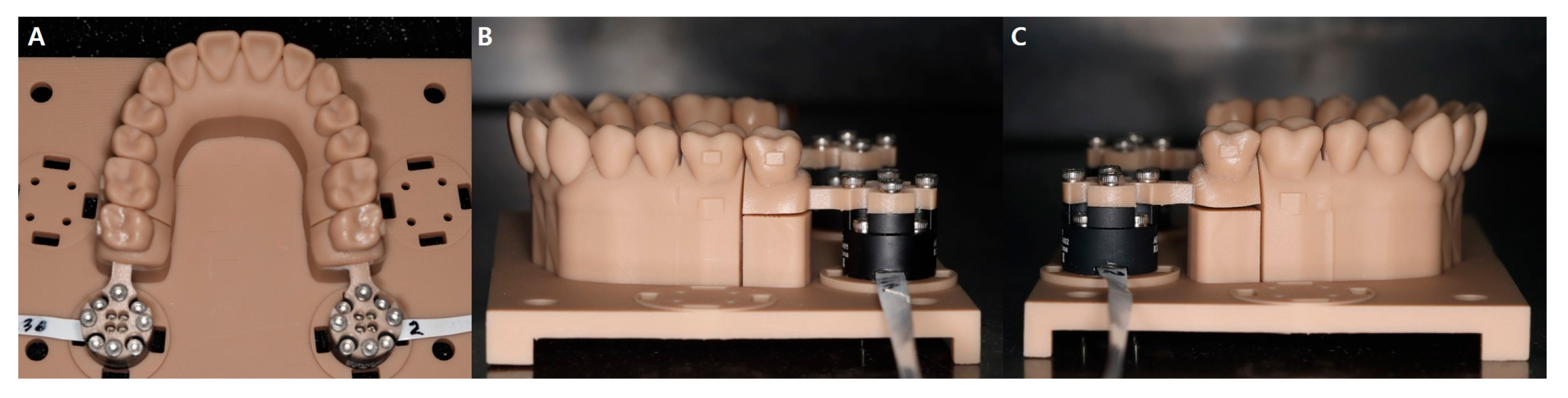
2.1. Design and Fabrication of a Force/Moment Measurement System

A 3D scanner (TRIOS 4; 3Shape, Copenhagen, Denmark) was employed to scan the Nissin dental model (NISSIN B3-305; Nissin Dental Product, Kyoto, Japan) to create a digital maxillary dentition model. This model was used to simulate the distal bodily movement of the maxillary second molar. To measure the forces and moments applied by the CA on the maxillary second molar, a six-axis miniature force/torque sensor (Aidin Robotics, Seoul, Republic of Korea) was designed to connect to the maxillary second molar, and the experimental device was 3D printed (Figure 1). Force/moment measurements were acquired by mounting the CA consisting of a thermoforming material (polyethylene terephthalate glycol; Easy-Vac; 3A MEDES, Goyang-si, Republic of Korea) onto the experimental device. The setup was maintained at 37 °C, simulating the oral cavity temperature, for 10 min in a Forced Convection Incubator (C-INDF; Changshin Science, Seoul, Republic of Korea). Average values of the measured forces and moments were recorded. Force/moment directions on different axes were differentiated using the +/− signs (Table 1). Each CA was placed on the measurement setup and left for approximately one minute until the values stabilized. The average of the stabilized force and moment readings was then recorded. The interquartile range (IQR) was also presented to indicate measurement variability. A six-axis force/torque sensor was used to measure forces and moments, with a resolution of 0.1 N for forces and 0.3 Nmm for torques, and measurement limits of ±25 N and ±150 Nmm, respectively, as specified by the manufacturer. The sensor was pre-calibrated, and all values were recorded digitally.

2.2. Design Modification and Fabrication of the Experimental CA for the Distal Bodily Movement of Maxillary Second Molars

Four CA groups with different designs were fabricated to measure the forces and moments on the maxillary second molars. The designs varied based on the presence or absence of attachments on the first molars and the addition of enhancement structures on the outer surfaces of CAs. The maxillary second molars were posteriorly moved by 0.25 mm in all groups (Figure 2). The groups were classified as follows:
  • Group 1: #6 attachment and non-enhanced structure
  • Group 2: #6 no attachment and non-enhanced structure
  • Group 3: #6 attachment and enhanced structure (addition of a direct flowable resin)
  • Group 4: #6 no attachment and enhanced structure (addition of a direct flowable resin)
All second molars and first molars in groups 1 and 3 had a vertical rectangular attachment (3.5 × 1.5 × 1 mm) digitally applied to the buccal surface of the molar, and a dental model of the applied attachment was 3D printed. From the 3D printed dental cast models, the experimental CAs were fabricated via thermoforming using 0.75 mm thick polyethylene terephthalate glycol. We calculated the sample size using a 5% significance level, a medium effect size (f = 0.25), and 90% power based on the Grant et al. study [18]. While the minimum required sample size per group was 20, we included 30 samples per group (120 total) to account for potential variability or data exclusion.
In groups with enhanced structures, a flowable resin (Charmfil; Denkist, Gunpo-si, Republic of Korea) was added to fill the embrasure between the first and second molars on the outer surface of the CA. The depth and width of the enhanced structure were based on the line connecting the most prominent areas of the first and second molars on the buccal and lingual sides from the occlusal plane, and the height was determined from the occlusal plane to the gingival margin. The enhancement procedure involved sandblasting for 10 s, followed by the application of an adhesive (3M Adper; 3M, St. Paul, MN, USA), light-curing for 20 s, and the addition of the light-curing flowable resin.

2.3. Statistical Analysis

The measured values did not exhibit a normal distribution and are represented as the medians and interquartile ranges. To compare the forces/moments applied on the maxillary second molars by different CAs, a Kruskal–Wallis test with Bonferroni correction was used for post hoc multiple comparisons based on the presence or absence of attachments and the addition of enhanced structures. All analyses were conducted using the R software (R 4.5.0 version, The R Foundation for Statistical Computing, Vienna, Austria), with statistical significance set at p < 0.05.

3. Results

This study analyzed the forces and moments in four experimental CA groups categorized by the presence or absence of attachments and reinforcement structures. Statistically significant differences in measured forces and moments were observed among all groups (p < 0.001; Table 2, Figure 3). Effect sizes (η2) were calculated for each Kruskal–Wallis test and are presented in Table 2 to indicate the magnitude of differences between groups.

3.1. Forces on Multi-Axes

Buccolingual force (Fx) was significantly lower in group 1 than in the other groups. Groups 2, 3, and 4 did not demonstrate any significant differences. Despite the addition of enhanced structures, groups 3 and 4 exhibited Fx values similar to those of group 2. Group 3 with attachments and enhanced structures exhibited a significantly higher mesiodistal force (Fy) value than the other three groups. Groups 2 and 4 without attachments displayed almost the same Fy values, regardless of the presence or absence of the enhanced structure. Interestingly, groups 4 (with enhanced structure alone) and 1 (with attachments alone) exhibited no significant differences in Fy values.
Groups 3 and 4 with enhanced structures demonstrated significantly higher intrusive force (lower vertical force [Fz]) values than those in groups 1 and 2. No statistically significant differences in Fz values were observed between groups 1 and 2. Groups 3 and 4 exhibited large negative forces in the z-axis direction, with group 4 displaying the lowest Fz value. Despite incorporating attachments, the Fz value of group 3 was similar to that of group 4 without attachments.

3.2. Moments on Multi-Axes

Significant differences in the mesiodistal angulation moment (Mx) were observed among all groups. Group 3, followed by group 4, exhibited a significantly higher Mx value than the other groups. Group 2 demonstrated a lower Mx value than group 1, indicating that the absence of attachment decreased the Mx value. However, this difference was less pronounced than the variation in moments generated by the addition of enhanced structures in groups 3 and 4. Group 3 exhibited a significantly larger lingual inclination moment (negative buccolingual inclination moment [My] value) than the other groups, which demonstrated similar My values without any significant differences.
Group 3 exhibited the highest positive mesiodistal rotation (Mz) value compared to the other groups. Group 2 generated the smallest Mz value of 0.07 Nmm, indicating minimal moment generation along the z-axis.

3.3. Mx/Fy Ration

Maintaining an appropriate distal rotation moment to mesiodistal force ratio is crucial for the effective posterior movement of second molars. Group 4 exhibited the highest Mx/Fy ratio, followed by groups 3, 1, and 2. Group 1 exhibited a slightly lower Mx/Fy ratio than the ratio in groups 3 and 4; however, the difference was not statistically significant. Group 2 without attachments and enhanced structures exhibited a significantly lower Mx/Fy ratio than the other groups.

4. Discussion

Many studies have investigated the effectiveness of maxillary molar distalization using CAs with considerably variable outcomes. Although some studies have demonstrated treatment accuracy rates of ≤87%, others have reported low accuracy rates of 36–42%, with many cases of intrusion and buccal tipping instead of the desired body movement [19,20,21]. Reciprocal forces pose a serious risk of anterior teeth retraction during molar distalization, possibly offsetting some of the achieved distalization [20]. These side effects and limitations of molar distalization with CAs are largely attributed to the lower stiffness of CAs than conventional wire-based orthodontic appliances, with less predictable biomechanics due to variable contact points with different parts of the teeth [12,13]. However, accurate bodily movement can be achieved if the elastic energy stored in CAs is efficiently transferred to the teeth.
Attachments increase the contact area between the CAs and teeth, facilitating the uniform and direct transmission of forces [21]. In this study, we also planned to place vertical attachments on the first and second molars to prevent tipping and achieve posterior bodily movement, following the literature that attachment design has a minimal effect on molar posterior movement [22]. However, excessive attachments can increase the difficulty of CA usage and feelings of foreignness [16]. The efficiency of CA treatment can be improved by varying the thickness. Studies have demonstrated a strong correlation between the CA thickness and the force transferred to the teeth [23]. However, increasing the overall thickness alters the mechanical properties of CAs, thereby affecting localized stress distribution in specific areas. Vacuum thermoforming results in non-uniform thickness across the appliances, leading to inconsistent force application [24]. This study is built upon the work of Jin X et al. [16], focusing on two specific strategies: the application of an enhanced structure (localized thickening) to the buccal areas of CAs to increase the local stiffness and the use of attachments on the first molars. We aimed to analyze the effects of these modifications on the bodily distalization of maxillary second molars.
Here, maxillary buccal tipping and intrusion trends were similar in all groups, consistent with the finite element method results reported by Mao et al. [25]. However, significant differences in force magnitude were observed between the groups with attachments and those with enhanced structures. In the mesiodistal direction (Fy), group 3 with attachments and enhanced structures exhibited the largest force. Vertical force (Fz) resulted in significant intrusive forces in groups 3 and 4 with enhanced structures, but the differences between groups with and without attachments were relatively small. These findings suggest that enhanced structures significantly affect force generation in Fy and Fz directions better than attachments, improving molar distalization, consistent with previous reports on increased forces with increased CA thickness [26]. The analysis of buccolingual forces (Fx) revealed buccolingual tipping in all groups, consistent with previous reports [20,25,27]. This is possibly due to greater force transmission anterior to the attachment on the buccal side of the second molar during distalization. These results suggest the positive effect of molar distalization on occlusion construction in Class II malocclusion treatment. Intrusive force (Fz) in CA treatment minimizes the increase in vertical height, which is a common issue observed with conventional fixed appliances and devices [28]. This intrusive force, possibly due to the CA thickness and “bite block” effect, is particularly beneficial to treat Class II malocclusions with a hyperdivergent vertical pattern.
The biomechanics of tooth movement are complex, depending on the center of resistance of the tooth and the forces acting on it [29]. In orthodontic treatment, uncontrolled tipping is the most common type of tooth movement due to the forces applied to the crown [30]. However, excessive uncontrolled tipping leads to unwanted 3D tooth movement; bodily movement or controlled tipping is desirable. Body movements require greater forces in the cervical region; therefore, thermoformed CAs are the most susceptible to deformation and irregularities in the cervical region, and the lack of stiffness limits body movements with CAs alone [21,27,30,31,32]. Although attachments are essential, they are limited in achieving bodily movements, requiring additional methods to increase stiffness (e.g., CA design modifications) [33]. In this attachment analysis, group 3 exhibited higher moments in all directions compared to other movements, requiring additional methods to increase the stiffness (e.g., CA design modifications). This suggests a synergistic effect between the attachment and enhanced structure on moment formation. The mesiodistal angulation moment (Mx) results demonstrated significantly high values in group 3, with a larger difference between groups 3 and 4 than between groups 1 and 2, indicating that the enhanced structure plays a more significant role than attachment in generating x-axis moments. The z-axis rotational moments exhibited strong buccal rotational moments in group 3, suggesting effective posterior movement of the second molars, whereas forces were relatively low in group 2, where neither attachment nor enhanced structure was applied. These findings are consistent with those of previous studies that evaluated tooth movement through aligner design modifications or thickness variations [24].
An appropriate moment-to-force ratio is crucial for minimizing uncontrolled tipping during molar distalization. Cattaneo et al. reported that a moment-to-force ratio of 10.7:1 was necessary for bodily movement in maxillary premolars, indicating that a larger moment was required compared to uncontrolled tipping movement [34]. In this study, we compared the ratio of mesiodistal angulation moment (Mx) to distal force (Fy) between groups, as this ratio is integral to efficient molar distalization. The results revealed a significantly low moment-to-force ratio in group 2, without attachment and enhanced structures. This finding suggests a high likelihood of uncontrolled tipping in this group. These findings underscore the importance of CA with a firm grasp on the teeth, either through attachment or an enhanced structure, to achieve effective molar distalization. Although IQRs were relatively wide for certain measurements (e.g., Fz in groups 3 and 4), effect size values were additionally calculated and included in the results to enhance interpretability. These values support the statistical significance and help to contextualize the biomechanical differences among groups, even in the presence of data variability.
This study identified that increased local CA thickness improved molar distalization when combined with attachments. Different CA fabrication methods have many limitations. Thermoforming leads to uneven thicknesses during fabrication. Direct 3D printing requires additional design, and the fabrication of new CAs for treatment is cumbersome. However, the enhanced structural method using a flowable resin proposed in this study offers several advantages. First, the technique is easily applicable in clinical settings, thereby reducing chair time. Second, the method facilitates easy adjustments during the treatment period, eliminating the need for new device fabrication. This approach, allowing adjustments without the need for new aligners, also enhances the patient’s comfort and convenience. These findings may offer practical value in clinical situations where attachment bonding is difficult, such as in patients with short clinical crowns or partially erupted molars. Incorporating reinforced designs could supplement conventional protocols, aiding clinicians in achieving efficient molar distalization under challenging conditions.
This study has several limitations. First, only the initial forces/moments applied by CAs on the teeth were measured and compared, without considering changes in orthodontic forces over time. Because measurements were obtained at a single time point (after 10 min at 37 °C), the time-dependent characteristics of force decay and potential plastic deformation of thermoformed CAs could not be captured. Second, while clinical orthodontic treatment involves complex scenarios with simultaneous movements of multiple teeth, this study focused solely on forces/moments on maxillary molars. Third, although the experimental setup maintained 37 °C to simulate intraoral temperature, the oral environment could not be fully reproduced. The in vitro setup used a static environment without incorporating functional loading or dynamic masticatory forces, which may limit the translation of these findings to clinical scenarios. Specifically, factors such as humidity, saliva, friction between CAs and teeth, occlusal forces from masticatory muscles, and biological responses including microbial and gingival interaction with the material were not incorporated. Furthermore, the experimental design simplified the dentition by isolating only the second molar, without including adjacent teeth or opposing occlusion, omitting key anatomical and functional interactions that affect force distribution and tooth movement. The exclusion of salivary flow, biofilm formation, and other biological responses also reduces the clinical relevance of the model.
Moreover, while the proposed chairside resin enhancement method showed biomechanical efficacy in vitro, its clinical practicality remains to be validated. This study did not assess patient comfort, the durability of CA materials after repeated resin modifications, or the feasibility of repeated resin applications throughout the course of treatment. Future research involving dynamic force measurements would provide valuable insight into how aligner materials behave under prolonged intraoral conditions, and studies should aim to incorporate these intraoral conditions to more closely simulate the clinical environment. Furthermore, although the enhanced structural modifications demonstrated improved biomechanical performance in vitro, their long-term clinical effectiveness and applicability across diverse patient anatomies remain uncertain. Factors such as patient compliance, intraoral variations, and long-term material behavior could affect real-world outcomes and should be addressed in future clinical studies.

5. Conclusions

This study analyzed the effectiveness of a combination of attachment and local reinforcing structures on molar distalization with CA. The main conclusions were
  • Distal, intrusive forces are generated during the posterior movement of the second molar.
  • The incorporation of local enhancing structures increases the molar distal force, moment, and moment-to-force ratio, which increases the tendency of the second molar posterior bodily movement.
  • The localized structural strengthening method proposed in this study is a practical approach that can be easily implemented for CA treatment in clinical practice, especially when bonding attachments are not feasible.
  • However, the conclusions should be interpreted with caution, as this in vitro study does not fully simulate the dynamic intraoral environment, including adjacent tooth interactions and biological factors such as saliva and occlusal forces.
  • Future clinical research is necessary to evaluate the long-term effectiveness, material durability, and patient comfort of this technique under realistic treatment conditions.

Author Contributions

Y.-K.C.: conceptualization; methodology; investigation; writing—original draft; writing—review and editing. M.-J.J., S.-H.K., S.-S.K. and S.-B.P.: writing—original draft; writing—review and editing. Y.-I.K.: conceptualization; methodology; writing—original draft; writing—review and editing. All authors have read and agreed to the published version of the manuscript.

Funding

This study was supported by the Busan-Gyeongnam-Ulsan branch of the Korean Association of Orthodontics.

Institutional Review Board Statement

Not applicable.

Informed Consent Statement

Not applicable.

Data Availability Statement

The data supporting the findings of this study will be available from the corresponding author upon reasonable request.

Conflicts of Interest

The authors declare no conflicts of interest or financial interest.

Abbreviations

The following abbreviation is used in this manuscript:
CAClear aligner

References

  1. Weir, T. Clear aligners in orthodontic treatment. Aust. Dent. J. 2017, 62, 58–62. [Google Scholar] [CrossRef] [PubMed]
  2. Kravitz, N.D.; Kusnoto, B.; BeGole, E.; Obrez, A.; Agran, B. How well does Invisalign work? A prospective clinical study evaluating the efficacy of tooth movement with Invisalign. Am. J. Orthod. Dentofac. Orthop. 2009, 135, 27–35. [Google Scholar] [CrossRef]
  3. Lombardo, L.; Arreghini, A.; Ramina, F.; Ghislanzoni, L.T.; Siciliani, G. Predictability of orthodontic movement with orthodontic aligners: A retrospective study. Prog. Orthod. 2017, 18, 35. [Google Scholar] [CrossRef] [PubMed]
  4. Galan-Lopez, L.; Barcia-Gonzalez, J.; Plasencia, E. A systematic review of the accuracy and efficiency of dental movements with Invisalign®. Korean J. Orthod. 2019, 49, 140–149. [Google Scholar] [CrossRef]
  5. Jedliński, M.; Mazur, M.; Greco, M.; Belfus, J.; Grocholewicz, K.; Janiszewska-Olszowska, J. Attachments for the orthodontic aligner treatment-state of the art-a comprehensive systematic review. Int. J. Environ. Res. Public. Health. 2023, 20, 4481. [Google Scholar] [CrossRef]
  6. Tepedino, M.; Paoloni, V.; Cozza, P.; Chimenti, C. Movement of anterior teeth using clear aligners: A three-dimensional, retrospective evaluation. Prog. Orthod. 2018, 19, 9. [Google Scholar] [CrossRef]
  7. Bechtold, T.E.; Park, Y.C.; Kim, K.H.; Jung, H.K.; Kang, J.Y.; Choi, Y.J. Long-term stability of miniscrew anchored maxillary molar distalization in Class II treatment. Angle Orthod. 2020, 90, 362–368. [Google Scholar] [CrossRef]
  8. D′Antò, V.; Valletta, R.; Ferretti, R.; Bucci, R.; Kirlis, R.; Rongo, R. Predictability of maxillary molar distalization and derotation with clear aligners: A prospective study. Int. J. Environ. Res. Public Health 2023, 20, 2941. [Google Scholar] [CrossRef] [PubMed]
  9. Ravera, S.; Castroflorio, T.; Garino, F.; Daher, S.; Cugliari, G.; Deregibus, A. Maxillary molar distalization with aligners in adult patients: A multicenter retrospective study. Prog. Orthod. 2016, 17, 12. [Google Scholar] [CrossRef]
  10. Caruso, S.; Nota, A.; Ehsani, S.; Maddalone, E.; Ojima, K.; Tecco, S. Impact of molar teeth distalization with clear aligners on occlusal vertical dimension: A retrospective study. BMC Oral Health 2019, 19, 182. [Google Scholar] [CrossRef]
  11. Miao, Z.; Zhang, H.; Yang, Y.; Han, Y.; Leng, J.; Wang, S. Influence of maxillary molar distalization with clear aligners on three-dimensional direction: Molar distal movement, intrusion, distal tip and crown buccal torque. Prog. Orthod. 2023, 24, 48. [Google Scholar] [CrossRef] [PubMed]
  12. Baldwin, D.K.; King, G.; Ramsay, D.S.; Huang, G.; Bollen, A.M. Activation time and material stiffness of sequential removable orthodontic appliances. Part 3: Premolar extraction patients. Am. J. Orthod. Dentofac. Orthop. 2008, 133, 837–845. [Google Scholar] [CrossRef]
  13. Bichu, Y.M.; Alwaf, A.; Liu, X.; Andrews, J.; Ludwig, B.; Bichu, A.Y.; Zou, B. Advances in orthodontic clear aligner materials. Bioact. Mater. 2022, 22, 384–403. [Google Scholar] [CrossRef]
  14. Cui, J.Y.; Ting, L.; Cao, Y.X.; Sun, D.X.; Bing, L.; Wu, X.P. Morphology changes of maxillary molar distalization by clear aligner therapy. Int. J. Morphol. 2022, 40, 920–926. [Google Scholar] [CrossRef]
  15. Fang, D.; Li, F.; Zhang, Y.; Bai, Y.; Wu, B.M. Changes in mechanical properties, surface morphology, structure, and composition of Invisalign material in the oral environment. Am. J. Orthod. Dentofac. Orthop. 2020, 157, 745–753. [Google Scholar] [CrossRef]
  16. Jin, X.; Tian, X.; Hui, V.L.Z.; Zheng, Y.; Song, J.; Han, X. The effect of enhanced structure in the posterior segment of clear aligners during anterior retraction: A three-dimensional finite element and experimental model analysis. Prog. Orthod. 2024, 25, 3. [Google Scholar] [CrossRef]
  17. Zhu, Y.; Hu, W.; Li, S. Force changes associated with differential activation of en-masse retraction and/or intrusion with clear aligners. Korean J. Orthod. 2021, 51, 32–42. [Google Scholar] [CrossRef] [PubMed]
  18. Grant, J.; Foley, P.; Bankhead, B.; Miranda, G.; Adel, S.M.; Kim, K.B. Forces and moments generated by 3D direct printed clear aligners of varying labial and lingual thicknesses during lingual movement of maxillary central incisor: An in vitro study. Prog. Orthod. 2023, 24, 23. [Google Scholar] [CrossRef] [PubMed]
  19. Simon, M.; Keilig, L.; Schwarze, J.; Jung, B.A.; Bourauel, C. Treatment outcome and efficacy of an aligner technique—regarding incisor torque, premolar derotation and molar distalization. BMC Oral Health 2014, 14, 68. [Google Scholar] [CrossRef]
  20. Li, L.; Guo, R.; Zhang, L.; Huang, Y.; Jia, Y.; Li, W. Maxillary molar distalization with a 2-week clear aligner protocol in patients with Class II malocclusion: A retrospective study. Am. J. Orthod. Dentofac. Orthop. 2023, 164, 123–130. [Google Scholar] [CrossRef]
  21. Upadhyay, M.; Arqub, S.A. Biomechanics of clear aligners: Hidden truths & first principles. J. World Fed. Orthod. 2022, 11, 12–21. [Google Scholar] [PubMed]
  22. Gao, J.; Guo, D.; Zhang, X.; Cheng, Y.; Zhang, H.; Xu, Y.; Jin, Z.; Ma, Y. Biomechanical effects of different staging and attachment designs in maxillary molar distalization with clear aligner: A finite element study. Prog. Orthod. 2023, 24, 43. [Google Scholar] [CrossRef]
  23. Kohda, N.; Iijima, M.; Muguruma, T.; Brantley, W.A.; Ahluwalia, K.S.; Mizoguchi, I. Effects of mechanical properties of thermoplastic materials on the initial force of thermoplastic appliances. Angle Orthod. 2013, 83, 476–483. [Google Scholar] [CrossRef]
  24. Palone, M.; Longo, M.; Arveda, N.; Nacucchi, M.; Pascalis, F.; Spedicato, G.A.; Siciliani, G.; Lombardo, L. Micro-computed tomography evaluation of general trends in aligner thickness and gap width after thermoforming procedures involving six commercial clear aligners: An in vitro study. Korean J. Orthod. 2021, 51, 135–141. [Google Scholar] [CrossRef] [PubMed]
  25. Mao, B.; Tian, Y.; Xiao, Y.; Li, J.; Zhou, Y. The effect of maxillary molar distalization with clear aligner: A 4D finite-element study with staging simulation. Prog. Orthod. 2023, 24, 16. [Google Scholar] [CrossRef]
  26. Li, N.; Wang, C.; Yang, M.; Chen, D.; Yang, M.; Li, D.; Qui, S.; Chen, Q.; Feng, Y. Effects of different tooth movement patterns and aligner thicknesses on maxillary arch expansion with clear aligners: A three-dimensional finite element study. Front. Bioeng. Biotechnol. 2024, 12, 1424319. [Google Scholar] [CrossRef] [PubMed]
  27. Guo, R.; Lam, X.Y.; Zhang, L.; Li, W.; Lin, Y. Biomechanical analysis of miniscrew-assisted molar distalization with clear aligners: A three-dimensional finite element study. Eur. J. Orthod. 2024, 46, 1–9. [Google Scholar] [CrossRef]
  28. Al-Thomali, Y.; Basha, S.; Mohamed, R.N. Pendulum and modified pendulum appliances for maxillary molar distalization in Class II malocclusion—A systematic review. Acta Odontol. Scand. 2017, 75, 394–401. [Google Scholar] [CrossRef]
  29. Nanda, R.S.; Tosun, Y.S. Biomechanics in Orthodontics: Principles and Practice; Quintessence Publishing Co.: Hanover Park, IL, USA, 2010. [Google Scholar]
  30. Yassir, Y.A.; Nabbat, S.A.; McIntyre, G.T.; Bearn, D.R. Clinical effectiveness of clear aligner treatment compared to fixed appliance treatment: An overview of systematic reviews. Clin. Oral Investig. 2022, 26, 2353–2370. [Google Scholar] [CrossRef]
  31. Elshazly, T.M.; Salvatori, D.; Elattar, H.; Bourauel, C.; Keilig, L. Effect of trimming line design and edge extension of orthodontic aligners on force transmission: A 3D finite element study. J. Mech. Behav. Biomed. Mater. 2023, 140, 105741. [Google Scholar] [CrossRef]
  32. Elshazly, T.M.; Keilig, L.; Salvatori, D.; Chavanne, P.; Aldesoki, M.; Bourauel, C. Effect of trimming line design and edge extension of orthodontic aligners on force transmission: An in vitro study. J. Dent. 2022, 125, 104276. [Google Scholar] [CrossRef] [PubMed]
  33. Rossini, G.; Schiaffino, M.; Parrini, S.; Sedran, A.; Deregibus, A.; Castroflorio, T. Upper second molar distalization with clear aligners: A finite element study. Appl. Sci. 2020, 10, 7739. [Google Scholar] [CrossRef]
  34. Cattaneo, P.M.; Dalstra, M.; Melsen, B. Moment-to-force ratio, center of rotation, and force level: A finite element study predicting their interdependency for simulated orthodontic loading regimens. Am. J. Orthod. Dentofac. Orthop. 2008, 133, 681–689. [Google Scholar] [CrossRef] [PubMed]
Figure 1. Experimental device design. (A) Maxillary right and left second molars are connected to a six-axis miniature force/torque sensor. (B,C) The maxillary right first molar exhibits a vertical rectangular attachment on the buccal surface, whereas the left first molar did not exhibit the attachment.
Applsci 15 06655 g001
Figure 2. Clear aligners design. (A,B) Occlusal view: (B) clear aligners with enhanced structures (addition of a direct flowable resin). (C,D) Sagittal view: clear aligners with non-enhanced structures. (E,F) Sagittal view: clear aligners with enhanced structures (addition of a direct flowable resin).
Applsci 15 06655 g002
Figure 3. Forces, moments, and the Mx/Fy ratio generated by different clear aligner groups. (A) Fx (N) values; (B) Fy (N) values; (C) Fz (N) values; (D) Mx (Nmm) values; (E) My (Nmm) values; (F) Mz (Nmm) values; (G) Mx/Fy ratio. Fx (+), buccal force; Fy (+), mesial force; Fz (+), extrusive force; Mx, mesiodistal angulation moment; My, buccolingual inclination moment; Mz, rotation moment.
Applsci 15 06655 g003
Table 1. Sign convention of force/moments measurement system.
ComponentDefinitionSign Convention
Force (X)Buccolingual(+) Buccal(−) Lingual
Force (Y)Mesiodistal(+) Distal(−) Mesial
Force (Z)Occlusogingival(+) Occlusal(−) Gingival
Moment (X)Angulation(+) Mesial tipping(−) Distal tipping
Moment (Y)Inclination(+) Buccal(−) Lingual
Moment (Z)Rotation(+) Distal rotation(−) Mesial rotation
Table 2. Comparative analysis of the forces and moments generated by different clear aligner groups.
MeasurementsGroup 1
(Median, IQR)
Group 2
(Median, IQR)
Group 3
(Median, IQR)
Group 4
(Median, IQR)
p-ValueEffect Size
2)
Fx (N)0.31, 0.30 a0.57, 0.13 b0.56, 0.26 b0.46, 0.34 bH = 30.086
<0.001 ***
0.284
Fy2.01, 1.80 a1.56, 1.01 a3.11, 0.93 b1.83, 0.98 aH = 46.654
<0.001 ***
0.194
Fz−0.60, 0.32 a−0.88, 0.66 a−1.23, 0.57 b−1.33, 0.35 bH = 59.582
<0.001 ***
0.088
Mx (Nmm)12.38, 6.76 a10.27, 7.58 b20.83, 4.74 c14.50, 5.36 dH = 65.121
<0.001 ***
0.536
My−3.23, 2.95 a−2.92, 2.55 a−6.06, 2.52 b−2.97, 2.28 aH = 24.937
<0.001 ***
0.337
Mz1.02, 2.51 a0.07, 1.78 a,c3.70, 3.99 b−0.57, 2.11 cH = 43.935
<0.001 ***
0.238
Mx/Fy ratio6.21, 3.55 a5.77, 1.72 b6.73, 0.98 a7.02, 2.10 aH = 10.807
0.003 **
0.319
Superscript letters indicate a significant difference (p < 0.05). ** Statistically different at p < 0.01, *** Statistically different at p < 0.001. The Kruskal–Wallis test with Bonferroni correction was used for post hoc multiple comparisons. Abbreviations: IQR, interquartile range; Fx (+), buccal force; Fy (+), mesial force; Fz (+), extrusive force; Mx, mesiodistal angulation moment; My, buccolingual inclination moment; Mz, rotation moment.
Disclaimer/Publisher’s Note: The statements, opinions and data contained in all publications are solely those of the individual author(s) and contributor(s) and not of MDPI and/or the editor(s). MDPI and/or the editor(s) disclaim responsibility for any injury to people or property resulting from any ideas, methods, instructions or products referred to in the content.

