Article

Comparison of the Fracture Resistance of Provisional Crowns and Fixed Partial Dentures Manufactured with Conventional, Milling, and 3D-Printing Techniques

by
Beyza Güney
*,
Asude Dilek Nalbant
and
Merve Bankoğlu Güngör
Department of Prosthodontics, Faculty of Dentistry, Gazi University, 06490 Ankara, Türkiye
*
Author to whom correspondence should be addressed.
Appl. Sci. 2025, 15(12), 6539; https://doi.org/10.3390/app15126539
Submission received: 3 May 2025 / Revised: 4 June 2025 / Accepted: 6 June 2025 / Published: 10 June 2025

Abstract

This study aimed to evaluate the effect of different manufacturing techniques and thermal aging on the fracture resistance of provisional crowns and fixed partial dentures. Methods: A total of 60 provisional crowns and 60 provisional fixed partial dentures were fabricated using three manufacturing techniques: conventional manufacturing (CM), subtractive manufacturing (SM), and additive manufacturing (AM). An index created from SM-manufactured restorations was used to produce the CM group. Artificial abutments were created by duplicating scanned phantom teeth using model resin. Half of the restorations (n = 10 per group) were subjected to thermal aging (5–55 °C, 5000 cycles). The fracture resistance values of the specimens were tested using a universal testing machine. Data were analyzed using a two-way ANOVA and Tukey’s post hoc tests (α = 0.05). The highest mean fracture resistance was observed in the SM group without aging, both for crowns (1645.4 ± 346.8 N) and fixed partial dentures (1291.13 ± 564.15 N). The two-way ANOVA revealed statistically significant differences among the groups, and thermal aging significantly reduced the fracture resistance (p < 0.05). Both the manufacturing method and thermal aging significantly influenced the fracture resistance of provisional crowns and fixed partial dentures. In fixed partial dentures, a significant effect of aging was associated with the reduced durability of restorations fabricated using the subtractive manufacturing method.

1. Introduction

Provisional crowns and fixed partial dentures are crucial in fixed prosthodontics, protecting pulpal and periodontal tissues, supporting tissue healing for an ideal emergence profile, and preventing abutment migration by preserving proper spacing. Furthermore, they help assess oral hygiene by enabling the evaluation of plaque accumulation and soft tissue response over time, while also contributing to establishing a stable occlusal relationship [1]. In addition to their protective and functional roles, provisional restorations play a key role in the diagnostic and functional assessment of the final prosthesis, enhancing the predictability of treatment outcomes. These temporary restorations must fulfill biological, mechanical, and esthetic criteria for clinical success. Their durability under functional loads, occlusal forces, and intraoral stresses is particularly crucial. Due to their exposure to temperature fluctuations, high humidity, and continuous loading, provisional restorations require sufficient mechanical strength to resist fractures and minimize the necessity for further clinical adjustments or repairs [2]. This is especially critical in clinical situations requiring long-term provisionalization, such as dental implant therapy or full-mouth rehabilitation involving extensive occlusal adjustments. In these cases, provisional restorations may be in place for extended durations, typically between 6 and 12 weeks or longer. Consequently, understanding the fracture resistance of the materials used for provisional restorations is essential. The fabrication technique and material composition significantly determine fracture resistance and clinical performance [3,4].
Various provisional materials are utilized in clinical treatments, with polymethyl methacrylate (PMMA) historically being the most widely used due to its favorable strength, esthetics, color stability, and ease of repair. However, conventional PMMA-based materials also present drawbacks, such as polymerization shrinkage, a high exothermic reaction, limited wear resistance, potential pulpal irritation from residual monomers, and a strong odor. Bis-acryl composite resins have emerged as an alternative to address these limitations, offering enhanced polishability and color stability, although they tend to exhibit lower fracture resistance than PMMA [5,6]. With the evolution of digital dentistry, computer-aided design and computer-aided manufacturing (CAD-CAM)-based additive and subtractive techniques are progressively replacing traditional methods, offering benefits such as superior precision, improved internal fit, and minimized marginal discrepancies. Moreover, CAD-CAM technology enables the swift reproduction of restorations in the event of fracture or loss, enhancing efficiency and standardization in clinical practice [7,8].
Restorations produced through subtractive manufacturing provide a more efficient workflow and excellent durability compared to conventional methods. The adoption of CAD-CAM techniques, particularly the milling of pre-polymerized PMMA disks, has notably enhanced the mechanical properties of provisional restorations. These high-pressure polymerized materials feature a highly cross-linked structure, leading to superior strength and wear resistance compared to chairside-fabricated alternatives. However, despite these benefits, challenges such as high costs, material waste, the potential for microcracks, and limitations in surface detail continue to hinder their widespread adoption. Therefore, a comprehensive evaluation of various materials is crucial to assess their long-term viability in clinical applications [9,10]. As an alternative, additive manufacturing has gained prominence in prosthodontics due to its numerous advantages, including excellent fit, high detail reproducibility, cost-effectiveness, efficiency, speed, accuracy, precise adaptability, uniform contact pressure, and favorable mechanical properties [11]. This technology is especially advantageous for producing restorations with intricate geometries or undercuts, where conventional manufacturing methods may be less effective. Three-dimensional printing, a significant innovation in additive manufacturing, enables the layer-by-layer fabrication of restorations using techniques such as fused deposition modeling (FDM), digital light processing (DLP), and stereolithography (SLA). DLP is particularly notable for its speed and precision, making it well-suited for dental applications. This technique allows for the production of highly detailed and durable prostheses without relying on traditional impression materials or extensive manual processing, thereby enhancing efficiency in clinical practice [12]. Despite these advantages, research on the mechanical properties of 3D-printed materials has yielded inconsistent and sometimes contradictory findings [13,14,15]. The mechanical behavior of 3D-printed restorations is affected by several parameters, including raw material composition, light-curing conditions, layer thickness, laser type and efficiency, printing orientation, and post-processing procedures, all of which significantly influence the final restoration’s mechanical properties [16].
Provisional restorative materials must retain their mechanical properties throughout their clinical lifespan, as exposure to intraoral conditions can significantly affect their performance. The storage conditions of these materials have been shown to impact their mechanical properties, particularly water absorption. PMMAs are susceptible to water uptake due to their linear polymer structure and polarity, making them prone to molecular infiltration. Additionally, the air bubbles introduced during manual mixing can further increase the susceptibility of chairside-fabricated PMMAs to water absorption, potentially leading to hydrolysis and a decline in mechanical properties. In contrast, dimethacrylate-based resins, with their highly cross-linked structure, demonstrate lower water absorption and are less prone to air entrapment when dispensed from cartridges. However, despite their improved resistance, dimethacrylates still absorb water over time, which can cause them to weaken and become more brittle after prolonged exposure to humid environments [5,7].
Several studies [13,14,17,18] have compared additive manufacturing techniques with subtractive and conventional manufacturing techniques in terms of the physical and mechanical properties of provisional crown and fixed partial denture materials. Research on fracture resistance has examined both aged and non-aged crowns and fixed partial dentures. However, there is a lack of studies investigating the impact of aging on restorations fabricated using different manufacturing techniques. Therefore, this study aims to evaluate this effect on crowns and fixed partial dentures. The purpose of this in vitro study was to compare the fracture resistance of provisional crowns and fixed partial dentures manufactured using three different techniques: conventional manufacturing (CM), subtractive manufacturing (SM), and additive manufacturing (AM). Additionally, the effect of thermal aging on fracture resistance was evaluated. The null hypothesis was that the manufacturing technique and thermal aging process would not significantly affect the fracture resistance of the provisional restorations.

2. Materials and Methods

In this in vitro study, the fracture resistances of provisional crowns and fixed partial dentures manufactured with different techniques, whether subjected to thermal cycling or not, were investigated.

2.1. Sample Size Calculation

A priori power analysis was conducted using G*Power (version 3.1.9.4) to determine the required sample size. Separate analyses were performed for crown and fixed partial dentures using a two-way ANOVA. With a significance level of α = 0.05, a power of 0.90, and an effect size (f) of 0.50, the minimum required sample size per group was calculated as 54. Thus, 120 specimens were included in the study (n = 10 per subgroup).

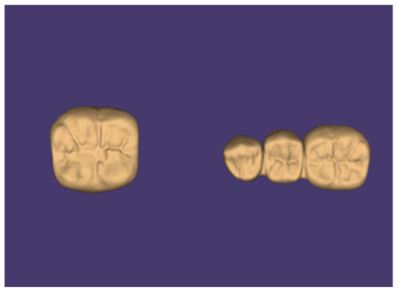
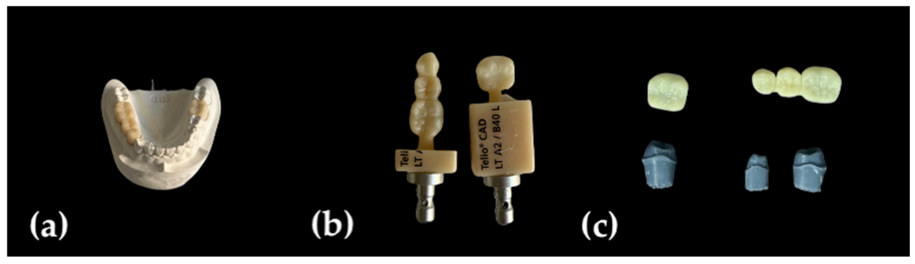
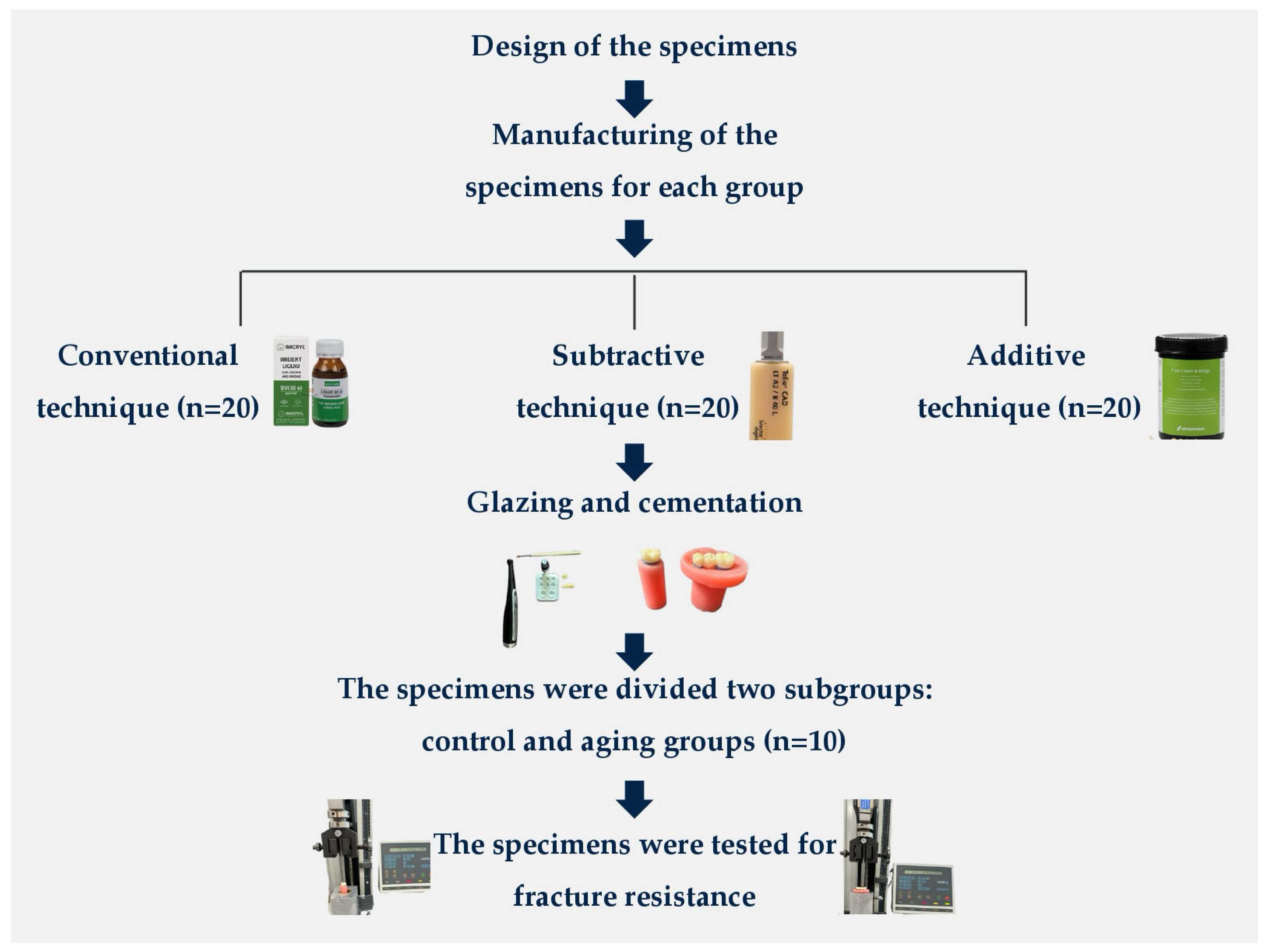
2.2. Specimen Preparation

Models were prepared using typodont teeth positioned in regions 36, 44, and 46, and Type III dental stone (Cerestone, İstanbul, Türkiye) was employed to create the models. The tooth at position 45 was removed from the gypsum model to mimic a clinical situation with a missing tooth. The gypsum study model and opposing models were mounted on a semi-adjustable articulator (Stratos 200, Ivoclar Vivadent, Schaan, Liechtenstein). The same operator prepared the teeth in positions 36, 44, and 46. The preparation included 1.5 mm of occlusal reduction, 1 mm of axial reduction, and 1 mm of chamfer finishing line at 1 mm above the cement–enamel junction, with a 6-degree convergence angle.
Three experimental groups were generated according to the manufacturing techniques for the provisional restorations (n = 20 for each type of production technique and restoration). The materials and their production techniques are presented in Table 1.
Group 1: Conventionally manufactured group (CM)
To simulate conventional manufacturing, one each of the milled crowns and fixed partial dentures were used as reference restorations. Impressions of these restorations were taken using a hand-mixed condensation silicone material (Oxasil, Kulzer GmbH, Hanau, Germany; lot number: K010162) with a double-mix technique to create a putty matrix. PMMA acrylic resin (Imicryl, Imident, Konya, Türkiye) in powder and liquid form was mixed for 1 min with a set powder/liquid ratio (2.4:1), following the manufacturer’s instructions, and applied to the putty matrix of the prepared abutments to fabricate crowns and fixed partial dentures (n = 20).
Group 2: Subtractively manufactured group (SM)
The prepared teeth (36, 44, and 46) and models were scanned separately using a lab scanner (Dental Wings 7 Series, Straumann, Basel, Switzerland) to reproduce the subtractive manufacturing method. Crown and fixed partial denture designs were created in a CAD program (Exocad DentalDB 2.4 Plovdiv, Darmstadt, Germany), allowing 1.5 mm occlusal and 1 mm marginal axial wall thickness. The connector cross-sectional area for the fixed partial dentures was designed to be 16 mm2. The digital designs are shown in Figure 1.
The cement space was set to 50 μm. To reproduce the subtractive manufacturing method, crowns and fixed partial dentures were fabricated with PMMA blocks (Telio CAD, Ivoclar Vivadent, Schaan, Liechtenstein) using a 5-axis milling machine (Redon Hybrid, Redon Group, İstanbul, Türkiye).
Group 3: Additively manufactured group (AM)
To reproduce the additive manufacturing method, crowns and fixed partial dentures were fabricated using methacrylate resin (P pro Crown & Bridge, Straumann AG, Basel, Switzerland). Slicing software (Netfabb 2023, Autodesk, San Francisco, CA, USA) was used to process the standard tessellation language (STL) file for 3D printing and to set the printing parameters as recommended by the provisional resin manufacturer. The printer (P30 DLP device, Rapid Shape, Heimsheim, Germany) settings were as follows: layer thickness of 50 µm, wavelength of 385 nm, resin color of A2, and degree orientation of 0°. After printing, the specimens were subjected to post-processing procedures. The specimens were washed in 99% isopropanol (P wash, Straumann Cares, Basel, Switzerland) for 10 min, followed by UV curing using the P cure (Straumann Cares, Basel, Switzerland), which utilizes an LED light source with a wavelength range of 385–405 nm and automatically evacuates oxygen via a vacuum to prevent the formation of an inhibition layer during curing. The resin type was selected from the device menu, and the curing process was performed automatically with a duration of 10 min. Subsequently, the specimens were cleaned with isopropyl alcohol and a brush for 2 min, dried with compressed air, and left to dry at room temperature for 30 min.
The manufactured crowns and FPDs for each manufacturing technique are shown in Figure 2.
The 36, 44, and 46 teeth (Frasaco, Tettnang, Germany) were scanned, and the dies were printed with digital light processing (P30 DLP device, Rapid Shape, Heimsheim, Germany) using model resin (P pro Master Model Grey, Straumann, Basel, Switzerland) for testing the fracture resistance of temporary restorations. A total of 60 3D-printed resin dies were created for each tooth. A surface sealant agent (Optiglaze, GC Dental, Tokyo, Japan) was applied to the temporary restorations using a soft brush in a thin, even layer in one direction, ensuring that no air bubbles were formed. Polymerization was completed with an LED light-curing device (Valo Cordless, Ultradent Products Inc., South Jordan, UT, USA) for 40 s, used according to the manufacturer’s instructions. The base and catalyst components of the cement were mixed for 30 s. All restorations were cemented onto the 3D-printed dies using temporary cement (Cavex, Harleem, The Netherlands) under 50 N of constant pressure from a metal pendulum for 6 min until a complete set in temporary cement was obtained. Excess cement was then removed with a dental probe. Each restoration was embedded into acrylic resin blocks to create testing specimens.

2.3. Aging and Fracture Resistance Test

The specimens were divided into control and aging groups (n = 10 each). The flowchart of the study is summarized in Figure 3.
Specimens being subjected to aging were placed in a thermal cycling device (Thermocycler SD Mechatronik, Fareham, UK) for 5000 cycles, alternating between water baths at 5 °C and 55 °C. After aging, the fracture resistance values of the specimens were determined under in vitro conditions. For the fixed partial dentures, a vertical load was applied at the central fossa of the pontic. For crowns, the load was applied at the central fossa. A universal testing machine (Lloyd LRX, Lloyd Instruments, Fareham, UK) with a crosshead speed of 1 mm/min and a tip diameter of 6 mm was used. The load at fracture values, measured in newtons (N), and the fracture displacement prior to rupture were recorded using Nexygen version 3.0 data analysis software.

2.4. Statistical Analysis

Data analysis was performed using IBM SPSS version 23.0, JASP ver. 0.12, and RStudio (ver. 2021.09.0). Descriptive statistics were evaluated, including mean ± standard deviation, median, interquartile range (Q1 and Q3), minimum, and maximum values. The effects of material type and aging conditions on fracture load measurements were analyzed using a two-way factorial ANOVA. Assumptions of multivariate normality and homogeneity of variances were checked using the “mvn” library in RStudio, as well as Levene’s test and the Fligner–Killeen test. Tukey post hoc tests were used for pairwise comparisons when significant differences were found. Statistical significance was set at p < 0.05.

3. Results

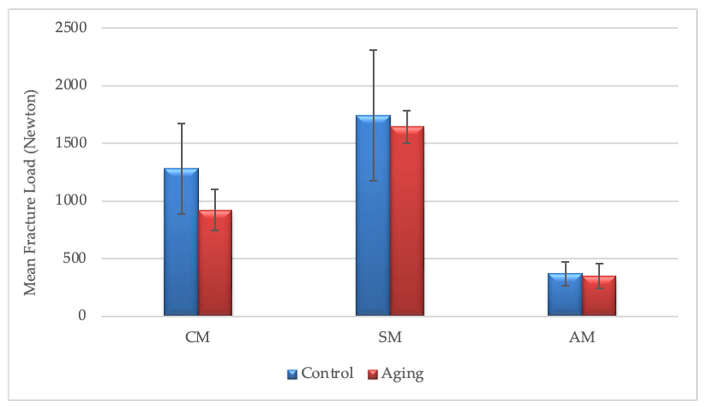
3.1. Fracture Resistance Values of the Crown Groups

The mean fracture resistance values for provisional crowns were found to be 1102.25 ± 379.76 N for conventional manufacturing (n = 20), 1693.62 ± 280.17 N for subtractive manufacturing (n = 20), and 360.08 ± 107.53 N for additive manufacturing (n = 20). The mean fracture resistance was 973.34 ± 603.51 N for aged specimens (n = 30) and 1130.62 ± 626.34 N for non-aged specimens (n = 30).
According to the results of the two-way ANOVA, the effects of different manufacturing techniques (p < 0.001) and aging (p = 0.025) on fracture resistance values were statistically significant. However, no interaction was observed between the manufacturing technique and aging factors (p = 0.116) (Table 2).
Descriptive and comparative statistics of the fracture resistance values for aged and non-aged crown groups are presented in Table 3 and illustrated in Figure 4 (n = 10). Tukey’s multiple comparison test showed statistically significant differences in fracture resistance between groups CM and SM (p < 0.001), CM and AM (p < 0.001), and SM and AM (p < 0.001).

3.2. Fracture Resistance Values of the Fixed Partial Denture Groups

The mean fracture resistance values for provisional fixed partial dentures were found to be 640.33 ± 130.63 N for conventional manufacturing (n = 20), 926.38 ± 547.83 N for subtractive manufacturing (n = 20), and 371.08 ± 102.86 N for additive manufacturing (n = 20).
The mean fracture resistance was 506.66 ± 147.89 N for aged specimens (n = 30) and 785.20 ± 509.02 N for non-aged specimens (n = 30).
According to the results of the two-way ANOVA, the effects of different manufacturing techniques (p < 0.001) and aging (p < 0.001) on fracture resistance values were statistically significant. Additionally, a significant interaction was observed between the manufacturing technique and aging factors (p < 0.001) (Table 4).
Descriptive and comparative statistics of the fracture resistance values for aged and non-aged fixed partial denture groups are presented in Table 5 and illustrated in Figure 5 (n = 10). Tukey’s multiple comparison test showed statistically significant differences in fracture resistance between the groups of CM and SM (p = 0.002), CM and AM (p = 0.004), and SM and AM (p < 0.001).
The Tukey multiple comparison test results for the combined effect of the manufacturing method and aging application revealed that aging significantly affected subtractively manufactured FPDs, as aged restorations exhibited significantly different fracture resistance values from their non-aged counterparts (p < 0.001). However, no statistically significant differences were found between aged and non-aged FPDs fabricated using additive methods (p = 1.000) and conventional methods (p = 0.925). Among aged FPDs, no significant differences were observed between any of the manufacturing methods. In contrast, for non-aged FPDs, subtractive manufacturing demonstrated significantly different fracture load values compared to both conventional (p < 0.001) and additive methods (p < 0.001), while no significant difference was found between conventional and additive FPDs (p = 0.062).

4. Discussion

Based on the results of this in vitro study, thermal aging and manufacturing techniques influence the fracture resistance of temporary crowns and fixed partial dentures, leading to the rejection of the null hypothesis.
Provisional restorations must withstand daily oral conditions and occlusal forces for a specific period, especially during full-mouth rehabilitation or dental implant treatments. Additionally, sufficient mechanical strength and stability are crucial to ensuring their functionality, as they are subjected to continuous stress in the oral environment [4]. Accordingly, in this study, both crowns and fixed partial dentures were produced to provide a more comprehensive understanding of their differences. In contrast, previous studies have evaluated the fracture resistance of temporary restorations by fabricating only crowns [2,8,19,20,21,22,23,24,25,26] or fixed partial dentures [3,6,7,10,27,28,29]. To address this gap, temporary crowns and fixed partial dentures were fabricated under identical conditions and subjected to the same procedures. The maximum fracture load at the fracture point was then evaluated. The findings indicated that restorations produced using the SM technique exhibited significantly higher fracture resistance than those fabricated using the CM and AM techniques, with the CM technique also demonstrating statistically higher fracture strength than the AM technique. This result is consistent with the findings of a previous systematic review, which reported that the flexural strength, hardness, and fracture resistance of AM-printed provisional prostheses were lower than those fabricated using conventional and subtractive methods and that their rigidity is insufficient to withstand masticatory forces over extended periods [30]. In line with this, a more recent systematic review concluded that 3D-printed resins exhibit lower flexural strength, fatigue resistance, and microhardness compared to other resin types used for both temporary and permanent restorations [31]. Numerous studies have reported that CM resin generally possesses inferior mechanical properties compared to SM materials [2,6,20,26,29,32,33,34,35]. Furthermore, most researchers have noted that AM provisional crown and fixed partial denture resin materials also demonstrate lower mechanical properties compared to SM materials [3,17,21,22,24,28,35,36]. Digholkar et al. [17] compared the flexural strength of provisional restoration materials and revealed that those produced using the SM technique demonstrated greater resistance. However, no significant differences were found between materials fabricated using the AM and CM techniques. Similarly, Park et al. [13] evaluated the flexural strength of provisional restorations fabricated using SLA, DLP, SM, and CM techniques. Their findings indicated that restorations manufactured using the CM technique exhibited lower flexural strength, whereas no significant difference was observed among the other techniques. Reeponmaha et al. [2] investigated the fracture strength of provisional crown restorations and reported no significant difference between SM and AM techniques. In contrast, Mayer et al. [36] and Abad-Coronel et al. [3] demonstrated that provisional fixed partial dentures manufactured using the AM technique exhibited lower fracture resistance than those manufactured using the SM technique. Therefore, evaluating the available materials based on their durability, surface properties, and mechanical performance is essential to determine if they can serve as reliable alternatives to conventionally or subtractively manufactured materials [9]. However, findings vary among those studies directly comparing AM and CM techniques. The reduced mechanical performance often observed in CM restorations may be attributed to the inherent characteristics of the material preparation process and its molecular structure, particularly the manual mixing of mono-methacrylate resins, which makes it difficult to control air bubbles and porosity, thereby potentially compromising mechanical strength [35]. In contrast, the prefabricated blocks used for SM techniques benefit from industrial manufacturing methods that allow for higher monomer conversion and a denser polymer network, thereby minimizing porosity, voids, and polymerization shrinkage [2,7]. The variability observed in 3D-printed restorations can be explained by findings from studies reporting that factors such as layer thickness, interlayer shrinkage, curing speed, intensity, angle, and printing direction influence the mechanical properties of 3D-printed materials [16,37]. Additionally, the lower resistance of printed restorations has been linked to material shrinkage during and after construction, as well as to data manipulation in the STL file, where conversion and formatting steps may introduce changes [3]. To address this drawback associated with additive manufacturing, this study utilized digital light processing (DLP) technology, a highly precise technique in which resin polymerization occurs layer by layer in a cross-sectional manner. Each layer is individually polymerized under controlled conditions within the 3D printer, ensuring accurate reproduction of fine details while minimizing volumetric shrinkage [24].
Aging is a crucial factor that significantly impacts the performance of provisional restorations. Provisional resins are prone to water absorption, which can lead to the hydrolysis of monomers and the degradation of polymeric chains, ultimately compromising the material’s mechanical properties. The strength of provisional restorations is highly influenced by water exposure, whether through storage in water, exposure to artificial saliva, or thermocycling [2]. According to the literature, 10,000 thermal cycles represent one year of clinical use; in the present study, half of the restorations underwent 5000 thermal cycles to simulate six months of clinical service [1,38]. As the temperature increases, water uptake also rises, and the polarity of resin materials allows water to penetrate the matrix, leading to the leaching of monomers and additives. This process weakens the polymer chains, reduces intermolecular forces, promotes crack formation, and causes bulk swelling, all of which adversely impact the mechanical performance of the material. Thermal cycling was applied to half of the specimens before the fracture resistance test to assess these effects according to the manufacturing technique. As was consistent with previous findings [2,5,7,25,38], thermocycling demonstrated a statistically significant impact on temporary crowns and fixed partial dentures. Considering that vertical forces during mastication typically range between 10 N and 50 N, with occasional peaks reaching up to 900 N in the posterior regions, depending on various factors [27], the fracture resistance values obtained in this study after aging may not be adequate to ensure long-term clinical durability without limitations. In temporary fixed partial dentures, the combined effect of aging and manufacturing methods revealed that non-aged restorations manufactured using the SM technique initially exhibited higher fracture strength compared to those manufactured by AM and CM techniques. However, following aging, SM restorations showed a marked reduction, resulting in no significant difference among the three methods. These findings align with Yao et al. [5], who observed that thermal cycling substantially diminished the flexural strength of SM restorations and eliminated the previously observed difference between the SM technique and the CM technique. However, the reduction in fracture strength for CM and AM restorations was not statistically significant, possibly because autopolymerization continued beyond the initial setting, thereby mitigating some of the detrimental effects of water absorption and hydrolysis. Repeated thermal expansion and contraction may also have facilitated molecular rearrangement within the resin. In contrast, the blocks used for the SM technique are fully polymerized and, thus, do not benefit from any residual polymerization effect [7,25]. These observations further suggest that the larger volume of fixed partial dentures, which requires a longer auto-polymerization time, could contribute to the differences observed in temporary crowns and fixed partial dentures. In a systematic review, it was concluded that AM-printed provisional crowns and FDPs could be considered alternatives to those manufactured by CM and SM methods for long-term provisional use [4]. In this study, as the restorations transitioned from single-unit to multi-unit designs after aging, the differences between manufacturing methods were no longer statistically significant. Therefore, the substantial decrease observed in SM fixed partial dentures post-aging raises concerns about their long-term clinical viability, despite their advantages for long-span interim treatments.
Reeponmaha et al. [2] contributed further insight by comparing the techniques of CM, SM, and AM for temporary crown restorations. They reported that SM and AM restorations exhibited higher fracture strength values than CM restorations. This difference may be attributed to the operator-dependent nature of the conventional method. The literature has demonstrated that conventional temporary restoration is technique- and experience-sensitive, and its mechanical properties may be influenced by the operator’s skill and experience, with potential manipulation errors impacting the outcomes [32]. Ibrahim et al. [19] also reported that temporary crowns fabricated using the AM technique exhibited significantly higher fracture resistance than those produced with the SM technique. However, a vertical printing orientation with supports on the buccal surface was applied in their study, whereas a 0-degree orientation with occlusal supports was used in the present study. Previous studies have shown that mechanical properties (e.g., wear volume, fracture load, flexural strength, and tensile strength) are generally superior in resin that is printed at a 0-degree orientation [39,40,41]. Accordingly, this orientation was selected, as the applied load remains perpendicular to the printing layer direction, potentially enhancing the strength of the printed object. Each printed layer is also thought to act as a barrier against complete fracture because a crack must propagate sequentially through each layer until failure occurs. Removing the occlusal supports, however, may induce microcracks and compromise fracture resistance in this region [1]. In addition, the lower fracture resistance values for Telio CAD (PMMA block), as observed by Ibrahim et al. [19], may be attributable to a more aggressive aging protocol. The added effect of mechanical aging could further weaken the material by promoting microcrack formation [42]. Similarly, Yıldırım et al. [25] reported that the AM technique exhibited the highest fracture strength values for temporary crown restorations, with no significant difference observed between the AM and SM techniques. In their study, crowns were produced using a 100 µm layer thickness and a 90-degree printing orientation. A lower layer thickness, however, has been associated with an increased number of layer-to-layer interfaces, which enhance polymerization and can positively influence mechanical performance [43]. In the present study, specimens were printed at a 50 µm layer thickness, and the use of different materials may also have contributed to variations in the outcomes. Discrepancies in the fracture resistance of AM-fabricated temporary restorations can arise from differences in material composition, polymerization protocols, and post-processing procedures [30]. The post-curing process plays a critical role in determining the mechanical properties of 3D-printed resins, with variations in post-curing time significantly affecting these characteristics [31]. Studies have shown that increasing the post-curing time improves the fracture resistance of 3D-printed restorations, particularly in oblique and horizontal orientations. While shorter curing times favor oblique prints, extending the duration to 30–60 min enhances strength across all orientations. However, a further extension to 90–120 min provides minimal benefit for vertically printed specimens. This may suggest that different layer orientations interact differently with residual monomer conversion, due to variations in the surface area of the laminated layers [16]. Some studies have further suggested that a 90-degree printing orientation contributes to higher fracture strength, potentially explaining the greater resistance reported with the AM technique in certain investigations [12,27]. A recent systematic review evaluating the impact of printing orientation on the physical and mechanical properties of fixed dental prostheses concluded that orientation does, indeed, affect the fracture resistance of resin crowns and prostheses. However, due to substantial heterogeneity among the included studies, in terms of materials, printing technologies, post-processing, and test methods, no definitive conclusion could be drawn regarding the optimal orientation [16].
In their study, Legaz et al. [44] used SLA and DLP-based AM technique to fabricate restorations from P pro Crown & Bridge (methacrylate resin). Compared to the present study, they obtained lower fracture resistance values, which may be attributed to their use of a 100 μm layer thickness, given that the degree of conversion depends on layer thickness. Moreover, in the present study, Optiglaze was applied to achieve a smoother surface; however, no surface treatment was mentioned in the study by Legaz et al. According to DLP principles, as each slice is cured from a single screen displayed by the chipset, fractures may occur more rapidly if the surface is rough [13].
Bhambu et al. [29] examined temporary fixed partial dentures fabricated using different manufacturing methods. Given that their study did not involve an aging process, the findings for non-aged restorations were consistent with the present study. However, the maximum fracture strength values they reported were higher than those observed in the present study, potentially due to the use of metal dies or variations in the materials tested. The material of the die can affect fracture resistance; therefore, resin dies were employed rather than metal dies to obtain values that were more representative of clinical reality [27]. Precision is critical when using a dental die, as it directly affects the design, fabrication, and assessment of a crown [45]. Unlike extracted teeth, which may vary in morphology and surface characteristics, 3D-printed resin dies provide uniformity and reproducibility, thus reducing the variability that might influence test outcomes [46]. Nonetheless, 3D-printed dies can exhibit significant distortion after one to three months of storage, especially under light exposure. Consequently, all detailed work on resin-based 3D-printed dies was performed within the first three weeks after manufacture, when dimensional changes were minimal [45,47].

Limitations and Future Perspectives

While this study provides insights into the fracture resistance of provisional restorations, several limitations should be acknowledged. Firstly, the experimental protocol involved only static loading to assess fracture resistance. Although this method offers a controlled means of evaluating mechanical performance, it does not fully replicate the complex biomechanical conditions experienced in the oral cavity, where restorations are subjected to dynamic, cyclic forces over extended periods. The absence of fatigue loading may limit the generalizability of the findings to real-world clinical scenarios. Secondly, the study design included only one representative material for each manufacturing method. This approach, while practical for initial comparison, may not yield information regarding the mechanical behaviors exhibited by different materials within the same category. Variations in composition, microstructure, and processing parameters across commercially available provisional materials could lead to significantly different outcomes.
In light of these limitations, further research is necessary to improve the clinical applicability of the results. Future studies should integrate thermomechanical aging procedures, such as cyclic loading and thermal cycling, to more accurately simulate the oral environment and assess the long-term durability of provisional restorations. Additionally, including a wider variety of materials within each manufacturing category would enable a more thorough investigation of material-dependent mechanical properties, leading to stronger, more generalizable conclusions about their suitability for clinical use. Such in-depth analyses are crucial for supporting informed, evidence-based decisions regarding the selection of materials for provisional restorative treatments.

5. Conclusions

Within the limitations of the present study, the following conclusions can be drawn:
For crowns, the manufacturing technique and thermal aging affected the fracture resistance. Subtractive manufacturing demonstrated the highest fracture resistance in both aged (1645.41 ± 346.82 N) and non-aged (1741.82 ± 200.64 N) restorations, followed respectively by conventional and additive manufacturing techniques (p < 0.001).
For fixed partial dentures, the manufacturing technique and thermal aging affected fracture resistance, with the significant effect of aging resulting from a substantial decrease in the fracture resistance of restorations manufactured by subtractive manufacturing (p < 0.001). In non-aged restorations, subtractive manufacturing (1291.13 ± 564.15 N) demonstrated the highest fracture resistance, similar to the manufactured crowns, followed, respectively, by conventional (695.6 ± 140.97 N) and additive manufacturing (368.87 ± 104.04 N) techniques. However, in aged restorations, no significant differences in fracture resistance were observed among the manufacturing techniques.

Author Contributions

Conceptualization, B.G., A.D.N. and M.B.G.; methodology, B.G., A.D.N. and M.B.G.; software, B.G.; validation, B.G., A.D.N. and M.B.G.; formal analysis, B.G.; investigation, B.G.; resources, B.G., A.D.N. and M.B.G.; data curation, B.G.; writing—original draft preparation, B.G.; writing—review and editing, A.D.N. and M.B.G.; visualization, B.G.; supervision, A.D.N. and M.B.G.; project administration, A.D.N. and M.B.G.; funding acquisition, B.G. and A.D.N. All authors have read and agreed to the published version of the manuscript.

Funding

This research was funded by the Scientific Research Projects Coordination Unit of Gazi University (grant no. TDH-2023-8910).

Institutional Review Board Statement

Not applicable.

Informed Consent Statement

Not applicable.

Data Availability Statement

The data presented in this study are available on request from the corresponding author.

Conflicts of Interest

The authors declare no conflicts of interest.

References

  1. Alkhateeb, R.I.; Algaoud, H.S.; Aldamanhori, R.B.; Alshubaili, R.R.; Alalawi, H.; Gad, M.M. Fracture load of 3D-printed interim three-unit fixed dental prostheses: Impact of printing orientation and post-curing time. Polymers 2023, 15, 1737. [Google Scholar] [CrossRef] [PubMed]
  2. Reeponmaha, T.; Angwaravong, O.; Angwarawong, T. Comparison of fracture strength after thermo-mechanical aging between provisional crowns made with CAD/CAM and conventional method. J. Adv. Prosthodont. 2020, 12, 218. [Google Scholar] [CrossRef] [PubMed]
  3. Abad-Coronel, C.; Carrera, E.; Mena Córdova, N.; Fajardo, J.I.; Aliaga, P. Comparative analysis of fracture resistance between CAD/CAM materials for interim fixed prosthesis. Materials 2021, 14, 7791. [Google Scholar] [CrossRef] [PubMed]
  4. Jain, S.; Sayed, M.E.; Shetty, M.; Alqahtani, S.M.; Al Wadei, M.H.D.; Gupta, S.G.; Othman, A.A.A.; Alshehri, A.H.; Alqarni, H.; Mobarki, A.H.; et al. Physical and mechanical properties of 3D-printed provisional crowns and fixed dental prosthesis resins compared to CAD/CAM milled and conventional provisional resins: A systematic review and meta-analysis. Polymers 2022, 14, 2691. [Google Scholar] [CrossRef]
  5. Yao, J.; Li, J.; Wang, Y.; Huang, H. Comparison of the flexural strength and marginal accuracy of traditional and CAD/CAM interim materials before and after thermal cycling. J. Prosthet. Dent. 2014, 112, 649–657. [Google Scholar] [CrossRef]
  6. Sadid-Zadeh, R.; Zirkel, C.; Makwoka, S.; Li, R. Fracture strength of interim CAD/CAM and conventional partial fixed dental prostheses. J. Prosthodont. 2021, 30, 720–724. [Google Scholar] [CrossRef]
  7. Henderson, J.Y.; Korioth, T.V.P.; Tantbirojn, D.; Versluis, A. Failure load of milled, 3D-printed, and conventional chairside-dispensed interim 3-unit fixed dental prostheses. J. Prosthet. Dent. 2022, 127, 275.e1–275.e7. [Google Scholar] [CrossRef]
  8. Bein, L.; Rauch, A.; Schmidt, M.; Rosentritt, M. In vitro fatigue and fracture testing of temporary materials from different manufacturing processes in implant-supported anterior crowns. Clin. Oral Investig. 2023, 27, 4215–4224. [Google Scholar] [CrossRef]
  9. Sulaiman, T.A. Materials in digital dentistry: A review. J. Esthet. Restor. Dent. 2020, 32, 171–181. [Google Scholar] [CrossRef]
  10. Suralik, K.M.; Sun, J.; Chen, C.Y.; Lee, S.J. Effect of fabrication method on fracture strength of provisional implant supported fixed dental prostheses. Prosthesis 2020, 2, 325–332. [Google Scholar] [CrossRef]
  11. Tian, Y.; Chen, C.; Xu, X.; Wang, J.; Hou, X.; Li, K.; Lu, X.; Shi, H.; Lee, E.S.; Jiang, H.B. A review of 3D printing in dentistry: Technologies, affecting factors, and applications. Scanning 2021, 2021, 9950131. [Google Scholar] [CrossRef] [PubMed]
  12. Kaiahara, F.H.; Pizi, E.C.G.; Straioto, F.G.; Galvani, L.D.; Kuga, M.C.; Arrué, T.A.; Junior, A.R.; Só, M.V.R.; Pereira, J.R.; Vidotti, H. Influence of Printing Orientation on the Mechanical Properties of Provisional Polymeric Materials Produced by 3D Printing. Polymers 2025, 17, 265. [Google Scholar] [CrossRef]
  13. Park, S.M.; Park, J.M.; Kim, S.K.; Heo, S.J.; Koak, J.Y. Flexural strength of 3D-printing resin materials for provisional fixed dental prostheses. Materials 2020, 13, 3970. [Google Scholar] [CrossRef]
  14. Taşın, S.; Ismatullaev, A. Comparative evaluation of the effect of thermocycling on the mechanical properties of conventionally polymerized, CAD-CAM milled, and 3D-printed interim materials. J. Prosthet. Dent. 2022, 127, 173.e1–173.e8. [Google Scholar] [CrossRef]
  15. Al-Wahadni, A.; Abu Rashed, B.O.; Al-Fodeh, R.; Tabanjah, A.; Hatamleh, M. Marginal and Internal Gaps, Surface Roughness and Fracture Resistance of Provisional Crowns Fabricated with 3D Printing and Milling Systems. Oper. Dent. 2023, 48, 464–471. [Google Scholar] [CrossRef]
  16. Alghauli, M.A.; Almutairi, S.; Almuzaini, S.; Aljohani, R.; Aljohani, W.; Alqutaibi, A.Y. Properties and behavior of additively manufactured provisional fixed dental prostheses: A systematic review on 3D printing orientations relative to applied materials and postprocessing. J. Esthet. Restor. Dent. 2025, 37, 373–381. [Google Scholar] [CrossRef]
  17. Digholkar, S.; Madhav, V.N.V.; Palaskar, J. Evaluation of the flexural strength and microhardness of provisional crown and bridge materials fabricated by different methods. J. Indian Prosthodont. Soc. 2016, 16, 328–334. [Google Scholar] [CrossRef]
  18. Pantea, M.; Ciocoiu, R.C.; Greabu, M.; Ripszky Totan, A.; Imre, M.; Țâncu, A.M.C.; Sfeatcu, R.; Spînu, T.C.; Ilinca, R.; Petre, A.E. Compressive and flexural strength of 3D-printed and conventional resins designated for interim fixed dental prostheses: An in vitro comparison. Materials 2022, 15, 3075. [Google Scholar] [CrossRef]
  19. Ibrahim, A.; El Shehawy, D.; El-Naggar, G. Fracture resistance of interim restoration constructed by 3D printing versus CAD/CAM technique (In vitro study). Ain Shams Dent. J. 2020, 23, 13–20. [Google Scholar] [CrossRef]
  20. Karaman, T.; Eser, B.; Altintas, E.; Atala, M.H. Evaluation of the effects of finish line type and width on the fracture strength of provisional crowns. Odontology 2021, 109, 76–81. [Google Scholar] [CrossRef]
  21. Martín-Ortega, N.; Sallorenzo, A.; Casajús, J.; Cervera, A.; Revilla-León, M.; Gómez-Polo, M. Fracture resistance of additive manufactured and milled implant-supported interim crowns. J. Prosthet. Dent. 2022, 127, 267–274. [Google Scholar] [CrossRef] [PubMed]
  22. Othman, A.; Sandmair, M.; Alevizakos, V.; von See, C. The fracture resistance of 3D-printed versus milled provisional crowns: An in vitro study. PLoS ONE 2023, 18, e0285760. [Google Scholar] [CrossRef] [PubMed]
  23. Rosentritt, M.; Rauch, A.; Hahnel, S.; Schmidt, M. In-vitro performance of subtractively and additively manufactured resin-based molar crowns. J. Mech. Behav. Biomed. Mater. 2023, 141, 105806. [Google Scholar] [CrossRef] [PubMed]
  24. Elsareef, S.S.; Azer, A.S.; Morsy, N. Evaluation of fracture resistance and marginal fit of implant-supported interim crowns fabricated by conventional, additive and subtractive methods. BMC Oral Health 2024, 24, 852. [Google Scholar] [CrossRef]
  25. Yıldırım, M.; Aykent, F.; Özdoğan, M.S. Comparison of fracture strength, surface hardness, and color stain of conventionally fabricated, 3D printed, and CAD-CAM milled interim prosthodontic materials after thermocycling. J. Adv. Prosthodont. 2024, 16, 115. [Google Scholar] [CrossRef]
  26. Tüfekçi, B.B.; Yeşil, Z. Examination of the effect of aging process on marginal fit and fracture strength of temporary crowns prepared from different materials. Heliyon 2024, 10, e26737. [Google Scholar] [CrossRef]
  27. Reymus, M.; Fabritius, R.; Keßler, A.; Hickel, R.; Edelhoff, D.; Stawarczyk, B. Fracture load of 3D-printed fixed dental prostheses compared with milled and conventionally fabricated ones: The impact of resin material, build direction, post-curing, and artificial aging—An in vitro study. Clin. Oral Investig. 2020, 24, 701–710. [Google Scholar] [CrossRef]
  28. Nold, J.; Wesemann, C.; Rieg, L.; Binder, L.; Witkowski, S.; Spies, B.C.; Kohal, R.J. Does printing orientation matter? In-vitro fracture strength of temporary fixed dental prostheses after a 1-year simulation in the artificial mouth. Materials 2021, 14, 259. [Google Scholar] [CrossRef]
  29. Bhambhu, S.; Sahni, S.; Padiyar, N.; Kaurani, P. Comparison of fracture resistance of three-unit provisional fixed dental prostheses fabricated using conventional and digital methods. Int. J. Prosthodont. Restor. Dent. 2023, 12, 59–63. [Google Scholar] [CrossRef]
  30. Valenti, C.; Federici, M.I.; Masciotti, F.; Marinucci, L.; Xhimitiku, I.; Cianetti, S.; Pagano, S. Mechanical properties of 3D printed prosthetic materials compared with milled and conventional processing: A systematic review and meta-analysis of in vitro studies. J. Prosthet. Dent. 2024, 132, 381–391. [Google Scholar] [CrossRef]
  31. Abad-Coronel, C.; Durán Urdiales, D.; Benalcázar Arias, M.V.; Córdova, A.K.; Medina, M.S.; Bravo Torres, W. Flexural strength, fatigue behavior, and microhardness of three-dimensional (3D)-printed resin material for indirect restorations: A systematic review. Materials 2025, 18, 556. [Google Scholar] [CrossRef] [PubMed]
  32. Rayyan, M.; Aboushelib, M.; Sayed, N.M.; Ibrahim, A.; Jimbo, R. Comparison of interim restorations fabricated by CAD/CAM with those fabricated manually. J. Prosthet. Dent. 2015, 114, 414–419. [Google Scholar] [CrossRef]
  33. Karaokutan, I.; Sayin, G.; Kara, O. In vitro study of fracture strength of provisional crown materials. J. Adv. Prosthodont. 2015, 7, 27–31. [Google Scholar] [CrossRef]
  34. Alp, G.; Murat, S.; Yilmaz, B. Comparison of flexural strength of different CAD/CAM PMMA-based polymers. J. Prosthodont. 2019, 28, e491–e495. [Google Scholar] [CrossRef]
  35. Ellakany, P.; Fouda, S.M.; Mahrous, A.A.; AlGhamdi, M.A.; Aly, N.M. Influence of CAD/CAM milling and 3D-printing fabrication methods on the mechanical properties of 3-unit interim fixed dental prosthesis after thermo-mechanical aging process. Polymers 2022, 14, 4103. [Google Scholar] [CrossRef]
  36. Mayer, J.; Stawarczyk, B.; Vogt, K.; Hickel, R.; Edelhoff, D.; Reymus, M. Influence of cleaning methods after 3D printing on two-body wear and fracture load of resin-based temporary crown and bridge material. Clin. Oral Investig. 2021, 25, 5987–5996. [Google Scholar] [CrossRef]
  37. Alzahrani, S.J.; Hajjaj, M.S.; Azhari, A.A.; Ahmed, W.M.; Yeslam, H.E.; Carvalho, R.M. Mechanical properties of three-dimensional printed provisional resin materials for crown and fixed dental prosthesis: A systematic review. Bioengineering 2023, 10, 663. [Google Scholar] [CrossRef]
  38. Keßler, A.; Hickel, R.; Ilie, N. In vitro investigation of the influence of printing direction on the flexural strength, flexural modulus and fractographic analysis of 3D-printed temporary materials. Dent. Mater. J. 2021, 40, 641–649. [Google Scholar] [CrossRef]
  39. Turksayar, A.A.D.; Donmez, M.B.; Olcay, E.O.; Demirel, M.; Demir, E. Effect of printing orientation on the fracture strength of additively manufactured 3-unit interim fixed dental prostheses after aging. J. Dent. 2022, 124, 104155. [Google Scholar] [CrossRef]
  40. Al-Dulaijan, Y.A.; Alsulaimi, L.; Alotaibi, R.; Alboainain, A.; Akhtar, S.; Khan, S.Q.; Al-Ghamdi, M.; Gad, M.M. Effect of printing orientation and postcuring time on the flexural strength of 3D-printed resins. J. Prosthodont. 2023, 32, 45–52. [Google Scholar] [CrossRef]
  41. Paranna, S.; Thosar, N.; Kanitkar, A. Effect of build orientation on mechanical and physical properties of additively manufactured resins using digital light processing technology in dentistry: A systematic review. J. Contemp. Dent. Pract. 2024, 25, 892. [Google Scholar] [CrossRef]
  42. Elmokadem, M.I.; Haggag, K.M.; Mohamed, H.R. Effect of thermo-mechanical cycling on fracture resistance of different CAD/CAM crowns: An in vitro study. J. Contemp. Dent. Pract. 2024, 25, 29–34. [Google Scholar] [CrossRef] [PubMed]
  43. Tahayeri, A.; Morgan, M.; Fugolin, A.P.; Bompolaki, D.; Athirasala, A.; Pfeifer, C.S.; Ferracane, J.L.; Bertassoni, L.E. 3D printed versus conventionally cured provisional crown and bridge dental materials. Dent. Mater. 2018, 34, 192–200. [Google Scholar] [CrossRef] [PubMed]
  44. Legaz, J.; Sailer, I.; Mojon, P.; Lee, H.; Karasan, D. Mechanical properties of additively manufactured and milled interim 3-unit fixed dental prostheses. J. Prosthodont. 2023, 32, 234–243. [Google Scholar] [CrossRef]
  45. Sabbah, A.; Romanos, G.; Delgado-Ruiz, R. Impact of layer thickness and storage time on the properties of 3D-printed dental dies. Materials 2021, 14, 509. [Google Scholar] [CrossRef]
  46. Aktaş, N.; Akın, Y.; Ocak, M.; Atabek, D.; Bankoğlu Güngör, M. Marginal and internal adaptation and absolute marginal discrepancy of 3D-printed, milled, and prefabricated crowns for primary molar teeth: An in vitro comparative study. BMC Oral Health 2025, 25, 575. [Google Scholar] [CrossRef]
  47. Lai, Y.C.; Yang, C.C.; Levon, J.A.; Chu, T.M.G.; Morton, D.; Lin, W.S. The effects of additive manufacturing technologies and finish line designs on the trueness and dimensional stability of 3D-printed dies. J. Prosthodont. 2023, 32, 519–526. [Google Scholar] [CrossRef]
Figure 1. The digital designs of the crown and FPD.
Applsci 15 06539 g001
Figure 2. Manufactured crowns and FPDs: (a) conventional technique, (b) subtractive technique, and (c) additive technique.
Applsci 15 06539 g002
Figure 3. Flowchart of the study design.
Applsci 15 06539 g003
Figure 4. Visual comparison of the mean fracture load (N) of crowns. CM, conventional manufacturing; SM, subtractive manufacturing; AM, additive manufacturing.
Applsci 15 06539 g004
Figure 5. Visual comparison of the mean fracture load (N) of provisional fixed partial dentures. CM, conventional manufacturing; SM; subtractive manufacturing; AM, additive manufacturing.
Applsci 15 06539 g005
Table 1. The materials and production techniques used in the study.
MaterialContentBrandManufacturerLot Number
PMMA powder and liquidPowder: PMMA
Liquid: Methyl methacrylate (MMA)
ImicrylImident, Konya, TürkiyeB601, B633
PMMA blockPMMA
99.5% and Pigments (<1.0%)
Telio CADIvoclar Vivadent, Schaan, LiechtensteinZ04ZG3, Z04F2C
Methacrylate resinAcrylic resin urethane dimethacrylate (UDMA)
2,2-bis(acryloyloxymethyl)butyl acrylate; trimethylolpropane triacrylate
Diphenyl(2,4,6-trimethylbenzoyl) phosphine oxide)
P pro Crown & BridgeStraumann, Basel, Switzerland250415A
Table 2. Two-way ANOVA results for the tested crowns.
Source of VariationDegrees of Freedom (df)Sum of Squares (SS)Mean of
Squares (MS)
Fp
Manufacturing Method217,859,090.38,929,545.16127.984<0.001
Aging1371,044.301371,044.3015.3180.025
Manufacturing Method × Aging2312,515.861156,257.9302.2400.116
Error543,767,631.3569,770.951--
Total5922,310,281.8---
Table 3. Descriptive and comparative statistics for the fracture load values (N) of experimental crown groups.
Production MethodAging
AppliedNot Applied
Conventional
Manufacturing
Mean ± SD924.03 ± 325.561280.48 ± 357.61
Median
(Q1–Q3)
851.99
(701.75–1019.89)
1284.15
(1006.55–1599.73)
Min–Max530.79–1708.83645.30–1688.23
Subtractive
Manufacturing
Mean ± SD1645.41 ± 346.821741.82 ± 200.64
Median
(Q1–Q3)
1597.78
(1433.04–1725.63)
1675.99
(1585.92–1810.34)
Min–Max1293.02–2481.091551.66–2220.96
Additive
Manufacturing
Mean ± SD350.59 N ± 108.91369.56 ± 111.13
Median (Q1–Q3)377
(236.25–422.56)
367.08
(293.52–457.48)
Min–Max192.7–516.17212.5–533.02
Mean ± SD: mean and standard deviation; Median (Q1–Q3): median with first and third quartiles; Min–Max: minimum and maximum values.
Table 4. Two-way ANOVA results for the experimental fixed partial denture groups.
Source of VariationDegrees of Freedom (df)Sum of Squares (SS)Mean of
Squares (MS)
Fp
Manufacturing Method23,084,537.241,542,268.6223.758<0.001
Aging11,163,758.041,163,758.0317.927<0.001
Manufacturing Method × Aging21,558,295.57779,147.7912.003<0.001
Error543,505,416.6964,915.124------
Total599,312,007.54---------
Table 5. Descriptive and comparative statistics for the fracture load values (N) of the experimental fixed partial denture groups.
Production MethodAging
AppliedNot Applied
Conventional
Manufacturing
Mean ± SD585.06 ± 96.77695.60 ± 140.97
Median (Q1–Q3):587.99
(558.60–602.55)
724.68
(579.76–815.65)
Min–Max432.10–802.09450.27–858.72
Subtractive
Manufacturing
Mean ± SD561.63 ± 140.211291.13 ± 564.15
Median (Q1–Q3):592.71
(457.94–656.56)
993.20
(917.29–1692.83)
Min–Max277.86–745.65765.62–2447.01
Additive
Manufacturing
Mean ± SD373.29 ± 107.24368.87 ± 104.04
Median (Q1–Q3):377.54
(260.90–441.06)
372.11
(331.33–459.85)
Min–Max214.48–542.61189.48–479.42
Mean ± SD: mean and standard deviation; Median (Q1–Q3): median with first and third quartiles; Min–Max: minimum and maximum values.
Disclaimer/Publisher’s Note: The statements, opinions and data contained in all publications are solely those of the individual author(s) and contributor(s) and not of MDPI and/or the editor(s). MDPI and/or the editor(s) disclaim responsibility for any injury to people or property resulting from any ideas, methods, instructions or products referred to in the content.

