Article

Comparative Assessment of an IMU-Based Wearable Device and a Marker-Based Optoelectronic System in Trunk Motion Analysis: A Cross-Sectional Investigation

by
Fulvio Dal Farra
1,
Serena Cerfoglio
2,3,
Micaela Porta
4,
Massimiliano Pau
4,
Manuela Galli
2,
Nicola Francesco Lopomo
5,† and
Veronica Cimolin
2,3,*,†
1
Department of Information Engineering, University of Brescia, 25123 Brescia, Italy
2
Department of Electronics, Information and Bioengineering, Politecnico di Milano, Piazza Leonardo da Vinci 32, 20133 Milano, Italy
3
IRCCS Istituto Auxologico Italiano, San Giuseppe Hospital, Strada Luigi Cadorna 90, 28824 Piancavallo, Italy
4
Department of Mechanical, Chemical and Materials Engineering, University of Cagliari, 09123 Cagliari, Italy
5
Department of Design, Politecnico di Milano, Piazza Leonardo da Vinci 32, 20133 Milan, Italy
*
Author to whom correspondence should be addressed.
These authors contributed equally to this work.
Appl. Sci. 2025, 15(11), 5931; https://doi.org/10.3390/app15115931 (registering DOI)
Submission received: 11 April 2025 / Revised: 20 May 2025 / Accepted: 22 May 2025 / Published: 24 May 2025

Abstract

:
Wearable inertial measurement units (IMUs) are increasingly used in human motion analysis due to their ability to measure movement in real-world environments. However, with rapid technological advancement and a wide variety of models available, it is essential to evaluate their performance and suitability for analyzing specific body regions. This study aimed to assess the accuracy and precision of an IMU-based sensor in measuring trunk range of motion (ROM). Twenty-seven healthy adults (11 males, 16 females; mean age: 31.1 ± 11.0 years) participated. Each performed trunk movements—flexion, extension, lateral bending, and rotation—while angular data were recorded simultaneously using a single IMU and a marker-based optoelectronic motion capture (MoCap) system. Analyses included accuracy indices, Root Mean Square Error (RMSE), Pearson’s correlation coefficient (r), concordance correlation coefficient (CCC), and Bland–Altman limits of agreement. The IMU showed high accuracy in rotation (92.4%), with strong correlation (r = 0.944, p < 0.001) and excellent agreement [CCC = 0.927; (0.977–0.957)]. Flexion (72.1%), extension (64.1%), and lateral bending (61.4%) showed moderate accuracy and correlations (r = 0.703, 0.564, and 0.430, p < 0.05). The RMSE ranged from 1.09° (rotation) to 3.01° (flexion). While the IMU consistently underestimated ROM, its accuracy in rotation highlights its potential as a cost-effective MoCap alternative, warranting further study for broader clinical use.

1. Introduction

For years, the quantitative analysis of human movement has been representing a crucial aspect in the rehabilitation domain, providing relevant insights into the mechanisms underlying physical impairments and supporting the definition of therapeutic plans [1,2]. In fact, detecting abnormal movement patterns, as well as changes in motor strategies resulting from physical and pharmacologic treatments, is of paramount importance for assessing, treating, and monitoring a wide range of clinical conditions in both neurological and musculoskeletal areas [3,4,5].
In this context, evaluating trunk movement with specific focus on the range of motion (ROM) is particularly important, as it provides valuable information on functional deficits and compensatory strategies in people with musculoskeletal disorders, such as low back pain (LBP) [6,7]. However, despite its importance, the ease of implementation and overall reliability of trunk ROM assessment remain questionable in both research and clinical settings [8,9].
In general, ROM refers to the angular extent of movement a joint can achieve from its starting position to its maximum range in a given direction. In clinical practice, ROM is often quantified using goniometric measurements, allowing to assess trunk mobility and its impact on functional performance [10]. While goniometers are affordable, portable, and widely used for direct ROM assessments, their overall reliability can be influenced by factors such as the examiner’s expertise, the specific joint being evaluated, and challenges in correctly positioning the device during movement [11,12]. To overcome these limitations, more advanced measurement tools have been developed, providing greater reliability in assessing joint ROM. Over time, marker-based optoelectronic motion capture (MoCap) systems have established themselves as the gold standard for quantitative movement analysis.
In general, these MoCap systems rely on cameras and reflective markers placed on specific anatomical landmarks to capture 3D kinematic data, providing reliable and accurate measurements of joint angles, velocities, accelerations, and spatio-temporal parameters [13]. Although these MoCap systems offer valuable insights into joint function and biomechanical performance, which is crucial for diagnosing and treating movement-related disorders, they present several limitations that hinder their widespread adoption. In fact, such systems are expensive. They require a controlled laboratory environment with trained specialized personnel and involve complex procedures for data acquisition and processing demanding both time and clinical expertise. Additionally, their use in controlled conditions often reduces their ecological validity, thus limiting their applicability in real-world contexts [14,15].
In recent years, wearable inertial measurement units (IMUs) have emerged as promising tools for human movement analysis [16]. These compact and lightweight devices typically embed various tri-axial sensors (i.e., accelerometers, gyroscopes, and magnetometers), whose data integration via on-board sensor fusion algorithms enables the direct and real-time estimation of key 3D kinematic parameters [17]. IMUs offer some advantages over marker-based MoCap systems, such as portability, ease of use, and the ability to measure movements in real-world environments [18]. However, they do have several limitations, including potential calibration errors and the need for complex algorithmic processing to interpret the data; in addition, several constraints are present in previous research, such as issues with ecological validity, calibration complexity, or limited validation in trunk motion [19,20,21]. Despite these limitations, IMUs have emerged as valuable tools for measuring trunk kinematics and ROM in various contexts, including clinical assessments and occupational studies [22]. Furthermore, the recent literature has highlighted the growing role of wearable IMU-based systems combined with AI techniques in enabling reliable motion tracking and remote assessment of motor function, addressing many of the limitations of traditional lab-based systems [23,24,25].
Concerning spine kinematics, the effectiveness and reliability of IMUs were evaluated by different studies, exploring their ability to provide accurate and consistent measurements when compared to gold standard systems. For instance, Liengswangwong et al. [26] assessed and confirmed the accuracy of IMUs in measuring cervical spine motion; this finding was further reinforced by Ali et al. [27], who reported a strong agreement between IMU measurements and marker-based MoCap data during walking, with thoracic trunk ROM values consistently ranging from of 2.4° to 2.6°. In addition, the effectiveness of IMUs in estimating spine kinematics was assessed in relation to marker-based MoCap across different movement planes. For instance, Schall and colleagues [22] suggested that IMUs can capture trunk posture with reasonable reliability. Similarly, Parrington et al. [28] reported that IMUs could estimate trunk ROM with moderate to excellent agreement under various conditions, such as standing and locomotion. This capability may be particularly beneficial for assessing populations with pathological conditions such as low back pain, where reliable measurement of trunk movements is essential for effective treatment planning [29].
In general, the literature underscores the potential of IMUs as a promising alternative to traditional motion capture technologies, providing a more accessible and portable solution for quantifying trunk overall kinematics and, more specifically, the ROMs. However, several challenges remain open. Factors such as the inherent differences in the measurements realized by different devices highlight the need for further research aimed at thoroughly evaluating the clinimetric properties of IMUs, such as reliability and validity of their use in different clinical scenarios.
In this context, the main objective of our research is threefold, including providing a systematic comparison between IMUs and marker-based MoCap systems for trunk ROM analysis in healthy individuals, identifying movement-specific differences in accuracy and agreement between the two approaches, and highlighting the clinical applicability of IMUs as a portable and cost-effective tool.
Therefore, we hypothesized that a wearable system based on a single IMU was able to provide reliable information concerning trunk range of motion in different planes. The novelty of this project lies in the potential of further bridging the gap between controlled laboratory-based human motion analysis and practical user-friendly solutions reliable and suitable for real-world applications.

2. Materials and Methods
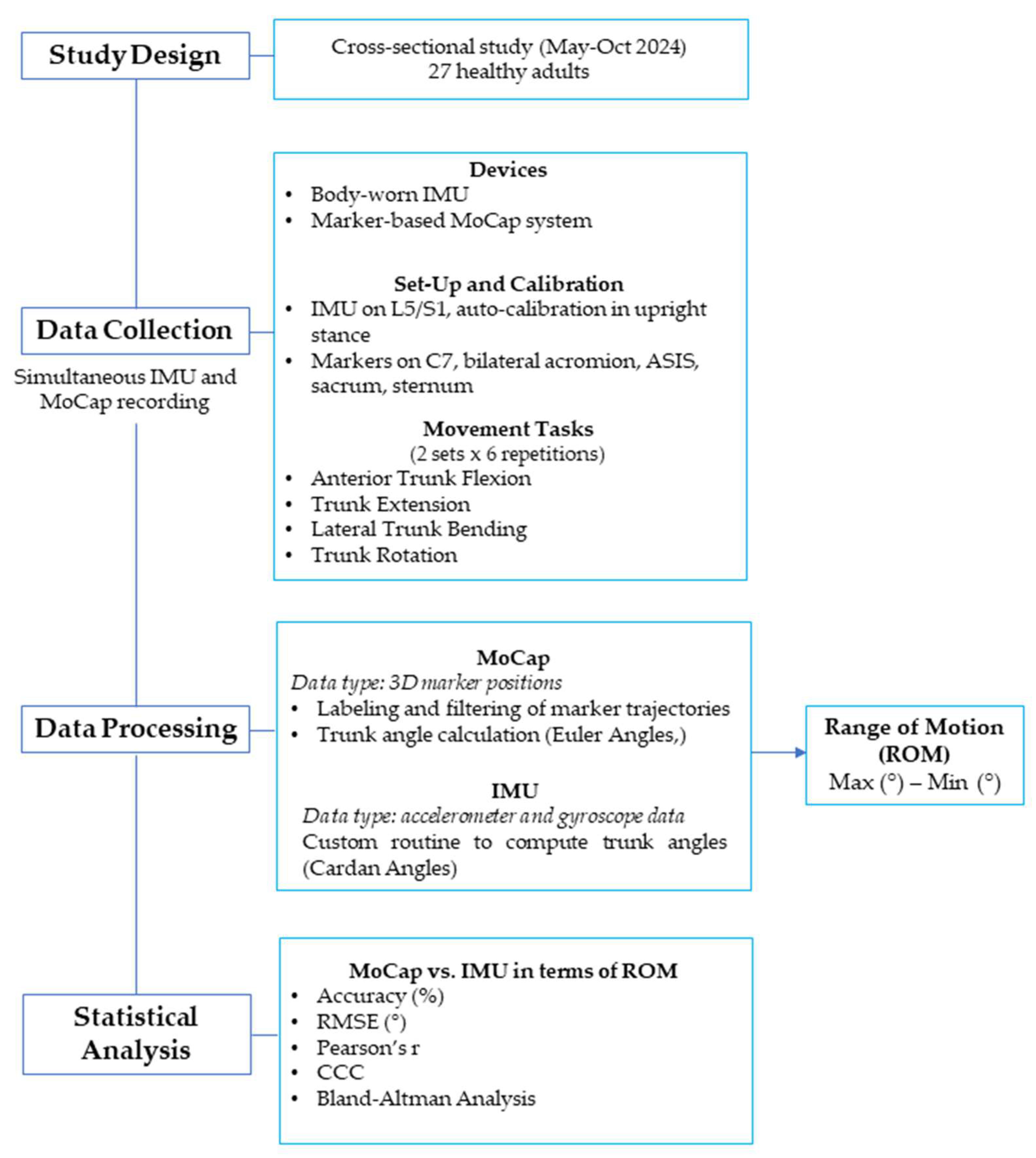
2.1. Study Design

The proposed investigation was designed as a cross-sectional study which took place from May 2024 to October 2024, aimed to assess precision and accuracy of a wearable IMU sensor (Baiobit, Rivelo Srl—BTS Bioengineering, Milan, Italy), compared with an optoelectronic marker-based MoCap system (SMART DX 400 system, BTS Bioengineering, Milan, Italy). This research was carried out in accordance with the Ethical Standards of the Institution and the 1964 Helsinki declaration and its latest amendments; it was approved by the Ethical Committees of Politecnico di Milano (22/2021, 14 June 2021). Written informed consent was signed by all participants. The reporting of the current study followed the updated STARD 2015 reporting guideline for diagnostic accuracy studies [30].

2.2. Participants

All the experimental procedures were carried out at the “Posture and Movement Analysis Laboratory Luigi Divieti” located at Politecnico di Milano, Italy.
Healthy subjects were consecutively recruited on a voluntary basis among the staff of the university campus. Inclusion criteria were aged 18 to 65 years, with a Body Mass Index (BMI) ranging from 18.5 to 24.9 kg/m2; exclusion criteria included the presence of systemic, neurological, and musculoskeletal conditions, and the inability to perform the requested tests for cognitive or psychiatric disorders that could prevent the completion of the required tests.

2.3. Sample Size Calculation
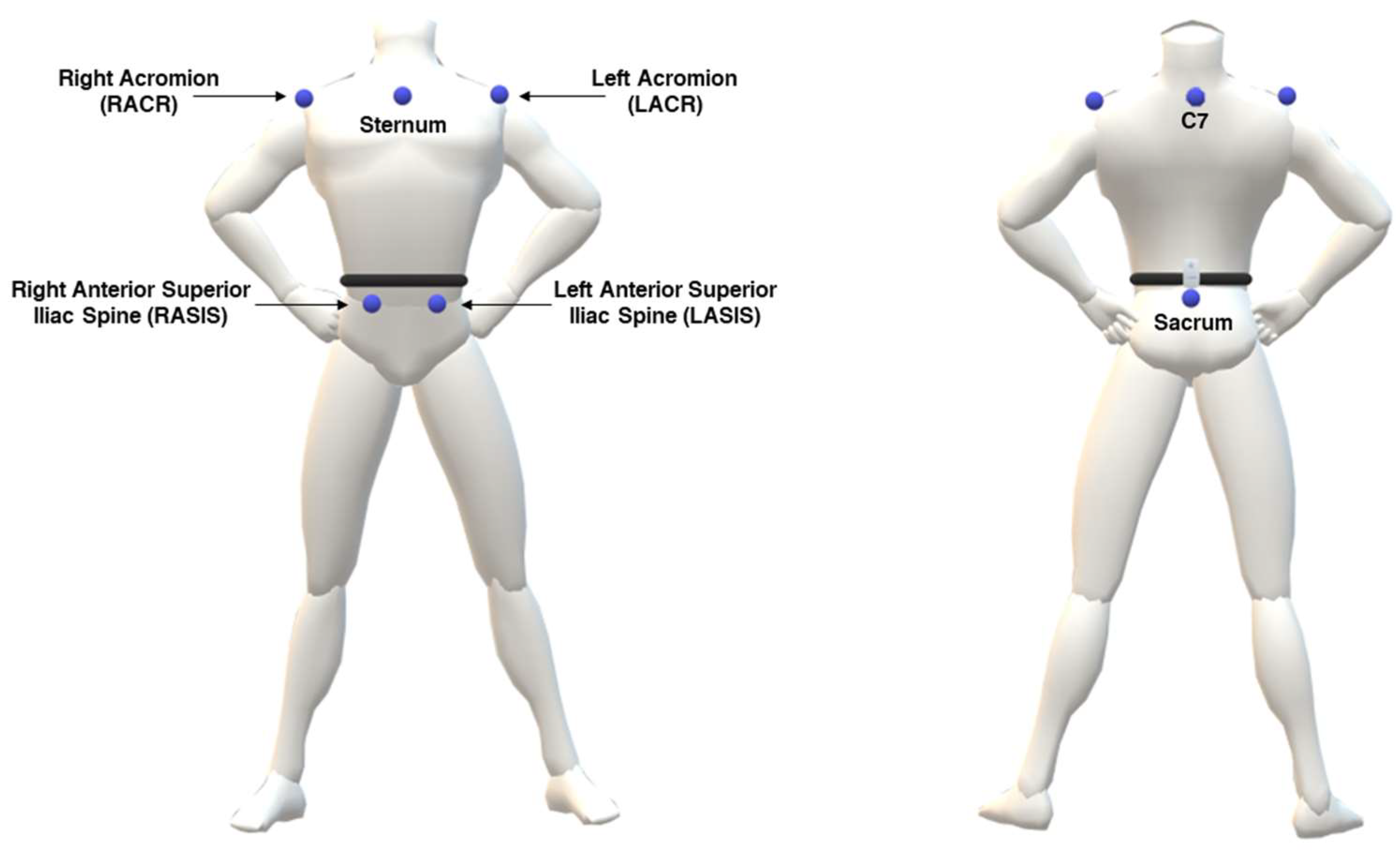
An a priori sample size calculation, assuming a strong correlation (r = 0.80), an alpha level of 0.05, and a statistical power of 0.80, suggested that a minimum of 15 participants would be sufficient to detect a significant correlation between the two technologies. Thence, a convenience sample of 27 healthy participants was recruited for this study, based on their availability to participate in the study. The full sample of 27 participants was retained to enhance the precision of the estimates and reduce the influence of outliers or missing data. This decision is consistent with methodological recommendations for reliability studies, which support the inclusion of larger samples when feasible [30].

2.4. MoCap System

The marker-based MoCap system employed for data acquisition was the SMART DX 400 (BTSBioengineering, Milan, Italy), equipped with 8 cameras operating at a sampling frequency of 100 Hz. Anthropometric measurements of the participants included height and weight. Passive markers were attached to anatomical landmarks on each participant, following a customized marker set for upper body derived from Davis protocol [31], considering markers on a bilateral acromion, seventh cervical vertebrae (C7), bilateral anterior superior iliac spine, and sacrum with the adjunction of marker on sternum (Figure 1). To maximize reliability of the procedure, the anatomical landmarks were identified manually through palpation by two different operators for each tested participant, focusing on regions with minimal soft tissue between the bone and skin.

2.5. IMU-Based System

The tested wearable IMU-based system (Baiobit, Rivelo Srl—BTS Bioengineering, Milan, Italy) was a medical device designed for clinical motor assessment. The device (dimensions: 70 × 40 × 18 mm, weight: 37 g; sample frequency: 100 Hz) integrates multiple sensors, including a tri-axial accelerometer, gyroscope, and magnetometer, enabling a detailed assessment of motion within its 3D local coordinate system. The IMU provides orientation data that allow the algorithm to compute roll (axial rotation), pitch (antero-posterior inclination), and yaw (lateral inclination) angles. The IMU is able to wirelessly communicate with a personal computer via BLE technology. The IMU can be attached to the user’s body via an adjustable elastic band to address different body locations, preventing unwanted sensor displacement throughout the movement; the tightness of the band was manually adjusted for each participant to ensure comfort and stability.
Calibration of the IMU was performed according to the manufacturer’s instructions, with participants standing in a quiet, upright stance while the sensor automatically calibrated itself. This process allowed the device to detect a static, zero-motion condition and establish a baseline orientation, correcting for sensor bias to ensure accurate motion data during subsequent recordings.

2.6. Testing Procedures

For data collection, each participant was simultaneously equipped with the previously described marker set and the IMU. According to the manufacturer’s instructions, the IMU was placed approximately at the L5/S1 vertebrae level using a customized elastic belt.
Starting from a neutral standing position, each participant was then required to perform the following tasks (Figure 2):
  • Trunk flexion (Figure 2a): participants bent forward at the waist level while maintaining a neutral spine, aiming to reach toward the floor without knee flexion;
  • Trunk extension (Figure 2b): participants extended the trunk backward while ensuring hip stability;
  • Lateral bending toward right/left (Figure 2c): participants performed a lateral bending movement at the waist, lowering one arm toward the corresponding leg while maintaining pelvic stability;
  • Trunk rotation toward right/left (Figure 2d): participants rotated the upper body to one side while keeping the hips oriented forward.
Each task was performed in two sets of six repetitions (per side for bilateral movements). Participants were instructed to perform the movements at their natural, self-selected pace.

2.7. Data Analysis and Processing

Raw data acquired with the marker-based MoCap system were initially processed using the proprietary software, i.e., SMARTtracker (version: 1.10.465.0; BTS Bioengineering, Milan, Italy). This step involved labeling each marker so as to assign it to its corresponding anatomical landmark. In particular, for this evaluation the attention was focused on the following markers: C7, left and right acromion, left and right antero-superior iliac spines, sacrum.
Subsequently, the processed data were imported into SMARTanalyzer software (version: 1.10.465.0; BTS Bioengineering, Milan, Italy) for further analysis through custom routines designed to extract quantitative measurements. The 3D marker coordinates underwent linear interpolation and were filtered using a 5 Hz low-pass Butterworth filter to minimize noise. Task-specific routines were employed to calculate relevant rotation angles and their corresponding ROM. In particular, trunk orientation was calculated with respect to the fixed laboratory coordinate system, providing an absolute measure of the movement. The angles were computed using Euler angles, ensuring a precise evaluation of trunk kinematics within the global reference frame.
Repetitions for each task were identified by locating the maximum and minimum values along the curve corresponding to the anatomical angles. For each repetition, ROM was determined as the difference in degrees (°) between these maximum and minimum values. These values were extracted from the time series of angular data by applying a peak detection method to identify local extrema.
IMU data were processed through an ad hoc and custom MATLAB (R2025a) (Mathworks Inc., Natick, MA, USA) routine. Raw data, including accelerometer and gyroscope readings preprocessed by a Digital Motion Processor (DMPTM), which provided rotational angles (i.e., roll, pitch, and yaw), were processed to obtain Cardan angles referred to the global reference system. Cardan angles about the IMU axes were calculated using the YZX sequence (i.e., flexion–extension around Y; lateral bending around Z axis; axial rotation around X axis) as generalized by Cole [32] using as a reference the mean angles calculated during the stabilization phase defined as a 5-second window in which acceleration variations fell below 0.2 m/s2. Functional angles were then extracted so as to quantify lateral bending, flexion/extension, and axial rotation, from which the ROM was calculated as the difference in degrees (°) between the maximum and minimum values.

2.8. Statistical Analysis

The normality of the ROM data was assessed using a Shapiro–Wilk test, and a normal distribution was confirmed for the variables of interest; consequently, variables were statistically described as mean and standard deviation.
The comparison of ROM values obtained from the marker-based MoCap and IMU was evaluated by assessing the accuracy and the Root Mean Square Error (RMSE), calculated as per the following equations:
A c c u r a c y = 1 | R O M I M U R O M M o C a p | R O M M o C a p 100
R M S E = i = 1 n ( y i ^ y i ) 2 n = i = 1 n e i 2 n
In Equation (2), y i ^ represents the ROM values derived from the marker-based MoCap system, yi refers to the ROM values from the IMU system, ei denotes the error, and n is the total number of observations or participants.
To evaluate the agreement between the two systems, Pearson’s correlation coefficient (r) and the concordance correlation coefficient (CCC) were calculated. In fact, Pearson’s correlation coefficient quantifies the strength and direction of a linear relationship between two continuous variables. The value of r ranges from −1 to 1, where the extreme values indicate a strong (positive or negative) correlation, whereas values around zero suggest the absence of linear relationship. The strength of the correlation was classified as follows: |r| ≤ 0.4, weak; 0.4 < |r| ≤ 0.6, moderate; 0.6 < |r| ≤ 0.8, strong; and |r| > 0.8, very strong [33]. The CCC evaluates the agreement between two continuous variables by combining measures of precision and accuracy. This coefficient provides a combined assessment of both accuracy (i.e., proximity to the values provided by the reference method) and precision (i.e., consistency of measurements); it ranges from −1 to +1, where 1 suggests perfect agreement, 0 indicates no agreement, and −1 implies disagreement [34].
Additionally, Bland–Altman (BA) plots were used to visualize the level of agreement (LoA) between the measurements [35].
All statistical tests were performed with a significance level set at α = 0.05.
The methodological workflow and timeline of the study are detailed in Figure 3.

3. Results

The study included 27 participants, with a male/female ratio of 11/16. The participants’ age ranged from 20 to 61 years (age: 31.1 ± 11.0 years), while the BMI spanned from 17.3 to 25.4 kg/m2. All the main characteristics of the sample are detailed in Table 1
The mean values for ROM measured by the marker-based MoCap and IMU systems were, respectively, as follows: flexion (78.5° ± 9.8° vs. 57.4° ± 14.4°), extension (21.2° ± 8.14° vs. 14.7° ± 5.92°), lateral bending (27.2° ± 6.93° vs. 16.7° ± 4.76°), and rotation (113° ± 28.3° vs. 108° ± 27.0°). Standard deviations showed greater variability in IMU measurements for flexion (14.4°) compared to MoCap (9.80°); conversely, similar trends were observed for other motions. ROM distribution by movement and measurement system are reported in Figure 4.

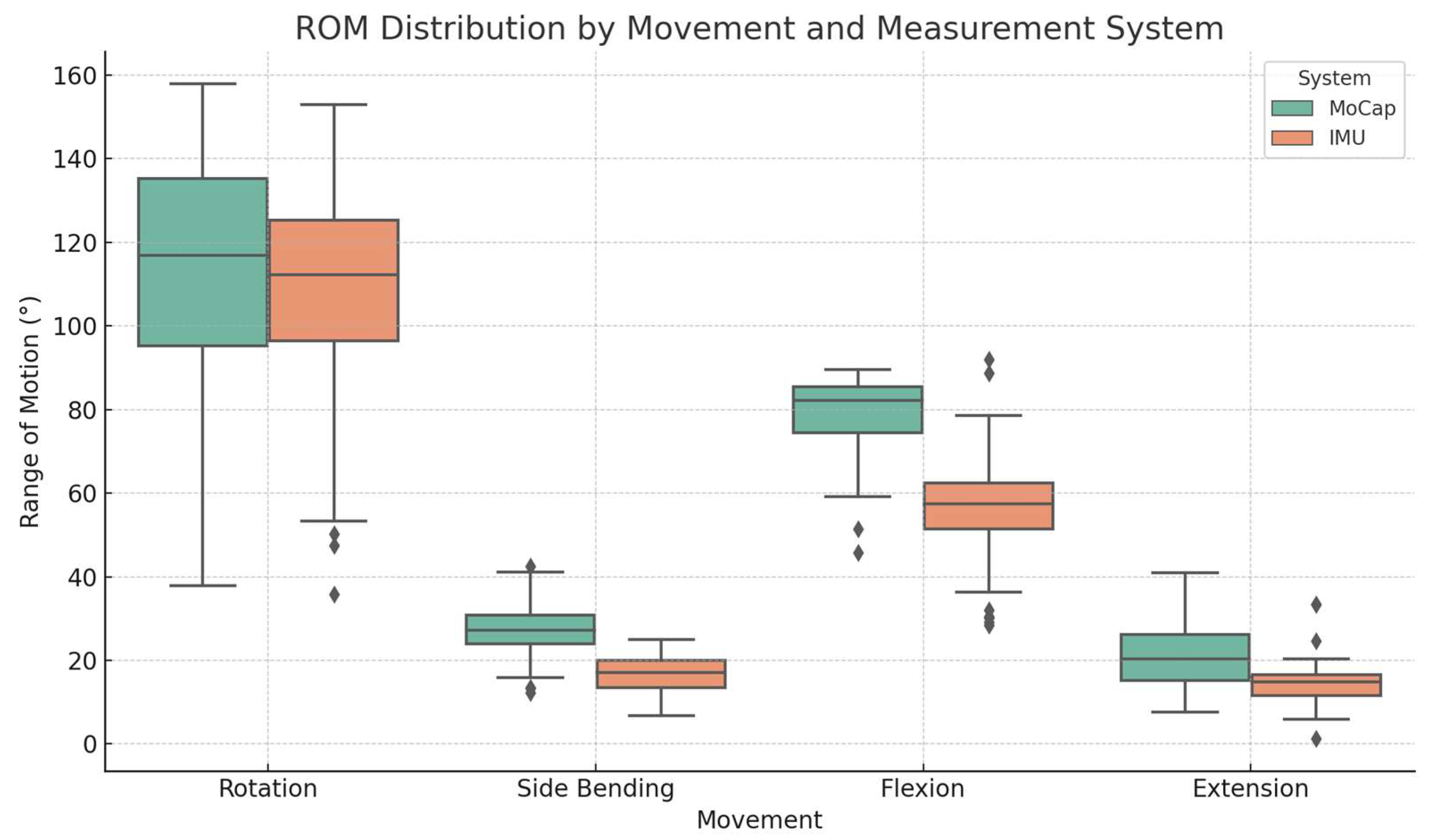
3.1. Accuracy and RMSE

The mean percentage of accuracy values for IMU measurements showed varying degrees of agreement with MoCap data: flexion 72.1% (SD: 12.7%), extension 64.1% (SD: 23.5%), lateral bending 61.4% (SD: 16.8%), and rotation 92.4% (SD: 7.61%). These findings highlight that accuracy ranges from moderate levels for lateral bending to good or optimal levels for rotation. RMSE values highlighted discrepancies across movements, with the largest error in flexion: 3.01° (SD: 1.32°), and the smallest in rotation: 1.09° (SD: 1.01°). The results indicate that the agreement between the two measurement systems ranged from acceptable (flexion) to negligible (other tasks) error across all analyzed movements. Data are summarized in Table 2.

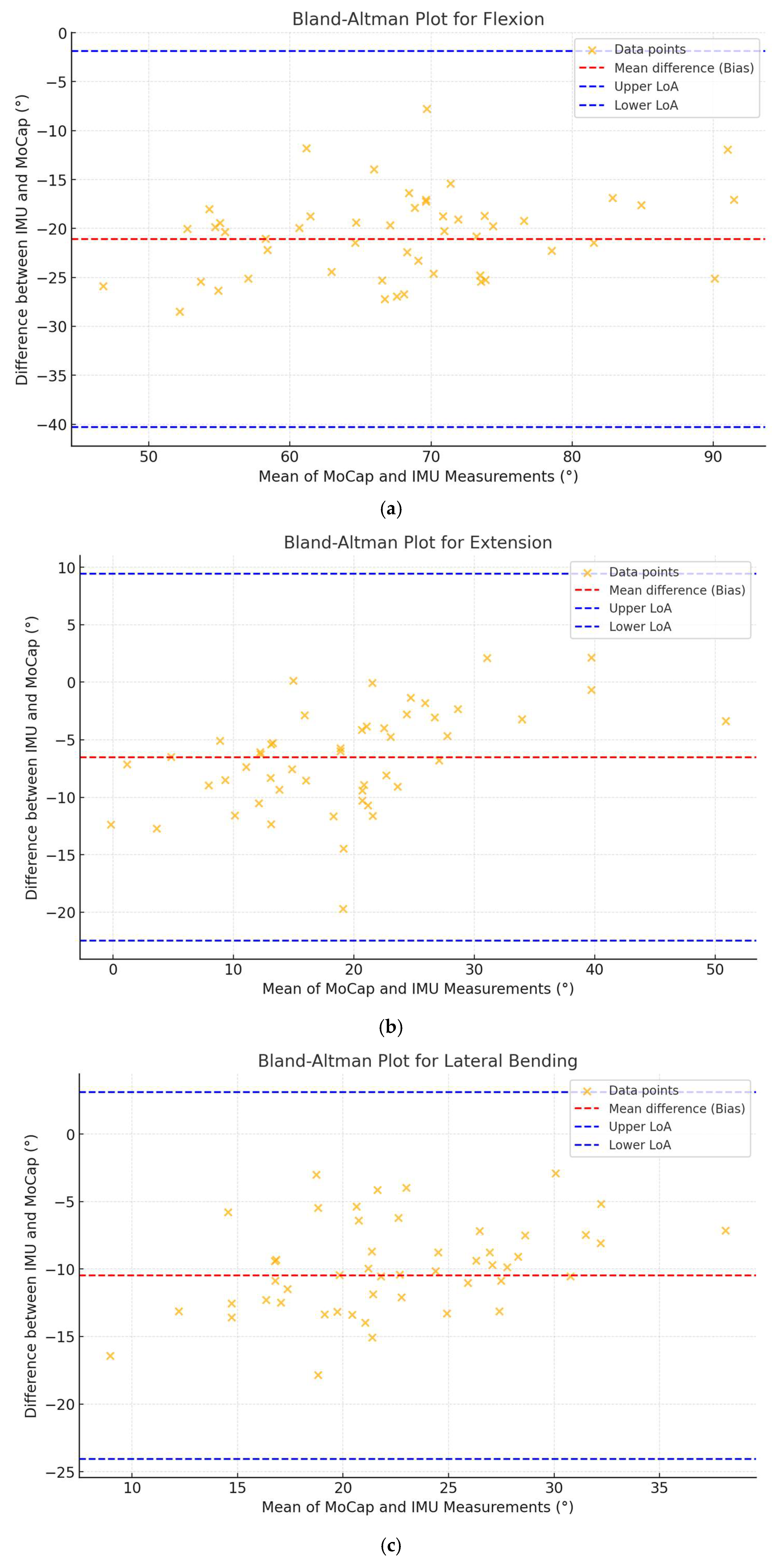
3.2. Correlation and Agreement Analysis

Pearson’s correlation and CCC values are reported in Table 3. The analysis showed strong to very strong relationships between IMU and MoCap for flexion and rotation (r = 0.703, p < 0.001 and r = 0.944, p < 0.001, respectively), correlations for extension (r = 0.564, p < 0.001), and lateral bending (r = 0.430, p = 0.003) were moderate. The CCC was highest for rotation (CCC = 0.927, 95% CI: 0.877–0.957), indicating strong agreement, while lower values were observed for lateral bending (CCC = 0.155, 95% CI: 0.046–0.260). Flexion and extension showed moderate agreement with CCC values of 0.262 and 0.375, respectively.
Bland–Altman analyses (Figure 5) revealed biases ranging from −21.09° for flexion to −5.12° for rotation, with narrower limits of agreement in rotation) compared to flexion (−23.56° to 13.31° and −41.18° to −1.01°, respectively).

4. Discussion

This study aimed to assess the overall reliability of an IMU-based wearable device in measuring trunk range of motion (ROM), by comparing it with respect to a marker-based optoelectronic MoCap system, which can be considered the actual gold standard in human motion analysis. Our findings provide relevant insights into the applicability and limitations of the IMU-based system utilized for trunk motion analysis.
The results showed that the IMU device provided comparable but consistently underestimated ROM values across all movements, when compared to the marker-based MoCap systems. Notably, systematic underestimation was observed in both flexion and lateral bending, suggesting that intrinsic factors, such as sensor drift, soft tissue artifacts, and sensor alignment, could contribute to these discrepancies [36,37]. Similar trends were reported in earlier studies using IMUs, especially in quantifying anterior trunk flexion, where the IMU-based measurements were either overestimated [18] or underestimated [38]. The difference in directionality (i.e., underestimation vs. overestimation) may also be attributed to differences in sensor’s configuration, calibration protocols, and biomechanical modeling approaches used across studies.
Despite these discrepancies, the high accuracy observed for trunk rotation (92.4%) in the present study highlights the potential of IMU technology for specific movement planes, possibly due to the more consistent inertial signatures associated with axial rotation. This finding aligns with the literature, suggesting that rotational movements are often less susceptible to soft tissue motion artifacts and sensor misalignment, making them more reliably captured by inertial sensors [39].
The RMSE analysis further revealed variable agreement levels between the two systems, with the highest RMSE found in flexion (3.01°) and the lowest in rotation (1.09°). This variability could be explained by the physiological and biomechanical characteristics of the different trunk movements, which might affect the reliability of the IMU. This variability is consistent with findings of Lee et al. (2023), who reported RMSE values ranging from 1.6° to 2.9° for trunk motions, and reinforces the idea that biomechanical characteristics of different trunk movements can lead to variability in measurement accuracy [39]. In the same way, Khobkhun et al. demonstrated how accelerometers and gyroscopes can be influenced by the subject’s posture and movement dynamic [40]. Additionally, the higher variability in IMU measurements, particularly for flexion (SD = 14.4°), may highlight the influence of measurement noise [41].
Furthermore, it should be considered that misalignment errors are possible, indeed [42,43]. In the presented study, the IMU was placed to the lumbo-sacral region of each participant, using an elastic belt; although this belt was necessary to fix the device to the trunk, a certain degree of relative motion occurred between the participant’s body and the sensor itself—due also to the presence of possible soft tissue artifacts—potentially accounting for some measurement accuracy issues, which were clearly observed both during flexion and lateral bending tasks.
The Pearson correlation analysis demonstrated strong relationships between the IMU and the marker-based MoCap system, particularly for flexion (r = 0.703) and rotation (r = 0.944). However, moderate correlations for extension (r = 0.564) and lateral bending (r = 0.430) are encouraging but suggest that certain motions may be more challenging to capture accurately with the IMU system. Once again, this finding can be interpreted in light of the potential displacements of the IMU device during various movements, particularly those involving flexion, extension, and lateral bending, as opposed to rotation. Additionally, the Bland–Altman analysis provided further insight into the agreement between the two systems, showing narrower limits of agreement for rotation compared to flexion, indicating better consistency for rotational movements. Furthermore, for each movement, the data points were randomly distributed around the mean difference (bias), suggesting the absence of any specific angle-dependent discrepancy between the two technologies, particularly during rotational movements. Finally, the mean difference values between the two measures are relatively low and appear to correlate with the amplitude of each movement, i.e., higher for flexion and lateral bending, and lower for extension and rotation. This aspect was already reported by some authors [43], though there is no complete agreement in the literature [44].
The CCC values supported these findings, with rotation showing the highest level of agreement (CCC = 0.927), when side-bending had the lowest (CCC = 0.155). This suggests that while the IMU-based system performs well for specific motions, it may require further adjustment to improve its performance for others, such as lateral bending and extension. In these last tasks, the results we obtained reflect both systematic bias and considerable variability. This confirms that although several specific motions can be measured reliably by using an IMU (e.g., rotation), others are prone to discrepancies due to sensor limitations, such as drift, as well as artifacts related to sensor’s placement and soft tissue motion.
It is worth noting that, unlike more complex IMU systems employing multiple sensor units for improved segmental tracking and sensor fusion, the system evaluated in this study used a single IMU configuration. While this design choice enhances practicality and reduces setup time, it may limit the accuracy of kinematic estimations, particularly for movements involving multiple degrees of freedom or compound trunk dynamics. In this respect, the performance observed here remains promising, especially considering the simplicity of the setup and the constraints of single-sensor systems. The previous literature has shown that multi-IMU configurations generally outperform single-IMU setups in terms of accuracy and precision, but at the cost of increased complexity and user burden [18].
Overall, our findings suggested that the assessment of rotational tasks appeared to be more reliable with respect to the other analyzed movements. From a biomechanical point of view, axial rotation involves less deformation of soft tissues in the lumbo-sacral area, reducing the impact of motion artifacts. Technically, rotational measurements primarily rely on the gyroscope signal (i.e., angular velocity), which tends to be more reliable and less affected by gravitational interference compared to accelerometer-based estimations used in flexion and lateral bending [45]. The different performance across the tasks thus underscore the need for task-specific validation when using IMUs, particularly in contexts where precise motion characterization is required.
These findings substantially overlap with previous research highlighting the advantages of IMU-based systems, such as portability, ease of use, and real-time data acquisition [32,40]. However, the observed limitations emphasize the need for further improvements at different levels (i.e., compliance to soft tissue artifacts) to strengthen overall reliability and reduce errors, and the use of machine learning and artificial neural networks may be promising tools [46,47]. Future works may explore the use of machine learning approaches, such as classification and regression machine learning approaches (e.g., Support Vector Regression), and neural network models (e.g., feedforward or LSTM architectures), to improve the prediction of joint kinematics from IMU data, potentially enhancing accuracy and generalizability across diverse movement patterns.
Importantly, the lower ecological validity of laboratory-based MoCap systems remains a significant limitation, and the IMU capacity for real-world application makes it an interesting alternative [36,48].
The IMU-based wearable device demonstrates promising potential as a cost-effective and practical tool for trunk ROM analysis. Our findings suggest that it cannot yet fully replace the precision of a gold-standard marker-based MoCap system; however, its application in clinical and real-world settings offers substantial advantages, warranting further development and assessment [37]. As future perspectives, the implementation of IMU-based assessments in outpatient and real-world settings represents a promising direction. Future protocols may incorporate simplified motor tasks, integration with smart garments or wearable technologies, and mobile-based data collection tools to enhance feasibility and user compliance in clinical practice. This could pave the way for a wider adoption of wearable technologies in daily rehabilitation practices, enabling clinicians to monitor and assess patients’ motor performance with greater efficiency [49,50,51].
However, in clinical populations, the accuracy and interpretability of IMU-derived kinematic data may be influenced by altered movement strategies, such as compensatory trunk patterns and postural asymmetries. These factors can affect the alignment between the sensor and the underlying anatomical segments, potentially introducing bias in the computed joint angles. Despite this limitation, IMUs retain strong potential for use in clinical contexts and future studies should consider these population-specific characteristics and validate IMU-based assessments accordingly.
This study has several limitations that should be acknowledged. First, it was conducted on a relatively small sample size, which may limit the generalizability of the findings. A post hoc sensitivity analysis showed that our sample size (N = 27) was sufficient to detect correlations of 0.51 or higher with 80% power (α = 0.05). This suggests that movements showing weaker correlations, such as lateral bending, may not have been adequately powered, possibly limiting the ability to detect relevant relationships. Therefore, caution is needed when interpreting these specific results, and future studies with larger samples are needed to strengthen these findings. Second, the study included only healthy participants, which does not allow for conclusions regarding the applicability of these tools in clinical populations, or individuals with movement disorders. Although the inclusion of 27 healthy adults provides initial validation and meets the recommended criteria for agreement studies, the lack of clinical populations limits generalizability. Moreover, a relatively small sample may not capture the intrinsic variability in trunk kinematics that can emerge in healthy and clinical populations, particularly in the presence of pain, compensation, or postural control deficits. Third, potential errors may have occurred due to a lack of precise instructions provided to participants, prior to performing the motor tasks. Finally, the elastic belt used to secure the IMU sensor may have allowed slight displacements during the execution of motor tasks, potentially affecting the precision of the collected data. To reduce artifacts, participants were asked to perform test trials, and visual and manual inspections were conducted to verify proper sensor placement and stability. While we did not directly quantify soft tissue artifacts, we attempted to minimize their influence by positioning the sensor over a bony landmark (lumbo-sacral junction), verifying sensor stability during trial preparation, and repeating trials when visible sensor shifts were noticed. Although no quantitative correction for soft tissue artifacts was applied, we acknowledge this limitation that might have potential implications in the generalization of our findings. Future studies are needed to consider biomechanical compensation methods or improved fixation systems to mitigate this issue.
In addition, while functional calibration procedures and a quantitative assessment of belt fixation pressure were not implemented in the current protocol, we are aware of their potential to enhance anatomical alignment and data accuracy; these represent a valuable direction for methodological refinement in future investigations.
Since this study exclusively focused on healthy participants, future research should investigate the performance of IMU sensors for trunk movements in individuals with musculoskeletal disorders, where compensatory movements and functional limitations might further require their use in the clinical field [52]. Lastly, refining the calibration procedures and algorithms for the IMU system may help reduce measurement discrepancies [53].
Future research should focus on expanding the scope of IMU-based evaluations to also include other acceleration-derived metrics (e.g., root mean square, improved harmonic ratio and jerk). These indicators might be indicative for postural stability, movement symmetry, and smoothness, which are critical for understanding trunk movement dynamics [54]. Investigating these parameters in populations with musculoskeletal disorders would provide further essential insights into their clinical relevance and the potential for targeted interventions [55]. Additionally, studies involving larger and more heterogeneous cohorts, including individuals with specific pathologies, are needed to further validate the utility of IMU sensors in complex clinical scenarios.

5. Conclusions

This study confirmed that an IMU-based wearable sensor can provide a feasible and cost-effective alternative to marker-based MoCap systems for quantitatively analyzing the trunk range of motion. Among the movements tested, rotational tasks showed the highest accuracy and agreement, whereas flexion, extension, and lateral bending were more affected by variability and underestimation, likely due to sensor relative displacement and soft tissue artifacts.
Despite these limitations, including a small sample size and the exclusive inclusion of healthy participants, this study offers a proper preliminary validation of the clinical potential of the proposed approach. Future research should focus on clinical populations, where compensatory strategies and postural alterations may impact measurement accuracy. Refining calibration procedures and improving sensor fixation methods will also be essential to increase reliability in real-world and outpatient settings.

Author Contributions

Conceptualization, F.D.F. and V.C.; methodology, V.C. and N.F.L.; formal analysis, F.D.F., S.C. and V.C.; investigation, F.D.F. and S.C.; resources, V.C.; data curation, F.D.F., S.C. and V.C.; writing—original draft preparation, F.D.F., S.C., M.P. (Micaela Porta), M.P. (Massimiliano Pau) and M.G.; writing—review and editing, F.D.F., V.C., M.G. and N.F.L.; supervision, V.C. and M.P. (Massimiliano Pau), M.G. and N.F.L. All authors have read and agreed to the published version of the manuscript.

Funding

The study was supported by the MUSA—Multilayered Urban Sustainability Action—project funded by the European Union’s NextGenerationEU under the National Recovery and Resilience Plan (NRRP) Mission 4, Component 2, Investment Line 1.5: Strengthening of research structures and creation of R&D “innovation ecosystems” of “territorial leaders in R&D”.

Institutional Review Board Statement

The study was conducted in accordance with the Declaration of Helsinki and approved by the Ethics Committee of POLITECNICO OF MILAN (protocol 22/2021, 14 June 2021).

Informed Consent Statement

Informed consent was obtained from all subjects involved in the study.

Data Availability Statement

Data collected and analyzed during the current study are available from the corresponding author upon reasonable request.

Acknowledgments

The authors would like to thank Alexia Lanzaro for her valuable help in data acquisition and processing.

Conflicts of Interest

The authors declare no conflicts of interest.

Abbreviations

The following abbreviations are used in this manuscript:
ROMRange of Motion
MoCapMotion Capture
IMUInertial Measurement Unit
LBPLow Back Pain

References

  1. Benedetti, M.G.; Negrini, S. Instrumental motion analysis: From the research laboratory to the rehabilitation clinic. Eur. J. Phys. Rehabil. Med. 2016, 52, 557–559. [Google Scholar] [PubMed]
  2. Quinn, L.; Riley, N.; Tyrell, C.M.; Judd, D.L.; Gill-Body, K.M.; Hedman, L.D.; Packel, A.; Brown, D.A.; Nabar, N.; Scheets, P. A Framework for Movement Analysis of Tasks: Recommendations From the Academy of Neurologic Physical Therapy’s Movement System Task Force. Phys. Ther. 2021, 101, pzab154. [Google Scholar] [CrossRef]
  3. Martin, C.; Phillips, B.A.; Kilpatrick, T.J.; Butzkueven, H.; Tubridy, N.; McDonald, E.; Galea, M.P. Gait and balance impairment in early multiple sclerosis in the absence of clinical disability. Mult. Scler. J. 2006, 12, 620–628. [Google Scholar] [CrossRef]
  4. Stolze, H.; Klebe, S.; Baecker, C.; Zechlin, C.; Friege, L.; Pohle, S.; Deuschl, G. Prevalence of gait disorders in hospitalized neurological patients. Mov. Disord. 2004, 20, 89–94. [Google Scholar] [CrossRef]
  5. Dal Farra, F.; Arippa, F.; Carta, G.; Segreto, M.; Porcu, E.; Monticone, M. Sport and non-specific low back pain in athletes: A scoping review. BMC Sports Sci. Med. Rehabil. 2022, 14, 216. [Google Scholar] [CrossRef] [PubMed]
  6. Reis, F.J.J.d.; Macedo, A.R.d. Influence of hamstring tightness in pelvic, lumbar and trunk range of motion in low back pain and asymptomatic volunteers during forward bending. Asian Spine J. 2015, 9, 535. [Google Scholar] [CrossRef]
  7. Dal Farra, F.; Arippa, F.; Arru, M.; Cocco, M.; Porcu, E.; Tramontano, M.; Monticone, M. Effects of exercise on balance in patients with non-specific low back pain: A systematic review and meta-analysis. Eur. J. Phys. Rehabil. Med. 2022, 58, 423–434. [Google Scholar] [CrossRef]
  8. VanDijk, M.; Smorenburg, N.; Visser, B.; Heerkens, Y.F.; Der Sanden, M.W.G.N.-V. How clinicians analyze movement quality in patients with non-specific low back pain: A cross-sectional survey study with Dutch allied health care professionals. BMC Musculoskelet. Disord. 2017, 18, 288. [Google Scholar]
  9. Schlager, A.; Ahlqvist, K.; Rasmussen-Barr, E.; Bjelland, E.K.; Pingel, R.; Olsson, C.; Nilsson-Wikmar, L.; Kristiansson, P. Inter- and intra-rater reliability for measurement of range of motion in joints included in three hypermobility assessment methods. BMC Musculoskelet. Disord. 2018, 19, 376. [Google Scholar] [CrossRef]
  10. Cano-de-la-Cuerda, R.; Vela, L.; Moreno-Verdú, M.; Ferreira-Sánchez, M.d.R.; Macías-Macías, Y.; Miangolarra-Page, J.C. Trunk range of motion is related to axial rigidity, functional mobility and quality of life in parkinson’s disease: An exploratory study. Sensors 2020, 20, 2482. [Google Scholar] [CrossRef]
  11. Shamsi, M.; Mirzaei, M.; Khabiri, S. Universal goniometer and electro-goniometer intra-examiner reliability in measuring the knee range of motion during active knee extension test in patients with chronic low back pain with short hamstring muscle. BMC Sports Sci. Med. Rehabil. 2019, 11, 4. [Google Scholar] [CrossRef] [PubMed]
  12. Kim, S.; Kim, E. Test-retest reliability of an active range of motion test for the shoulder and hip joints by unskilled examiners using a manual goniometer. J. Phys. Ther. Sci. 2016, 28, 722–724. [Google Scholar] [CrossRef]
  13. McGinley, J.L.; Baker, R.; Wolfe, R.; Morris, M.E. The reliability of three-dimensional kinematic gait measurements: A systematic review. Gait Posture 2009, 29, 360–369. [Google Scholar] [CrossRef]
  14. Alarcón-Aldana, A.C.; Callejas-Cuervo, M.; Bó, A.P.L. Upper limb physical rehabilitation using serious videogames and motion capture systems: A systematic review. Sensors 2020, 20, 5989. [Google Scholar] [CrossRef]
  15. Moro, M.; Marchesi, G.; Hesse, F.; Odone, F.; Casadio, M. Markerless vs. marker-based gait analysis: A proof of concept study. Sensors 2022, 22, 2011. [Google Scholar] [CrossRef] [PubMed]
  16. Poitras, I.; Dupuis, F.; Bielmann, M.; Campeau-Lecours, A.; Mercier, C.; Bouyer, L.J.; Roy, J. Validity and reliability of wearable sensors for joint angle estimation: A systematic review. Sensors 2019, 19, 1555. [Google Scholar] [CrossRef] [PubMed]
  17. Fong, D.T.; Chan, Y.M. The use of wearable inertial motion sensors in human lower limb biomechanics studies: A systematic review. Sensors 2010, 10, 11556–11565. [Google Scholar] [CrossRef]
  18. Cerfoglio, S.; Capodaglio, P.; Rossi, P.; Conforti, I.; D’Angeli, V.; Milani, E.; Galli, M.; Cimolin, V. Evaluation of upper body and lower limbs kinematics through an imu-based medical system: A comparative study with the optoelectronic system. Sensors 2023, 23, 6156. [Google Scholar] [CrossRef]
  19. Tadano, S.; Takeda, R.; Miyagawa, H. Three dimensional gait analysis using wearable acceleration and gyro sensors based on quaternion calculations. Sensors 2013, 13, 9321–9343. [Google Scholar] [CrossRef]
  20. Li, H.; Khoo, S.; Yap, H.J. Differences in motion accuracy of baduanjin between novice and senior students on inertial sensor measurement systems. Sensors 2020, 20, 6258. [Google Scholar] [CrossRef]
  21. Manupibul, U.; Tanthuwapathom, R.; Jarumethitanont, W.; Kaimuk, P.; Limroongreungrat, W.; Charoensuk, W. Integration of force and imu sensors for developing low-cost portable gait measurement system in lower extremities. Sci. Rep. 2023, 13, 10653. [Google Scholar] [CrossRef] [PubMed]
  22. Schall, M.C.; Fethke, N.B.; Chen, H.; Oyama, S.; Douphrate, D.I. Accuracy and repeatability of an inertial measurement unit system for field-based occupational studies. Ergonomics 2015, 59, 591–602. [Google Scholar] [CrossRef]
  23. Zhang, H.; Tao, Y.; Shi, K.; Li, J.; Shi, J.; Xu, S.; Guo, Y. Multi-Object Recognition and Motion Detection Based on Flexible Pressure Sensor Array and Deep Learning. Appl. Sci. 2025, 15, 3302. [Google Scholar] [CrossRef]
  24. Zhu, P.; Niu, M.; Liang, S.; Yang, W.; Zhang, Y.; Chen, K.; Pan, Z.; Mao, Y. Non-hand-worn, load-free VR hand rehabilitation system assisted by deep learning based on ionic hydrogel. Nano Res. 2025, 18, 94907301. [Google Scholar] [CrossRef]
  25. Lu, L.; Hu, G.; Liu, J.; Yang, B. 5G NB-IoT System Integrated with High-Performance Fiber Sensor Inspired by Cirrus and Spider Structures. Adv. Sci. (Weinh. Baden-Wurtt. Ger.) 2024, 11, e2309894. [Google Scholar] [CrossRef] [PubMed]
  26. Liengswangwong, W.; Lertviboonluk, N.; Yuksen, C.; Jamkrajang, P.; Limroongreungrat, W.; Mongkolpichayaruk, A.; Jenpanitpong, C.; Watcharakitpaisan, S.; Palee, C.; Thaipasong, S.; et al. Validity of inertial measurement unit (imu sensor) for measurement of cervical spine motion, compared with eight optoelectronic 3d cameras under spinal immobilization devices. Med. Devices Evid. Res. 2024, 17, 261–269. [Google Scholar] [CrossRef]
  27. Ali, F.; Hogen, C.A.; Miller, E.J.; Kaufman, K.R. Validation of Pelvis and Trunk Range of Motion as Assessed Using Inertial Measurement Units. Bioengineering 2024, 11, 659. [Google Scholar] [CrossRef]
  28. Parrington, L.; Jehu, D.A.; Fino, P.C.; Pearson, S.; El-Gohary, M.; King, L.A. Validation of an inertial sensor algorithm to quantify head and trunk movement in healthy young adults and individuals with mild traumatic brain injury. Sensors 2018, 18, 4501. [Google Scholar] [CrossRef]
  29. Abdollahi, M.; Ashouri, S.; Abedi, M.; Azadeh-Fard, N.; Parnianpour, M.; Khalaf, K.; Rashedi, E. Using a motion sensor to categorize nonspecific low back pain patients: A machine learning approach. Sensors 2020, 20, 3600. [Google Scholar] [CrossRef]
  30. Bossuyt, P.M.; Reitsma, J.B.; Bruns, D.E.; Gatsonis, C.A.; Glasziou, P.P.; Irwig, L.; Lijmer, J.G.; Moher, D.; Rennie, D.; de Vet, H.C.W.; et al. STARD 2015: An Updated List of Essential Items for Reporting Diagnostic Accuracy Studies. Radiology 2015, 277, 826–832. [Google Scholar] [CrossRef]
  31. Davis, R.B.; Õunpuu, S.; Tybursky, D.; Gage, J.R. A gait analysis data collection and reduction technique. Hum. Mov. Sci. 1991, 10, 575–587. [Google Scholar] [CrossRef]
  32. Cole, G.K.; Nigg, B.M.; Ronsky, J.L.; Yeadon, M.R. Application of the joint coordinate system to three-dimensional joint attitude and movement representation: A standardization proposal. J. Biomech. Eng. 1993, 115, 344–349. [Google Scholar] [CrossRef]
  33. Akoglu, H. User’s guide to correlation coefficients. Turk. J. Emerg. Med. 2018, 18, 91–93. [Google Scholar] [CrossRef]
  34. Hiriote, S.; Chinchilli, V.M. Matrix-based concordance correlation coefficient for repeated measures. Biometrics 2011, 67, 1007–1016. [Google Scholar] [CrossRef] [PubMed]
  35. Zaki, R.A.; Bulgiba, A.; Ismail, R.; Ismail, N.A. Statistical methods used to test for agreement of medical instruments measuring continuous variables in method comparison studies: A systematic review. PLoS ONE 2012, 7, e37908. [Google Scholar] [CrossRef] [PubMed]
  36. Al-Amri, M.; Nicholas, K.; Button, K.; Sparkes, V.; Sheeran, L.; Davies, J. Inertial measurement units for clinical movement analysis: Reliability and concurrent validity. Sensors 2018, 18, 719. [Google Scholar] [CrossRef] [PubMed]
  37. Cerfoglio, S.; Lopomo, N.F.; Capodaglio, P.; Scalona, E.; Monfrini, R.; Verme, F.; Galli, M.; Cimolin, V. Assessment of an IMU-Based Experimental Set-Up for Upper Limb Motion in Obese Subjects. Sensors 2023, 23, 9264. [Google Scholar] [CrossRef]
  38. Brouwer, N.P.; Yeung, T.; Bobbert, M.F.; Besier, T.F. 3D trunk orientation measured using inertial measurement units during anatomical and dynamic sports motions. Scand. J. Med. Sci. Sports. 2021, 31, 358–370. [Google Scholar] [CrossRef]
  39. Lee, R.; Akhundov, R.; James, C.; Edwards, S.; Snodgrass, S.J. Variations in concurrent validity of two independent inertial measurement units compared to gold standard for upper body posture during computerised device use. Sensors 2023, 23, 6761. [Google Scholar] [CrossRef]
  40. Khobkhun, F.; Hollands, M.A.; Richards, J.; Ajjimaporn, A. Can we accurately measure axial segment coordination during turning using inertial measurement units (imus)? Sensors 2020, 20, 2518. [Google Scholar] [CrossRef]
  41. Suvorkin, V.; Garcia-Fernandez, M.; González-Casado, G.; Li, M.; Rovira-Garcia, A. Assessment of noise of mems imu sensors of different grades for gnss/imu navigation. Sensors 2024, 24, 1953. [Google Scholar] [CrossRef] [PubMed]
  42. Zhu, K.; Li, J.; Li, D.; Fan, B.; Shull, P.B. Imu shoulder angle estimation: Effects of sensor-to-segment misalignment and sensor orientation error. IEEE Trans. Neural Syst. Rehabil. Eng. 2023, 31, 4481–4491. [Google Scholar] [CrossRef]
  43. Lebleu, J.; Gosseye, T.; Detrembleur, C.; Mahaudens, P.; Cartiaux, O.; Penta, M. Lower limb kinematics using inertial sensors during locomotion: Accuracy and reproducibility of joint angle calculations with different sensor-to-segment calibrations. Sensors 2020, 20, 715. [Google Scholar] [CrossRef]
  44. Fang, Z.; Woodford, S.C.; Senanayake, D.; Ackland, D.C. Conversion of upper-limb inertial measurement unit data to joint angles: A systematic review. Sensors 2023, 23, 6535. [Google Scholar] [CrossRef] [PubMed]
  45. Pasciuto, I.; Ligorio, G.; Bergamini, E.; Vannozzi, G.; Sabatini, A.; Cappozzo, A. How angular velocity features and different gyroscope noise types interact and determine orientation estimation accuracy. Sensors 2015, 15, 23983–24001. [Google Scholar] [CrossRef] [PubMed]
  46. Almassri, A.M.M.; Shirasawa, N.; Purev, A.; Uehara, K.; Oshiumi, W.; Mishima, S.; Wagatsuma, H. Artificial neural network approach to guarantee the positioning accuracy of moving robots by using the integration of imu/uwb with motion capture system data fusion. Sensors 2022, 22, 5737. [Google Scholar] [CrossRef]
  47. Adans-Dester, C.; Hankov, N.; O’Brien, A.; Vergara-Diaz, G.; Black-Schaffer, R.M.; Zafonte, R.; Dy, J.; Lee, S.I.; Bonato, P. Enabling precision rehabilitation interventions using wearable sensors and machine learning to track motor recovery. NPJ Digit. Med. 2020, 3, 121. [Google Scholar] [CrossRef] [PubMed]
  48. Park, S.; Yoon, S. Validity evaluation of an inertial measurement unit (imu) in gait analysis using statistical parametric mapping (spm). Sensors 2021, 21, 3667. [Google Scholar] [CrossRef]
  49. Jiménez-Olmedo, J.M.; Pueo, B.; Mossi, J.M.; Villalón-Gasch, L. Concurrent validity of the inertial measurement unit vmaxpro in vertical jump estimation. Appl. Sci. 2023, 13, 959. [Google Scholar] [CrossRef]
  50. Komaris, D.; Tarfali, G.; O’Flynn, B.; Tedesco, S. Unsupervised imu-based evaluation of at-home exercise programmes: A feasibility study. BMC Sports Sci. Med. Rehabil. 2022, 14, 28. [Google Scholar] [CrossRef]
  51. Lee, S.I.; Adans-Dester, C.; O’Brien, A.; Vergara-Diaz, G.; Black-Schaffer, R.M.; Zafonte, R.; Dy, J.G.; Bonato, P. Predicting and monitoring upper-limb rehabilitation outcomes using clinical and wearable sensor data in brain injury survivors. IEEE Trans. Biomed. Eng. 2021, 68, 1871–1881. [Google Scholar] [CrossRef] [PubMed]
  52. Dal Farra, F.; Arippa, F.; Arru, M.; Cocco, M.; Porcu, E.; Solla, F.; Monticone, M. Is dynamic balance impaired in people with non-specific low back pain when compared to healthy people? A systematic review. Eur. J. Phys. Rehabil. Med. 2025, 61, 72–81. [Google Scholar] [CrossRef] [PubMed]
  53. Hu, Q.; Liu, L.; Mei, F.; Yang, C. Joint constraints based dynamic calibration of imu position on lower limbs in imu-mocap. Sensors 2021, 21, 7161. [Google Scholar] [CrossRef]
  54. Tramontano, M.; Orejel Bustos, A.S.; Montemurro, R.; Vasta, S.; Marangon, G.; Belluscio, V.; Morone, G.; Modugno, N.; Buzzi, M.G.; Formisano, R.; et al. Dynamic Stability, Symmetry, and Smoothness of Gait in People with Neurological Health Conditions. Sensors 2024, 24, 2451. [Google Scholar] [CrossRef] [PubMed]
  55. Castiglia, S.F.; Dal Farra, F.; Trabassi, D.; Turolla, A.; Serrao, M.; Nocentini, U.; Brasiliano, P.; Bergamini, E.; Tramontano, M. Discriminative ability, responsiveness, and interpretability of smoothness index of gait in people with multiple sclerosis. Arch. Physiother. 2025, 15, 9–18. [Google Scholar] [CrossRef]
Figure 1. Visual representation of markers and IMU sensor placement on the body used for data collection.
Applsci 15 05931 g001
Figure 2. Visual representation of the performed trunk motion tasks. Flexion (a); extension (b); lateral bending (c); and rotation (d).
Applsci 15 05931 g002
Figure 3. Diagram illustrating the methodological workflow and timeline of the study.
Applsci 15 05931 g003
Figure 4. ROM distribution by movement and measurement system (MoCap and IMU).
Applsci 15 05931 g004
Figure 5. Bland–Altman plots for MoCap and IMU measurements. (a) Flexion; (b) extension; (c) lateral bending; (d) rotation. The plots show the agreement between the two measurement systems. The solid line represents the mean difference (bias), while the dashed lines indicate the 95% limits of agreement (mean ± 1.96 SD), within which most differences between the two methods are expected to lie.
Applsci 15 05931 g005aApplsci 15 05931 g005b
Table 1. Summary of descriptive statistics for demographic and anthropometric variables of interest expressed as mean (standard deviation).
Gender N (M/F)27 (11/16)
Age (years)31.1 (11.0)
Body mass (kg)64.9 (9.68)
Height (cm)171 (8.46)
BMI (kg/m2)22.1 (2.16)
Data are expressed in mean and standard deviation ().
Table 2. Descriptive statistics, accuracy, and Root Mean Square Error (RMSE) for the motion assessed with IMU and MoCap technologies.
Analyzed MovementMoCap (°)IMU (°)Accuracy (%)RMSE (°)
Flexion78.5 (9.8)57.4 (14.4)72.1 (12.7)3.01 (1.32)
Extension21.2 (8.14)14.7 (5.92)64.1 (23.5)1.15 (0.83)
Lateral bending27.2 (6.93)16.7 (4.76)61.4 (16.8)1.59 (0.84)
Rotation113.0 (28.3)108.0 (27.0)92.4 (7.61)1.09 (1.01)
Data are expressed in mean and standard deviation (). Abbreviations. MoCap: ROM detected using marker-based motion capture system; IMU: ROM detected using inertial measurement units sensor; Accuracy: accuracy index expressed in percentage; RMSE: Root Mean Square Error between the two technologies.
Table 3. Correlation and Bland–Altman statistics for the motion assessed with IMU and MoCap technologies.
MotionPearson “r”p-ValueCCC (95%CI)Bias°LoA Lower°LoA Upper°
Flexion0.703<0.0010.262 (0.156–0.363)−21.09−41.18−1.01
Extension0.564<0.0010.375 (0.187–0.537)−6.53−19.966.91
Lat. Bending0.4300.0030.155 (0.004–0.260)−10.48−23.232.26
Rotation0.944<0.0010.927 (0.877–0.957)−5.12−23.5613.31
Abbreviations. “r”: Pearson coefficient correlation (−1/+1); CCC: concordance correlation coefficient (−1/+1); CI: confidence interval; Bias: systematic error between the two measurement systems; LoA: limit of agreement.
Disclaimer/Publisher’s Note: The statements, opinions and data contained in all publications are solely those of the individual author(s) and contributor(s) and not of MDPI and/or the editor(s). MDPI and/or the editor(s) disclaim responsibility for any injury to people or property resulting from any ideas, methods, instructions or products referred to in the content.

