Article

Texture Analysis of Near-Infrared Vein Images During Reactive Hyperemia in Healthy Subjects

by
Henrique Silva
1,2,3,* and
Carlota Rezendes
2
1
Research Institute for Medicines (iMed.ULisboa), Faculdade de Farmácia, Universidade de Lisboa, Av. Prof. Gama Pinto, 1649-003 Lisbon, Portugal
2
Department of Pharmacy, Pharmacology and Health Technologies, Faculdade de Farmácia, Universidade de Lisboa, Av. Prof. Gama Pinto, 1649-003 Lisbon, Portugal
3
Biophysics and Biomedical Engineering Institute (IBEB), Faculdade de Ciências, Universidade de Lisboa, Campo Grande, 1749-016 Lisbon, Portugal
*
Author to whom correspondence should be addressed.
Appl. Sci. 2025, 15(10), 5702; https://doi.org/10.3390/app15105702
Submission received: 17 April 2025 / Revised: 14 May 2025 / Accepted: 15 May 2025 / Published: 20 May 2025

Abstract

:

Featured Application

This work shows that near-infrared vein finder devices, typically used for vascular access, can be repurposed for low-cost, non-invasive functional assessment of superficial venous responses. The integration of texture and morphological analysis may support novel tools for vascular screening, rehabilitation monitoring, or hemodynamic research in outpatient settings.

Abstract

Venous perfusion plays a crucial role in vascular health, yet functional assessment of superficial veins remains limited. Near-infrared reflectance imaging (NIRI) devices, commonly used for vein visualization, may offer untapped potential in this context. We investigated whether texture analysis (TA) applied to NIRI-based vein finder images can detect dynamic changes in superficial venous structure during reactive hyperemia. Fourteen healthy adults underwent a suprasystolic occlusion protocol, with real-time images acquired from the hand dorsum. From defined regions of interest, we extracted classical texture parameters (e.g., contrast, correlation, entropy, energy, fractal dimension, and lacunarity) and vein width. While vein width significantly increased during occlusion (p < 0.001), most individual texture parameters remained stable. Notably, correlation increased during occlusion (p = 0.023), and lacunarity decreased during recovery (p = 0.024). We developed composite indices combining texture and morphological features. Entropy-to-width and correlation-to-width ratios decreased during occlusion (p < 0.001), while total entropic content rose (p < 0.001). A modest increase in the correlation-to-entropy ratio during recovery (p = 0.026) suggested delayed reorganization of venous texture. These findings indicate that TA of vein finder images captures functional vascular responses beyond morphology alone. Composite parameters enhance sensitivity and may support the development of non-invasive, low-cost tools for assessing venous function.

1. Introduction

The evaluation of venous structure and function is a fundamental, yet often underestimated, component of cardiovascular assessment [1]. In pathological conditions, such as chronic venous insufficiency [2,3], venous compression syndromes [4], phlebitis [5], and thrombosis [6], veins are subjected to both morphological and functional changes, including impaired flow, reduced compliance, or increased pressure. Despite their clinical relevance, such alterations are not routinely assessed outside specialized vascular laboratories. Conventional imaging techniques, including Doppler ultrasound [7], computed tomography (CT) [4,8], magnetic resonance imaging (MRI) [9], and phlebography (venography) [10], provide detailed vascular information but are often costly, time-consuming, and operator-dependent. These limitations hinder their widespread use in primary care, bedside assessments, and low-resource settings, where simplified approaches could offer substantial clinical value [11]. In this context, near-infrared imaging devices, commonly known as ‘vein finders’, have gained popularity as non-invasive tools for visualizing superficial veins. Their current applications remain mostly restricted to procedural assistance and educational use, particularly in venipuncture [12,13,14,15,16,17]. However, they have also proven useful in assisting the identification of venous malformations [18] and in intraoperative applications, such as locating veins during specific phases of surgery [19]. The real-time images generated by these devices display a rich texture profile and high contrast between vascular and non-vascular tissues, raising the possibility of broader diagnostic applications. Specifically, they may enable the quantification of superficial venous morphology and perfusion, thus transforming vein finders from simple guidance tools into instruments of functional vascular assessment.
Texture analysis (TA) offers a powerful computational approach to characterize spatial variations in gray-level distributions within an image [20]. It has been extensively used in medical imaging to extract diagnostic-relevant features that may not be perceptible to the human eye, enhancing objectivity and reproducibility. Applications of TA in mammography [21], ultrasound [22], CT [23], and MRI [24] have contributed to improved diagnostic sensitivity and the development of quantitative biomarkers. In the specific area of venous assessment, texture analysis has been employed for the assessment of venous invasion of tumor masses [25], characterization of thrombosis [26], and assessment of venous malformations [27]. Yet, despite this growing interest, the use of texture analysis in venous imaging applied to vein finder data remains largely unexplored.
To the authors’ knowledge, no previous studies have assessed the combination of TA and vein finder imaging in a dynamic physiological condition. Furthermore, no validated texture parameters or reference trajectories currently exist for the characterization of venous perfusion over time. This study therefore aims not only to evaluate the feasibility of this methodological pairing but also to contribute to the development of a quantitative framework for venous texture analysis. Such an approach could help fill a critical gap in the vascular imaging field, particularly with regard to non-invasive, low-cost tools suitable for functional assessment. To explore this potential, we applied a standardized vascular occlusion protocol to the upper limb of healthy young subjects, using a portable vein finder to capture images throughout the occlusion and reperfusion phases. This physiological maneuver generates transient venous and arterial congestion followed by reactive hyperemia [28], providing a dynamic model to test whether TA can detect perfusion-related changes in real time. We hypothesize that texture parameters extracted from vein finder images are sensitive to perfusion changes induced by venous occlusion and reactive hyperemia and that these parameters follow a reproducible temporal pattern throughout the occlusion–reperfusion cycle.

2. Materials and Methods

2.1. Subjects

A convenience sample of fourteen young, healthy subjects (21.5 ± 4.2 y.o.) participated in this study after giving informed written consent. All subjects complied with previously defined inclusion and non-inclusion criteria. The inclusion criteria were male or female (non-pregnant), between 18 and 35 years old, non-obese (body mass index < 30 kg/m2), and normotensive (blood pressure < 130/90 mmHg). The defined exclusion criteria were the current or history of cardiovascular, metabolic, neurologic, or psychiatric diseases; taking vasoactive medications (except contraceptives) or dietary supplements; and tobacco smoking. Subjects were asked to refrain from drinking caffeinated beverages and from performing physical exercise 12 h prior to the procedures. The characteristics of the assessed subjects are presented in Table 1.

2.2. Technologies

The device emits near-infrared (NIR) light through LEDs, which is more strongly absorbed by hemoglobin in circulating red blood cells than by the surrounding skin. This differential absorption enhances the visual contrast of superficial veins. The reflected radiation is captured by the device and processed by a connected computer, which identifies areas of higher absorption corresponding to veins with higher concentrations of deoxyhemoglobin [29]. A contrast image is then projected on the screen in real time, allowing for clear visualization of the venous pattern.The LEDs were placed approximately 30 cm above the subjects’ hands, and images were acquired continuously throughout the entire procedure, generating a video file for each subject at a rate of 60 frames per second.

2.3. Methodology

All procedures were approved by the local Ethics Committee of the Faculty of Pharmacy (no. 11/2024). The procedures were carried out in a room with controlled temperature and humidity conditions (24 ± 1 °C; 40–60%). The room had moderate ambient lighting, which remained constant throughout the procedure. To avoid potential interference with the active near-infrared illumination system of the imaging device, no external infrared sources or natural sunlight were present during data acquisition. Skin temperature is known to influence superficial vein diameter [30]. However, room temperature was maintained constant, and no significant intra-individual variation in skin temperature was observed during the protocol. Subjects were lightly clothed, sitting upright with back support and with both feet flat on the floor (Figure 1). Both arms were supported at heart level to eliminate the hydrostatic effect. A pneumatic cuff was fitted around a randomly chosen arm, approximately 2 cm above the elbow. A baseline recording was made for 5 min (phase I—baseline), after which the cuff was rapidly (<10 s) inflated to 200 mmHg for 3 min (phase II—occlusion). Finally, the cuff was rapidly deflated, and the recording was maintained for a further 5 min (phase III—recovery).

2.4. Analytics

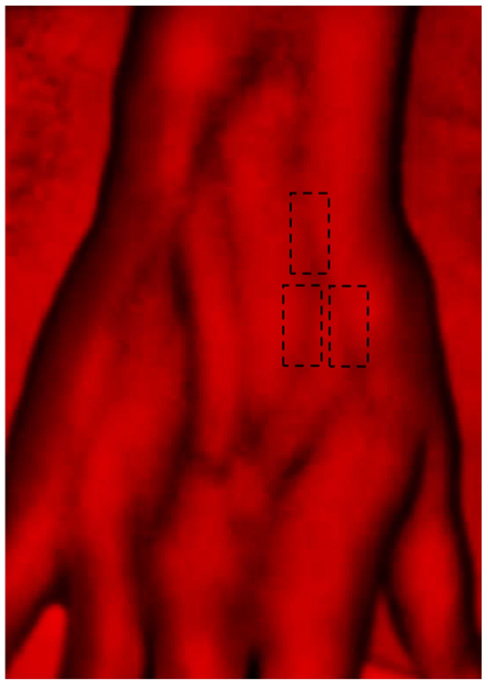
The video files of the participants were decomposed into individual frames using Matlab R2015a (MathWorks, Natick, MA, USA). Given the high anatomical variability in the number, size, and arrangement of superficial veins on the dorsal hand, a standardized and systematic approach to region of interest (ROI) selection was adopted. In each participant, three clearly visible dorsal metacarpal veins were identified, along with two associated tributary veins. For each of these veins, a rectangular ROI was manually selected in every frame, resulting in a total of 126 ROIs analyzed across the dataset (Figure 2). In each ROI, the width perpendicular to the vein axis was manually measured using ImageJ software (version 1.54g; National Institutes of Health, Bethesda, MD, USA) at seven predefined time points: three times during baseline (minutes 1, 3, and 5), twice during occlusion (minutes 6 and 7), and three times during recovery (minutes 9, 11, and 13). These time points were selected to capture representative phases of the protocol. Each ROI was converted to a grayscale image, from which a set of textural parameters was extracted, namely contrast, correlation, energy, homogeneity, and entropy, as well as fractal dimension and lacunarity. No pre-processing of images was performed before assessing textural parameters. Texture features were extracted from grayscale ROIs using standard gray-level co-occurrence matrix (GLCM) methodology. The co-occurrence matrices were normalized as probability distributions, but no normalization of grayscale intensity values was applied to the input ROIs prior to texture analysis. The spatial resolution of the images was estimated at approximately 0.15 mm/pixel, based on calibration using a reference object within the imaging field. This resolution was consistent across all frames and participants due to the fixed position of the imaging system. All analyses were conducted on rectangular ROIs of identical pixel dimensions, manually positioned using consistent anatomical landmarks to ensure reproducibility and minimize variability due to lighting or movement.
Recognizing that isolated texture parameters might not fully capture the venous response to congestion and reperfusion, we additionally computed derived indices that combine morphological (vein width) and textural information. This approach was motivated by the observation that correlation increased during occlusion, while entropy remained globally stable. Based on this physiological rationale, the composite indices were constructed heuristically rather than derived from data-driven multivariate models. Therefore, we hypothesized that expressing texture in relative terms, normalized to vein size or entropy, could offer further insight into vascular adaptation. Accordingly, we computed the following composite indices: entropy-to-width ratio, correlation-to-width ratio, total entropic content, and correlation-to-entropy ratio. These derived parameters were designed to capture the relationship between internal texture organization and structural expansion during venous filling. Due to the manual nature of width measurements, these indices were calculated at discrete time points rather than continuously across the full sequence. Other first-order histogram features such as mean intensity, skewness, and kurtosis were explored but did not reveal meaningful phase-dependent changes and were thus not included in the final analysis. A full description of all extracted and derived variables is presented in Table 2. Normality of the data were assessed using the Shapiro–Wilk test, which indicated that most parameters did not follow a normal distribution. Therefore, comparisons between experimental phases were performed using the Wilcoxon signed-rank test for related samples. A significance level of 0.05 was adopted for all statistical analyses.

3. Results

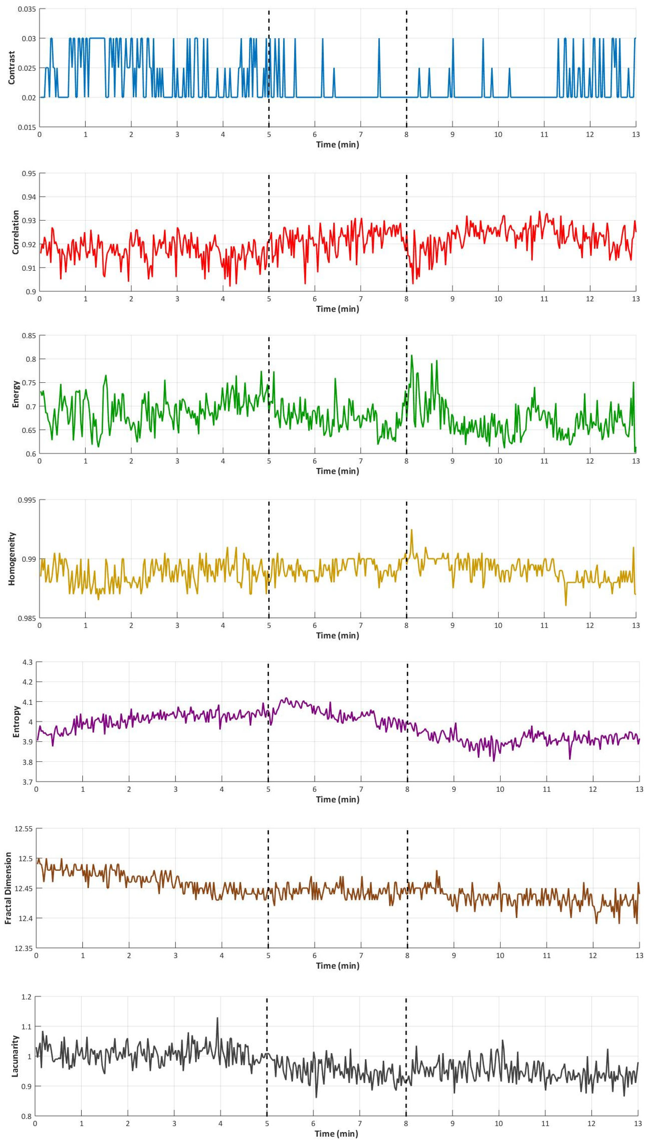
Table 3 presents the median and 95% confidence interval values for vein width and all calculated textural parameters across the different phases of the reactive hyperemia procedure. Figure 3 illustrates the temporal profile of each textural parameter throughout the procedure. Vein width increased significantly during occlusion (p < 0.001). However, it did not fully return to baseline during the recovery phase, remaining significantly higher than at baseline (p = 0.020). Conversely, most individual textural parameters, including contrast, energy, entropy, homogeneity, fractal dimension, and lacunarity, did not exhibit significant changes during occlusion. The exception was correlation, which increased significantly (p = 0.023) and returned to baseline levels during recovery. Lacunarity demonstrated a progressive decline over time, reaching a value significantly lower than baseline during recovery (p = 0.024). Among the composite indices, both entropy-to-width and correlation-to-width ratios decreased significantly with occlusion (p < 0.001 for both) and did not recover to baseline values. Conversely, total entropic content increased significantly with occlusion (p < 0.001) and remained elevated during recovery.

4. Discussion

4.1. Interpretation of Classical Texture Parameters

Several imaging modalities, such as computed tomography (CT), magnetic resonance imaging (MRI), and phlebography, have been used to detect venous congestion in specific clinical contexts [34,35,36]. However, their use is often limited by availability, cost, or invasiveness. In contrast, simpler and noninvasive techniques such as Doppler ultrasound [37] and photoplethysmography (PPG) [38,39] are widely applied in clinical and research settings to assess venous congestion and refill dynamics. While Doppler ultrasound and PPG are valuable for capturing hemodynamic parameters such as flow velocity or volume changes, they do not provide spatial or structural insights into superficial venous patterns. The present approach may therefore serve as a complementary tool in vascular research, particularly when integrated with established flow-based techniques in future validation studies. The NIRI-based vein finder used in this study offers a complementary perspective by capturing structural and textural changes in superficial veins in response to congestion. It produces images that reflect the amount of infrared light absorbed by the skin [40], where veins appear darker due to higher absorption by hemoglobin, while the overlying skin appears in varying shades of gray. Even though venous occlusion can be achieved with lower cuff pressures [41,42], we aimed at evoking a reactive hyperemia phenomenon, since this maneuver creates a faster venous filling, which could also change textural characteristics. As expected, the venous congestion resulting from occlusion at 200 mmHg translated to the significant increase in vein width. Here, we integrated textural analysis, aiming to understand whether passive venous filling alters the internal texture of vein projections. This approach is not only novel but also potentially more accessible than manual geometric quantifications.
Among the texture parameters assessed, only correlation changed significantly during occlusion. This parameter, derived from the GLCM, reflects the predictability or regularity of gray-level patterns [43,44,45]. Its increase likely reflects a smoother, more homogeneous distribution of intensities within the distended veins, possibly due to greater intraluminal filling. Conversely, the contrast, energy, homogeneity, and entropy parameters did not change statistically. This suggests that despite mechanical distension, the overall variability and intensity gradients within the vein ROIs remained relatively stable. Similarly, fractal dimension, a measure of geometric complexity and self-similarity, did not vary, indicating preservation of the hierarchical organization of the vein pattern across scales. Notably, lacunarity, which captures heterogeneity and the distribution of gaps within a structure, decreased steadily throughout the protocol, with a significant reduction in the recovery phase. This may indicate a more uniform and less fragmented appearance of venous filling after reactive hyperemia, although further investigation is required to interpret this behavior physiologically.

4.2. Divergence Between Morphology and Texture

Notably, none of the classical texture parameters followed the same dynamic profile as vein width throughout the protocol. Rather than indicating a limitation, this divergence highlights that texture and morphometry may capture different aspects of the venous response. While vein width reflects macroscopic expansion, texture parameters may be more sensitive to microstructural or organizational features that evolve independently. This supports the idea that structural and textural analyses are not redundant, but complementary, each offering a distinct physiological lens [46,47]. Despite significant venous dilation, most texture parameters remained globally stable, indicating that the grayscale distribution within the vein projections was not fundamentally altered by passive filling. However, the isolated increase in correlation, alongside the stable entropy, hints at localized reorganization rather than global disruption. Correlation captures regularity in pixel transitions, while entropy reflects overall disorder. Their divergence highlights how different aspects of texture may respond independently to hemodynamic changes. This divergence motivated the development of composite indices that integrate texture parameters with vein size. Our hypothesis was that single texture parameters may not fully capture the complexity of venous adaptation, especially when structure and signal organization evolve at different scales. By expressing textural parameters relative to morphological change, these derived parameters aimed to uncover hidden relationships between expansion and internal signal structure.

4.3. Composite Parameters and Their Physiological Meaning

To better capture how venous texture evolves with morphological changes, we developed composite parameters that integrate both dimensions [48]. Rather than assessing whether isolated parameters change, these indices explore how texture behaves relative to vein expansion. Among them, the entropy-to-width ratio reflects the informational complexity per unit of venous cross-sectional size, essentially, how “densely disorganized” the signal is. Its significant decrease during occlusion and incomplete recovery thereafter suggest that while entropy remained stable, the enlarged vein diluted the local complexity. This may reflect a more homogeneous, less chaotic signal within the distended structure, consistent with smoother intraluminal filling. Similarly, the correlation-to-width ratio declined with occlusion. Although absolute correlation increased, indicating more regular pixel relationships, this index adjusts for the simultaneous growth in vein size. The decline implies that regularity did not scale proportionally with expansion, suggesting that the additional volume was more uniform but not more ordered at the microtextural level, which may reflect different venous filling profiles across veins of varying diameters [49].
In contrast, total entropic content increased significantly during occlusion and remained elevated during recovery. This metric captures the total informational complexity across the entire venous cross-section. The rise suggests that while local entropy per pixel did not change, the overall signal complexity increased, likely due to the greater tissue volume or variability between vein lumens. Finally, the correlation-to-entropy ratio rose modestly, reaching statistical significance only during the recovery phase. This may indicate a physiological shift toward a more structured organization relative to disorder, possibly reflecting a reduction in flow velocity. Although we did not directly measure venous flow, one possible interpretation is that the delayed rise in this index reflects gradual reorganization of the venous signal as congestion resolves. Interestingly, the pattern observed in Figure 3 mirrors this temporal behavior—the ratio declines during baseline, possibly reflecting reduced venous return due to immobility [50], then increases with occlusion-induced volume accumulation, and rises further after cuff release, approaching baseline only after several minutes. Taken together, these composite indices revealed phase-dependent changes not detected by single features. By expressing texture in relation to size and complexity, they offered a more sensitive and physiologically meaningful characterization of venous adaptation to congestion and reperfusion.

4.4. Possible Relation with Movement Artifacts

Although entropy has previously been interpreted as a marker of local structural complexity potentially influenced by venous occlusion, our current findings revealed no statistically significant variation in this parameter across the phases of the protocol. This stability might initially suggest the absence of relevant physiological changes in pixel-level complexity. However, it also invites reflection on potential confounding factors, particularly motion-related artifacts, which are common in superficial imaging of the hand dorsum. In this context, minor involuntary movements or positional adjustments, especially under cuff-induced pressure or transient discomfort, could introduce small displacements of the skin surface. Such movements might affect pixel alignment within the ROIs and generate slight fluctuations in grayscale distribution, which are known to impact texture-derived measures such as entropy [51]. Yet, the absence of significant entropy variation in our results may suggest that any motion-related variability was minimal or effectively neutralized by averaging across frames and subjects. Moreover, supporting the robustness of our measurements, no parallel changes were observed in other texture features known to be sensitive to image disruption, namely contrast, energy, and homogeneity. This pattern strengthens the interpretation that the image texture remained globally stable and that movement was unlikely to have masked substantial entropy dynamics. Nonetheless, the possibility that motion artifacts subtly influenced image consistency cannot be entirely excluded. To further reduce this potential source of noise in future studies, we recommend implementing motion correction algorithms or image registration techniques [52], particularly when working with small ROIs and manually defined regions. Additionally, using high-frame-rate imaging or mechanical stabilization of the hand could enhance the temporal resolution and minimize intra-procedural variability. Ultimately, the lack of significant entropy variation, despite clear changes in vein width and correlation, likely reflects a true physiological decoupling. Alternatively, it may highlight the need for composite indices, such as entropy-to-width ratios, which more effectively capture the dynamic interplay between texture and morphology.

4.5. Study Limitations and Future Directions

A key limitation of this study is the small and homogeneous sample, composed exclusively of healthy young adults. While sufficient to assess intra-subject dynamics and methodological feasibility, this limited cohort restricts the generalizability of our findings. Caution is therefore warranted when extrapolating these results to broader or clinical populations. Additionally, while the composite indices proposed in this study were based on physiological reasoning and observed signal dynamics, they were constructed heuristically. No multivariate techniques were applied, as the limited sample size would not support stable dimensionality reduction. Future studies with larger cohorts should explore data-driven approaches to validate or refine these indices. Future research should apply the proposed texture-based parameters to diverse populations, particularly those with known vascular dysfunction such as diabetes or chronic venous insufficiency. Additionally, longitudinal studies could assess the stability and sensitivity of these parameters over time, supporting their potential use as non-invasive biomarkers for vascular health monitoring.

5. Conclusions

This study shows that texture analysis of vein images during reactive hyperemia can reveal functional aspects of venous perfusion not captured by morphological measures alone. While vein width increased during occlusion, most texture parameters remained stable, suggesting that structural and organizational changes may occur independently. Composite indices combining texture and geometry, such as entropy-to-width and correlation-to-entropy ratios, were more sensitive to dynamic changes and may serve as accessible markers of venous function. These findings support the potential of vein finder devices as low-cost tools for functional vascular assessment. Future studies should validate this approach in clinical populations with known vascular dysfunctions, such as diabetes or chronic venous insufficiency, where texture-based parameters may offer diagnostic or monitoring value.

Author Contributions

Conceptualization, H.S.; methodology, H.S. and C.R.; software, H.S.; validation, H.S.; formal analysis, H.S.; investigation, H.S. and C.R.; resources, H.S.; data curation, H.S.; writing—original draft preparation, H.S.; writing—review and editing, H.S. and C.R.; visualization, H.S.; supervision, H.S.; project administration, H.S. All authors have read and agreed to the published version of the manuscript.

Funding

This research received no external funding.

Institutional Review Board Statement

The study was conducted in accordance with the Declaration of Helsinki and approved by the Ethics Committee for Human Research (CEISH) of the Faculty of Pharmacy of the University of Lisbon (protocol no. 11/2024, approved on 11 October 2024).

Informed Consent Statement

Informed consent was obtained from all subjects involved in the study.

Data Availability Statement

The raw data supporting the conclusions of this article will be made available by the authors on request.

Acknowledgments

The authors thank all volunteers for their participation in this study.

Conflicts of Interest

The authors declare no conflicts of interest.

References

  1. Cleman, J.; Xia, K.; Haider, M.; Nikooie, R.; Scierka, L.; Romain, G.; Attaran, R.R.; Grimshaw, A.; Mena-Hurtado, C.; Smolderen, K.G. A state-of-the-art review of quality-of-life assessment in venous disease. J. Vasc. Surg. Venous Lymphat. Disord. 2024, 12, 101725. [Google Scholar] [CrossRef]
  2. Skoog, J.; Nelzén, O.; Zachrisson, H. Venous Compliance in Great Saphenous Vein Incompetence: Pre- and Post-interventional Changes. EJVES Vasc. Forum 2020, 47, 78–82. [Google Scholar] [CrossRef]
  3. Spiridon, M.; Corduneanu, D. Chronic Venous Insufficiency: A Frequently Underdiagnosed and Undertreated Pathology. Maedica 2017, 12, 59–61. [Google Scholar]
  4. Zhang, F.; Song, H.-X.; He, Z.-P.; Zheng, L.-H.; Han, Y.-R.; Wang, B.-Y.; Liu, P. Analysis of computed tomography venography for the diagnosis and endovascular treatment of iliac venous compression syndrome with venous leg ulcers: A retrospective study. Sci. Rep. 2024, 14, 22314. [Google Scholar] [CrossRef]
  5. Zhu, L.-L.; Wang, Y.-H.; Zhou, Q. Progress in Research on the Mechanisms and Interventions of Phlebitis from the Perspective of Vascular Endothelial Cell and Signaling Pathway. J. Inflamm. Res. 2023, 16, 6469–6481. [Google Scholar] [CrossRef]
  6. Schulman, S.; Makatsariya, A.; Khizroeva, J.; Bitsadze, V.; Kapanadze, D. The Basic Principles of Pathophysiology of Venous Thrombosis. Int. J. Mol. Sci. 2024, 25, 11447. [Google Scholar] [CrossRef]
  7. Kassaï, B.; Boissel, J.-P.; Cucherat, M.; Sonie, S.; Shah, N.R.; Leizorovicz, A. A systematic review of the accuracy of ultrasound in the diagnosis of deep venous thrombosis in asymptomatic patients. Thromb. Haemost. 2004, 91, 655–666. [Google Scholar]
  8. Burnside, P.R.; Green, E.; Kline, J.A. Indirect computed tomography venography: A report of vascular opacification. Emerg. Radiol. 2010, 17, 195–201. [Google Scholar] [CrossRef]
  9. Arnoldussen, C.W.K.P.; De Graaf, R.; Wittens, C.H.A.; De Haan, M.W. Value of magnetic resonance venography and computed tomographic venography in lower extremity chronic venous disease. Phlebol. J. Venous Dis. 2013, 28, 169–175. [Google Scholar] [CrossRef]
  10. Kistner, R.L. Diagnosis of chronic venous insufficiency. J. Vasc. Surg. 1986, 3, 185–188. [Google Scholar] [CrossRef]
  11. Aulagnier, J.; Hoc, C.; Mathieu, E.; Dreyfus, J.F.; Fischler, M.; Le Guen, M. Efficacy of Accuvein to facilitate peripheral intravenous placement in adults presenting to an emergency department: A randomized clinical trial. Acad. Emerg. Med. 2014, 21, 858–863. [Google Scholar] [CrossRef]
  12. Pan, C.-T.; Francisco, M.D.; Yen, C.-K.; Wang, S.-Y.; Shiue, Y.-L. Vein pattern locating technology for cannulation: A review of the low-cost vein finder prototypes utilizing near infrared (NIR) light to improve peripheral subcutaneous vein selection for phlebotomy. Sensors 2019, 19, 3573. [Google Scholar] [CrossRef]
  13. Francisco, M.D.; Chen, W.-F.; Pan, C.-T.; Lin, M.-C.; Wen, Z.-H.; Liao, C.-F.; Shiue, Y.-L. Competitive real-time near infrared (NIR) vein finder imaging device to improve peripheral subcutaneous vein selection in venipuncture for clinical laboratory testing. Micromachines 2021, 12, 373. [Google Scholar] [CrossRef]
  14. Vyas, V.; Sharma, A.; Goyal, S.; Kothari, N. Infrared vein visualization devices for ease of intravenous access in children: Hope versus hype. Anaesthesiol. Intensiv. Ther. 2021, 53, 69–78. [Google Scholar] [CrossRef] [PubMed]
  15. Zhang, Z.; Wang, X.; Zhang, L.; Lou, X.; Su, X.; Wang, X.; Sun, F.; He, X. Infrared Vein Imaging for Insertion of Peripheral Intravenous Catheter for Patients Requiring Isolation for Severe Acute Respiratory Syndrome Coronavirus 2 Infection: A Nonrandomized Clinical Trial. J. Emerg. Nurs. 2022, 48, 159–166. [Google Scholar] [CrossRef] [PubMed]
  16. Renno, I.; Horch, R.E.; Ludolph, I.; Cai, A.; Arkudas, A. Vein Visualization With a Near Infrared Imaging Device and Its Impact on Students’ and Nurses’ Skills in an Academic Teaching University Hospital. J. Infus. Nurs. 2024, 47, 249–254. [Google Scholar] [CrossRef]
  17. Fraifeld, A.D.; Thompson, J.A. Incorporating Near Infrared Light Vein Visualization Technology Into Peripheral Intravenous Access Protocols. J. Infus. Nurs. 2023, 46, 313–319. [Google Scholar] [CrossRef]
  18. Gupta, S.; Mehta, N.; Bajpai, M.; Gaurav, V. Near-infrared vein finder for assessment of superficial venous malformations. Indian Dermatol. Online J. 2023, 14, 448–449. [Google Scholar] [CrossRef]
  19. Goldschmidt, E.; Faraji, A.H.; Jankowitz, B.T.; Gardner, P.; Friedlander, R.M. Use of a near-infrared vein finder to define cortical veins and dural sinuses prior to dural opening. J. Neurosurg. 2020, 133, 1202–1209. [Google Scholar] [CrossRef]
  20. Castellano, G.; Bonilha, L.; Li, L.M.; Cendes, F. Texture analysis of medical images. Clin. Radiol. 2004, 59, 1061–1069. [Google Scholar] [CrossRef]
  21. Li, Z.; Yu, L.; Wang, X.; Yu, H.; Gao, Y.; Ren, Y.; Wang, G.; Zhou, X. Diagnostic Performance of Mammographic Texture Analysis in the Differential Diagnosis of Benign and Malignant Breast Tumors. Clin. Breast Cancer 2018, 18, e621–e627. [Google Scholar] [CrossRef]
  22. Jan, Y.-K.; Hung, I.Y.-J.; Cheung, W.C. Texture Analysis in Musculoskeletal Ultrasonography: A Systematic Review. Diagnostics 2025, 15, 524. [Google Scholar] [CrossRef]
  23. Lubner, M.G.; Smith, A.D.; Sandrasegaran, K.; Sahani, D.V.; Pickhardt, P.J. CT texture analysis: Definitions, applications, biologic correlates, and challenges. Radiographics 2017, 37, 1483–1503. [Google Scholar] [CrossRef]
  24. Kunimatsu, A.; Yasaka, K.; Akai, H.; Sugawara, H.; Kunimatsu, N.; Abe, O. Texture Analysis in Brain Tumor MR Imaging. Magn. Reson. Med. Sci. 2022, 21, 95–109. [Google Scholar] [CrossRef] [PubMed]
  25. Fang, J.; Sun, W.; Wu, D.; Pang, P.; Guo, X.; Yu, C.; Lu, W.; Tang, G. Value of texture analysis based on dynamic contrast-enhanced magnetic resonance imaging in preoperative assessment of extramural venous invasion in rectal cancer. Insights Into Imaging 2022, 13, 1–11. [Google Scholar] [CrossRef]
  26. Canellas, R.; Mehrkhani, F.; Patino, M.; Kambadakone, A.; Sahani, D. Characterization of portal vein thrombosis (neoplastic versus bland) on CT images using software-based texture analysis and thrombus density (hounsfeld units). Am. J. Roentgenol. 2016, 207, W81–W87. [Google Scholar] [CrossRef]
  27. Ito, K.; Muraoka, H.; Hirahara, N.; Sawada, E.; Tokunaga, S.; Kaneda, T. Texture analysis of low-flow vascular malformations in the oral and maxillofacial region: Venous malformation vs. lymphatic malformation. Pol. J. Radiol. 2022, 87, 494–499. [Google Scholar] [CrossRef]
  28. Rosenberry, R.; Nelson, M.D. Reactive hyperemia: A review of methods, mechanisms, and considerations. Am. J. Physiol. Integr. Comp. Physiol. 2020, 318, R605–R618. [Google Scholar] [CrossRef]
  29. Marathe. A Novel Wireless Vein Finder. In International Conference on Circuits, Communication, Control and Computing; IEEE: Piscataway, NJ, USA, 2014. [Google Scholar]
  30. Shourav, M.K.; Choi, J.; Kim, J.K. Visualization of superficial vein dynamics in dorsal hand by near-infrared imaging in response to elevated local temperature. J. Biomed. Opt. 2021, 26, 026003. [Google Scholar] [CrossRef]
  31. Gonzalez, R.C.; Woods, R.E.; Eddins, S.L. Digital Image Processing Using MATLAB; Pearson/Prentice Hall: Upper Saddle River, NJ, USA, 2004; 609p. [Google Scholar]
  32. Gould, D.J.; Vadakkan, T.J.; Poché, R.A.; Dickinson, M.E. Multifractal and Lacunarity Analysis of Microvascular Morphology and Remodeling. Microcirculation 2011, 18, 136–151. [Google Scholar] [CrossRef]
  33. Myint, S.W.; Lam, N. A study of lacunarity-based texture analysis approaches to improve urban image classification. Comput. Environ. Urban Syst. 2005, 29, 501–523. [Google Scholar] [CrossRef]
  34. Kara, M.; Pradel, C.; Phan, C.; Miquel, A.; Arrivé, L. CT features of vertebral venous congestion simulating sclerotic metastases in nine patients with thrombosis of the superior vena cava. Am. J. Roentgenol. 2016, 207, 80–86. [Google Scholar] [CrossRef]
  35. Asciutto, G.; Mumme, A.; Marpe, B.; Köster, O.; Asciutto, K.; Geier, B. MR Venography in the Detection of Pelvic Venous Congestion. Eur. J. Vasc. Endovasc. Surg. 2008, 36, 491–496. [Google Scholar] [CrossRef] [PubMed]
  36. Steenbeek, M.P.; van der Vleuten, C.J.M.; Schultze Kool, L.J.; Nieboer, T.E. Noninvasive diagnostic tools for pelvic congestion syndrome: A systematic review. Acta Obstet. Et. Gynecol. Scand. 2018, 97, 776–786. [Google Scholar] [CrossRef] [PubMed]
  37. Galindo, P.; Gasca, C.; Argaiz, E.R.; Koratala, A. Point of care venous Doppler ultrasound: Exploring the missing piece of bedside hemodynamic assessment. World J. Crit. Care Med. 2021, 10, 310–322. [Google Scholar] [CrossRef]
  38. Darvall, K.; Sam, R.; Bate, G.; Adam, D.; Silverman, S.; Bradbury, A. Photoplethysmographic venous refilling times following ultrasound guided foam sclerotherapy for symptomatic superficial venous reflux: Relationship with clinical outcomes. Eur. J. Vasc. Endovasc. Surg. 2010, 40, 267–272. [Google Scholar] [CrossRef] [PubMed]
  39. Guven, H. Plethysmography: A game changer for chronic venous insufficiency diagnosis, treatment, and follow-up. Vascular 2024, 33, 192–199. [Google Scholar] [CrossRef]
  40. Kim, D.; Kim, Y.; Yoon, S.; Lee, D. Preliminary study for designing a novel vein-visualizing device. Sensors 2017, 17, 304. [Google Scholar] [CrossRef]
  41. Okazaki, K.; Fu, Q.; Martini, E.R.; Shook, R.; Conner, C.; Zhang, R.; Crandall, C.G.; Levine, B.D. Vasoconstriction during venous congestion: Effects of venoarteriolar response, myogenic reflexes, and hemodynamics of changing perfusion pressure. Am. J. Physiol. Regul. Integr. Comp. Physiol. 2005, 289, 1354–1359. [Google Scholar] [CrossRef]
  42. Fujii, N.; McGarr, G.W.; McNeely, B.D.; Ichinose, M.; Nishiyasu, T.; Kenny, G.P. KCa and KV channels modulate the venoarteriolar reflex in non-glabrous human skin with no roles of KATP channels, NOS, and COX. Eur. J. Pharmacol. 2020, 866, 172828. [Google Scholar] [CrossRef]
  43. Vironicka, A.S.; Sathiaseelan, J.G.R. Texture feature extraction with medical image using glcm and machine learning techniques. Ictact J. Image Video Process. 2022, 4. [Google Scholar] [CrossRef]
  44. Indra, Z.; Jusman, Y. Performance of GLCM Algorithm for Extracting Features to Differentiate Normal and Abnormal Brain Images. IOP Conf. Ser. Mater. Sci. Eng. 2021, 1082, 012011. [Google Scholar] [CrossRef]
  45. Soh, L.-K.; Tsatsoulis, C. Texture analysis of SAR sea ice imagery using gray level co-occurrence matrices. IEEE Trans. Geosci. Remote Sens. 1999, 37, 780–795. [Google Scholar] [CrossRef]
  46. Depeursinge, A.; Foncubierta-Rodriguez, A.; Van De Ville, D.; Müller, H. Three-dimensional solid texture analysis in biomedical imaging: Review and opportunities. Med. Image Anal. 2014, 18, 176–196. [Google Scholar] [CrossRef]
  47. Materka, A. Texture analysis methodologies for magnetic resonance imaging. Dialog-Clin. Neurosci. 2004, 6, 243–250. [Google Scholar] [CrossRef] [PubMed]
  48. Lee, J.; Jain, R.; Khalil, K.; Griffith, B.; Bosca, R.; Rao, G.; Rao, A. Texture feature ratios from relative CBV maps of perfusion MRI are associated with patient survival in glioblastoma. Am. J. Neuroradiol. 2016, 37, 37–43. [Google Scholar] [CrossRef] [PubMed]
  49. Persichini, R.; Lai, C.; Teboul, J.-L.; Adda, I.; Guérin, L.; Monnet, X. Venous return and mean systemic filling pressure: Physiology and clinical applications. Crit. Care 2022, 26, 150. [Google Scholar] [CrossRef]
  50. Silva, H.; Rezendes, C. Revisiting the Venoarteriolar Reflex–Further Insights from Upper Limb Dependency in Healthy Subjects. Biology 2024, 13, 715. [Google Scholar] [CrossRef]
  51. Rantalainen, T.; Chivers, P.; Beck, B.R.; Robertson, S.; Hart, N.H.; Nimphius, S.; Weeks, B.K.; McIntyre, F.; Hands, B.; Siafarikas, A. Please Don’t Move-Evaluating Motion Artifact From Peripheral Quantitative Computed Tomography Scans Using Textural Features. J. Clin. Densitom. 2018, 21, 260–268. [Google Scholar] [CrossRef]
  52. Alizadeh, M.; Conklin, C.J.; Middleton, D.M.; Shah, P.; Saksena, S.; Krisa, L.; Finsterbusch, J.; Faro, S.H.; Mulcahey, M.; Mohamed, F.B. Identification of ghost artifact using texture analysis in pediatric spinal cord diffusion tensor images. Magn. Reson. Imaging 2018, 47, 7–15. [Google Scholar] [CrossRef]
Figure 1. Posture adopted by the participants throughout the procedure. The NIRI device lamp was placed 30 cm above the test hand of the participants.
Applsci 15 05702 g001
Figure 2. NIRI image from a representative subject, showing a region of venous confluence on the dorsal hand. Three rectangular regions of interest are marked, corresponding to a superficial metacarpal vein and its two associated tributaries, which were selected for texture analysis.
Applsci 15 05702 g002
Figure 3. Median values of textural parameters extracted from the regions of interest in near-infrared vein images over time. Parameters include contrast (blue), correlation (red), energy (green), homogeneity (yellow), entropy (purple), fractal dimension (brown), and lacunarity (grey). Dashed vertical lines indicate the beginning and end of suprasystolic occlusion (minute 5 to minute 8).
Applsci 15 05702 g003
Table 1. Characteristics of the sample data subjects, expressed in mean ± standard deviation.
TotalFemalesMales
N1486
Age21.5 ± 4.221.5 ± 1.221.0 ± 6.4
Height (m)1.73 ± 0.11.69 ± 0.11.75 ± 0.1
Body mass (kg)63.0 ± 8.561.0 ± 6.065.0 ± 10.5
Body mass index (kg/m2)23.1 ± 2.323.2 ± 2.121.0 ± 3.0
Systolic blood pressure (mmHg)111.0 ± 8.5107.5 ± 8.7114.0 ± 6.8
Diastolic blood pressure (mmHg)74.0 ± 6.776.0 ± 7.474.0 ± 5.7
Menstrual cycle duration (days)-28 ± 5-
Menstrual cycle day-8 ± 15-
Table 2. Description of the texture parameters of hand dorsum veins throughout the procedure (adapted from Gonzalez [31], Gould [32] and Myint [33]. P(i,j) is the (i,j)-th entry of the normalized GLCM, representing the joint probability of two pixels with gray levels i and j at a defined offset. N is the number of gray levels. μ and σ represent the mean and standard deviation of the marginal distributions of P(i,j). Definitions are provided for contrast, correlation, energy, entropy, homogeneity, fractal dimension, and lacunarity.
ParameterDescriptionEquation
ContrastA measure of the intensity difference between neighboring pixels; indicates how much texture varies locally. i = 0 N 1 j = 0 N 1 i j 2 . P ( i , j )
CorrelationA measure of how strongly the intensity of one pixel is related to its neighbor; reflects linear patterns in texture. i = 0 N 1 j = 0 N 1 i µ i ( j µ j ) . P ( i , j ) σ i . σ j
EnergyA measure of image uniformity; calculated as the sum of squared values in the co-occurrence matrix. i = 0 N 1 j = 0 N 1 P i , j 2
HomogeneityA measure of the similarity between neighboring pixel intensities; higher values indicate smoother and more uniform textures. i = 0 N 1 j = 0 N 1 P ( i , j ) 1 + i j
EntropyA measure of randomness or complexity in the distribution of co-occurring gray levels within the GLCM; higher values indicate greater texture irregularity and disorder. i = 0 N 1 j = 0 N 1 P i , j . l o g 2 P ( i , j )
Fractal dimensionA non-integer exponent that describes how detail in a pattern changes with scale. Calculated with the box-counting method, where ε represents the size (side length) of the box used to cover the structure and N(ε) is the number of boxes of size ε that are needed to completely cover the object lim ε 0 l o g N ( ε ) l o g ( 1 ε )
LacunarityA scalar value that characterizes the distribution of gaps or voids in a binary structure. It is calculated as the ratio between the square of the variance (σ) and the square of the mean (µ) of the pixel intensities. σ µ 2 2
Entropy-to-width ratioA measure of the amount of texture detail present in relation to the visible size of the vein.Entropy/Width
Correlation-to-width ratioA measure of the consistency of the texture pattern relative to the vein’s width.Correlation/Width
Total entropic contentA measure of the overall texture variation and visual complexity across the entire vein image.Entropy × Width
Correlation-to-entropy ratioA measure of the amount of order in the texture in relation to its level of complexity.Correlation/Entropy
Table 3. Median and 95% confidence interval values for the vein width and textural parameters in the different phases of the reactive hyperemia procedure (* p < 0.05).
ParametersBaseline
(Phase I)
Occlusion
(Phase II)
Recovery
(Phase III)
p-Value
(O vs. B)
p-Value
(R vs. B)
Contrast0.021 (0.015; 0.025)0.020 (0.015; 0.024)0.021 (0.017; 0.025)0.3970.737
Correlation0.902 (0.882; 0.922)0.913 (0.900; 0.929)0.920 (0.914; 0.946)0.023 *0.074
Energy0.699 (0.628; 0.786)0.661 (0.540; 0.716)0.696 (0.631; 0.795)0.6000.157
Homogeneity0.989 (0.987; 0.992)0.990 (0.988; 0.992)0.989 (0.987; 0.991)0.5120.912
Entropy4.25 (4.15; 4.42)4.16 (4.00; 4.34)4.18 (4.11; 4.42)0.1980.083
Fractal dimension12.462 (12.439; 12.480)12.463 (12.429; 12.63)12.463 (12.423; 12.485)0.0600.319
Lacunarity1.06 (0.95; 1.15)0.98 (0.72; 1.05)0.97 (0.79; 1.05)0.1360.024 *
Vein width (mm)2.19 (2.12; 2.28)2.55 (2.34; 2.54)2.28 (2.14; 2.33)<0.001 *0.020 *
Entropy-to-width ratio1.87 (1.69; 2.01)1.67 (1.57; 1.79)1.80 (1.67; 1.91)<0.001 *0.228
Correlation-to-width ratio0.403 (0.375; 0.420)0.368 (0.350; 0.388)0.404 (0.379; 0.412)<0.001 *0.641
Total entropic content9.03 (8.50; 9.48)10.09 (9.37; 10.68)8.99 (8.26; 9.95)<0.001 *0.858
Correlation-to-entropy ratio0.213 (0.205; 0.223)0.217 (0.207; 0.229)0.221 (0.210; 0.230)0.5400.026 *
Disclaimer/Publisher’s Note: The statements, opinions and data contained in all publications are solely those of the individual author(s) and contributor(s) and not of MDPI and/or the editor(s). MDPI and/or the editor(s) disclaim responsibility for any injury to people or property resulting from any ideas, methods, instructions or products referred to in the content.