Article

Evaluating the Impact of Artificial Saliva Formulations on Stainless Steel Integrity

by
Daniela Laura Buruiana
,
Nicoleta Lucica Bogatu
,
Alina Crina Muresan
,
Elena Emanuela Herbei
,
Constantin Trus
and
Viorica Ghisman
*
Interdisciplinary Research Centre in the Field of Eco-Nano Technology and Advance Materials CC-ITI, Faculty of Engineering, “Dunarea de Jos” University of Galati, 47 Domneasca, 800008 Galati, Romania
*
Author to whom correspondence should be addressed.
Appl. Sci. 2025, 15(10), 5345; https://doi.org/10.3390/app15105345
Submission received: 14 April 2025 / Revised: 2 May 2025 / Accepted: 7 May 2025 / Published: 10 May 2025

Abstract

Featured Application

This study supports the selection of 316L stainless steel used in orthodontic and dental applications by evaluating its corrosion behavior when exposed to artificial salivas of varying chemical compositions. The findings can guide material engineers and dental professionals in selecting or surface-modifying metallic biomaterials to enhance their long-term performance and biocompatibility in the oral environment.

Abstract

The biocompatibility and long-term stability of stainless steel orthodontic devices are critically influenced by their corrosion resistance in the oral environment. This study evaluates the effect of three artificial saliva formulations—Afnor (pH 7.64), Fletcher (pH 8.07, fluoride-containing), and Fusayama/Meyer (pH 6.34, acidic)—on the surface integrity and chemical behavior of 316L stainless steel over 7 and 28 days. A multi-technique approach was employed, including SEM imaging, EDX elemental mapping, XRF analysis, microhardness testing (Vickers), and the monitoring of key physico-chemical parameters (pH, conductivity, salinity, and TDS). The results indicate that Afnor saliva maintains alloy stability with minimal surface damage while Fusayama/Meyer promotes pitting corrosion and selective leaching of Fe and Ni. Fletcher saliva led to the formation of crystalline corrosion products and significant surface hardening, likely due to the interaction of fluoride with the passive layer. Microhardness values increased across all samples after 28 days, most notably in the Fletcher condition (from 191.3 HV to 256.9 HV). These findings provide valuable insights into the time-dependent degradation mechanisms of orthodontic stainless steel in varied salivary environments, emphasizing the importance of simulating realistic oral conditions in corrosion testing. The study contributes to the optimization of material selection and surface treatment strategies for improved biocompatibility in dental applications.

1. Introduction

Biomaterials science is an interdisciplinary field that demands a deep understanding of engineering, biology, and medical sciences. It involves both fundamental and applied research focused on exploring interactions between living systems and biomaterials, designing medical devices to support biological structures, and addressing the needs of patients and healthcare professionals [1,2]. Stainless steels are among the most commonly used metallic biomaterials, extensively employed in the manufacturing of medical implants and various biomedical devices. In particular, 316L stainless steel, characterized by its low carbon content (<0.03%) and molybdenum addition (~2–3%), is widely utilized in biomedical applications due to its outstanding properties, such as excellent mechanical performance and high biocompatibility [3,4]. Medical-grade stainless steel (SS) is a type of metal widely used in manufacturing biomedical devices and implants [5]. The surface properties of a material play a crucial role in influencing its biocompatibility, corrosion resistance, antibacterial activity, and tissue regeneration potential [6]. Monik et al. [7] examined the corrosion behavior of stainless steel in artificial saliva with varying fluoride concentrations. Their findings revealed that exposure to fluoride ions leads to an increase in corrosion current density within the passivated region. Additionally, the presence of fluoride expands the passivation range of stainless steel alloys. The significance of pH in metal corrosion is well established, as dietary habits, including the consumption of carbonated beverages and desserts, can influence intraoral pH. The pH in the oral cavity is known to fluctuate considerably depending on factors such as food and beverage intake, microbial metabolism, and systemic health conditions. Reports in the literature indicate that salivary pH can range from highly acidic values around 4.0 (due to frequent sugar consumption or gastroesophageal reflux) to alkaline conditions exceeding 8.5, particularly after exposure to bicarbonate-rich saliva or antibacterial rinses [8,9]. These pH variations play a critical role in modulating the corrosion behavior of dental materials and should be considered when simulating in vitro environments. Močnik observed that a reduction in pH narrows the passive range in the cyclic potentiodynamic curves of stainless steel dental arch wires, suggesting that the stability of the passivated film diminishes in acidic environments [7]. Research by Arab-Nozari et al. [10] found that stainless steel prefabricated crowns submerged in artificial saliva with a lower pH released higher amounts of nickel (Ni). Zhao et al. [11] found that copper-containing 304 stainless steel exhibits significantly greater biofilm inhibition compared to copper-free 304 stainless steel. Additionally, in S. mutans-containing artificial saliva, the Cu-containing alloy demonstrated a lower corrosion current density (i_corr), higher polarization resistance (R_p), and greater charge transfer resistance (R_ct). N. Simionescu et al. evaluated the corrosion resistance of 316L stainless steel in two artificial saliva solutions with varying pH and chloride concentrations, aiming to assess its suitability for orthodontic applications. The study concluded that an increase in pH combined with a higher chloride concentration resulted in the lowest corrosion resistance, whereas a decrease in pH with a lower chloride concentration led to improved corrosion resistance [12].
The aim of this study is to investigate the effects of three different artificial saliva formulations on the structural and surface characteristics of stainless steel. By analyzing variations in pH, chloride concentration, and other relevant parameters, this research seeks to assess the material’s suitability for biomedical applications, particularly in dental and orthodontic environments.

2. Materials and Methods

The research was performed on medical-grade 316L stainless steel with a chemical composition (wt%) of 0.03% C, 16.5–18.5% Cr, 11–14% Ni, 2.0–2.5% Mo, 1.0% Si, 2.0% Mn, and 61.97–67.47% Fe. The formulations of the artificial saliva used in this research were Afnor [13], Fusayama/Meyer [14,15], and Fletcher [16]. Three artificial saliva formulations were prepared to simulate different oral conditions: Afnor (neutral pH, reference model), Fletcher (alkaline pH with fluoride ions), and Fusayama/Meyer (acidic pH with urea and sulfides). Although the original recipes for these solutions were introduced more than two decades ago [13,14,15,16], they remain widely accepted in contemporary corrosion and biomaterial research due to their reproducibility and physiological relevance. Recent studies continue to employ these formulations to assess corrosion resistance and ion release in orthodontic alloys [17,18]. The timespans of 7 and 28 days were selected to represent short- and medium-term exposure periods, which are commonly used in biomaterial corrosion studies and simulate early clinical conditions. This approach also enables future comparison with longer term tests. The samples were stored in artificial saliva at a 37 °C temperature in order to simulate the oral environment.
Scanning Electron Microscopy (SEM): The surface morphology of the AISI 316L stainless steel samples was analyzed using the Tescan Vega Scanning Electron Microscope. This instrument represents the fourth generation of SEM systems, combining high-resolution imaging with real-time elemental composition analysis. The integrated software provides a highly efficient analytical platform suitable for quality control, failure analysis, and routine material inspections in research laboratories. One of its key advantages is the vacuum damper, which significantly reduces the operating time of the rotary vacuum pump, providing both economic and biological benefits. Additionally, the Vega Compact chamber allows for the rapid handling and analysis of multiple samples due to its large capacity.
Fourier Transform Infrared (FTIR) Spectroscopy: FTIR analysis was conducted using a Shimadzu spectrophotometer equipped with a QATR-S accessory. This accessory includes a diamond crystal prism, enabling the detection of wavelengths down to 400 cm−1. The device accurately measured the absorbance of infrared photons by the 316L stainless steel samples immersed in three types of artificial saliva. The resulting spectra revealed the presence of various chemical functional groups formed on the sample surfaces as a result of interaction with the simulated biological media.
X-ray Fluorescence (XRF) Spectroscopy: Elemental composition was determined using an Olympus portable XRF analyzer, which employs X-ray fluorescence spectroscopy to detect and quantify chemical elements. The analyzer uses high-energy radiation to stimulate emission from the sample surface and is capable of detecting elements ranging from magnesium to uranium. A major advantage of this device is its portability, which allows for fast and flexible sample analysis. In this study, the control sample’s spectrum matched the standard chemical composition of AISI 316L stainless steel.
Microhardness Testing (Vickers Method): Mechanical surface properties were assessed using the Insize Vickers microhardness tester(INSIZE Co., Ltd., Suzhou, Jiangsu, China). Microhardness was measured at three different points on each sample, and the average Vickers and Rockwell values were reported. This method determines material hardness based on the diagonal measurements of the indentation left by a diamond indenter under a controlled load. The tester includes an integrated microscope offering 100× and 200× magnification, and allows for testing within the range of 5 HV to 3000 HV. The hardness data provided insights into surface strengthening and material response to long-term exposure to various artificial saliva environments.
The selected characterization techniques were chosen to provide a comprehensive assessment of surface, chemical, and mechanical changes induced by corrosion. SEM and EDX were used to evaluate morphological degradation and elemental distribution. XRF offered rapid quantitative analysis of elemental composition changes. FTIR enabled the detection of new surface compounds formed after immersion, while microhardness testing (Vickers method) was employed to assess changes in surface mechanical properties caused by corrosion or mineral deposition. This multi-analytical approach ensures robust interpretation of time-dependent interactions between stainless steel and artificial saliva.

3. Results and Discussion

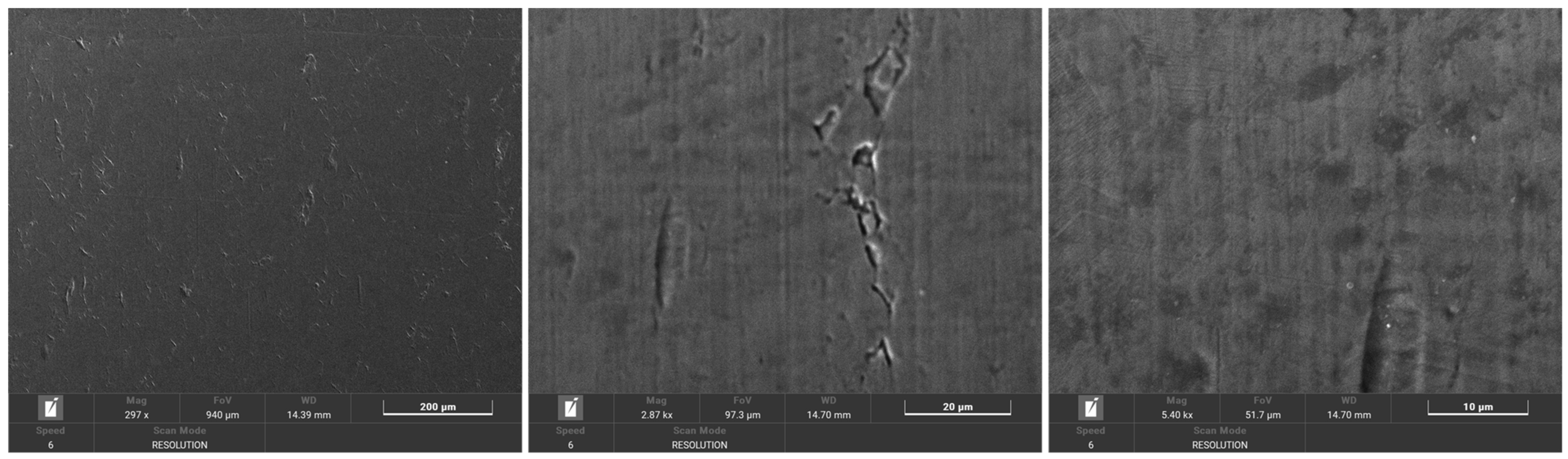
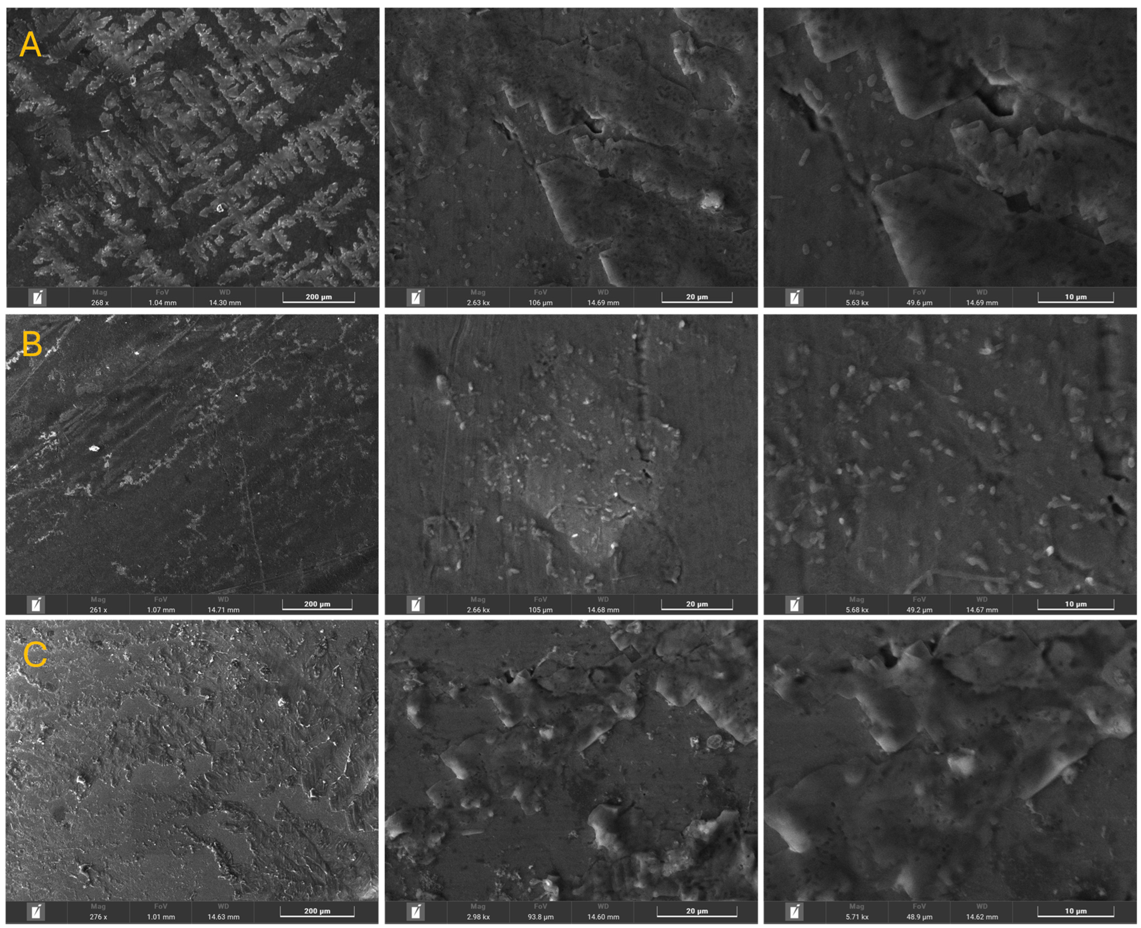
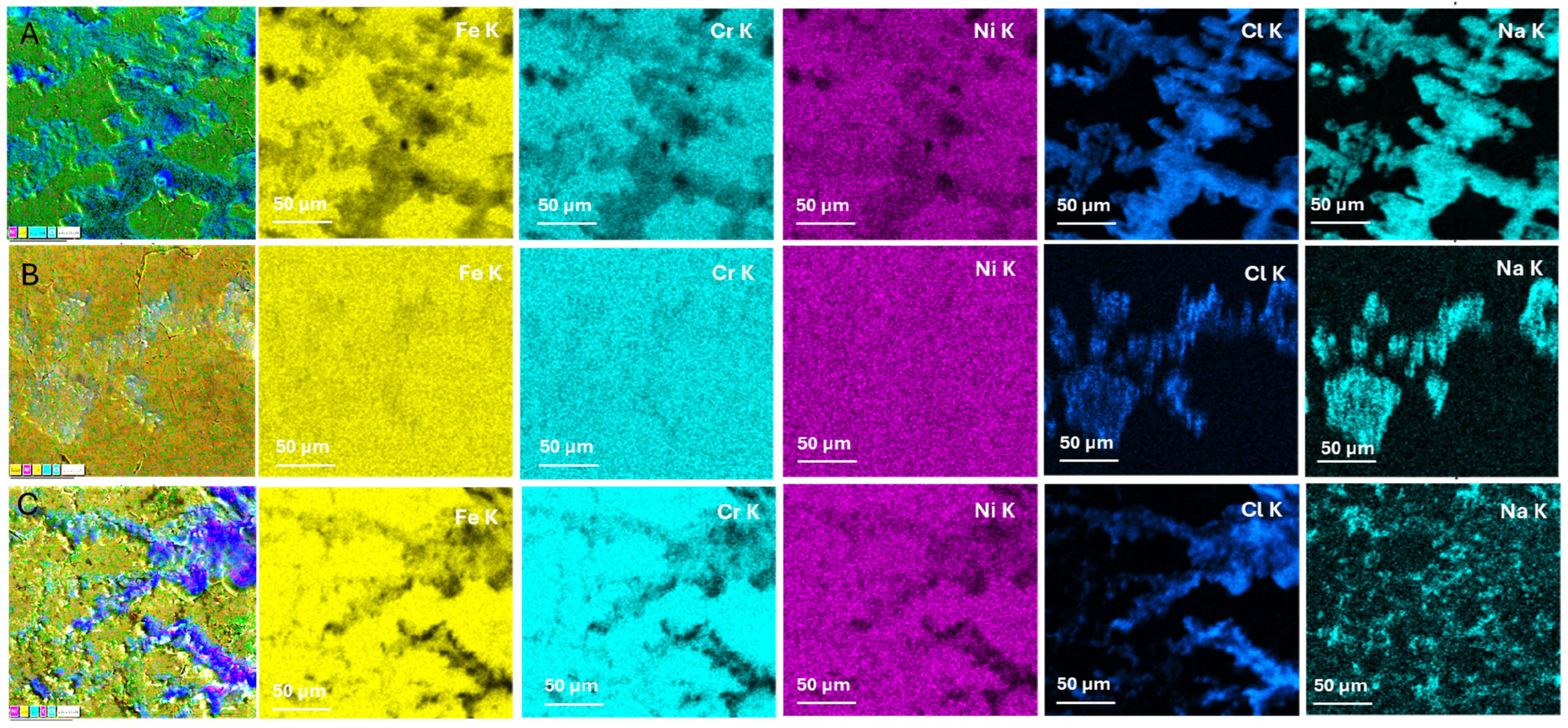
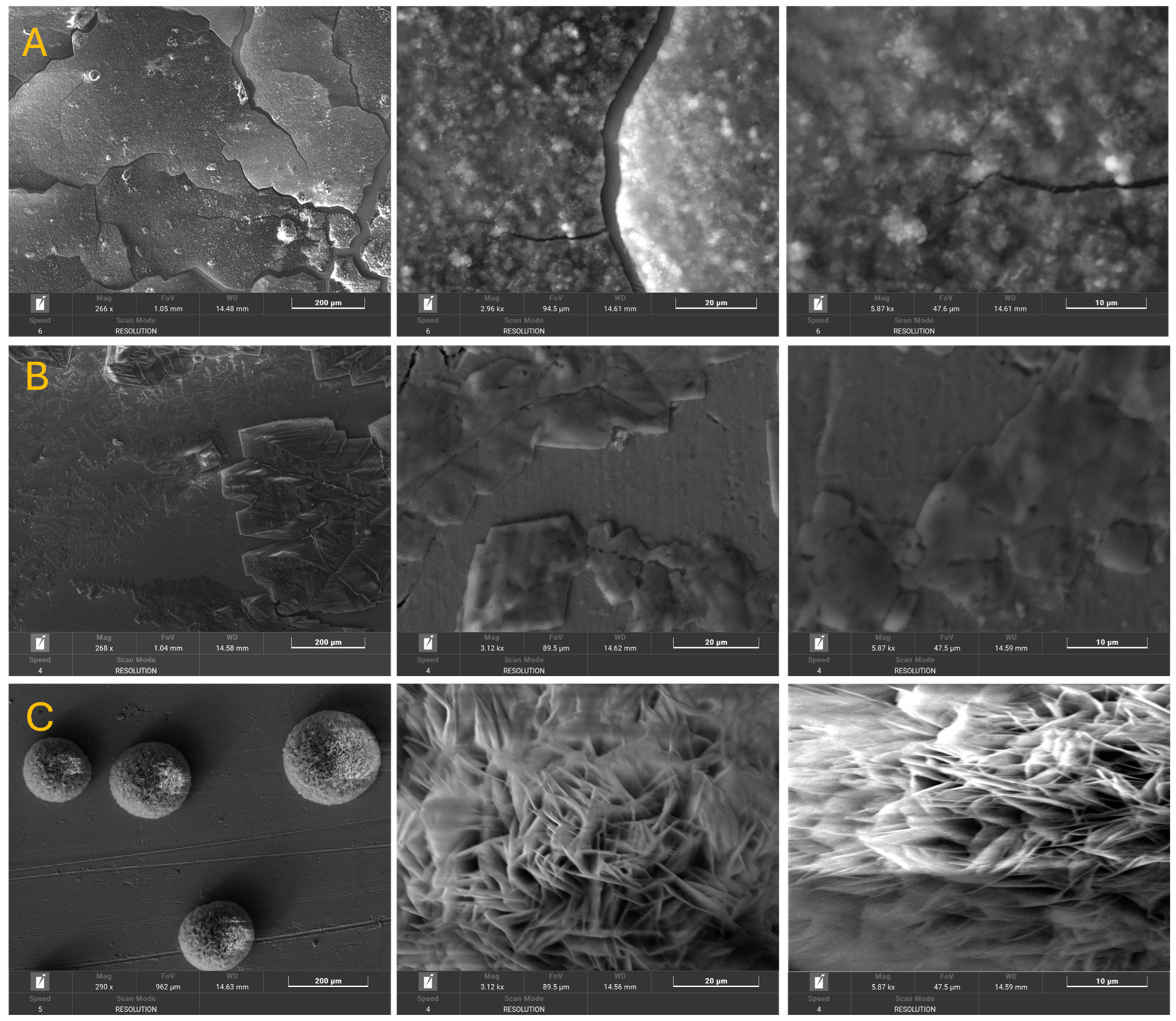
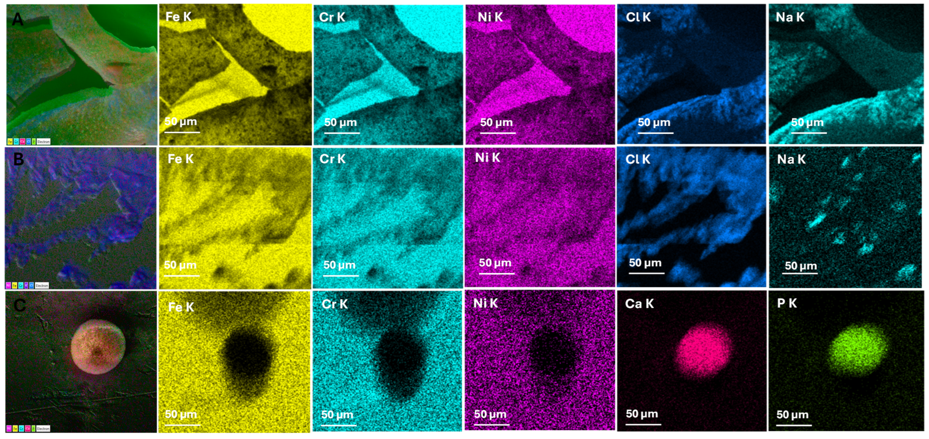
3.1. Morphological and Chemical Composition

Figure 1 presents the SEM analysis of sample 316L before immersion in artificial saliva. The surface appears smooth and homogeneous, with no evidence of pitting, delamination, or significant defects. Minor parallel striations suggest mechanical polishing marks from sample preparation, which are typical and do not indicate surface damage. At higher magnification, elongated surface grooves and shallow micro-scratches become visible. These features are consistent with surface finishing and have not yet initiated corrosion or passive layer breakdown. The surface remains intact and free from corrosion pits. Fine features such as polishing textures and grain boundary traces can be discerned. No surface oxidation or particulate deposits are detected. The uniform contrast suggests a stable passive oxide film, typical for 316L in ambient conditions. These baseline images establish a reference morphology for direct comparison with post-immersion samples. The absence of corrosion features supports the initial chemical stability of the 316L alloy and underscores the subsequent changes observed in artificial saliva as the effects of environmental interaction rather than pre-existing flaws.
Figure 2 presents the SEM surface analysis of the 316L stainless steel samples after 7 days of immersion in three types of artificial saliva—Afnor (neutral pH), Fletcher (basic with fluoride), and Fusayama/Meyer (acidic). The results are compared at three magnifications per condition and are supported by visual observations related to corrosion or surface alterations. For Figure 2A, immersed in Afnor saliva (pH 7.64, neutral), at a low magnification the surface appears clean and well-preserved, with visible machining or polishing marks. No major pitting, cracks, or corrosion products are visible. At medium and high magnification, the surface is smooth, with minor scratches and very limited particulate accumulation. No significant pitting or localized attacks are evident. The passive Cr2O3 layer likely remained intact, showing minimal signs of corrosion. In the case of Figure 2B with Fletcher saliva (pH 8.07, basic with fluoride), at a low magnification, a slightly rougher texture compared to A can be seen. Fine scratches appear to be more irregular and there are hints of localized dull patches. At a medium magnification, the surface shows early signs of corrosion or film breakdown, with uneven morphology, but at a high magnification the nanometer- to micrometer-scale roughening and etching-like features suggest fluoride attack. This surface shows incipient degradation under fluoride exposure, with signs of passive film destabilization. For Figure 2C— Fusayama/Meyer Saliva (pH 6.34, mildly acidic)—at a low magnification, the sample shows a clearly rougher and duller surface, suggesting more widespread corrosion. Machining marks are nearly obliterated. At a medium magnification, the widespread roughening and etching are consistent with acid-induced attack, and at a high magnification the corrosion morphology is well developed, with multiple small pits or corrosion product clusters. Fusayama/Meyer saliva shows the strongest corrosive effect, with visible damage and pitting after 7 days.
Figure 3 shows the energy dispersive X-ray (EDX) mapping results for 316L stainless steel samples immersed for 7 days in three types of artificial saliva: Afnor (A), Fletcher (B), and Fusayama/Meyer (C). The elemental distributions of Fe, Cr, Ni, Cl, and Na were observed to assess the surface integrity and elemental leaching that resulted from electrochemical interactions in each environment. In the case of samples stored in Afnor saliva (Figure 3A), it can be observed that Fe is uniformly distributed, indicating no selective leaching; Cr is homogeneously present, suggesting an intact passive layer; Ni is evenly spread, with no major depletion; and Cl and Na are present superficially, not penetrating deeply. It can be concluded that the surface remained stable and that corrosion resistance was maintained. For the sample stored in Fletcher saliva (Figure 3B), regarding its elemental composition mapping, Fe presents minor surface irregularities, suggesting early interaction; Cr is slightly disrupted with potential signs of passive film attack; Ni shows slight depletion in localized areas; Cl displays structured deposition patterns, possibly along defects or grain boundaries; and Na has accumulation in defined areas, likely in its salt form. From the figure, it can be seen that early passive-layer breakdown is likely caused by an interaction between fluoride and chloride.
Figure 3C shows EDX mapping of the sample stored in Fusayama/Meyer saliva (pH 6.34, acidic) after 7 days. As can be seen, the Fe is strongly depleted in certain areas and has an uneven distribution. Cr is fragmented and less intense, showing damage to the passive layer, and Ni is less uniform, which is indicative of elemental leaching. Cl shows strong, widespread penetration indicating corrosive attack and Na is dispersed broadly, which is linked with corrosion activity. There is clear evidence of aggressive corrosion and the loss of protective elements. The Fusayama/Meyer saliva shows the strongest corrosive effect, with visible damage and pitting after 7 days.
SEM images of samples stored for 28 days in artificial saliva—Afnor, Fletcher, and Fusayama/Meyer —can be seen in Figure 4. Regarding Figure 4A—Afnor Saliva (pH 7.64, neutral)—it can be observed that at a low magnification the sample shows mild intergranular corrosion and micro-pits and, at a medium/high magnification, slight surface roughening and grain boundary exposure are visible, but the sample still remains relatively intact, which can be interpreted as minor surface evolution and the passive film being mostly preserved. As for the Fletcher saliva (pH 8.07, with Fluoride)—Figure 4B shows that crystalline corrosion products are clearly visible, possibly fluoride-related salts, and a high-magnification reveals pitting and passive film disruption. With respect to the Fusayama/Meyer saliva (pH 6.34, acidic) after 28 days (Figure 4C), large spherical deposits are present, covered with spiky, filamentous structures and severe corrosion with precipitated compounds suggests a loss of the original alloy material. This can be interpreted as strong acidic corrosion with aggressive material degradation.
Figure 5 presents EDX elemental mapping (28 days) for 316L stainless steel stored in Afnor, Fletcher, and Fusayama/Meyer artificial saliva. When considering the Afnor saliva (pH 7.64, neutral) (Figure 5A), the Fe, Cr, and Ni remain uniform with only minor signal dimming, but the Cl and Na remain at superficial levels, not indicating deep corrosion. The EDX mapping shows that the composition remains mostly stable; excellent passivation is retained. Regarding the Fletcher saliva (pH 8.07, with Fluoride) (Figure 5B), the Fe and Ni signals show slight depletion, the Cr is slightly disrupted, and the Cl and Na are more intense and diffuse compared to the 7-day data; thus, it can be concluded that surface corrosion is progressing and fluoride likely plays a key role. When considering the Fusayama/Meyer saliva (pH 6.34, acidic) (Figure 5C), the Fe, Cr, and Ni are depleted in spherical regions, suggesting material loss and the presence of strong Ca and P signals corresponding to the precipitation of new compounds with evidence of advanced corrosion and bio-like precipitation (e.g., calcium phosphate).
Table 1 presents the X-ray fluorescence (XRF) results for 316 stainless steel (an Fe–Cr–Ni–Mo alloy) that were compared before exposure (initial) and after 7 and 28 days of immersion in three artificial saliva solutions: Afnor (pH 7.64, neutral/slightly basic), Fusayama/Meyer (pH 6.34, mildly acidic), and Fletcher (pH 8.07, basic with fluoride). The focus is on changes in key alloy elements—iron (Fe), chromium (Cr), nickel (Ni), molybdenum (Mo), copper (Cu), and cobalt (Co)—to infer corrosion or leaching behavior under each condition. Elemental changes have been highlighted to emphasize surface interactions. Slight decreases in Fe and Cr, particularly in the Fletcher samples, suggest possible leaching or passive-layer disruption. The Co and Mo showed slight increases, indicating possible surface enrichment or stabilization. The presence of P in all of the exposed samples points to ionic adsorption from the artificial saliva, especially in fluoride- and phosphate-containing environments. Table 2 summarizes the approximate compositional changes, followed by detailed interpretations for each environment.
For the Afnor artificial saliva (Neutral pH 7.6), the 316 steel initially contains ~67–68% Fe, ~17% Cr, ~12–13% Ni, ~2% Mo, and only trace levels of Cu and Co (in the order of 0.1% or less). These are typical levels for 316 stainless steel, with Fe as the balance and Cr, Ni, and Mo as major alloying elements that form a passive oxide film (rich in Cr2O3 with Mo oxides) for corrosion resistance. Seven-Day Exposure: After 1 week in Afnor saliva (near-neutral pH, containing chloride, phosphate, bicarbonate, and thiocyanate ions), the XRF shows very minor changes. Fe remains about the same (within 0.1–0.2% of the initial ~67.8%), and Ni and Cr also stay essentially unchanged (changes in the order of a few hundredths of a percent). Any differences at 7 days are near the margin of error, indicating minimal general corrosion in this solution. The passive Cr-oxide film likely remains intact, preventing significant metal leaching. Twenty-Eight-Day Exposure: By 28 days, the Afnor-exposed sample still shows only slight shifts in composition relative to the initial immersion. The Fe content decreases by only a few tenths of a percent (still ~67.5–67.6%), and Ni shows a similarly tiny decrease (in the order of 0.1%). Cr conversely appears to increase slightly (to ~17.2%, a few tenths higher than initial). This subtle Cr enrichment with a correspondingly slight Fe/Ni reduction suggests that any corrosion that did occur was extremely mild and selective—the more active elements, such as the Fe (and to some extent the Ni), may have leached in minute amounts, leaving the surface enriched in Cr [19]. Such Cr enrichment is expected because chromium is less readily dissolved—it stays behind as part of the protective oxide film, which raises the measured Cr fraction at the surface as other elements depart [20]. Mo remains roughly constant or is up by a negligible amount (~2.2% vs. 2.1% initially), indicating it too is retained in the passive film. Trace Cu and Co show no meaningful change (both remain at or near their initial ~0.2% and ~0.06% levels, respectively), which is not surprising given their very low levels—any slight dissolution or concentration is within the bounds of experimental noise. Interpretation: Overall, Afnor saliva (pH ~7.6) has a negligible corrosive impact on 316 stainless steel over 28 days. The alloy’s composition is essentially stable, reflecting that the neutral pH and lack of aggressive ions (no fluoride and moderate chloride) enable the stainless steel’s passive layer to remain intact. The presence of thiocyanate (SCN) did not produce any obvious effect on the bulk composition; any influence (SCN can in some cases promote corrosion) was minimal in this static exposure. There is no strong evidence of leaching of Fe, Ni, or other elements in the Afnor solution—only a hint of Cr enrichment and Fe/Ni reduction is evident by 28 days, which indicates very slight selective dissolution. In practical terms, the steel’s elemental stability in Afnor saliva suggests excellent corrosion resistance in a neutral saliva environment, with no significant depletion of important elements.
Regarding the Fusayama/Meyer artificial saliva (acidic pH 6.3), after 7 days of exposure in a more acidic medium—containing chlorides, sulfide (S2−), phosphate, urea, etc.—the XRF changes are still relatively small but are more noticeable than in Afnor. Fe remains high (~67.8% → ~67.8% at 7 days, essentially unchanged), but Ni shows a slight decrease even at 7 days (a drop in the order of 0.1–0.2%). A small Ni loss early on is significant because Ni is known to dissolve out of stainless steel more readily under acidic conditions [20]. Cr at 7 days is roughly constant or up slightly, again hinting that a bit of Fe/Ni has been removed, leaving the surface richer in Cr. These 7-day results suggest the onset of corrosion in acidic saliva, albeit limited—Ni (and possibly some Fe) is beginning to leach, while Cr is left behind in the passive film. Twenty-Eight-Day Exposure: By 28 days, the effect of the acidic environment becomes clearer. The Fe content of the steel drops modestly (to ~67.3%, a few tenths below the initial immersion), and Ni decreases more noticeably (down to roughly 12.0%, compared to ~12.7% initially). This Ni depletion is a strong indicator of corrosion—nickel is being released from the alloy into the solution. In fact, studies have shown that a low pH greatly accelerates Ni ion release from stainless steel, much more so than a neutral pH [20]. Our results align with that—Ni is evidently one of the more “mobile” elements under acidic conditions. Fe also decreases, though to a lesser extent. The concurrent rise in Cr content (to ~17.5% vs. 17.1% initially) confirms that Cr is preferentially retained on the corroded surface [21]. In essence, as Ni and some Fe atoms leach out, the remaining surface matrix becomes enriched in the more corrosion-resistant Cr (and to a degree Mo). Mo shows a slight uptick (perhaps ~2.3% vs. 2.1% initial), suggesting that Mo is not leaching but is instead staying or even concentrating in corrosion products—consistent with Mo’s known role in reinforcing the passive film by forming insoluble compounds [22]. With respect to Fletcher artificial saliva (basic pH 8.07 with fluoride), after 7 days of exposure, which is basic and contains fluoride (NaF), the composition already shows some early signs of interaction—Fe drops slightly from the initial 67.92% to 67.64%, more than in Afnor or Fusayama/Meyer at this point. Ni decreases modestly from 10.238% to 10.270%, then is up slightly—possibly due to minor measurement variation. Cr slightly decreases from 17.167% to 16.955%. Mo increases from 2.067% to 2.140%, and Co increases more visibly from 0.263% to 0.315%. This suggests a shift in surface composition, possibly due to fluoride ions attacking the passive film, leading to selective leaching. After 28 days, the effects of fluoride and basic pH are more pronounced—Fe further decreases to 67.45%—the largest Fe loss among the three media. Cr drops from 17.167% to 16.813%, more significantly than in other media—indicating that Cr is dissolving, which is atypical and concerning. Ni increases slightly, but still fluctuates near the baseline (~10.310%). Mo increases to 2.175%, suggesting it is retained or even concentrating due to other elements dissolving. Co also increases to 0.386%, more than in any other media. These trends suggest that fluoride ions destabilize the passive Cr oxide layer, enabling Fe and Cr to dissolve. While Cr is normally stable, basic environments with fluoride are known to promote Cr dissolution, especially in dental alloys. The increased Co and Mo levels indicate that these elements are retained or even enriched as Fe and Cr leach away.

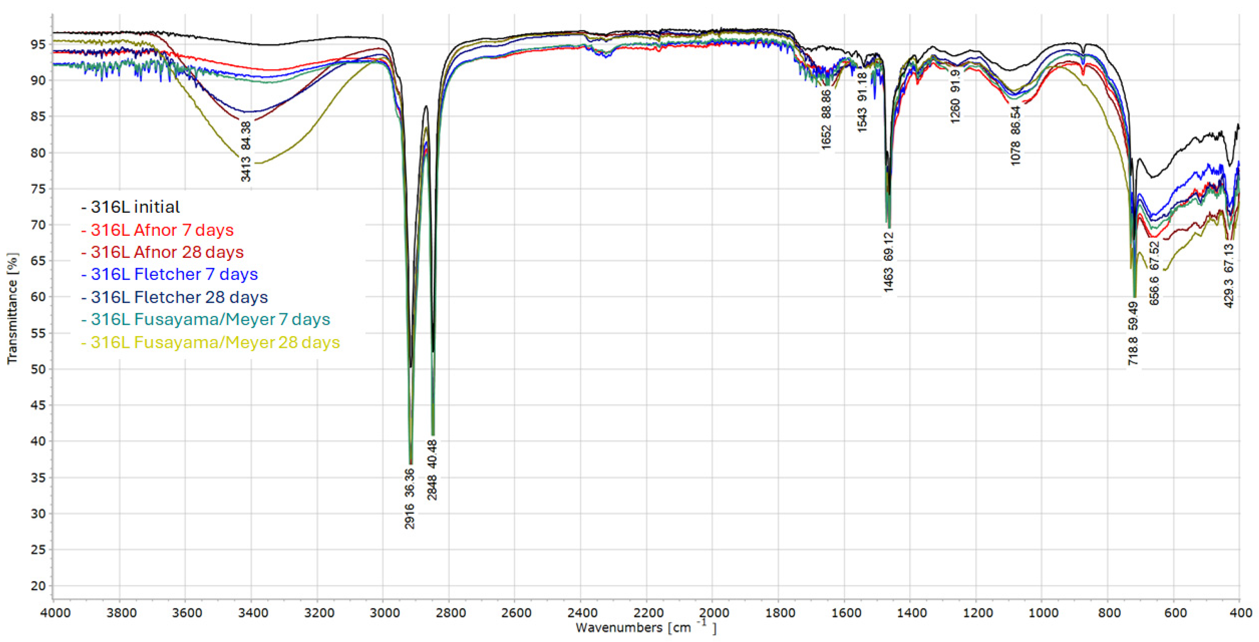
3.2. Structural Analysis

Fourier-transform infrared (FTIR) spectroscopy was conducted to identify chemical changes at the surface of AISI 316L stainless steel after immersion in three artificial saliva types over 7 and 28 days (Figure 6). The transmittance spectra were compared with the initial untreated sample to assess modifications due to corrosion, adsorption, and product formation. In the case of the sample stored in Afnor (neutral pH) artificial saliva, it can be observed that the FTIR spectra show only minor changes compared to the initial 316L sample—slight increases in O–H and carboxyl bands suggest mild hydration or oxidation and indicates a stable passive film with minimal corrosion. For the sample immersed in Fletcher (basic with fluoride) artificial saliva, an increased intensity is observed in the phosphate and metal–O regions, especially at 28 days with bands around 669–718 cm−1 broadened, consistent with fluoride/chloride interaction, which suggests the formation of inorganic corrosion products due to fluoride-driven surface reactions. On the other hand, the sample stored in Fusayama/Meyer (acidic) artificial saliva shows the most intense O–H and phosphate bands among all of the samples and the new peaks suggest the precipitation of calcium phosphate and significant surface alteration, which confirms severe corrosion, leaching, and chemical reactivity at the surface. The interpretation of FTIR spectra with possible assignment for each value of wavenumber is presented in Table 3.

3.3. Hardness Analysis

The hardness analysis of the stainless steel samples was conducted at three points for each sample. For the 316 stainless steel blank sample, we obtained an indentation with the following diagonal dimensions D1 = 100 µm and D2 = 99.51 µm and the hardness obtained is 186.3 HV; the Rockwell conversion is approximately 55.3 HRA which is within the range of typical values for stainless steel. Table 4 presents the results of the Vickers hardness (HV) and Rockwell hardness (HRA) tests for samples stored in artificial saliva by Afnor, Fletcher, and Fusayama/Meyer after 7 and 28 days. All of the samples showed an increase in Vickers hardness (HV) and Rockwell hardness (HRA) after 28 days compared to 7 days. In the case of the Afnor sample, the increase was from 196.1 HV to 234.9 HV, suggesting moderate hardening, possibly due to surface passivation or precipitation. The Fusayama/Meyer sample showed a smaller hardness increase (from 191.1 HV to 204.7 HV), which may be linked to corrosion product layer formation with a limited strengthening effect. In the case of the Fletcher saliva sample, this showed the most pronounced increase (from 191.3 HV to 256.9 HV), indicating the possible deposition of hard crystalline corrosion products or sub-surface hardening due to localized attack.
These trends correlate with the SEM and EDX observations—the fluoride-rich Fletcher environment promoted crystalline surface alteration.

4. Conclusions

This study comprehensively investigated the corrosion behavior and surface transformations of AISI 316L stainless steel after immersion in three artificial saliva formulations (Afnor, Fletcher, and Fusayama/Meyer) over 7 and 28 days. The Afnor solution (neutral pH) maintained the integrity of the stainless steel surface with minimal morphological or compositional changes, indicating a stable passive film. The Fletcher solution (basic with fluoride) promoted moderate surface degradation, including the formation of crystalline corrosion products and selective elemental depletion, particularly of Fe and Cr. The Fusayama/Meyer solution (acidic) caused the most aggressive corrosion, with evident pitting, surface roughening, and significant leaching of Fe and Ni, as confirmed by the SEM, EDX, and XRF analyses. The FTIR spectroscopy revealed increasing surface hydroxylation and phosphate-related bands, particularly in the Fusayama/Meyer and Fletcher conditions, indicating the formation of corrosion byproducts and reprecipitated compounds. The EDX mapping highlighted chloride and sodium accumulation in all environments, with calcium and phosphorus precipitation notably observed in the acidic environment (Fusayama/Meyer), suggesting secondary surface mineralization. The Vickers and Rockwell microhardness measurements showed an overall increase in surface hardness over time in all conditions. The most pronounced hardening occurred in the Fletcher solution, likely due to the formation of hard crystalline corrosion products or sub-surface phase changes. The findings underline the critical influence of the chemistry of artificial saliva on the degradation behavior of stainless steel in simulated oral environments. These insights are directly applicable to the design, selection, and performance evaluation of orthodontic and dental metallic materials intended for prolonged use in the oral cavity. Future work may focus on surface coatings or alloy modifications to enhance resistance in more aggressive media.

Author Contributions

Conceptualization, D.L.B. and V.G.; methodology, A.C.M., N.L.B. and E.E.H.; formal analysis and investigation, C.T., D.L.B. and V.G.; writing—original draft preparation, D.L.B. and V.G. All authors have read and agreed to the published version of the manuscript.

Funding

This research was funded by “Dunarea de Jos” University of Galati, Romania, grant research no. 7954/31.03.2025.

Institutional Review Board Statement

Not applicable.

Informed Consent Statement

Not applicable.

Data Availability Statement

All data analyzed in this study are included in the published article.

Conflicts of Interest

The authors declare no conflicts of interest.

References

  1. Holzapfel, B.M.; Reichert, J.C.; Schantz, J.T.; Gbureck, U.; Rackwitz, L.; Nöth, U.; Jakob, F.; Rudert, M.; Groll, J.; Hutmacher, D.W. How Smart Do Biomaterials Need to Be? A Translational Science and Clinical Point of View. Adv. Drug Deliv. Rev. 2013, 65, 581–603. [Google Scholar] [CrossRef] [PubMed]
  2. Masaeli, R.; Zandsalimi, K.; Tayebi, L. Biomaterials Evaluation: Conceptual Refinements and Practical Reforms. Ther. Innov. Regul. Sci. 2019, 53, 120–127. [Google Scholar] [CrossRef]
  3. Harun, W.S.W.; Kamariah, M.S.I.N.; Muhamad, N.; Ghani, S.A.C.; Ahmad, F.; Mohamed, Z. A Review of Powder Additive Manufacturing Processes for Metallic Biomaterials. Powder Technol. 2018, 327, 128–151. [Google Scholar] [CrossRef]
  4. Shahmir, H.; Al-Asadi, N.K.F.; Bani-Asad, Z.J.A.A. Comparison of Microstructure, Mechanical Properties and Biocompatibility of CoCrFeNiMn High-Entropy Alloy with 316L Stainless Steel. Intermetallics 2024, 167, 108215. [Google Scholar] [CrossRef]
  5. Bekmurzayeva, A.; Duncanson, W.J.; Azevedo, H.S.; Kanayeva, D. Surface Modification of Stainless Steel for Biomedical Applications: Revisiting a Century-Old Material. Mater. Sci. Eng. C 2018, 93, 1073–1089. [Google Scholar] [CrossRef]
  6. Xu, Y.; Li, Y.; Chen, T.; Dong, C.; Zhang, K.; Bao, X. A Short Review of Medical-Grade Stainless Steel: Corrosion Resistance and Novel Techniques. J. Mater. Res. Technol. 2024, 29, 2788–2798. [Google Scholar] [CrossRef]
  7. Mocnik, P.; Kosec, T.; Kovac, J.; Bizjak, M. The Effect of pH, Fluoride and Tribocorrosion on the Surface Properties of Dental Archwires. Mater. Sci. Eng. C 2017, 78, 682–689. [Google Scholar] [CrossRef]
  8. Dawes, C.; Wong, S.F. The Effects of Consumption of Acidic Beverages on Salivary pH and Dental Erosion. J. Dent. 2019, 87, 103158. [Google Scholar] [CrossRef]
  9. Lenander-Lumikari, M.; Loimaranta, V. Saliva and Oral Microflora in Health and Disease. Adv. Dent. Res. 2000, 14, 40–47. [Google Scholar] [CrossRef]
  10. Arab-Nozari, M.; Shokrzadeh, M.; Zamehran, N.; Charati, J.Y.; Nahvi, A. Trimming and pH Effects on Nickel Ion Release from Stainless Steel Crowns of Primary Teeth. J. Dent. Indones. 2020, 27, 125–130. [Google Scholar] [CrossRef]
  11. Zhao, H.; Sun, Y.; Yin, L.; Yuan, Z.; Lan, Y.; Xu, D.; Yang, C.; Yang, K. Improved Corrosion Resistance and Biofilm Inhibition Ability of Copper-Bearing 304 Stainless Steel Against Oral Microaerobic Streptococcus mutans. J. Mater. Sci. Technol. 2021, 66, 112–120. [Google Scholar] [CrossRef]
  12. Simionescu, N.; Ravoiu, A.; Benea, L. Electrochemical In Vitro Properties of 316L Stainless Steel for Orthodontic Applications. Rev. Chim. 2019, 70, 1144–1148. [Google Scholar] [CrossRef]
  13. Elagli, K.; Traisnel, M.; Hildebrand, H.F. Electrochemical behaviour of titanium and dental alloys in artificial saliva. Electrochim. Acta 1993, 38, 1769–1774. [Google Scholar] [CrossRef]
  14. Fusayama, T.; Katayori, T.; Nomoto, S. Corrosion of Gold and Gold Alloys. J. Dent. Res. 1963, 42, 1183–1197. [Google Scholar] [CrossRef] [PubMed]
  15. Meyer, J.M. Saliva Artificial Preparation for Corrosion Studies. Corros. Sci. 1977, 17, 971–982. [Google Scholar] [CrossRef]
  16. Amin, H.F.; Shehab, G.; Rahman, S. The Effect of Fluoride Ion Concentration and pH on the Corrosion and Cytotoxic Behavior of Ni–Cr Alloy. Corr. Prev. Control 2002, 49, 105–116. [Google Scholar]
  17. Marques, I.D.S.; Pereira, F.A.; Conti, P.C.R.; Oltramari-Navarro, P.V.P.; Navarro, R.L. Artificial Saliva Formulations: A Review of Their Characteristics and Clinical Indications. Gerodontology 2022, 39, 265–274. [Google Scholar] [CrossRef]
  18. Khairnar, M.R.; Wadkar, P.; Aher, G.; Sonawane, K.; Das, S. In Vitro Evaluation of Corrosion Behavior of Orthodontic Stainless Steel Wires in Artificial Saliva. J. Orthod. Sci. 2021, 10, 24. [Google Scholar]
  19. Wang, R.; Chen, W.; Wang, J.; Zhang, Y.; Liu, H. Influence of Fluoride and pH on the Corrosion Behavior of Dental Alloys in Artificial Saliva. J. Mater. Sci. Mater. Med. 2020, 31, 41. [Google Scholar]
  20. Chiba, A.; Sakakura, S.; Kobayashi, K.; Kusayanagi, K. Dissolution Amounts of Nickel, Chromium and Iron from SUS 304, 316 and 444 Stainless Steels in Sodium Chloride Solutions. J. Mater. Sci. 1997, 32, 1995–2000. [Google Scholar] [CrossRef]
  21. Narmada, I.B.; Alida; Farha, N.J.; Virgianti, I.D.; Larasati, P.P.; Nugraha, A.P.; Noor, T.N.E.B.T.A. Release of Nickel and Chromium Ions from Stainless Steel Orthodontics Bracket: A Review. Res. J. Pharm. Technol. 2023, 16, 4935–4942. [Google Scholar] [CrossRef]
  22. Lv, R.; Tang, X.; Ying, Z.; Ai, H.; Sun, H.; Zhang, W.; Wang, Y.; Cheng, J.; Yan, L. Corrosion Mechanism and Properties of 316L Stainless Steel in NaCl–KCl Molten Salt at High Temperatures. Crystals 2025, 15, 280. [Google Scholar] [CrossRef]
Figure 1. SEM images of 316L stainless steel before immersion in artificial saliva.
Applsci 15 05345 g001
Figure 2. SEM images of 316L stainless steel stored for 7 days in artificial saliva by Afnor (A), Fletcher (B) and Fusayama/Meyer (C).
Applsci 15 05345 g002
Figure 3. EDX mapping of 316L stainless steel stored for 7 days in artificial saliva by Afnor (A), Fletcher (B), and Fusayama/Meyer (C).
Applsci 15 05345 g003
Figure 4. SEM images of 316L stainless steel stored for 28 days in artificial saliva by Afnor (A), Fletcher (B), and Fusayama/Meyer (C).
Applsci 15 05345 g004
Figure 5. EDX mapping of 316L stainless steel stored for 28 days in artificial saliva by Afnor (A), Fletcher (B), and Fusayama/Meyer (C).
Applsci 15 05345 g005
Figure 6. FTIR spectra of 316L stainless steel initially and stored for 7 and 28 days in artificial saliva by Afnor, Fletcher, and Fusayama/Meyer.
Applsci 15 05345 g006
Table 1. XRF results for stainless steel 316 initial and after storage in artificial saliva recipes for 7 and 28 days. (red for decrease, green for increase, blue for newly detected elements).
ElementInitial Sample (%)Afnor Saliva (%)Fusayama/Meyer Saliva (%)Fletcher Saliva (%)
7 Days28 Days7 Days28 Days7 Days28 Days
Fe 67.92067.77067.84067.86067.85067.64067.450
Cr17.16717.10517.11817.15217.06116.95516.813
Mn1.2691.2731.2991.2661.2541.2711.299
Ni 10.23810.26010.18010.12110.1210.27010.310
Cu 0.3680.3870.3880.3800.3850.3980.390
Nb 0.0130.0150.0140.0150.0140.0150.015
Mo 2.0672.0612.0652.0572.0532.1402.175
Si 0.5800.5200.4800.5300.5200.6100.590
V 0.0740.0700.0700.0730.0740.0610.061
Co 0.2630.2860.2950.3250.2950.3150.386
W 0.0340.0390.0440.0430.0440.0420.045
P-0.1750.2000.1820.3270.2810.465
S-0.039-----
Table 2. Trends of compositional changes in XRF results after 28 days of storage in artificial saliva.
ElementInitial (%)Afnor 28 Days (%)Fusayama/Meyer 28 Days (%)Fletcher 28 Days (%)Trend (28 Days vs. Initial)
Fe~67.8~67.6~67.3~67.7Slight decrease in acidity; ~stable in neutral/basic
Cr~17.1~17.2~17.5~17.1Slight increase in acidity (enriched); ~stable elsewhere
Ni~12.7~12.6~12.0~12.6Notable decrease in acidity; minimal change in others
Mo~2.1~2.2~2.3~2.1Slight increase in acidity; ~stable in neutral/basic
Cu~0.2~0.2~0.2~0.2Essentially no change (trace element)
Co~0.06~0.06~0.05~0.06Essentially no change (trace element)
(Values are approximate XRF weight percentages. “~” indicates values rounded for clarity. Changes from initial immersion to 28 days are given in parentheses where notable).
Table 3. Interpretation of FTIR spectra for samples stored in artificial saliva.
Wavenumber (cm−1)Possible AssignmentInterpretation
~3414, 3438O–H stretching (H2O, hydroxyls)Surface hydration or hydroxylation, strongest in Fusayama/Meyer for 28 days
2916, 2848C–H stretchingPossible organic residue adsorption from artificial saliva
1652, 1543Amide I/II, H–O–H bendingAssociated with water or organic layer accumulation
1463, 1386C–H bending, COOPresence of degraded organics or adsorbed molecules
1230, 1078P=O, P–O–C, S=OPhosphate/sulfate species, intense in Fusayama/Meyer
718, 669Metal–O, ClCr–O, Fe–O, or chloride species, evident in Fletcher
429, 671Metal oxide latticeBroadening indicates surface oxide changes over time
Table 4. Hardness results for samples stored in artificial saliva for 7 and 28 days.
Saliva TypeD1 (7 Days) [µm]D2 (7 Days) [µm]HV (7 Days)HRA (7 Days)D1 (28 Days) [µm]D2 (28 Days) [µm]HV (28 Days)HRA (28 Days)
Afnor98.2596.19196.156.575.75101.92234.960.3
Fusayama/Meyer96.82100.18191.155.993.2297.09204.757.5
Fletcher97.9798.90191.355.990.6279.27256.962.1
Disclaimer/Publisher’s Note: The statements, opinions and data contained in all publications are solely those of the individual author(s) and contributor(s) and not of MDPI and/or the editor(s). MDPI and/or the editor(s) disclaim responsibility for any injury to people or property resulting from any ideas, methods, instructions or products referred to in the content.

