Review

Microbiome and Phageome: Key Factors in Host Organism Function and Disease Prevention in the Context of Microbiome Transplants

by
Wojciech Jankowski
1,
Małgorzata Mizielińska
1 and
Paweł Nawrotek
2,*
1
Center for Bioimmobilisation and Innovative Packaging Materials, Faculty of Food Sciences and Fisheries, West Pomeranian University of Technology in Szczecin, Janickiego 35, 71-270 Szczecin, Poland
2
Center for Nanotechnology Research and Education, Department of Microbiology and Biotechnology, Faculty of Biotechnology and Animal Husbandry, West Pomeranian University of Technology in Szczecin, Piastów Avenue 45, 70-311 Szczecin, Poland
*
Author to whom correspondence should be addressed.
Appl. Sci. 2025, 15(10), 5330; https://doi.org/10.3390/app15105330 (registering DOI)
Submission received: 20 March 2025 / Revised: 25 April 2025 / Accepted: 8 May 2025 / Published: 10 May 2025

Abstract

:
The study of interactions between gut microbiota and the well-being of the host has become increasingly popular in the last decades. Growing interest in gut microbiota–host interactions has brought attention to faecal microbiota transplantation (FMT) as a clinically effective, though still debated, therapeutic approach. This review discusses how limitations in the characterisation of gut bacteriomes—particularly interindividual variation and methodological inconsistencies—may influence the outcomes of FMT. The concept of enterotypes is considered as a framework that could support more refined stratification of donors and recipients, offering a possible route toward greater precision in microbiota-based interventions. Further on, the review touches on the subject of interactions among the host, the bacteriome, and the phageome—the community of bacteriophages—with specific focus on the presence and intriguing distribution patterns of crAssviruses. The final chapters are dedicated to discussing the current state of the FMT procedure and its variations, as well as the possibility of performing faecal virome transplants (FVTs) as a potentially safer and equally efficient alternative.

1. Introduction

The direct, and yet complicated, correlation between gut microbiome and host health is well-established in scientific the literature [1]. There are numerous ways to influence the microbial composition of the gut to exert beneficial impact on the host. This includes the usage of probiotics—bacterial preparations containing isolated strains of symbiotic bacteria—as well as prebiotics, which are various substances that can avoid digestion in the upper parts of the gastrointestinal tract and provide energy to microorganisms already present in the colon [2]. However, these preparations are not always efficient enough to facilitate the restitution of a disturbed microbiome, nor are they able to effectively treat diseases caused by gut dysbiosis, such as Clostridioides difficile infections and irritable bowel syndrome. This is why in recent years, special attention was given to a procedure dubbed ‘faecal microbiota transplant’ (FMT), the central premise of which is the transfer of the entire stool microbiome from a donor to a recipient. The procedure is characterized by consistently high efficacy [3], yet its reliance on the transfer of unfiltered donor faecal material has raised both clinical and public concerns. In response, several modified approaches have been developed to improve safety and refine microbial composition. One such approach is faecal virome transplantation (FVT), which involves isolating and transferring the viral fraction—primarily bacteriophages—from donor material [4]. By excluding bacteria, FVT may reduce the risk of transmitting antibiotic-resistant or pathogenic strains while retaining the potential to modulate bacterial populations indirectly through phage–bacteria interactions. Moreover, as phages can selectively target specific bacterial taxa, FVT may offer a more controlled and mechanistically defined intervention. These features have positioned FVT as a promising alternative or complement to traditional FMT, especially in settings where microbiota-targeted precision is needed. However, appropriate material selection for both these procedures requires deep and thorough understanding of interactions between the host, the microbiome and the microorganisms themselves, which in many regards we still apparently lack due to reasons described in the following sections.

2. Diversity of Gut Prokaryotes and the Principles of Microbiota Transfer

2.1. Challenges in Defining the Structure of Gut Microbiota

A significant obstacle in projecting potential outcomes of FMT or FVT is the overwhelming diversity of microorganisms, both between individual hosts and within them. A general overview of publications describing a typical human gut microbiota composition reveals that the most ubiquitous phyla, frequently found to constitute over 90% of all bacteria in the gut, are Bacteroidota (also known as Bacteroidetes) and Bacillota (also recognised as Firmicutes) [5,6]. Bacteroidota, encompassing species such as Bacteroides fragilis or Prevotella ruminicola, are well-known proteo- and saccharolytic agents in the gut [7,8]. In the phylum Bacillota, prevalent bacteria include different species within the genus Clostridium, among which there exist both symbiotic and potentially pathogenic strains. A crucial species of Clostridium is C. butyricum, known for exerting immunoregulatory effects and tightening the gastrointestinal barrier through production of short-chained fatty acids (SCFAs) such as butyric acid and acetic acid [9]. While the beneficial effects of C. butyricum presence in the gut has made it a promising component of probiotic preparations [10], it is important to note that some of its strains can actually be pathogenic. For example, certain strains of C. butyricum can cause botulism—traditionally associated with C. botulinum—possibly because of horizontal, intraspecific transfer of botulin-coding genes [11,12]. On the other hand, while particular species related to Clostridium such as Clostridioides difficile are generally considered pathogenic because of the enzymes and toxins produced by proliferating cells during an infection [13], there are also nonpathogenic strains of these species that, because of their intraspecific competition with toxigenic varieties, can be administered therapeutically to significantly reduce the reoccurrence rate of infections [14].
It is therefore vital to note that in many cases, differences between the role and functionality of specific strains within the same species can be significant, complicating the already cumbersome challenge of characterising the microbial composition of the gut using species-specific quantification methods. In fact, the very concept of a ‘species’, central to modern classification of organisms into higher clades, had for decades been subjected to discussion and critique [15], and while it remains a crucial and essential part of modern taxonomy, its shortcomings are especially apparent when dealing with prokaryotes of different strains occupying various ecological niches [16]. Therefore, some researchers have proposed altering the approach to characterising gut microbial communities by focusing on their metabolomic profile, which is supposed to more accurately describe the actual functionality of the gut microorganism consortium and its potential effect on the host. In fact, in many cases significant similarities between metabolic profiles of phylogenetically distant species, as well as stark metabolic differences between closely related strains, were observed [17], which complements the aforementioned examples of C. butyricum and C. difficile.
While the pursuit of other forms of characterisation—including proteomics and metabolomics—might be a promising direction in gut science, the concept of a species was, is, and will probably remain the basic unit for describing gut microbiotas. Because of the difficulties with applying genus-based metrics to very complex microbiomes, such as bacterial consortia in the gut, higher-order clades or synthetic functionality-based groups can be used to describe them. Many studies have emphasised the utilisation of Firmicutes/Bacteroidetes (Bacillota/Bacteroidota) ratio as a useful metric for assessing microbiome–host interactions frequently associated with the production of specific SCFAs by specific phyla [18,19,20]. Yet there are findings that cast doubts on the traditional differentiation between butyrate-producing Firmicutes and propionate-and-acetate-producing Bacteroidetes, as pathways for producing these compounds are actually present in species belonging to both of these groups [21,22]. In fact, host dietary patterns can significantly affect the abundance of crucial gut microbiome members belonging the same phylum—for example, one paper found that a diet that included high levels of proteins and animal fats was strongly correlated with increased presence of Bacteroides but significantly decreased the amount of Prevotella [23]. Therefore, a possibly more suitable way of characterizing populations of gut-dwelling bacteria is to find relevant macro-scale patterns shared among individuals. While this goal is certainly ambitious, recent advances in gut microbiology have allowed for defining certain patterns of microbial clusters present in the gut, called enterotypes.

2.2. Enterotypes of Human and Animal Guts

The concept of enterotypes seems to be one of the more intriguing ideas in gut microbial characterization in recent years. They were described in 2011 as discrete gut microbiota profiles characterised by increased abundances of specific genera (such as Prevotella, Bacteroides, and Ruminococcus), which in turn positively or negatively affected other bacterial taxa [23,24]. While the establishment of well-defined microbial clusters shared between individuals would indubitably be of incredible importance for microbiologists and dieticians alike, some later reports have challenged its central premise and postulated that the profiles are of more gradual nature and are not distinctive enough to be used to accurately describe gut microbiotas [25,26]. Yet the concept seems to have gained significant popularity in the scientific community, and in recent years many papers have been published that present this idea in favourable light [27,28]. The prevalence of Bacteroides—dubbed Enterotype 1—has been linked to the characteristics of a typical Western-style diet, rich in lipids and protein, which is theorized to negatively affect Prevotella growth through several mechanisms, including the stimulation of bile production and lower bicarbonate levels in the gut [29,30]. Conversely, Enterotype 2—associated with Prevotella dominance—is most commonly found in communities and individuals following diets that are heavily reliant on plant consumption [31]. Yet there are many obstacles to characterising enterotypes in a simple and concise way—as an example, one paper described Prevotella strains linked to high-fat, high-protein diets, as well as Bacteroides strains flourishing in the gut of vegetarians and vegans [32]. Several models for enterotype classification have been proposed; the original paper from 2011 postulated the distinctiveness of a Ruminococcus-associated enterotype [24], and a subsequent publication by many of the original authors tried to reformulate the division between Bacteroides- and Prevotella-dominated enterotypes by taking additional factors into account [33]. Some authors also propose the existence of other enterotypes, such as an Escherichia-dominated enterotype often identified in Asian countries [34]. However, it is crucial to note that enterotype-oriented research initiatives often utilise significantly different methodologies [27], while the common denominator of essentially all these studies is the usage of faeces as the source of research material [29,34,35,36] Conversely, enterotype-oriented studies that focus on the mucosal microbiota of humans are virtually nonexistent, mostly because of issues with material collection. As the mucosal microbiota of the gut is quite different from the faecal one [37], it would certainly be an interesting avenue for future research.
While the results of research oriented towards exploring human enterotypes remain ambiguous, animal enterotypes seem to be even less well-explored. However, even the scarce results that are currently available show some interesting similarities between species while also providing initial insight into enterotypic characterisations of certain animals. According to an early study, wild house mice tend to exhibit two well-defined enterotypes—one dominated by Bacteroides, and one dominated by Robinsoniella and other Lachnospiraceae [38]. The authors, taking into consideration the close phylogenetic relationship between Lachnospiraceae and Ruminococcus, drew parallels between proposed murine and human enterotypes, noticing the absence of a Prevotella-derived enterotype in mice. Unfortunately, the availability of enterotype-oriented analyses of murine gut microbiotas is quite low, despite the importance of the mouse as a model organism. It is worth noting that some later publications oftentimes suggested different ways of characterising murine enterotypes, proposing clusters centred around Alistipes, Akkermansia, and Clostridium [39]; however, it has to be clarified that this particular study relied on an external dataset [40] that utilised material from murine faeces instead of caecal content, as was the case with the paper published by Wang and colleagues.
When it comes to bacterial clusters inhabiting porcine guts, an early study describing microbiotas in young pigs found two clear enterotypes: one exhibiting the domination of Ruminococcus and Treponema, and the other, Prevotella and Mitsuokella [41]. Apart from the interesting resemblance to human microbial profiles, it was observed that the second enterotype was associated with significantly higher growth rates and total body weight after sixty days, which has obvious implications for the economy of animal husbandry. A somewhat similar result was achieved by a different group, which managed to classify porcine enterotypes into a Treponema-dominated and a Prevotella-dominated one, observing slightly improved feed efficiency in the second group that approached the traditional threshold of statistical significance [42]. However, other publications have identified different compositions of porcine gut microbiotas, with one study reporting consistent dominance of Clostridium with increased presence of either Prevotella or Escherichia [43]. Another study found two enterotypes at 52 days of age: one dominated by Prevotella and Sarcinia and the other by Lactobacillus; further divergence was observed later on, with Lactobacillus and Turicibacter–Clostridium enterotypes becoming the most common during subsequent measurements at 99 and 154 days of age, along with frequent enterotypic switches undergone by individual animals [44].
Studies on avian enterotypes have demonstrated their impact on host metabolism and phenotype. In broiler chickens, three enterotypes (ET1, ET2, ET3) have been identified, each dominated by distinct bacterial genera such as Bacteroides, Rhodococcus, and Akkermansia. ET2 was associated with increased triglyceride levels and fat deposition, with Rhodococcus and Ochrobactrum correlating with efficient plant polysaccharide degradation and lipid metabolism. These findings highlight the role of enterotypes in avian species, extending their impact beyond metabolism to production traits [45].
In Muscovy ducks, enterotype classification has also been linked to physiological traits, particularly in relation to carcass composition. Analysis of ileal microbiota in 200 ducks identified three enterotypes (ET1, ET2, and ET3), dominated by Streptococcus, Candidatus Arthromitus, and Bacteroides, respectively. Unlike in broilers, where enterotypes influenced lipid metabolism, enterotypic classification in ducks correlated significantly with carcass traits, including eviscerated yield and leg muscle weight. ET3, characterized by higher microbial diversity, exhibited the highest leg muscle weight and percentage of leg muscle, suggesting potential microbiota-mediated modulation of muscle development. These findings indicate that enterotypes in avian species may influence traits beyond digestion and metabolism, with potential implications for production efficiency [46].
Enterotype differentiation in marine fish is less studied than that in terrestrial and avian species, despite the recognized role of gut microbiota in host physiology. A study on Mediterranean scorpionfishes (Scorpaena spp.) revealed that both host phylogeny and geographic origin influence gut microbiota composition. Analysis of gut mucosal communities in three Scorpaena species identified distinct core microbiota profiles, with Photobacterium, Enterovibrio, and Vibrio dominating in S. notata while Clostridium sensu stricto 1 was the only consistently present genus in S. scrofa. Notably, the microbial composition exhibited signs of phylosymbiosis, where host evolutionary history correlated with gut bacterial diversity. However, geographic location exerted a stronger influence on microbiota structure than host species, suggesting that environmental factors play a predominant role in structuring gut microbial communities. These findings extend enterotype research to marine teleosts, demonstrating that both intrinsic and extrinsic factors contribute to microbiome differentiation in wild fish populations [47].
Recent studies have demonstrated the influence of habitat preferences on gut microbiota composition in fish. A study on fish from Lake Sanjiao showed that vertical habitat preferences—whether fish inhabit pelagic or benthic zones—play a primary role in shaping gut microbial communities. High-throughput sequencing and stable isotope analysis revealed that gut microbiota of pelagic fish exhibited greater similarity to water microbiota, while benthic fish showed a stronger association with sediment microbiota. Classification models confirmed that habitat preference was a more significant determinant of gut microbiota composition than host taxonomy or trophic level. Additionally, co-occurrence network analysis indicated that the gut microbiota of pelagic fish was more stable than that of benthic fish, likely because of the homogeneity of microbial communities in the water column compared with the higher microbial diversity and variability in sediments. These findings support the idea that environmental factors, rather than host genetics alone, are key determinants of fish gut microbiota composition, contributing to the broader concept of enterotype differentiation across animal species [48].
Although the lack of uniformity in defining enterotypes—both in animals and, as previously noted, in humans—may be confounding, it is vital to consider the methodology behind specific studies. The rapid development of sequencing methods in the last decade causes difficulties in comparing papers published even a few years apart from each other, as the depth of metagenomic analysis can be significantly different. In many cases, a significant share of reads was left unassigned to any known phylum, obfuscating the actual distribution of microorganisms and possibly leading to over- or underrepresentation of some of them. In many studies, operational taxonomic units were also defined in quite a narrow way, which might have led to claims of enterotypes not being detectable in surveyed microbiota, as grouping microorganisms into broader taxa seems to delineate specific enterotypes much more clearly [49]. Apart from these issues, it is once again vital to note that material processing procedures in various studies are far from standardised and often exhibit vast differences in terms of sourcing, collection, storage, and forms of microbial identification.
While appropriately describing and modelling gut microbiotas will probably remain a significant challenge, it is equally important to focus on the potential benefits of using these systems of classification as reference points in therapy, disease prevention, and improving overall well-being. Apart from the aforementioned phenotypic changes in animals, certain effects were also observed in humans. One study found a significant correlation between enterotype and stool consistency—a Prevotella enterotype was linked to looser stools, while a different microbial profile, defined by the authors as a RuminococcaceaeBacteroides enterotype, yielded harder and dryer stools [50]. Different enterotypes may also affect digestion efficiency—the Bacteroides-dominated enterotype is characterised by greater alginate breakdown capabilities compared with Prevotella-dominated and Escherichia-dominated enterotypes [35]. Certain microbiome configurations can also be considered risk factors for diseases, as is the case with type 2 diabetes and the Bacteroides enterotype [36]. Therefore, it seems that devising reliable ways to predictably affect the enterotype—and the gut microbiome as a whole—could supplement current therapeutic and preventive approaches in medicine and dietetics. Currently, the procedure of faecal microbiota transplant seems to be one of those promising approaches which allow for enterotypic conversion and/or stabilisation of the intestinal microbiome.

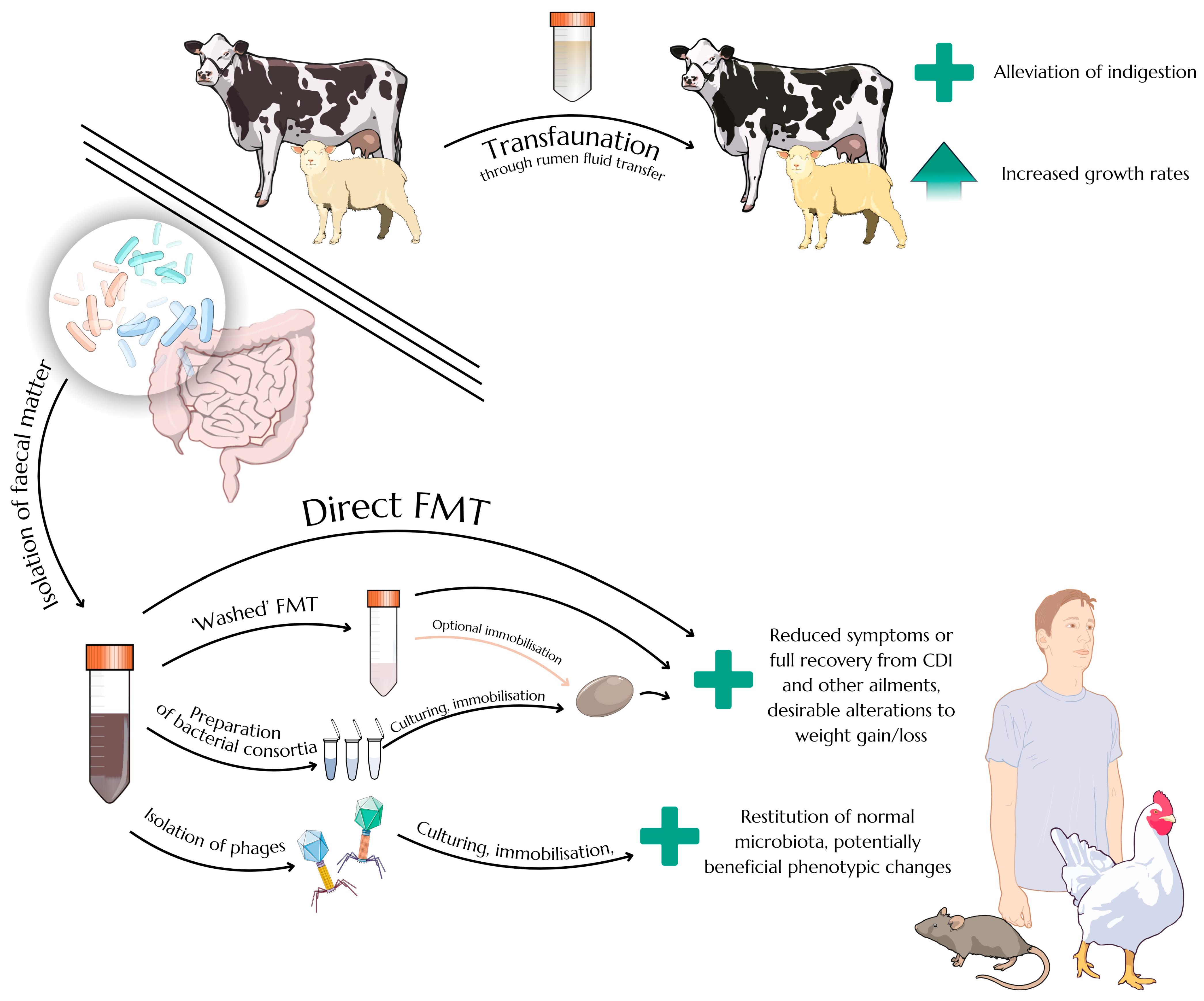
2.3. Faecal Microbiota Transplantation as an Efficient Therapeutic Approach

Faecal microbiota transplantation is not a strictly novel approach, as its early forms date back to the seventeenth century in Europe and the third century in Asia [51]. The procedure remains controversial despite its high efficacy in resolving gastrointestinal disorders, as aside from the natural scepticism expressed by patients towards the transfer of faecal matter between individuals, current data simply do not sufficiently explain all aspects of recipient response, and there are many well-founded doubts about biosafety, potential pathogen transmission, and long-term effects of altering the gut microbiota [52]. While the process—as all clinical interventions do—does carry some inherent risks, current research supports the notion that it is a safe form of treatment. A meta-analysis of studies performed in the first two decades of the XXI century found that adverse effects related to FMT occurred in 19 percent of patients, but these were mostly mild symptoms such as diarrhoea, cramping, or abdominal pain/discomfort. Serious adverse effects such as infection occurred in 1.4 percent of patients, and 0.12% of patients died after the procedure; however, all patients in these groups had been dealing with mucosal barrier injury, which presumably increased their susceptibility to pathogens [53]. Taking into consideration the high overall mortality rate of people suffering from CDI, sometimes estimated to be as high as 37% within one year after diagnosis [54], FMT does seem to be a preferable therapeutic alternative. Even though the incidence of serious adverse effects is indeed relatively small, additional steps aimed towards pathogen removal substantially contribute to the safety of the procedure [55].
Faecal microbiota transplantation has demonstrated high efficacy in treating recurrent Clostridioides difficile infection, while its role in other conditions is still being explored and remains to be clearly established. These include Clostridioides difficile infections (CDIs), a very common cause of contagious diarrhoea in hospital settings, and the possibility of effectively curing CDI and/or preventing its reoccurrence in patients (along with decreasing mortality rate in severe cases of CDI) has been proven by both numerous individual studies [3,56] and meta-analyses [57,58]. FMT can also be used to successfully treat irritable bowel syndrome (IBS) symptoms [59]. There are also some claims of FMT’s usefulness in treating other conditions, such as epilepsy [60], depression [61] or autism [62]. While certainly promising, further studies are needed to assess the validity of these findings and elucidate specific mechanism of FMT’s influence on human behaviour and mental well-being.
FMT is also the subject of research oriented towards improving the health, well-being, growth rate and final product quality of various animals. There exists a procedure called transfaunation, which has been common in animal husbandry for at least a few hundred years, that is based on transferring cud or rumen fluid from healthy animals to individuals suffering from indigestion [63]. While these substances are not faeces per se, the principle of transferring beneficial biological factors remains essentially the same. Available studies on transfaunation are surprisingly scarce given the long history of the procedure, but the current literature confirms its efficiency in treating indigestion caused by various factors [64]. Another study showed that transfaunation can significantly increase growth rates in lambs [65]; interestingly enough, this effect was observed after supplementation not only with sheep rumen fluid but with cow rumen fluid, suggesting an intrinsic potential of interspecific alimentary tract microbiota transfers that may also be applicable in the context of FMT.
Even though improving the digestive capabilities of ruminants can be achieved with transfaunation, some grazers—such as horses—are hindgut fermenters, and therefore standard FMT may be useful in their case. One study confirmed the effectivity of FMT procedures in reducing diarrhoea in horses [66]. Similar results can be seen in pig rearing, where FMT with was successfully utilised to increase average daily gains and decrease the diarrhoea susceptibility of piglets [67]; these results are especially intriguing because of the fact that while the piglets belonged to a crossbreed of Duroc × Landrace × Yorkshire, donor material was taken from local Jinhua variety. Another study introduced warthog FM to domestic pigs, which apparently resulted in increased resistance to African swine fever virus [68]. However, a different study found that FMT with highly feed-efficient pigs as donors significantly decreased growth rates, both in piglets subjected to FMT and in the offspring of FMT-treated sows [69]. Yet it is crucial to remark that the FMT procedure in this case involved the removal of the original gut microbiota by administration of an antibiotic cocktail followed by 36 h of fasting, and a similar routine was not applied to the control group, which might have substantially affected the end results. Nevertheless, all these findings highlight the importance of donor selection but show that FMT can be effective even when performed between individuals of different breeds or species, identifying promising avenues for future research aimed at conferring desirable traits on domesticated animals.
In terms of the technical aspects of the procedure, traditional approaches to FMT therapy in human patients have been centred on utilising crude (often blended, sieved, and mixed with saline) donor faeces that were screened to obtain safe and effective transplant material [55]. However, other concepts have also been proposed, some of which are illustrated in Figure 1. A procedure dubbed “washed faecal microbiota transplantation” is based on repeated filtration, centrifugation, and resuspension of bacteria obtained from faeces, which is supposed to significantly decrease the amount of potentially toxic substances in the preparation and can apparently lower the occurrence rate of adverse effects after microbiota transplantation [70]. Other approaches—still classified as FMT without any additional designations in the name—include lyophilising bacterial suspensions and administering the powder inside capsules, with no apparent loss of efficacy [71]. There were also solutions that advocated for the usage of standardised, lab-cultured bacterial formulations that consisted of dozens of artificially selected strains of microbes; these preparations lack any perceived resemblance to faeces, potentially reducing the psychological discomfort of patients, and offer supreme control over their final composition [72]. There, perhaps, lies the future of FMT—selecting the crucial biological components of faeces to then culture, purify, and administer in a safe, controlled, precise, and repeatable manner. Capsules containing a carefully designed consortium of microorganisms could be fine-tuned by chemical alterations or coating to make them more suitable for feeding specific animals in order to achieve better yields. This includes hard, hydrophobic capsules for birds that exhibit pecking while scavenging for food or soft, hydrophilic capsules for aquatic animals such as fish. Naturally, the development of such products will have to be preceded by extensive research to identify the most suitable bacterial strains.

3. Intestinal Bacteriophages and Faecal Virome Transplants

3.1. The Phageome of the Gut and the Surprising Prevalence of crAssviridae

The number of studies describing the gut virome—or, more specifically, the phageome, as most viruses in the human gut are bacteriophages [73]—is not very high. However, current publications shed some light on this issue, revealing some incredibly interesting facts that might be crucial for our understanding of the virus–prokaryote–host dynamics. A superficial glance at the numerosity and diversity of gut phages might lead the observer to question their supposed stabilizing effect on the gut microflora—after all, according to the standard Lotka–Volterra model of predation, one might expect to observe peaks of bacterial numerosity followed by sharp drops as viral counts begin to increase and subsequently peak. While this Lotka–Volterra dynamic is in fact present in the gut of infants [74], the phageome (and indeed, in many cases, the entire microbiome) of the gut tends to remain stable during adulthood [75,76]. Therefore, a different model for characterising interactions between prokaryotes and viruses in the gut has been developed, which postulates that gut phages in the outer layers of gut mucosa in healthy, adult hosts are mostly temperate viruses, which tend to integrate with the bacterial genome through the lysogenic cycle and only rarely cause cell death and virion release through cell lysis [77]. These temperate phages can protect commensal bacteria from becoming infected by other phages through a mechanism known as superinfection exclusion [78]. According to the model proposed by Silveira and Rohwer, the nutrient-rich environment of the outer mucosa may contribute to rather low lytic activity of the temperate phages. At the same time, virions that do get released after the occasional reactivation of a temperate phage and subsequent lysis of the cell may be deposited in significant numbers in the middle mucus layer, where they are more likely to cause a lytic infection of bacteria that are able to penetrate the mucosa, therefore forming a barrier against pathogens. This characterisation of the role of temperate phages is supported by the fact that they tend to be highly conserved within the microbiome of an individual. According to one study, over 80% of viral contigs found in the gut of a particular adult individual could be identified again after 850 days, and the contigs belonging to temperate phages were characterised by significantly lower nucleotide substitution rates than lytic phages [75]. This might have been caused by the relatively high precision of bacterial polymerases that replicate viral sequences integrated with bacterial genomes and the lower overall replication frequency.
Gut viromes of different hosts, similarly to gut bacteriomes, tend to be highly diversified. However (and perhaps surprisingly, given the difficulty in characterising common patterns shared between bacteriomes), there are reports of certain phages that are identifiable in most, if not all, samples taken from different hosts, forming the core phageome of a population. One study identified 23 core bacteriophages that could be found in more than 50% of healthy individuals from several cohorts [79]; a significant decrease in core bacteriophage presence was observed among individuals with ulcerative colitis or Crohn’s disease. Even though the results are certainly promising, it is important to note that the data utilised in the study came from typically Western cohorts, geographically located in Boston, Chicago, and Cambridge.
One of the most prevalent phage genomes identified in the study, along with three similar genomes, was a virus discovered two years earlier [80] dubbed “crAssphage”, which owes its name to the “crAss” v. 1.2 cross-assembly software used to facilitate its identification in metagenomic data. This prototypical crAssphage has been subsequently reclassified as Carjivirus communis [81], and it is now known that the crAssphage family is a highly diverse taxonomic unit, mostly including difficult-to-culture phages that tend to be found in the human gut in large quantities, being routinely identified in about 50% of individuals in certain populations and sometimes making up nearly 90% of the total viral DNA load in faeces [80,82]. Members of this unit have also been identified in many other habitats, from the guts of termites [83] to the waters of the Arctic Ocean [84]. CrAssphages are also characterised by somewhat unique host–predator dynamics that are—similarly to the previously mentioned phage interactions—consistently inconsistent with the Lotka–Voltera model. Perhaps surprisingly, crAssphages, while usually not inducing the formation of plaques in plaque assays, like most lytic viruses, were generally thought to be incapable of classic lysogeny, lacking suitable lysogeny-associated genes [85], and therefore their ample presence and persistence within the gut had to be explained by other mechanisms. An interesting host–phage relationship can be observed in ΦCrAss001, now known as Kehishuvirus primarius [81], which was the first crAssphage family member to be successfully isolated and cultured [85]. The virus infects Bacteroides intestinalis, but a certain share of B. intestinalis subpopulations can spontaneously convert to a subtype expressing a different capsular polysaccharide, which prevents infection by Kehishuvirus crAssphage—thus, at all times, a portion of the bacterial population is susceptible to the virus and the lysis of its members yields new virions, while another portion remains resistant to infection and can proliferate undisturbed [86]. The mechanism of switching between capsular polysaccharides, dubbed phase variation, has been observed before and is apparently regulated by genetic or epigenetic factors [87]. This explains the difficulty in identifying the phage in vitro. Even though the original study describing Kehishuvirus primarius did report the observation of clear plaques on agar media in some circumstances, in others—such as after prolonged coculture of host and phage—the opacity caused by the continuous growth of resistant wild-type B. intestinalis lineages within the plaques makes them barely noticeable.
Some crAssviruses have not been observed to form visible plaques at all, as is the case with ΦCrAss002, now known as Jahgtovirus secundus, which infects Bacteroides xylanisolvens [81,88]. This virus exhibits a different, perhaps even paradoxical, dynamic with its host—in in vitro cultures, the titre of the introduced virus does not increase (and, in fact, tends to decrease) for up to 2 or 3 days after the introduction of viral lysate into the culture until its count finally rebounds, achieving relatively high values of 108–109 copies per millilitre; the titre then remains essentially the same for weeks [88]. Moreover, neither this initial lag in viral proliferation nor the subsequent drastic increase in viral count seem to affect the numerosity of host bacteria. Therefore, faced with these confusing reports about a group of phages that should supposedly be strictly lytic given their apparent lack of lysogeny-enabling genes, other models have to be considered. A recent publication postulated that the prototypical Carjivirus communis crAssphage, while not integrating with the host chromosome in accordance with classic lysogeny models, is able to persist within the bacterial cell as a prophage-like plasmid molecule [89]. This is consistent with many previous reports of phages that exist as plasmids instead of integrating with the host chromosome [90,91]. This discovery of the phage–plasmid lifestyle of Carjivirus communis—the first and to date only crAssphage proved to possess this characteristic—has tremendous implications for our understanding of phage–host coexistence in vivo. In the realm of phages specificity, the study has also managed to identify three separate hosts for Carjivirus communis, namely Phocaeicola vulgatus, Phocaeicola dorei, and Bacteroides stercoris, which proves that at least some crAssviruses are characterised by relatively low host specificity and therefore could potentially engage in regulating gut microbiota above species level. During the aforementioned study, no apparent effect of phage addition on bacterial numerosity was noticed, which was consistent with observations about Jahgtovirus secundus by Guerin and colleagues. Considering that these viral species are closely related and that both belong to the Alphacrassvirinae subfamily [88], it seems reasonable to consider Jahgtovirus secundus to potentially be a phage–plasmid as well. While further studies are definitely necessary to confirm this hypothesis, it would explain many of the unusual traits of the virus mentioned before, including not forming plaques and not decreasing bacterial counts in liquid cultures in a noticeable way despite high viral titres. This sheds new light on the influence of phages in the gut, as it is possible that many of these are in fact phage–plasmids, therefore possessing high capacity for carrying genetic elements which can significantly influence their hosts—and, by extension, affect the entire microbiome.
Many phage–plasmids are capable of conferring antibiotic resistance [92], and some can possess genes aiding infection, functional CRISPR systems to protect the bacterium from further infections, genes encoding crucial enzymes for the biosynthesis of certain metabolites, plasmid maintenance genes and many others [93] Carjivirus communis itself carries infection-supporting genes, a repL gene implicated in plasmid replication, as well as genes predicted to encode a toxin–antitoxin (TA) system that eliminates bacterial progeny which do not inherit the viral plasmid [89]. Moreover, it also contains a fragment which is highly similar to the MobC relaxosome protein gene, crucial for the formation of sex pili in bacteria [89]. It can be therefore assumed that the phage propagates intensely through conjugation, especially when the bacterial population is not under high environmental stress. Further studies are definitely necessary to confirm the presence of similar genes in other crAssviruses, their effective expression in infected cells, and the general prevalence of phage–plasmid lifestyle among gut viruses, especially in animals. Applying this new knowledge to review, reconsider, and possibly update the previously discussed concept of immunity mediated by mucosa-dwelling phages would also be desirable. Nonetheless, utilising the concept of the phage–plasmid lifestyle to explain the previously mentioned peculiarities of crAssviruses seems to be a reasonable approach that reflects their characterisation as persistent and able to significantly influence the gut microflora, therefore justifying the consideration of using virome transplants to stabilise intestinal microbial communities.
The presence of viruses exhibiting a phage–plasmid lifestyle in the human gut—capable of both vertical and horizontal gene transmission—suggests their potential role in maintaining microbial network resilience and adaptability. In the context of FVT, their transfer could contribute to restoring functional microbial dynamics, particularly in microbiomes disrupted by antibiotics or infection, even if their direct therapeutic role remains to be fully elucidated. It is also worth reiterating that plaque assays, though widely used, fail to detect many abundant gut phages, including crAsslike and temperate viruses, which often do not form plaques. This limitation highlights the need for metagenomic and alternative culture-independent approaches better suited to capturing the core human gut virome.

3.2. Advantages and Potential Benefits of Faecal Virome Transplants

In the light of various adaptations and optimisations of FMT techniques briefly described above, it can be assumed that broadening their usage in the future will lead to safer and more effective procedures. However, considering faecal virome transplantation as an alternative to FMT would mean that the issue of introducing potentially pathogenic bacteria into the gut of the patient is essentially removed from the equation, which makes FVT a promising candidate for a novel therapeutic approach. In fact, while FVT can be still considered to be in its infancy, there are studies that were already able to highlight its efficiency in altering gut microbiota and affecting the metabolism of animals. A FVT using material from mice with high gut abundance of Akkermansia muciniphilia, a known probiotic species, was able to significantly increase the prevalence of this bacteria in mice naturally deficient in it; the same study also identified significantly increased fertility of the mice as a surprising and yet unexplained side effect of the FVT procedure [94]. Another study discovered that FVT based on material from obese mice on a high-fat diet can, aside from noticeably influencing the gut microbiota of the recipient mice, significantly affect their growth rates and induce an obese phenotype, even though the recipients were continuously fed a regular diet; a reverse dynamic was also observed, where mice fed a high-fat diet gained weight more slowly after a gavage of FVT from mice on a regular diet [95]. In a study conducted on broilers with LPS-induced intestinal injury, FVT proved to have significant effect on immune response within the gastrointestinal tract, effectively decreasing expression of IL-8 and IFN-γ on par with an FMT procedure [4]. These findings have tremendous implications, as they show that FVT can be an effective tool for altering gut microbiotas and influencing animal phenotypes, making it possible to confer positive traits not only between closely related individuals but—as is already the case with FMT—between species. However, FVT can also be used to restabilise GIT microbiotas without needing to source material from other individuals. This was shown by a study on autochthonous viral transplants in which antibiotic-treated mice were administered with phage-containing filtrate from faeces that had been collected before the start of the treatment. The GIT microbial composition of FVT-recipient mice was highly similar to their original microbiome, while the microbiome of mice that had not received the filtrate of their own faeces after the administration of antibiotics was noticeably more different [96]. There are also interesting reports of phage resistance being correlated with increased susceptibility to antibiotics in and/or decreased ability to colonise the gut in certain bacteria [73], possibly implying that bacteriophage presence may lead to passive selection of strains with decreased potential for pathogenicity.
Even though it may be tempting to debate the superiority of one therapeutic approach over another, it seems more rational to instead think of FMT and FVT as similarly valid counterparts that can both be further developed in different directions. However, utilising FVT as a substitute for FMT might have certain advantages, such as the previously mentioned elimination of potentially pathogenic bacteria and toxigenic substances from the transferred material—provided, of course, that suitable purification and quality control steps are undertaken. In this regard, it resembles the “washed” microbiota transplant procedure [70]. Moreover, identifying bacteriophage species that are crucial for maintaining gut stability and devising efficient, sustainable ways of mass-producing specific bacteriophage strains could prove to be economical enough for such preparations to be used not only in therapeutic approaches but in feed supplementation and therefore enhance the well-being of animals and the quality of animal-derived products. This perspective can be compared to the usage of the already-mentioned artificially defined bacterial formulations, but the amount of biomass that has to be introduced could potentially be lower in FVT procedures. On top of this, novel state-of-the-art approaches can significantly enhance the bactericidal effect of phages through genetic modification based on integrating a CRISPR-Cas system into their genome [97]. While such solutions seem to resemble targeted phage therapy more than typical FVT, they remain a fascinating approach that should not be overlooked.
Phages can be administered in suspension directly into the alimentary tract [95] or immobilised in many different mediums that are fine-tuned to achieve desired results, as can be seen in studies focused on immobilising specific phage species. The usage of liposome particles is an effective way of securing the passage of bacteriophages through the acidic and protease-rich contents of the stomach [98], although liposomes can also be utilised to increase the retention time and viability of bacteriophage preparations used topically for wound treatment [99]. Phages can also be freeze- or spray-dried and then encapsulated [100,101] to obtain products that can be stored for months in nonrefrigerated conditions with a relatively low decrease in viral viability levels. For now, there are no studies that have focused on utilising such techniques to immobilise an entire intestinal phageome of a donor, and the physicochemical qualities of individual viruses—such as their size, charge, and presence or absence of a lipid envelope—would definitely pose a challenge in devising a suitable method that maximises both the practicality and viability of the resulting preparation. Yet long shelf-life in easy-to-achieve storage conditions is a prominent advantage of phage preparations, one that is especially noticeable when compared with the shelf-life of spray-dried preparations of Gram-negative bacteria kept in nonrefrigerated environments, which rarely exceed a few weeks [102]. This may have specific implications for future ways of regulating Bacteroidota populations in the gut, as even though Bacteroides-based probiotics are currently being developed [103], the production and storage of such preparations will probably require overcoming the same challenges encountered when processing other Gram-negative microorganisms. If these challenges are not resolved in a way that allows for logistically sound storage and distribution of bacterial preparations, then perhaps phages could prove to be a viable and scalable alternative.
Moreover, when considering FVT approaches, it is vital to note that bacteriophages can directly affect not only bacteria but the host itself. Surprisingly, bacteriophages are actually able to penetrate the intestinal epithelium using a variety of mechanisms and then directly and indirectly influence the cells of the immune system [104]. A paper by Górski et al. [105] provided several examples of bacteriophages decreasing elevated cytokine levels, both in vitro and in vivo, and described their ability to affect leukocytes in gut-associated lymphoid tissue. Some of these interactions may not necessarily be beneficial, as there are also reports of bacteriophages inciting immune responses rather than quenching them [106]. However, future research will hopefully elucidate the interactions between bacteriophages and the host immune system to help create efficient therapies that holistically integrate direct antipathogen action with desirable forms of immunomodulation.

4. Conclusions

Recent advancements have deepened our understanding of the gut microbiome’s complexity and its pivotal role in host health. Investigations into prokaryotic diversity have identified distinct enterotypes across various species, underscoring both inter- and intraindividual variations in microbial composition. Growing evidence also points to the important role of the human gut phageome in shaping microbial communities and host interactions.
Notably, the gut virome, particularly the prevalence of crAsslike phages (classified under the order Crassvirales), has garnered significant attention. Initially discovered in human faecal metagenomes, these bacteriophages predominantly infect bacteria within the phylum Bacteroidota. Emerging evidence suggests that crAsslike phages are not exclusive to humans; they have also been detected in the gut microbiota of other animals, including termites, as well as in ocean water samples, indicating a broader ecological distribution.
Faecal microbiota transplantation has emerged as an effective therapeutic strategy for restoring gut microbial balance, particularly in cases of recurrent Clostridioides difficile infections. Beyond bacterial components, the potential benefits of transferring viral communities, or the virome, are being explored. This approach aims to reestablish a comprehensive microbial ecosystem, offering promising avenues for treating a variety of conditions. Possible future solutions, including genetically engineering microbes to affect their metabolism and interactions with other organisms, could provide additional avenues for efficient, targeted therapy. However, much remains to be done to better understand the interactions among bacteria, viruses, and the host—at this moment, not much is known about bacteriophages that could infect probiotic bacteria and disrupt the healthy microbiome of the gut, therefore acting as indirect human and animal pathogens. The data on long-term results of the procedures—especially FVT—also seem to be lacking, so there is definitely significant potential for studies that could prove the permanence of beneficial changes in the intestinal microbiota.
In summary, integrating knowledge of gut microbial diversity, enterotype classification, and the therapeutic potential of microbiota and virome transplants offers new perspectives in medical science. Continued research in these domains is essential for developing personalized therapies and promoting optimal gut health across different species.

Author Contributions

Conceptualization, W.J., M.M., and P.N.; writing—original draft preparation, W.J.; writing—review and editing, W.J., M.M., and P.N.; visualization, W.J.; supervision, M.M. and P.N.; funding acquisition, P.N. All authors have read and agreed to the published version of the manuscript.

Funding

This research received no external funding.

Institutional Review Board Statement

Not applicable.

Informed Consent Statement

Not applicable.

Data Availability Statement

Not applicable.

Acknowledgments

The authors would like to thank the following content creators for distributing the works that were used for the preparation of diagrams under Creative Commons licenses: Ryan Kissinger (NIH BIOART)—human intestines, bacteria, Gram-negative bacterium, cow, rat (cropped, modified colours)—CC0 1.0; Andi-Wilson (bioicons.com)—Eppendorf tubes—CC-BY 4.0; Diogo Losch de Oliveira (scidraw.io)—Falcon tube (cropped, modified colours)—CC-BY 4.0; James-Lloyd (scidraw.io)—oblong bacterium with flagella—CC0 1.0; B-Gideon-Bergheim (scidraw.io)—E. coli—CC0 1.0; DBCLS (scidraw.io)—plasmid—CC-BY 1.0; Contributors from Servier Medical Art—bacteriophages, capsule (modified colours)—CC-BY 4.0; Contributors from NIAID (NIH BIOART)—chicken (modified colours)—CC0 1.0; LadyOfHats (Wikimedia Commons)—sheep (modified colours)—CC0 1.0. Figures in this manuscript were prepared in Canva (Canva Pty. Ltd.).

Conflicts of Interest

The authors declare no conflicts of interest.

Abbreviations

The following abbreviations are used in this manuscript:
MDPIMultidisciplinary Digital Publishing Institute
DOAJDirectory of Open Access Journals
FMTFaecal microbiota transplant
FVTFaecal virome transplant
CDIClostridioides difficile infection
IBSIrritable bowel syndrome

References

  1. Simon, J.-C.; Marchesi, J.R.; Mougel, C.; Selosse, M.-A. Host-Microbiota Interactions: From Holobiont Theory to Analysis. Microbiome 2019, 7, 5. [Google Scholar] [CrossRef]
  2. Pandey, K.; Naik, S.; Vakil, B. Probiotics, Prebiotics and Synbiotics—A Review. J. Food Sci. Technol. 2015, 52, 7577. [Google Scholar] [CrossRef]
  3. Hocquart, M.; Lagier, J.-C.; Cassir, N.; Saidani, N.; Eldin, C.; Kerbaj, J.; Delord, M.; Valles, C.; Brouqui, P.; Raoult, D.; et al. Early Fecal Microbiota Transplantation Improves Survival in Severe Clostridium difficile Infections. Clin. Infect. Dis. 2018, 66, 645. [Google Scholar] [CrossRef]
  4. Wu, D.; Liang, S.; Du, X.; Xiao, J.; Feng, H.; Ren, Z.; Yang, X.; Yang, X. Effects of Fecal Microbiota Transplantation and Fecal Virome Transplantation on LPS-Induced Intestinal Injury in Broilers. Poult. Sci. 2024, 103, 103316. [Google Scholar] [CrossRef] [PubMed]
  5. Qin, J.; Li, R.; Raes, J.; Arumugam, M.; Burgdorf, K.S.; Manichanh, C.; Nielsen, T.; Pons, N.; Levenez, F.; Yamada, T.; et al. A Human Gut Microbial Gene Catalogue Established by Metagenomic Sequencing. Nature 2010, 464, 59–65. [Google Scholar] [CrossRef]
  6. Romaní-Pérez, M.; Líebana-García, R.; Flor-Duro, A.; Bonillo-Jiménez, D.; Bullich-Vilarrubias, C.; Olivares, M.; Sanz, Y. Obesity and the Gut Microbiota: Implications of Neuroendocrine and Immune Signaling. FEBS J. 2024, 292, 1397–1420. [Google Scholar] [CrossRef] [PubMed]
  7. Paster, B.J.; Dewhirst, F.E.; Olsen, I.; Fraser, G.J. Phylogeny of Bacteroides, Prevotella, and Porphyromonas spp. and Related Bacteria. J. Bacteriol. 1994, 176, 725. [Google Scholar] [CrossRef]
  8. Wallace, R.J.; McKain, N.; Broderick, G.A.; Rode, L.M.; Walker, N.D.; Newbold, C.J.; Kopecny, J. Peptidases of the Rumen Bacterium, Prevotella ruminicola. Anaerobe 1997, 3, 35. [Google Scholar] [CrossRef] [PubMed]
  9. Stoeva, M.K.; Garcia-So, J.; Justice, N.; Myers, J.; Tyagi, S.; Nemchek, M.; McMurdie, P.J.; Kolterman, O.; Eid, J. Butyrate-Producing Human Gut Symbiont, Clostridium butyricum, and Its Role in Health and Disease. Gut Microbes 2021, 13, 1907272. [Google Scholar] [CrossRef]
  10. Kanai, T.; Mikami, Y.; Hayashi, A. A Breakthrough in Probiotics: Clostridium butyricum Regulates Gut Homeostasis and Anti-Inflammatory Response in Inflammatory Bowel Disease. J. Gastroenterol. 2015, 50, 928. [Google Scholar] [CrossRef]
  11. Hill, K.K.; Xie, G.; Foley, B.T.; Smith, T.J.; Munk, A.C.; Bruce, D.; Smith, L.A.; Brettin, T.S.; Detter, J.C. Recombination and Insertion Events Involving the Botulinum Neurotoxin Complex Genes in Clostridium botulinum Types A, B, E and F and Clostridium butyricum Type E Strains. BMC Biol. 2009, 7, 66. [Google Scholar] [CrossRef] [PubMed]
  12. Dykes, J.K.; Lúquez, C.; Raphael, B.H.; McCroskey, L.; Maslanka, S.E. Laboratory Investigation of the First Case of Botulism Caused by Clostridium butyricum Type e Toxin in the United States. J. Clin. Microbiol. 2015, 53, 3363. [Google Scholar] [CrossRef]
  13. Czepiel, J.; Dróżdż, M.; Pituch, H.; Kuijper, E.J.; Perucki, W.; Mielimonka, A.; Goldman, S.; Wultańska, D.; Garlicki, A.; Biesiada, G. Clostridium difficile Infection: Review. Eur. J. Clin. Microbiol. Infect. Dis. 2019, 38, 1211. [Google Scholar] [CrossRef]
  14. Gerding, D.N.; Meyer, T.; Lee, C.; Cohen, S.H.; Murthy, U.K.; Poirier, A.; Van Schooneveld, T.C.; Pardi, D.S.; Ramos, A.; Barron, M.A.; et al. Administration of Spores of Nontoxigenic Clostridium difficile Strain M3 for Prevention of Recurrent C. difficile Infection: A Randomized Clinical Trial. JAMA 2015, 313, 1719. [Google Scholar] [CrossRef]
  15. Dobzhansky, T. A Critique of the Species Concept in Biology. Philos. Sci. 1935, 2, 344. [Google Scholar] [CrossRef]
  16. Novick, A.; Doolittle, W.F. “Species” without Species. Stud. Hist. Philos. Sci. 2021, 87, 72. [Google Scholar] [CrossRef] [PubMed]
  17. Han, S.; Treuren, W.V.; Fischer, C.R.; Merrill, B.D.; DeFelice, B.C.; Sanchez, J.M.; Higginbottom, S.K.; Guthrie, L.; Fall, L.A.; Dodd, D.; et al. A Metabolomics Pipeline for Mechanistic Interrogation of the Gut Microbiome. Nature 2021, 595, 415. [Google Scholar] [CrossRef] [PubMed]
  18. Koliada, A.; Syzenko, G.; Moseiko, V.; Budovska, L.; Puchkov, K.; Perederiy, V.; Gavalko, Y.; Dorofeyev, A.; Romanenko, M.; Tkach, S.; et al. Association between Body Mass Index and Firmicutes/Bacteroidetes Ratio in an Adult Ukrainian Population. BMC Microbiol. 2017, 17, 120. [Google Scholar] [CrossRef]
  19. Magne, F.; Gotteland, M.; Gauthier, L.; Zazueta, A.; Pesoa, S.; Navarrete, P.; Balamurugan, R. The Firmicutes/Bacteroidetes Ratio: A Relevant Marker of Gut Dysbiosis in Obese Patients? Nutrients 2020, 12, 1474. [Google Scholar] [CrossRef]
  20. Tsai, Y.-C.; Tai, W.-C.; Liang, C.-M.; Wu, C.-K.; Tsai, M.-C.; Hu, W.-H.; Huang, P.-Y.; Chen, C.-H.; Kuo, Y.-H.; Yao, C.-C.; et al. Alternations of the Gut Microbiota and the Firmicutes/Bacteroidetes Ratio after Biologic Treatment in Inflammatory Bowel Disease. J. Microbiol. Immunol. Infect. 2024, 58, 62–69. [Google Scholar] [CrossRef]
  21. Reichardt, N.; Duncan, S.H.; Young, P.; Belenguer, A.; McWilliam Leitch, C.; Scott, K.P.; Flint, H.J.; Louis, P. Phylogenetic Distribution of Three Pathways for Propionate Production within the Human Gut Microbiota. ISME J. 2014, 8, 1323. [Google Scholar] [CrossRef] [PubMed]
  22. Fettig, N.M.; Osborne, L.C. Direct and Indirect Effects of Microbiota-Derived Metabolites on Neuroinflammation in Multiple Sclerosis. Microbes Infect. 2021, 23, 104814. [Google Scholar] [CrossRef] [PubMed]
  23. Wu, G.D.; Chen, J.; Hoffmann, C.; Bittinger, K.; Chen, Y.-Y.; Keilbaugh, S.A.; Bewtra, M.; Knights, D.; Walters, W.A.; Knight, R.; et al. Linking Long-Term Dietary Patterns with Gut Microbial Enterotypes. Science 2011, 334, 105. [Google Scholar] [CrossRef] [PubMed]
  24. Arumugam, M.; Raes, J.; Pelletier, E.; Le Paslier, D.; Yamada, T.; Mende, D.R.; Fernandes, G.R.; Tap, J.; Bruls, T.; Batto, J.-M.; et al. Enterotypes of the Human Gut Microbiome. Nature 2011, 473, 174. [Google Scholar] [CrossRef]
  25. Jeffery, I.B.; Claesson, M.J.; O’Toole, P.W.; Shanahan, F. Categorization of the Gut Microbiota: Enterotypes or Gradients? Nat. Rev. Microbiol. 2012, 10, 591. [Google Scholar] [CrossRef]
  26. Knights, D.; Ward, T.L.; McKinlay, C.E.; Miller, H.; Gonzalez, A.; McDonald, D.; Knight, R. Rethinking “Enterotypes”. Cell Host Microbe 2014, 16, 433. [Google Scholar] [CrossRef]
  27. Di Pierro, F. A Possible Perspective about the Compositional Models, Evolution, and Clinical Meaning of Human Enterotypes. Microorganisms 2021, 9, 2341. [Google Scholar] [CrossRef]
  28. Bartsch, M.; Hahn, A.; Berkemeyer, S. Bridging the Gap from Enterotypes to Personalized Dietary Recommendations: A Metabolomics Perspective on Microbiome Research. Metabolites 2023, 13, 1182. [Google Scholar] [CrossRef]
  29. David, L.A.; Maurice, C.F.; Carmody, R.N.; Gootenberg, D.B.; Button, J.E.; Wolfe, B.E.; Ling, A.V.; Devlin, A.S.; Varma, Y.; Fischbach, M.A.; et al. Diet Rapidly and Reproducibly Alters the Human Gut Microbiome. Nature 2014, 505, 559. [Google Scholar] [CrossRef]
  30. Hayashi, H.; Shibata, K.; Sakamoto, M.; Tomita, S.; Benno, Y. Prevotella copri sp. nov. and Prevotella stercorea sp. nov., Isolated from Human Faeces. Int. J. Syst. Evol. Microbiol. 2007, 57, 941. [Google Scholar] [CrossRef]
  31. Ley, R.E. Prevotella in the Gut: Choose Carefully. Nat. Rev. Gastroenterol. Hepatol. 2016, 13, 69. [Google Scholar] [CrossRef] [PubMed]
  32. De Filippis, F.; Pellegrini, N.; Laghi, L.; Gobbetti, M.; Ercolini, D. Unusual Sub-Genus Associations of Faecal Prevotella and Bacteroides with Specific Dietary Patterns. Microbiome 2016, 4, 57. [Google Scholar] [CrossRef] [PubMed]
  33. Costea, P.I.; Hildebrand, F.; Arumugam, M.; Bäckhed, F.; Blaser, M.J.; Bushman, F.D.; de Vos, W.M.; Ehrlich, S.D.; Fraser, C.M.; Hattori, M.; et al. Enterotypes in the Landscape of Gut Microbial Community Composition. Nat. Microbiol. 2017, 3, 8. [Google Scholar] [CrossRef]
  34. Liang, C.; Tseng, H.-C.; Chen, H.-M.; Wang, W.-C.; Chiu, C.-M.; Chang, J.-Y.; Lu, K.-Y.; Weng, S.-L.; Chang, T.-H.; Chang, C.-H.; et al. Diversity and Enterotype in Gut Bacterial Community of Adults in Taiwan. BMC Genomics 2017, 18, 932. [Google Scholar] [CrossRef]
  35. Fu, T.; Pan, L.; Shang, Q.; Yu, G. Fermentation of Alginate and its Derivatives by Different Enterotypes of Human Gut Microbiota: Towards Personalized Nutrition Using Enterotype-Specific Dietary Fibers. Int. J. Biol. Macromol. 2021, 183, 1649. [Google Scholar] [CrossRef] [PubMed]
  36. Wang, J.; Li, W.; Wang, C.; Wang, L.; He, T.; Hu, H.; Song, J.; Cui, C.; Qiao, J.; Qing, L.; et al. Enterotype Bacteroides Is Associated with a High Risk in Patients with Diabetes: A Pilot Study. J. Diabet. Res. 2020, 2020, 6047145. [Google Scholar] [CrossRef]
  37. Lepage, P.; Seksik, P.; Sutren, M.; de la Cochetière, M.-F.; Jian, R.; Marteau, P.; Doré, J. Biodiversity of the Mucosa-Associated Microbiota Is Stable Along the Distal Digestive Tract in Healthy Individuals and Patients With IBD. Inflamm. Bowel Dis. 2005, 11, 473. [Google Scholar] [CrossRef]
  38. Wang, J.; Linnenbrink, M.; Künzel, S.; Fernandes, R.; Nadeau, M.-J.; Rosenstiel, P.; Baines, J.F. Dietary History Contributes to Enterotype-like Clustering and Functional Metagenomic Content in the Intestinal Microbiome of Wild Mice. Proc. Natl. Acad. Sci. USA 2014, 111, E2703. [Google Scholar] [CrossRef]
  39. Li, X.; Liang, S.; Xia, Z.; Qu, J.; Liu, H.; Liu, C.; Yang, H.; Wang, J.; Madsen, L.; Hou, Y.; et al. Establishment of a Macaca Fascicularis Gut Microbiome Gene Catalog and Comparison with the Human, Pig, and Mouse Gut Microbiomes. GigaScience 2018, 7, 100. [Google Scholar] [CrossRef]
  40. Xiao, L.; Feng, Q.; Liang, S.; Sonne, S.B.; Xia, Z.; Qiu, X.; Li, X.; Long, H.; Zhang, J.; Zhang, D.; et al. A Catalog of the Mouse Gut Metagenome. Nat. Biotechnol. 2015, 33, 1103. [Google Scholar] [CrossRef]
  41. Ramayo-Caldas, Y.; Mach, N.; Lepage, P.; Levenez, F.; Denis, C.; Lemonnier, G.; Leplat, J.-J.; Billon, Y.; Berri, M.; Doré, J.; et al. Phylogenetic Network Analysis Applied to Pig Gut Microbiota Identifies an Ecosystem Structure Linked with Growth Traits. ISME J. 2016, 10, 2973. [Google Scholar] [CrossRef] [PubMed]
  42. Yang, H.; Huang, X.; Fang, S.; He, M.; Zhao, Y.; Wu, Z.; Yang, M.; Zhang, Z.; Chen, C.; Huang, L. Unraveling the Fecal Microbiota and Metagenomic Functional Capacity Associated with Feed Efficiency in Pigs. Front. Microbiol. 2017, 8, 1555. [Google Scholar] [CrossRef] [PubMed]
  43. Lu, D.; Tiezzi, F.; Schillebeeckx, C.; McNulty, N.P.; Schwab, C.; Shull, C.; Maltecca, C. Host Contributes to Longitudinal Diversity of Fecal Microbiota in Swine Selected for Lean Growth. Microbiome 2018, 6, 4. [Google Scholar] [CrossRef]
  44. Le Sciellour, M.; Renaudeau, D.; Zemb, O. Longitudinal Analysis of the Microbiota Composition and Enterotypes of Pigs from Post-Weaning to Finishing. Microorganisms 2019, 7, 622. [Google Scholar] [CrossRef]
  45. Yuan, Z.; Yan, W.; Wen, C.; Zheng, J.; Yang, N.; Sun, C. Enterotype Identification and Its Influence on Regulating the Duodenum Metabolism in Chickens. Poult. Sci. 2020, 99, 1515. [Google Scholar] [CrossRef]
  46. Fan, Q.; Xu, Y.; Xiao, Y.; Yang, C.; Lyu, W.; Yang, H. Linking Growth Performance and Carcass Traits with Enterotypes in Muscovy Ducks. Anim. Biosci. 2024, 37, 1213. [Google Scholar] [CrossRef] [PubMed]
  47. Lilli, G.; Sirot, C.; Campbell, H.; Brophy, D.; Graham, C.T.; George, I.F. Geographic Origin and Host’s Phylogeny Are Predictors of the Gut Mucosal Microbiota Diversity and Composition in Mediterranean Scorpionfishes (Scorpaena spp.). Front. Mar. Sci. 2023, 10, 1286706. [Google Scholar] [CrossRef]
  48. Zhang, B.; Xiao, J.; Liu, H.; Zhai, D.; Wang, Y.; Liu, S.; Xiong, F.; Xia, M. Vertical Habitat Preferences Shape the Fish Gut Microbiota in a Shallow Lake. Front. Microbiol. 2024, 15, 1341303. [Google Scholar] [CrossRef]
  49. Moeller, A.H.; Ochman, H. Microbiomes Are True to Type. Proc. Natl. Acad. Sci. USA 2014, 111, 9372. [Google Scholar] [CrossRef]
  50. Vandeputte, D.; Falony, G.; Vieira-Silva, S.; Tito, R.Y.; Joossens, M.; Raes, J. Stool Consistency Is Strongly Associated with Gut Microbiota Richness and Composition, Enterotypes and Bacterial Growth Rates. Gut 2016, 65, 57. [Google Scholar] [CrossRef]
  51. Zhang, F.; Luo, W.; Shi, Y.; Fan, Z.; Ji, G. Should We Standardize the 1700-Year-Old Fecal Microbiota Transplantation? Off. J. Am. Coll. Gastroenterol.|ACG 2012, 107, 1755. [Google Scholar] [CrossRef] [PubMed]
  52. Park, S.-Y.; Seo, G.S. Fecal Microbiota Transplantation: Is It Safe? Clin. Endosc. 2021, 54, 157. [Google Scholar] [CrossRef]
  53. Marcella, C.; Cui, B.; Kelly, C.R.; Ianiro, G.; Cammarota, G.; Zhang, F. Systematic Review: The Global Incidence of Faecal Microbiota Transplantation-Related Adverse Events from 2000 to 2020. Aliment. Pharmacol. Ther. 2021, 53, 33. [Google Scholar] [CrossRef]
  54. Hensgens, M.P.M.; Goorhuis, A.; Dekkers, O.M.; van Benthem, B.H.B.; Kuijper, E.J. All-Cause and Disease-Specific Mortality in Hospitalized Patients With Clostridium difficile Infection: A Multicenter Cohort Study. Clin. Infect. Dis. 2013, 56, 1108–1116. [Google Scholar] [CrossRef] [PubMed]
  55. Ng, R.W.; Dharmaratne, P.; Wong, S.; Hawkey, P.; Chan, P.; Ip, M. Revisiting the Donor Screening Protocol of Faecal Microbiota Transplantation (FMT): A Systematic Review. Gut 2024, 73, 1029. [Google Scholar] [CrossRef]
  56. Konturek, P.C.; Dieterich, W.; Neurath, M.; Zopf, Y. Successful Therapy of Clostridium difficile Infection with Fecal Microbiota Transplantation. Gastroenterology 2017, 152, S341. [Google Scholar] [CrossRef]
  57. Quraishi, M.N.; Widlak, M.; Bhala, N.; Moore, D.; Price, M.; Sharma, N.; Iqbal, T.H. Systematic Review with Meta-Analysis: The Efficacy of Faecal Microbiota Transplantation for the Treatment of Recurrent and Refractory Clostridium difficile Infection. Aliment. Pharmacol. Ther. 2017, 46, 479. [Google Scholar] [CrossRef] [PubMed]
  58. Ianiro, G.; Maida, M.; Burisch, J.; Simonelli, C.; Hold, G.; Ventimiglia, M.; Gasbarrini, A.; Cammarota, G. Efficacy of Different Faecal Microbiota Transplantation Protocols for Clostridium difficile Infection: A Systematic Review and Meta-Analysis. United Eur. Gastroenterol. J. 2018, 6, 1232. [Google Scholar] [CrossRef]
  59. El-Salhy, M.; Hatlebakk, J.G.; Gilja, O.H.; Kristoffersen, A.B.; Hausken, T. Efficacy of Faecal Microbiota Transplantation for Patients with Irritable Bowel Syndrome in a Randomised, Double-Blind, Placebo-Controlled Study. Gut 2020, 69, 859. [Google Scholar] [CrossRef]
  60. He, Z.; Cui, B.-T.; Zhang, T.; Li, P.; Long, C.-Y.; Ji, G.-Z.; Zhang, F.-M. Fecal Microbiota Transplantation Cured Epilepsy in a Case with Crohn’s Disease: The First Report. World J. Gastroenterol. 2017, 23, 3565. [Google Scholar] [CrossRef]
  61. Cai, T.; Shi, X.; Yuan, L.; Tang, D.; Wang, F. Fecal Microbiota Transplantation in an Elderly Patient with Mental Depression. Int. Psychogeriatr. 2019, 31, 1525. [Google Scholar] [CrossRef]
  62. Kang, D.-W.; Adams, J.B.; Gregory, A.C.; Borody, T.; Chittick, L.; Fasano, A.; Khoruts, A.; Geis, E.; Maldonado, J.; McDonough-Means, S.; et al. Microbiota Transfer Therapy Alters Gut Ecosystem and Improves Gastrointestinal and Autism Symptoms: An Open-Label Study. Microbiome 2017, 5, 10. [Google Scholar] [CrossRef] [PubMed]
  63. DePeters, E.J.; George, L.W. Rumen Transfaunation. Immunol. Lett. 2014, 162, 69. [Google Scholar] [CrossRef]
  64. Steiner, S.; Linhart, N.; Neidl, A.; Baumgartner, W.; Tichy, A.; Wittek, T. Evaluation of the Therapeutic Efficacy of Rumen Transfaunation. J. Anim. Physiol. Anim. Nutr. 2020, 104, 56. [Google Scholar] [CrossRef]
  65. Elrawy, A.; Mohammed, A.; Sabra, M.S.; Mahmoud, U.; Darwish, M. Impact of Rumen Juice Transfaunation on Behavioral Activities, Performance Parameters and Kidney Function in Fattening Lambs. Assiut Vet. Med. J. 2023, 69, 74. [Google Scholar] [CrossRef]
  66. McKinney, C.A.; Bedenice, D.; Pacheco, A.P.; Oliveira, B.C.M.; Paradis, M.-R.; Mazan, M.; Widmer, G. Assessment of Clinical and Microbiota Responses to Fecal Microbial Transplantation in Adult Horses with Diarrhea. PLoS ONE 2021, 16, e0244381. [Google Scholar] [CrossRef] [PubMed]
  67. Hu, L.; Geng, S.; Li, Y.; Cheng, S.; Fu, X.; Yue, X.; Han, X. Exogenous Fecal Microbiota Transplantation from Local Adult Pigs to Crossbred Newborn Piglets. Front. Microbiol. 2018, 8, 02663. [Google Scholar] [CrossRef] [PubMed]
  68. Zhang, J.; Rodríguez, F.; Navas, M.J.; Costa-Hurtado, M.; Almagro, V.; Bosch-Camós, L.; López, E.; Cuadrado, R.; Accensi, F.; Pina-Pedrero, S.; et al. Fecal Microbiota Transplantation from Warthog to Pig Confirms the Influence of the Gut Microbiota on African Swine Fever Susceptibility. Sci. Rep. 2020, 10, 17605. [Google Scholar] [CrossRef]
  69. McCormack, U.M.; Curião, T.; Wilkinson, T.; Metzler-Zebeli, B.U.; Reyer, H.; Ryan, T.; Calderon-Diaz, J.A.; Crispie, F.; Cotter, P.D.; Creevey, C.J.; et al. Fecal Microbiota Transplantation in Gestating Sows and Neonatal Offspring Alters Lifetime Intestinal Microbiota and Growth in Offspring. mSystems 2018, 3, e00134-17. [Google Scholar] [CrossRef]
  70. Zhang, T.; Lu, G.; Zhao, Z.; Liu, Y.; Shen, Q.; Li, P.; Chen, Y.; Yin, H.; Wang, H.; Marcella, C.; et al. Washed Microbiota Transplantation vs. Manual Fecal Microbiota Transplantation: Clinical Findings, Animal Studies and in Vitro Screening. Protein Cell 2020, 11, 251. [Google Scholar] [CrossRef]
  71. Varga, A.; Kocsis, B.; Sipos, D.; Kása, P.; Vigvári, S.; Pál, S.; Dembrovszky, F.; Farkas, K.; Péterfi, Z. How to Apply FMT More Effectively, Conveniently and Flexible—A Comparison of FMT Methods. Front. Cell Infect. Microbiol. 2021, 11, 657320. [Google Scholar] [CrossRef]
  72. Petrof, E.O.; Gloor, G.B.; Vanner, S.J.; Weese, S.J.; Carter, D.; Daigneault, M.C.; Brown, E.M.; Schroeter, K.; Allen-Vercoe, E. Stool Substitute Transplant Therapy for the Eradication of Clostridium difficile Infection: ‘RePOOPulating’ the Gut. Microbiome 2013, 1, 3. [Google Scholar] [CrossRef] [PubMed]
  73. Sausset, R.; Petit, M.A.; Gaboriau-Routhiau, V.; De Paepe, M. New Insights into Intestinal Phages. Mucosal Immunol. 2020, 13, 205. [Google Scholar] [CrossRef] [PubMed]
  74. Lim, E.S.; Zhou, Y.; Zhao, G.; Bauer, I.K.; Droit, L.; Ndao, I.M.; Warner, B.B.; Tarr, P.I.; Wang, D.; Holtz, L.R. Early Life Dynamics of the Human Gut Virome and Bacterial Microbiome in Infants. Nat. Med. 2015, 21, 1228. [Google Scholar] [CrossRef] [PubMed]
  75. Minot, S.; Bryson, A.; Chehoud, C.; Wu, G.D.; Lewis, J.D.; Bushman, F.D. Rapid Evolution of the Human Gut Virome. Proc. Natl. Acad. Sci. USA 2013, 110, 12450. [Google Scholar] [CrossRef]
  76. Shkoporov, A.N.; Clooney, A.G.; Sutton, T.D.S.; Ryan, F.J.; Daly, K.M.; Nolan, J.A.; McDonnell, S.A.; Khokhlova, E.V.; Draper, L.A.; Forde, A.; et al. The Human Gut Virome Is Highly Diverse, Stable, and Individual Specific. Cell Host Microbe 2019, 26, 527. [Google Scholar] [CrossRef]
  77. Silveira, C.B.; Rohwer, F.L. Piggyback-the-Winner in Host-Associated Microbial Communities. NPJ Biofilms Microbiomes 2016, 2, 16010. [Google Scholar] [CrossRef]
  78. Obeng, N.; Pratama, A.A.; Elsas, J.D. van The Significance of Mutualistic Phages for Bacterial Ecology and Evolution. Trends Microbiol. 2016, 24, 440. [Google Scholar] [CrossRef]
  79. Manrique, P.; Bolduc, B.; Walk, S.T.; van der Oost, J.; de Vos, W.M.; Young, M.J. Healthy Human Gut Phageome. Proc. Natl. Acad. Sci. USA 2016, 113, 10400. [Google Scholar] [CrossRef]
  80. Dutilh, B.E.; Cassman, N.; McNair, K.; Sanchez, S.E.; Silva, G.G.Z.; Boling, L.; Barr, J.J.; Speth, D.R.; Seguritan, V.; Aziz, R.K.; et al. A Highly Abundant Bacteriophage Discovered in the Unknown Sequences of Human Faecal Metagenomes. Nat. Commun. 2014, 5, 4498. [Google Scholar] [CrossRef]
  81. Turner, D.; Shkoporov, A.N.; Lood, C.; Millard, A.D.; Dutilh, B.E.; Alfenas-Zerbini, P.; van Zyl, L.J.; Aziz, R.K.; Oksanen, H.M.; Poranen, M.M.; et al. Abolishment of Morphology-Based Taxa and Change to Binomial Species Names: 2022 Taxonomy Update of the ICTV Bacterial Viruses Subcommittee. Arch. Virol. 2023, 168, 74. [Google Scholar] [CrossRef] [PubMed]
  82. Guerin, E.; Shkoporov, A.; Stockdale, S.R.; Clooney, A.G.; Ryan, F.J.; Sutton, T.D.S.; Draper, L.A.; Gonzalez-Tortuero, E.; Ross, R.P.; Hill, C. Biology and Taxonomy of crAss-like Bacteriophages, the Most Abundant Virus in the Human Gut. Cell Host & Microbe 2018, 24, 653. [Google Scholar] [CrossRef]
  83. Chen, J.; Gissendanner, C.R.; Tikhe, C.V.; Li, H.-F.; Sun, Q.; Husseneder, C. Genomics and Geographic Diversity of Bacteriophages Associated With Endosymbionts in the Guts of Workers and Alates of Coptotermes Species (Blattodea: Rhinotermitidae). Front. Ecol. Evol. 2022, 10, 881538. [Google Scholar] [CrossRef]
  84. Piedade, G.J.; Schön, M.E.; Lood, C.; Fofanov, M.V.; Wesdorp, E.M.; Biggs, T.E.G.; Wu, L.; Bolhuis, H.; Fischer, M.G.; Yutin, N.; et al. Seasonal Dynamics and Diversity of Antarctic Marine Viruses Reveal a Novel Viral Seascape. Nat. Commun. 2024, 15, 9192. [Google Scholar] [CrossRef] [PubMed]
  85. Shkoporov, A.N.; Khokhlova, E.V.; Fitzgerald, C.B.; Stockdale, S.R.; Draper, L.A.; Ross, R.P.; Hill, C. ΦCrAss001 Represents the Most Abundant Bacteriophage Family in the Human Gut and Infects Bacteroides Intestinalis. Nat. Commun. 2018, 9, 4781. [Google Scholar] [CrossRef]
  86. Shkoporov, A.N.; Khokhlova, E.V.; Stephens, N.; Hueston, C.; Seymour, S.; Hryckowian, A.J.; Scholz, D.; Ross, R.P.; Hill, C. Long-Term Persistence of crAss-like Phage crAss001 Is Associated with Phase Variation in Bacteroides intestinalis. BMC Biol. 2021, 19, 163. [Google Scholar] [CrossRef]
  87. Porter, N.T.; Hryckowian, A.J.; Merrill, B.D.; Fuentes, J.J.; Gardner, J.O.; Glowacki, R.W.P.; Singh, S.; Crawford, R.D.; Snitkin, E.S.; Sonnenburg, J.L.; et al. Phase-Variable Capsular Polysaccharides and Lipoproteins Modify Bacteriophage Susceptibility in Bacteroides thetaiotaomicron. Nat. Microbiol. 2020, 5, 1170. [Google Scholar] [CrossRef]
  88. Guerin, E.; Shkoporov, A.N.; Stockdale, S.R.; Comas, J.C.; Khokhlova, E.V.; Clooney, A.G.; Daly, K.M.; Draper, L.A.; Stephens, N.; Scholz, D.; et al. Isolation and Characterisation of ΦcrAss002, a crAss-like Phage from the Human Gut That Infects Bacteroides Xylanisolvens. Microbiome 2021, 9, 89. [Google Scholar] [CrossRef]
  89. Schmidtke, D.T.; Hickey, A.S.; Liachko, I.; Sherlock, G.; Bhatt, A.S. Analysis and Culturing of the Prototypic crAssphage Reveals a Phage-Plasmid Lifestyle. bioRxiv 2024. [Google Scholar] [CrossRef]
  90. Ravin, N.V. N15: The Linear Phage–Plasmid. Plasmid 2011, 65, 102. [Google Scholar] [CrossRef]
  91. Pfeifer, E.; Moura de Sousa, J.A.; Touchon, M.; Rocha, E.P.C. Bacteria Have Numerous Distinctive Groups of Phage–Plasmids with Conserved Phage and Variable Plasmid Gene Repertoires. Nucleic Acids Res. 2021, 49, 2655. [Google Scholar] [CrossRef] [PubMed]
  92. Pfeifer, E.; Bonnin, R.A.; Rocha, E.P.C. Phage-Plasmids Spread Antibiotic Resistance Genes through Infection and Lysogenic Conversion. mBio 2022, 13, e01851-22. [Google Scholar] [CrossRef] [PubMed]
  93. Mullet, J.; Zhang, L.; Pruden, A.; Brown, C.L. Phage-Plasmid Hybrids Are Found Throughout Diverse Environments and Encode Niche-Specific Functional Traits. bioRxiv 2024. [Google Scholar] [CrossRef]
  94. Rasmussen, T.S.; Mentzel, C.M.J.; Danielsen, M.R.; Jakobsen, R.R.; Zachariassen, L.S.F.; Castro Mejia, J.L.; Brunse, A.; Hansen, L.H.; Hansen, C.H.F.; Hansen, A.K.; et al. Fecal Virome Transfer Improves Proliferation of Commensal Gut Akkermansia Muciniphila and Unexpectedly Enhances the Fertility Rate in Laboratory Mice. Gut Microbes 2023, 15, 2208504. [Google Scholar] [CrossRef]
  95. Borin, J.M.; Liu, R.; Wang, Y.; Wu, T.-C.; Chopyk, J.; Huang, L.; Kuo, P.; Ghose, C.; Meyer, J.R.; Tu, X.M.; et al. Fecal Virome Transplantation Is Sufficient to Alter Fecal Microbiota and Drive Lean and Obese Body Phenotypes in Mice. Gut Microbes 2023, 15, 2236750. [Google Scholar] [CrossRef] [PubMed]
  96. Draper, L.A.; Ryan, F.J.; Dalmasso, M.; Casey, P.G.; McCann, A.; Velayudhan, V.; Ross, R.P.; Hill, C. Autochthonous Faecal Viral Transfer (FVT) Impacts the Murine Microbiome after Antibiotic Perturbation. BMC Biol. 2020, 18, 173. [Google Scholar] [CrossRef]
  97. Selle, K.; Fletcher, J.R.; Tuson, H.; Schmitt, D.S.; McMillan, L.; Vridhambal, G.S.; Rivera, A.J.; Montgomery, S.A.; Fortier, L.-C.; Barrangou, R.; et al. In Vivo Targeting of Clostridioides difficile Using Phage-Delivered CRISPR-Cas3 Antimicrobials. mBio 2020, 11, e00019-20. [Google Scholar] [CrossRef]
  98. Colom, J.; Cano-Sarabia, M.; Otero, J.; Cortés, P.; Maspoch, D.; Llagostera, M. Liposome-Encapsulated Bacteriophages for Enhanced Oral Phage Therapy against Salmonella spp. Appl. Environ. Microbiol. 2015, 81, 4841. [Google Scholar] [CrossRef]
  99. Chadha, P.; Katare, O.P.; Chhibber, S. Liposome Loaded Phage Cocktail: Enhanced Therapeutic Potential in Resolving Klebsiella pneumoniae Mediated Burn Wound Infections. Burns 2017, 43, 1532. [Google Scholar] [CrossRef]
  100. Śliwka, P.; Skaradziński, G.; Dusza, I.; Grzywacz, A.; Skaradzińska, A. Freeze-Drying of Encapsulated Bacteriophage T4 to Obtain Shelf-Stable Dry Preparations for Oral Application. Pharmaceutics 2023, 15, 2792. [Google Scholar] [CrossRef]
  101. Yazdi, Z.R.; Leaper, M.C.; Malik, D.J. The Development of Oral Solid Dosage Forms Using the Direct-Compression Tableting of Spray-Dried Bacteriophages Suitable for Targeted Delivery and Controlled Release. Processes 2023, 11, 3146. [Google Scholar] [CrossRef]
  102. Kawakita, R.; Leveau, J.H.J.; Jeoh, T. Optimizing Viability and Yield and Improving Stability of Gram-Negative, Non-Spore Forming Plant-Beneficial Bacteria Encapsulated by Spray-Drying. Bioprocess. Biosyst. Eng. 2021, 44, 2289–2301. [Google Scholar] [CrossRef] [PubMed]
  103. Tufail, M.A.; Schmitz, R.A. Exploring the Probiotic Potential of Bacteroides Spp. Within One Health Paradigm. Probiotics Antimicro. Prot. 2025, 17, 681–704. [Google Scholar] [CrossRef]
  104. Podlacha, M.; Grabowski, Ł.; Kosznik-Kawśnicka, K.; Zdrojewska, K.; Stasiłojć, M.; Węgrzyn, G.; Węgrzyn, A. Interactions of Bacteriophages with Animal and Human Organisms—Safety Issues in the Light of Phage Therapy. Int. J. Mol. Sci. 2021, 22, 8937. [Google Scholar] [CrossRef] [PubMed]
  105. Górski, A.; Dąbrowska, K.; Międzybrodzki, R.; Weber-Dąbrowska, B.; Łusiak-Szelachowska, M.; Jończyk-Matysiak, E.; Borysowski, J. Phages and Immunomodulation. Future Microbiol. 2017, 12, 905–914. [Google Scholar] [CrossRef]
  106. Champagne-Jorgensen, K.; Luong, T.; Darby, T.; Roach, D.R. Immunogenicity of Bacteriophages. Trends Microbiol. 2023, 31, 1058–1071. [Google Scholar] [CrossRef]
Figure 1. The diagram presents a simplified summary of pipelines for FMT and its derivatives, as well as for FVT, which is described in the following section.
Applsci 15 05330 g001
Disclaimer/Publisher’s Note: The statements, opinions and data contained in all publications are solely those of the individual author(s) and contributor(s) and not of MDPI and/or the editor(s). MDPI and/or the editor(s) disclaim responsibility for any injury to people or property resulting from any ideas, methods, instructions or products referred to in the content.

