

Application of Digital Holographic Imaging to Monitor Real-Time Cardiomyocyte Hypertrophy Dynamics in Response to Norepinephrine Stimulation


Abstract
1. Introduction
2. Materials and Methods
2.1. Animal Breeding
2.2. Cardiomyocyte Isolation and Culture
2.3. Chemical Treatments
2.4. Digital Holographic Time-Lapse Imaging
2.5. Single-Cell Tracking of Surface Area and Optical Volume Dynamics
2.6. Statistical Analysis
3. Results
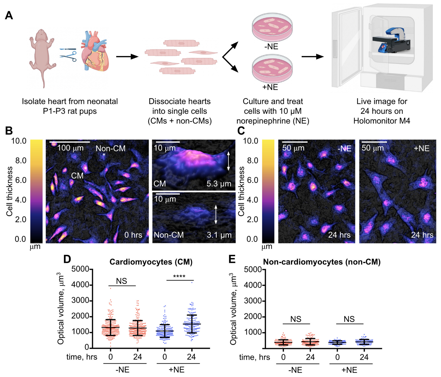
3.1. Validation of the Holomonitor M4 Digital Holographic Imaging System to Detect Norepinephrine-Induced Cardiomyocyte Hypertrophic Growth
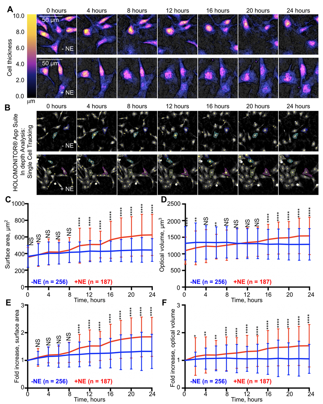
3.2. Application of the Holomonitor M4 Digital Holographic Imaging System to Monitor Real-Time Cardiomyocyte Hypertrophic Growth Dynamics
4. Discussion
5. Conclusions
Supplementary Materials
Author Contributions
Funding
Institutional Review Board Statement
Informed Consent Statement
Data Availability Statement
Acknowledgments
Conflicts of Interest
References
- Hill, J.A.; Olson, E.N. Cardiac Plasticity. N. Engl. J. Med. 2008, 358, 1370–1380. [Google Scholar] [CrossRef]
- Adzika, G.K.; Machuki, J.O.; Shang, W.; Hou, H.; Ma, T.; Wu, L.; Geng, J.; Hu, X.; Ma, X.; Sun, H. Pathological Cardiac Hypertrophy: The Synergy of Adenylyl Cyclases Inhibition in Cardiac and Immune Cells during Chronic Catecholamine Stress. J. Mol. Med. 2019, 97, 897–907. [Google Scholar] [CrossRef]
- Liu, X.; Li, H.; Hastings, M.H.; Xiao, C.; Damilano, F.; Platt, C.; Lerchenmüller, C.; Zhu, H.; Wei, X.P.; Yeri, A.; et al. miR-222 Inhibits Pathological Cardiac Hypertrophy and Heart Failure. Cardiovasc. Res. 2024, 120, 262–272. [Google Scholar] [CrossRef] [PubMed]
- Frey, N.; Katus, H.A.; Olson, E.N.; Hill, J.A. Hypertrophy of the Heart: A New Therapeutic Target? Circulation 2004, 109, 1580–1589. [Google Scholar] [CrossRef]
- Heineke, J.; Molkentin, J.D. Regulation of Cardiac Hypertrophy by Intracellular Signalling Pathways. Nat. Rev. Mol. Cell Biol. 2006, 7, 589–600. [Google Scholar] [CrossRef] [PubMed]
- Bueno, O.F.; De Windt, L.J.; Tymitz, K.M.; Witt, S.A.; Kimball, T.R.; Klevitsky, R.; Hewett, T.E.; Jones, S.P.; Lefer, D.J.; Peng, C.F.; et al. The MEK1-ERK1/2 Signaling Pathway Promotes Compensated Cardiac Hypertrophy in Transgenic Mice. EMBO J. 2000, 19, 6341–6350. [Google Scholar] [CrossRef]
- Molkentin, J.D.; Lu, J.R.; Antos, C.L.; Markham, B.; Richardson, J.; Robbins, J.; Grant, S.R.; Olson, E.N. A Calcineurin-Dependent Transcriptional Pathway for Cardiac Hypertrophy. Cell 1998, 93, 215–228. [Google Scholar] [CrossRef] [PubMed]
- Li, F.; Wang, X.; Capasso, J.M.; Gerdes, A.M. Rapid Transition of Cardiac Myocytes from Hyperplasia to Hypertrophy during Postnatal Development. J. Mol. Cell. Cardiol. 1996, 28, 1737–1746. [Google Scholar] [CrossRef]
- Nakano, H.; Minami, I.; Braas, D.; Pappoe, H.; Wu, X.; Sagadevan, A.; Vergnes, L.; Fu, K.; Morselli, M.; Dunham, C.; et al. Glucose Inhibits Cardiac Muscle Maturation through Nucleotide Biosynthesis. eLife 2017, 6, e29330. [Google Scholar] [CrossRef]
- Satoh, H.; Delbridge, L.M.; Blatter, L.A.; Bers, D.M. Surface:Volume Relationship in Cardiac Myocytes Studied with Confocal Microscopy and Membrane Capacitance Measurements: Species-Dependence and Developmental Effects. Biophys. J. 1996, 70, 1494–1504. [Google Scholar] [CrossRef]
- Watkins, S.J.; Borthwick, G.M.; Oakenfull, R.; Robson, A.; Arthur, H.M. Angiotensin II-Induced Cardiomyocyte Hypertrophy in Vitro Is TAK1-Dependent and Smad2/3-Independent. Hypertens. Res. 2012, 35, 393–398. [Google Scholar] [CrossRef] [PubMed]
- Mölder, A.; Sebesta, M.; Gustafsson, M.; Gisselson, L.; Wingren, A.G.; Alm, K. Non-Invasive, Label-Free Cell Counting and Quantitative Analysis of Adherent Cells Using Digital Holography. J. Microsc. 2008, 232, 240–247. [Google Scholar] [CrossRef] [PubMed]
- Park, S.; Huang, H.; Ross, I.; Moreno, J.; Khyeam, S.; Simmons, J.; Huang, G.N.; Payumo, A.Y. Quantitative Three-Dimensional Label-Free Digital Holographic Imaging of Cardiomyocyte Size, Ploidy, and Cell Division. bioRxiv 2023. 2023.11.02.565407. [Google Scholar] [CrossRef]
- Schlaich, M.P.; Kaye, D.M.; Lambert, E.; Sommerville, M.; Socratous, F.; Esler, M.D. Relation Between Cardiac Sympathetic Activity and Hypertensive Left Ventricular Hypertrophy. Circulation 2003, 108, 560–565. [Google Scholar] [CrossRef] [PubMed]
- Simpson, P. Norepinephrine-Stimulated Hypertrophy of Cultured Rat Myocardial Cells Is an Alpha 1 Adrenergic Response. J. Clin. Investig. 1983, 72, 732–738. [Google Scholar] [CrossRef] [PubMed]
- Blondel, B.; Roijen, I.; Cheneval, J.P. Heart Cells in Culture: A Simple Method for Increasing the Proportion of Myoblasts. Experientia 1971, 27, 356–358. [Google Scholar] [CrossRef] [PubMed]
- Sen, A.; Dunnmon, P.; Henderson, S.A.; Gerard, R.D.; Chien, K.R. Terminally Differentiated Neonatal Rat Myocardial Cells Proliferate and Maintain Specific Differentiated Functions Following Expression of SV40 Large T Antigen. J. Biol. Chem. 1988, 263, 19132–19136. [Google Scholar] [CrossRef] [PubMed]
- Orita, H.; Fukasawa, M.; Hirooka, S.; Uchino, H.; Fukui, K.; Washio, M. Modulation of Cardiac Myocyte Beating Rate and Hypertrophy by Cardiac Fibroblasts Isolated from Neonatal Rat Ventricle. Jpn. Circ. J. 1993, 57, 912–920. [Google Scholar] [CrossRef] [PubMed]
- Logg, K.; Bodvard, K.; Blomberg, A.; Käll, M. Investigations on Light-Induced Stress in Fluorescence Microscopy Using Nuclear Localization of the Transcription Factor Msn2p as a Reporter. FEMS Yeast Res. 2009, 9, 875–884. [Google Scholar] [CrossRef]
- Wagner, M.; Weber, P.; Bruns, T.; Strauss, W.S.L.; Wittig, R.; Schneckenburger, H. Light Dose Is a Limiting Factor to Maintain Cell Viability in Fluorescence Microscopy and Single Molecule Detection. Int. J. Mol. Sci. 2010, 11, 956–966. [Google Scholar] [CrossRef]
- Moon, I.; Jaferzadeh, K.; Ahmadzadeh, E.; Javidi, B. Automated Quantitative Analysis of Multiple Cardiomyocytes at the Single-Cell Level with Three-Dimensional Holographic Imaging Informatics. J. Biophotonics 2018, 11, e201800116. [Google Scholar] [CrossRef] [PubMed]
- Rohr, S. Role of Gap Junctions in the Propagation of the Cardiac Action Potential. Cardiovasc. Res. 2004, 62, 309–322. [Google Scholar] [CrossRef] [PubMed]
Disclaimer/Publisher’s Note: The statements, opinions and data contained in all publications are solely those of the individual author(s) and contributor(s) and not of MDPI and/or the editor(s). MDPI and/or the editor(s) disclaim responsibility for any injury to people or property resulting from any ideas, methods, instructions or products referred to in the content. |
© 2024 by the authors. Licensee MDPI, Basel, Switzerland. This article is an open access article distributed under the terms and conditions of the Creative Commons Attribution (CC BY) license (https://creativecommons.org/licenses/by/4.0/).
Share and Cite
Akter, W.; Huang, H.; Simmons, J.; Payumo, A.Y. Application of Digital Holographic Imaging to Monitor Real-Time Cardiomyocyte Hypertrophy Dynamics in Response to Norepinephrine Stimulation. Appl. Sci. 2024, 14, 3819. https://doi.org/10.3390/app14093819
Akter W, Huang H, Simmons J, Payumo AY. Application of Digital Holographic Imaging to Monitor Real-Time Cardiomyocyte Hypertrophy Dynamics in Response to Norepinephrine Stimulation. Applied Sciences. 2024; 14(9):3819. https://doi.org/10.3390/app14093819
Chicago/Turabian StyleAkter, Wahida, Herman Huang, Jacquelyn Simmons, and Alexander Y. Payumo. 2024. "Application of Digital Holographic Imaging to Monitor Real-Time Cardiomyocyte Hypertrophy Dynamics in Response to Norepinephrine Stimulation" Applied Sciences 14, no. 9: 3819. https://doi.org/10.3390/app14093819
APA StyleAkter, W., Huang, H., Simmons, J., & Payumo, A. Y. (2024). Application of Digital Holographic Imaging to Monitor Real-Time Cardiomyocyte Hypertrophy Dynamics in Response to Norepinephrine Stimulation. Applied Sciences, 14(9), 3819. https://doi.org/10.3390/app14093819