

An Assessment of the Natural Radioactivity Content in Pigments and an Estimation of the Radiological Health Risk for the Public

 ,
, 
 , ,
, , 
 and
and 
Abstract
1. Introduction
2. Materials and Methods
2.1. Description of the Samples
2.2. HPGE γ-Spectrometry Measurements
2.3. Assessment of Radiological Hazards Effects
2.3.1. Absorbed γ-Dose Rate (D)
2.3.2. Annual Effective Dose Equivalent Outdoor (AEDEout) and Indoor (AEDEin)
2.3.3. Activity Concentration Index (I)
2.4. Statistical Treatments
3. Results and Discussion
3.1. The Specific Activity of the Radionuclides
3.2. Dose Rate and Dose Assessment and Hazard Indices
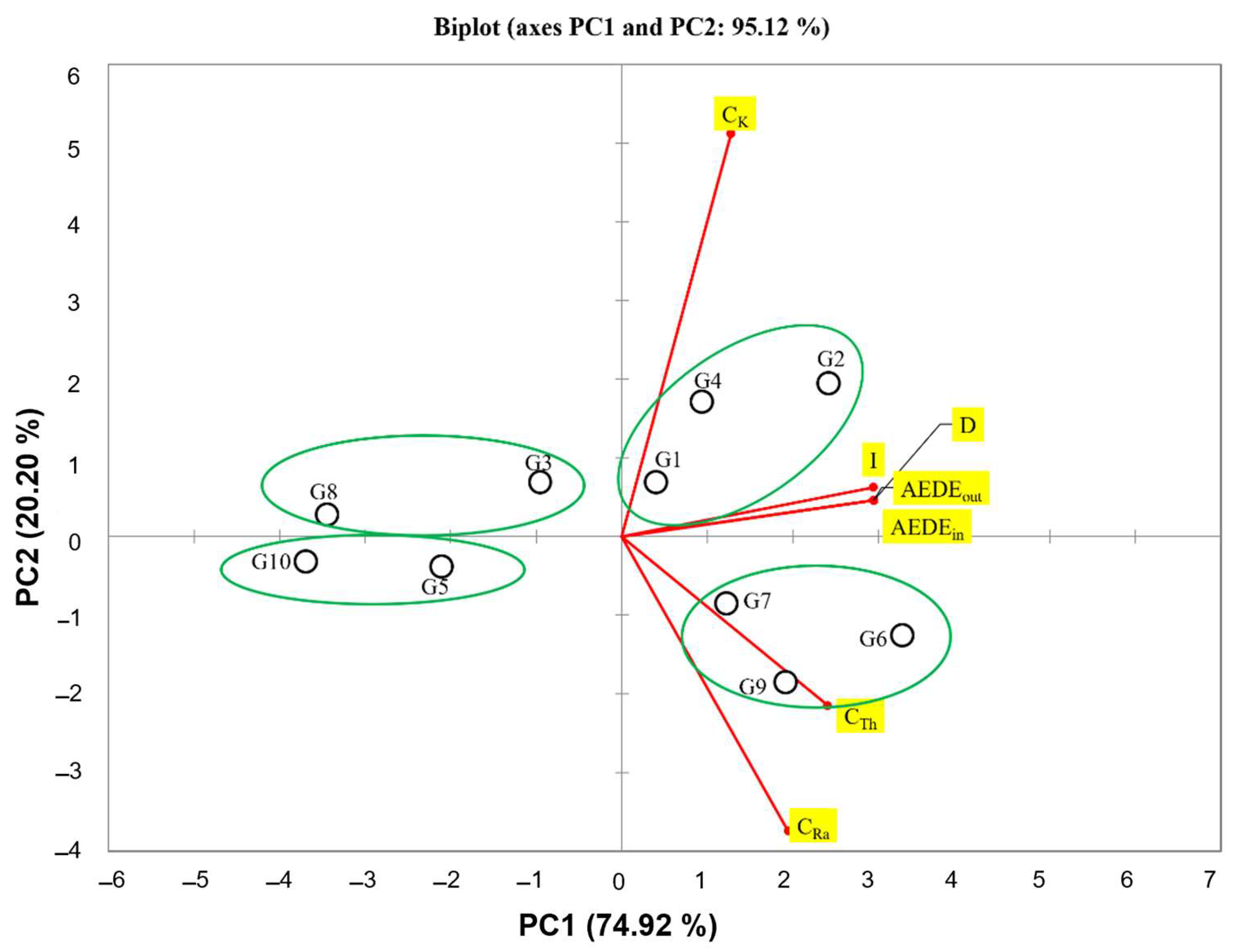
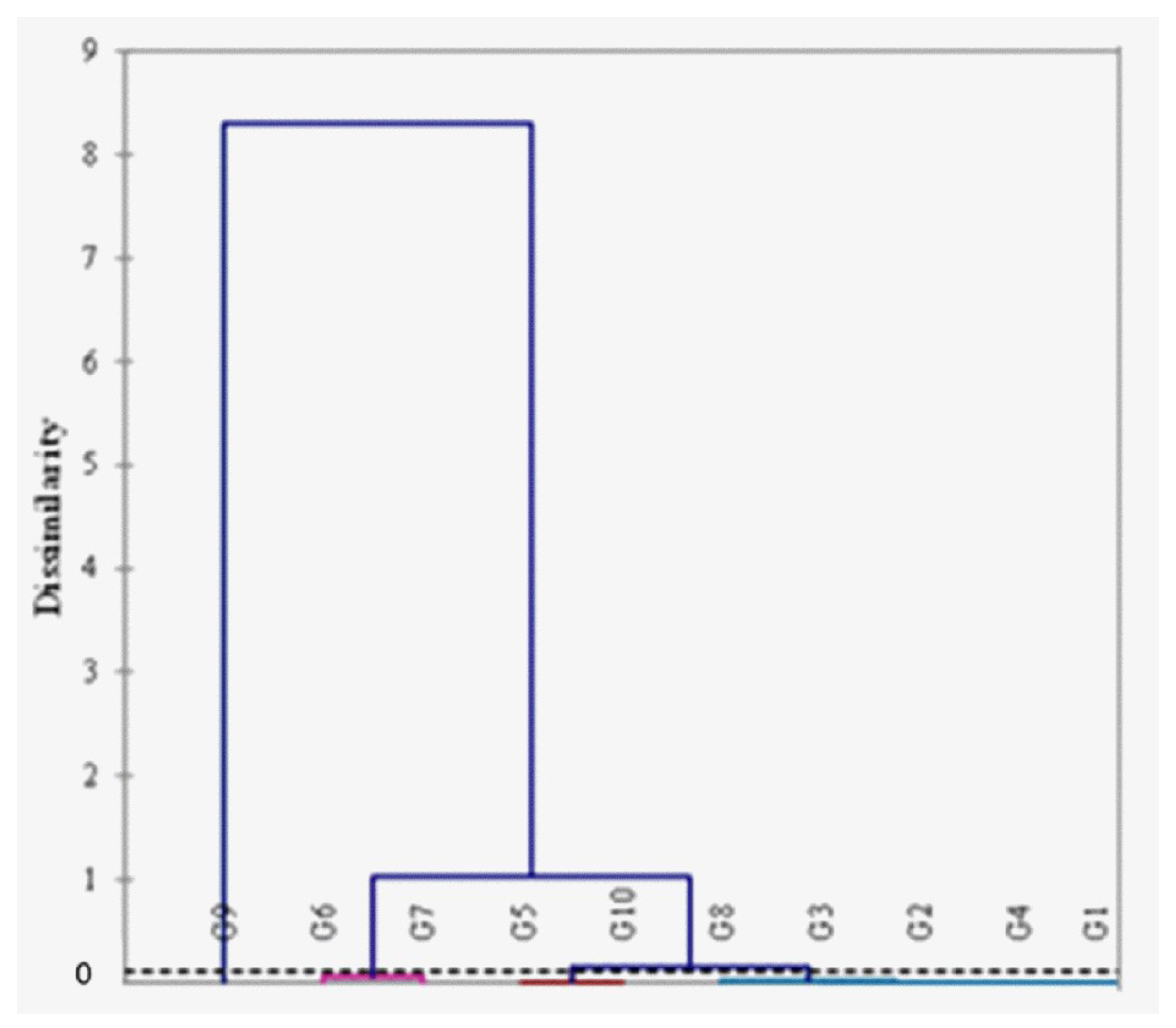
3.3. Statistical Features
4. Conclusions
Author Contributions
Funding
Institutional Review Board Statement
Informed Consent Statement
Data Availability Statement
Conflicts of Interest
References
- Gargano, M.; Bonizzoni, L.; Grifoni, E.; Melada, J.; Guglielmi, V.; Bruni, S.; Ludwig, N. Multi-analytical investigation of panel, pigments and varnish of The Martyirdom of St. Catherine by Gaudenzio Ferrari (16th century). J. Cult. Herit. 2020, 46, 289–297. [Google Scholar] [CrossRef]
- United Nations Scientific Committee on the Effects of Atomic Radiation. Sources and Effects of Ionizing Radiation: Report to the General Assembly, with Scientific Annexes; United Nations Scientific Committee on the Effects of Atomic Radiation: Vienna, Austria, 2000; Volume I, ISBN 92-1-142238-8. [Google Scholar]
- Amadori, M.L.; Barcelli, S.; Poldi, G.; Ferrucci, F.; Andreotti, A.; Baraldi, P.; Colombini, M.P. Invasive and non-invasive analyses for knowledge and conservation of Roman wall paintings of the Villa of the Papyri in Herculaneum. Microchem. J. 2015, 118, 183–192. [Google Scholar] [CrossRef]
- Comite, V.; Bergomi, A.; Lombardi, C.A.; Fermo, P. Characterization of soluble salts on the frescoes by Saturnino Gatti in the church of San Panfilo in Villagrande di Tornimparte (L’Aquila). Appl. Sci. 2023, 13, 6623. [Google Scholar] [CrossRef]
- Henshilwood, C.S.; d’Errico, F.; Watts, I. Engraved ochres from the Middle Stone Age levels at Blombos Cave, South Africa. J. Hum. Evol. 2009, 57, 27–47. [Google Scholar] [CrossRef] [PubMed]
- Gliozzo, E.; Ionescu, C. Pigments—Lead-based whites, reds, yellows and oranges and their alteration phases. Archaeol. Anthropol. Sci. 2021, 14, 17. [Google Scholar] [CrossRef]
- Fermo, P.; Mearini, A.; Bonomi, R.; Arrighetti, E.; Comite, V. An integrated analytical approach for the characterization of repainted wooden statues dated to the fifteenth century. Microchem. J. 2020, 157, 105072. [Google Scholar] [CrossRef]
- La Russa, M.F.; Belfiore, C.M.; Fichera, G.V.; Maniscalco, R.; Calabrò, C.; Ruffolo, S.A.; Pezzino, A. The behaviour to weathering of the Hyblean limestone in the Baroque architecture of the Val di Noto (SE Sicily): An experimental study on the “calcare a lumachella” stone. Constr. Build. Mater. 2015, 77, 7–19. [Google Scholar] [CrossRef]
- Watts, I.; Chazan, M.; Wilkins, J. Early evidence for brilliant ritualized display: Specularite use in the Northern Cape (South Africa) between ∼500 and ∼300 ka. Curr. Anthropol. 2016, 57, 287–310. [Google Scholar] [CrossRef]
- Wolf, S.; Conard, N.J.; Floss, H.; Dapschauskas, R.; Velliky, E.C.; Kandel, A.W. The Use of Ochre and Painting During the Upper Paleolithic of the Swabian Jura in the Context of the Development of Ochre Use in Africa and Europe. Open Archaeol. 2018, 4, 185–205. [Google Scholar] [CrossRef]
- Domingo, I.; Chieli, A. Characterizing the pigments and paints of prehistoric artists. Archaeol. Anthropol. Sci. 2021, 13, 196. [Google Scholar] [CrossRef]
- Rosso, D.E.; d’Errico, F.; Zilhão, J. Stratigraphic and spatial distribution of ochre and ochre processing tools at Porc-Epic Cave, Dire Dawa, Ethiopia. Quat. Int. 2014, 343, 85–99. [Google Scholar] [CrossRef]
- Steelman, K.L.; Boyd, C.E.; Allen, T. Two independent methods for dating rock art: Age determination of paint and oxalate layers at Eagle Cave, TX. J. Archaeol. Sci. 2021, 126, 105315. [Google Scholar] [CrossRef]
- Salomon, H.; Vignaud, C.; Coquinot, Y.; Beck, L.; Stringer, C.; Strivay, D.; D’Errico, F. Selection and heating of colouring materials in the Mousterian level of Es-Skhul (c. 100,000 years bp, Mount Carmel, Israel). Archaeometry 2012, 54, 698–722. [Google Scholar] [CrossRef]
- Caridi, F.; Marguccio, S.; Durante, G.; Trozzo, R.; Fullone, F.; Belvedere, A.; D’Agostino, M.; Belmusto, G. Natural radioactivity measurements and dosimetric evaluations in soil samples with a high content of NORM. Eur. Phys. J. Plus 2017, 132, 56. [Google Scholar] [CrossRef]
- Torrisi, L.; Caridi, F.; Picciotto, A.; Margarone, D.; Borrielli, A. Particle emission from tantalum plasma produced by 532 nm laser pulse ablation. J. Appl. Phys. 2006, 100, 093306. [Google Scholar] [CrossRef]
- Reisz, J.A.; Bansal, N.; Qian, J.; Zhao, W.; Furdui, C.M. Effects of ionizing radiation on biological molecules—Mechanisms of damage and emerging methods of detection. Antioxid. Redox Signal. 2014, 21, 260–292. [Google Scholar] [CrossRef] [PubMed]
- Mohan, S.; Chopra, V. Chapter 18—Biological effects of radiation. In Woodhead Publishing Series in Electronic and Optical Materials; Dhoble, S., Chopra, V., Nayar, V., Kitis, G., Poelman, D., Swart, H.B.T.-R.D.P., Eds.; Woodhead Publishing: Sawston, UK, 2022; pp. 485–508. ISBN 978-0-323-85471-9. [Google Scholar]
- Hall, E.J.; Kereiakes, J.G. 69—Effects of Ionizing Radiation on Cells. In Cell Physiology Source Book, 3rd ed.; Academic Press: San Diego, CA, USA, 2001; pp. 1185–1201. ISBN 978-0-12-656976-6. [Google Scholar]
- Lindop, P.J. Biological Effects of Ionizing Radiations. Nature 1963, 200, 1036. [Google Scholar] [CrossRef]
- Caridi, F.; Messina, M.; Belvedere, A.; D’Agostino, M.; Marguccio, S.; Settineri, L.; Belmusto, G. Food salt characterization in terms of radioactivity and metals contamination. Appl. Sci. 2019, 9, 2882. [Google Scholar] [CrossRef]
- Pacheco, R.; Stock, H. Effects of Radiation on Bone. Curr. Osteoporos. Rep. 2013, 11, 299–304. [Google Scholar] [CrossRef] [PubMed]
- Grabham, P.; Sharma, P. The effects of radiation on angiogenesis. Vasc. Cell 2013, 5, 19. [Google Scholar] [CrossRef] [PubMed]
- Chadwick, D.R.; Abrahams, S.P. Biological Effects of Radiation. Arch. Environ. Health Int. J. 1964, 9, 643–648. [Google Scholar] [CrossRef] [PubMed]
- Aslam, H.; Marx, W.; Rocks, T.; Loughman, A.; Chandrasekaran, V.; Ruusunen, A.; Dawson, S.L.; West, M.; Mullarkey, E.; Pasco, J.A.; et al. The effects of dairy and dairy derivatives on the gut microbiota: A systematic literature review. Gut Microbes 2020, 12, 1799533. [Google Scholar] [CrossRef] [PubMed]
- Ravisankar, R.; Chandrasekaran, A.; Vijayagopal, P.; Venkatraman, B.; Senthilkumar, G.; Eswaran, P.; Rajalakshmi, A. Natural radioactivity in soil samples of Yelagiri Hills, Tamil Nadu, India and the associated radiation hazards. Radiat. Phys. Chem. 2012, 81, 1789–1795. [Google Scholar] [CrossRef]
- Picciotto, A.; Krása, J.; Láska, L.; Rohlena, K.; Torrisi, L.; Gammino, S.; Mezzasalma, A.M.; Caridi, F. Plasma temperature and ion current analysis of gold ablation at different laser power rates. Nucl. Instr. Meth. Phys. Res. B 2006, 247, 261–267. [Google Scholar] [CrossRef]
- Albergamo, A.; Mottese, A.F.; Bua, G.; Caridi, F.; Sabatino, G.; Barrega, L.; Costa, R.; Dugo, G. Discrimination of the Sicilian Prickly Pear (Opuntia ficus-indica L., CV. Muscaredda) According to the Provenance by Testing Unsupervised and Supervised Chemometrics. J. Food Sci. 2018, 83, 2933–2942. [Google Scholar] [CrossRef] [PubMed]
- Avwiri, G.O.; Egieya, J.M. Radiometric assay of hazard indices and excess lifetime cancer risk due to natural radioactivity in soil profile in Ogba/Egbema/Ndoni local government area of Rivers state, Nigeria. Acad. Res. Int. 2013, 4, 54–65. [Google Scholar]
- Ispra. 2021. Available online: https://www.isprambiente.gov.it/it (accessed on 2 January 2024).
- Caridi, F.; Paladini, G.; Marguccio, S.; Belvedere, A.; D’Agostino, M.; Messina, M.; Crupi, V.; Venuti, V.; Majolino, D. Evaluation of Radioactivity and Heavy Metals Content in a Basalt Aggregate for Concrete from Sicily, Southern Italy: A Case Study. Appl. Sci. 2023, 13, 4804. [Google Scholar] [CrossRef]
- Eckert & Ziegler Calibration Sources. Available online: https://www.ezag.com/fileadmin/user_upload/isotopes/isotopes/Isotrak/isotrak-pdf/Product_literature/EZIPL/EZIP_catalogue_reference_and_calibration_sources.pdf (accessed on 2 January 2024).
- Ortec Gamma Vision Software User Manual. 2020. Available online: https://www.ortec-online.com/-/media/ametekortec/manuals/a/a66-mnl.pdf?la=en&revision=dd63bfec-f5cd-4579-8201-e81a52f7fb78 (accessed on 3 January 2024).
- Caridi, F.; Torrisi, L.; Mezzasalma, A.M.; Mondio, G.; Borrielli, A. Al2O3 plasma production during pulsed laser deposition. Eur. Phys. J. D 2009, 54, 467–472. [Google Scholar] [CrossRef]
- Margarone, D.; Torrisi, L.; Borrielli, A.; Caridi, F. Silver plasma by pulsed laser ablation. Plasma Sources Sci. Technol. 2008, 17, 035019. [Google Scholar] [CrossRef]
- ACCREDIA. Available online: https://www.accredia.it/ (accessed on 3 January 2024).
- Stark, K.; Goméz-Ros, J.M.; Vives I Batlle, J.; Lindbo Hansen, E.; Beaugelin-Seiller, K.; Kapustka, L.A.; Wood, M.D.; Bradshaw, C.; Real, A.; McGuire, C.; et al. Dose assessment in environmental radiological protection: State of the art and perspectives. J. Environ. Radioact. 2017, 175–176, 105–114. [Google Scholar] [CrossRef] [PubMed]
- Steinhauser, G.; Brandl, A.; Johnson, T.E. Comparison of the Chernobyl and Fukushima nuclear accidents: A review of the environmental impacts. Sci. Total Environ. 2014, 470–471, 800–817. [Google Scholar] [CrossRef] [PubMed]
- UNSCEAR. United Nations Scientific Committee on the Effects of Atomic Radiation, Vol. I, Annex B: Exposure of the Public and Workers from Various Sources of Radiation; United Nations: New York, NY, USA, 2008. [Google Scholar]
- Italian Legislation D.Lgs. 101/20. Available online: www.normattiva.it (accessed on 4 January 2024).
- Kayo, S.A.; Moyo, M.N.; Shouop, C.J.G.; Mekontso, É.J.N.; Motapon, O. Multivariate statistical assessment of natural radioactivity and radiological hazards data of cement building materials mainly used in Cameroon. Arab. J. Geosci. 2021, 14, 2487. [Google Scholar] [CrossRef]
- Shahrokhi, A.; Adelikhah, M.; Chalupnik, S.; Kovács, T. Multivariate statistical approach on distribution of natural and anthropogenic radionuclides and associated radiation indices along the north-western coastline of Aegean Sea, Greece. Mar. Pollut. Bull. 2021, 163, 112009. [Google Scholar] [CrossRef] [PubMed]
- Caridi, F.; Spoto, S.E.; Paladini, G.; Faggio, G.; D’Agostino, M.; Marguccio, S.; Belvedere, A.; Crupi, V.; Majolino, D.; Venuti, V. Measurement of the natural radioactivity content of earth pigments and evaluation of radiological health risks. In Proceedings of the 2022 IMEKO TC4 International Conference on Metrology for Archaeology and Cultural Heritage, Calabria, Italy, 19–21 October 2022; pp. 411–415. [Google Scholar] [CrossRef]
- Morelli, D.; Immé, G.; Cammisa, S.; Catalano, R.; Mangano, G.; La Delfa, S.; Patanè, G. Radioactivity measurements in volcano-tectonic area for geodynamic process study. EPJ Web Conf. 2012, 24, 5009. [Google Scholar] [CrossRef]
- Mohd Razali, N.; Yap, B. Power Comparisons of some selected normality tests. In Proceedings of the Regional Conference on Statistical Sciences 2010 (RCSS’10), Kota Bharu, Malaysia, 3–14 June 2010; Volume 2, pp. 126–138. [Google Scholar]
- Seier, E. Normality Tests: Power Comparison BT—International Encyclopedia of Statistical Science; Lovric, M., Ed.; Springer: Berlin/Heidelberg, Germany, 2011; pp. 1000–1003. ISBN 978-3-642-04898-2. [Google Scholar]
- Weide, A.C.; Beauducel, A. Varimax Rotation Based on Gradient Projection Is a Feasible Alternative to SPSS. Front. Psychol. 2019, 10, 424873. [Google Scholar] [CrossRef] [PubMed]
- Freeman, J.J.; Wang, A.; Kuebler, K.E.; Jolliff, B.L.; Haskin, L.A. Characterization of natural feldspars by raman spectroscopy for future planetary exploration. Can. Mineral. 2008, 46, 1477–1500. [Google Scholar] [CrossRef]
- Zinin, P.; Tatsumi-Petrochilos, L.; Bonal, L.; Acosta, T.; Hammer, J.; Gilder, S.; Fuller, M. Raman spectroscopy of titanomagnetites: Calibration of the intensity of Raman peaks as a sensitive indicator for their Ti content. Am. Mineral. 2011, 96, 1537–1546. [Google Scholar] [CrossRef]
- Buzatu, A.; Buzgar, N. The raman study of single-chain silicates. Analele Stiint. ale Univ. “Al. I. Cuza” din Iasi. Ser. Geol. 2010, LVI, 107–125. [Google Scholar]
- Aiuppa, A.; Allard, P.; D’Alessandro, W.; Agnès, M.; Parello, F.; Treuil, M.; Valenza, M. Mobility and fluxes of major, minor and trace metals during basalt weathering and groundwater transport at Mt. Etna volcano (Sicily). Geochim. Cosmochim. Acta 2000, 64, 1827–1841. [Google Scholar] [CrossRef]


| Group ID | Chemical Characterization | Origin | Number of Samples | |
|---|---|---|---|---|
| G1 | Raw Sienna | Fe2O3 ∙ nH2O + Al2O3 ∙ MnO2 + SiO2 ∙ H2O | Italy | 5 |
| G2 | Burnt Sienna | Fe2O3 ∙ nH2O + Al2O3 ∙ MnO2 | Italy | 5 |
| G3 | Raw Umber | CaSO4 · 2H2O + CaSO4 + CaCO3 + FeO(OH) + clay | Italy | 5 |
| G4 | Burnt Umber | CaSO4 · 2H2O + CaSO4 + Fe2O3 + CaCO3 + clay | Italy | 5 |
| G5 | Yellow Ochre | Fe2O3 ∙ n H2O | Italy | 5 |
| G6 | French Ochre | SiO2 + Al2O3 + Fe2O3 | Germany | 5 |
| G7 | Green Earth from Verona | K(Mg,Fe2+)(Fe3+, Al)[Si4O10](OH)2 + (K,Na)(Fe3+,Al,Mg)2 (Si,Al)4O10(OH)2 | Germany | 5 |
| G8 | Terra Pozzuoli | Fe2O3 · nH2O | Germany | 5 |
| G9 | Titanium Orange | Ti-Sb-Cr-O Rutile | Germany | 5 |
| G10 | Titanium White | TiO2 | Germany | 5 |
| HPGe Detector | |
|---|---|
| Full Width at Half Maximum | 1.94 keV |
| Peak-to-Compton ratio | 65:1 |
| Relative efficiency | 37.5% (at the 1.33 MeV 60Co γ-line) |
| Bias voltage | −4800 V |
| Energy range | 5 keV–2 MeV |
| Group ID | CRa (Bq kg−1 d.w.) | CTh (Bq kg−1 d.w.) | CK (Bq kg−1 d.w.) |
|---|---|---|---|
| G1 | 12.1 ± 3.8 | 23.1 ± 6.5 | 546 ± 67 |
| G2 | 15.3 ± 4.9 | 23.0 ± 6.4 | 1022 ± 116 |
| G3 | 13.5 ± 4.1 | 5.8 ± 2.9 | 486 ± 56 |
| G4 | 8.3 ± 3.1 | 15.2 ± 4.8 | 873 ± 98 |
| G5 | 10.6 ± 3.3 | 13.3 ± 3.9 | 104 ± 21 |
| G6 | 55.9 ± 6.8 | 54.1 ± 6.7 | 196 ± 38 |
| G7 | 26.5 ± 4.8 | 48.5 ± 5.9 | 136 ± 25 |
| G8 | 3.5 ± 1.8 | 1.5 ± 0.9 | 43.7 ± 5.8 |
| G9 | 80.8 ± 9.8 | 26.4 ± 4.8 | 32.4 ± 5.1 |
| G10 | 0.75 ± 0.18 | 1.1 ± 0.2 | 7.6 ± 3.8 |
| Group ID | D (nGy h−1) | AEDEout (mSv y−1) | AEDEin (mSv y−1) | I |
|---|---|---|---|---|
| G1 | 42.3 | 0.052 | 0.208 | 0.34 |
| G2 | 63.6 | 0.078 | 0.312 | 0.51 |
| G3 | 30.1 | 0.037 | 0.148 | 0.24 |
| G4 | 49.4 | 0.061 | 0.244 | 0.39 |
| G5 | 17.3 | 0.021 | 0.084 | 0.14 |
| G6 | 66.6 | 0.082 | 0.328 | 0.52 |
| G7 | 47.2 | 0.058 | 0.232 | 0.38 |
| G8 | 4.4 | 0.005 | 0.020 | 0.03 |
| G9 | 54.6 | 0.067 | 0.268 | 0.41 |
| G10 | 1.3 | 0.002 | 0.008 | 0.01 |
| Average | 37.7 | 0.046 | 0.185 | 0.30 |
| PC1 | PC2 | PC3 |
|---|---|---|
| 5.244 | 1.414 | 0.341 |
| 74.917 | 20.203 | 4.874 |
| 74.917 | 95.121 | 99.995 |
Disclaimer/Publisher’s Note: The statements, opinions and data contained in all publications are solely those of the individual author(s) and contributor(s) and not of MDPI and/or the editor(s). MDPI and/or the editor(s) disclaim responsibility for any injury to people or property resulting from any ideas, methods, instructions or products referred to in the content. |
© 2024 by the authors. Licensee MDPI, Basel, Switzerland. This article is an open access article distributed under the terms and conditions of the Creative Commons Attribution (CC BY) license (https://creativecommons.org/licenses/by/4.0/).
Share and Cite
Caridi, F.; Mottese, A.F.; Paladini, G.; Marguccio, S.; D’Agostino, M.; Belvedere, A.; Majolino, D.; Venuti, V. An Assessment of the Natural Radioactivity Content in Pigments and an Estimation of the Radiological Health Risk for the Public. Appl. Sci. 2024, 14, 3021. https://doi.org/10.3390/app14073021
Caridi F, Mottese AF, Paladini G, Marguccio S, D’Agostino M, Belvedere A, Majolino D, Venuti V. An Assessment of the Natural Radioactivity Content in Pigments and an Estimation of the Radiological Health Risk for the Public. Applied Sciences. 2024; 14(7):3021. https://doi.org/10.3390/app14073021
Chicago/Turabian StyleCaridi, Francesco, Antonio Francesco Mottese, Giuseppe Paladini, Santina Marguccio, Maurizio D’Agostino, Alberto Belvedere, Domenico Majolino, and Valentina Venuti. 2024. "An Assessment of the Natural Radioactivity Content in Pigments and an Estimation of the Radiological Health Risk for the Public" Applied Sciences 14, no. 7: 3021. https://doi.org/10.3390/app14073021
APA StyleCaridi, F., Mottese, A. F., Paladini, G., Marguccio, S., D’Agostino, M., Belvedere, A., Majolino, D., & Venuti, V. (2024). An Assessment of the Natural Radioactivity Content in Pigments and an Estimation of the Radiological Health Risk for the Public. Applied Sciences, 14(7), 3021. https://doi.org/10.3390/app14073021