Article

Effects of Temperature and Salinity on the LMS (Lysosomal Membrane Stability) Biomarker in Clams Donax trunculus and Chamelea gallina

by
Paula Soms-Molina
1,
Concepción Martínez-Gómez
2,
Esther Zuñiga
1,
Miguel Rodilla
1 and
Silvia Falco
1,*
1
Institut d’Investigació per a la Gestió Integrada de Zones Costaneres (IGIC), Universitat Politècnica de València, Paranimf 1, Grau de Gandia, 46730 Valencia, Spain
2
Instituto Español de Oceanografía (IEO, CSIC), Varadero 1, San Pedro del Pinatar, 30740 Murcia, Spain
*
Author to whom correspondence should be addressed.
Appl. Sci. 2024, 14(7), 2712; https://doi.org/10.3390/app14072712
Submission received: 29 January 2024 / Revised: 13 March 2024 / Accepted: 20 March 2024 / Published: 23 March 2024

Abstract

:
Population of clams Donax trunculus and Chamelea gallina have been declining significantly in recent decades, and environmental pollution and accelerated global warming have been proposed as contributing factors to this decline, in addition to overfishing. Lysosomal membrane stability (LMS) is a sensitive indicator of health status of the organisms. In this study, we investigate the LMS in these species after exposure for 21 days to nine combined conditions of water temperature (12, 20, and 27.5 °C) and salinity ranges (27–28, 32–33, and 37–38). LMS was assessed in living hemocytes by using the neutral red retention assay. Mortality and the condition index of the organisms were evaluated as supporting parameters. The results indicated interspecies differences in the LMS under similar environmental conditions. Overall, LMS was found to be more sensitive to temperature than to salinity changes. Although both species can tolerate changes in either salinity or temperature seawater conditions, the tolerance range is narrower for D. trunculus, showing a significant cytotoxicity (NRRT < 50 min) at temperatures above 27.5 °C and salinities above 32, and 100% mortality at 27.5 °C and a low salinity range (27–28). This study is the first to assess the combined effect of temperature and salinity on the LMS in C. gallina and D. trunculus, and provide necessary information before using LMS as contaminant-related biomarker in field studies with these species.

1. Introduction

In the western Mediterranean Sea, the wedge clam (Donax trunculus) and the striped venus clam (Chamelea gallina), among other species, hold significant socio-economic value as fishing resources and consequently face high fishing pressure [1,2]. These filter-feeding bivalve mollusks inhabit fine sandy bottoms [3,4]. D. trunculus exclusively resides in the biocenosis of superficial fine sands [5] with strong dependence on the nature and composition of the sediment [6]. On the other hand, the striped venus clam is preferentially distributed in the coastal biocenosis of well-selected fine sand [7], typically found at depths of 3 to 12 m [8], while wedge clams are predominantly distributed between 0 and 2 m deep [9].
Over the recent few decades, the fisheries of these clam species have witnessed a significant decline in the Spanish Mediterranean Region [1,2,10]. This decline has led to the gradual closure of fishing grounds by local authorities or the abandonment of fishing activities in some regions [1]. Management measures, such as legal minimum size requirements or spatial-temporal closures to avoid the fishing during the spawning season, have been implemented to address this situation [2,11]. Despite these efforts, the populations of wedge clams and striped venus clams have not recovered [1], resulting in severe consequences, including the loss of employment for hundreds of small-scale fishermen [1].
Marine ecosystems, particularly in the Mediterranean Sea, are experiencing the impacts of global climate change [12], chemical pollution [13], and biological pollution [14], resulting in significant effects on marine biodiversity [15,16]. While the ocean absorbs both CO2 and heat, slowing atmospheric warming, it also undergoes changes such as increased water temperature, reduced pH, decreased dissolved oxygen, salinity fluctuations, heatwaves, rising sea levels, and alterations in ocean currents [17]. Each of these stressors has its own effect, acting both independently [18] and synergistically [19,20]. Warming, acidification, deoxygenation, and salinity changes occur more rapidly in shallow water habitats, influenced by continental factors. Other anthropogenic pressures such as high urban pressure, mass tourism, overfishing, increased maritime traffic, sewage disposal, and extensive use of fertilizers are added to these coastal marine zones [21]. Consequently, the Mediterranean Sea is being pushed into a new and, at least, uncertain state by all these stress sources [14].
Rivetti et al. [22] linked changes in the temperature of surface waters of the Mediterranean Sea to mass mortality events of invertebrates, including mollusks, concluding that the frequency of mass mortality of benthic invertebrates is increasing and correlates with positive thermal anomalies. In the Mediterranean, marine heat waves are not only more frequent but also more intense and of longer duration [23]. Therefore, the two species under study may struggle to maintain good health at high temperatures. Moschino and Marin [24] demonstrated that C. gallina at temperatures of 28 ° C increased their respiration rate, potentially leading to metabolic imbalances, while Widdows and Staff [25] suggested that wild clams may experience high stress levels at such temperatures potentially causing death [26]. In the case of D. trunculus, Ansell et al. [27] observed a high thermal tolerance for this species, reflecting its distribution in shallower waters; however, their determinations were carried out working with short exposure periods of up to 96 h.
Marine bivalves are osmoconformers capable of maintaining metabolic function despite changes in osmolarity and cytosol composition during changes in salinity [28]. Closing and sealing of the valves are vital responses to stressful salinity [29]. Despite these adaptations, salinity variations can also cause mortality of benthic bivalves [30]. Although D. trunculus is a euryhaline species, prolonged exposure to low salinities slows its movements, which can affect its survival in the environment [31]. Matozzo et al. [32] suggest that salinity variations above and below 34 allow for the observation of quantifiable alterations in the immune responses of C. gallina.
When investigating the biological effects of environmental factors, the alterations in certain physiological processes or subcellular responses can be used as biomarkers of organism health condition. In this sense, lysosomal membrane stability (LMS) represents the most sensitive biomarker of stress at cellular level. LMS in bivalve hemocytes is a core biomarker of general stress of chemical pollution that has been widely recommended by different organizations, such as the Barcelona Convention, International Council for Exploration of the Sea (ICES), and Oslo and Paris (OSPAR) Conventions, and implemented in mussel monitoring programs of chemical pollution [33,34,35]. Given the fact that a variety of factors in addition to overfishing, such as environmental pollution, have been proposed to explain the population significant declines of Donax trunculus and Chamelea gallina mollusks in the Mediterranean Region, interest and opportunities arise to use LMS as a general stress biomarker of chemical environmental pollution in these species. However, the utility of this biomarker in other bivalve species than Mytilus sp. has to be supported by knowledge on how they respond to extrinsic abiotic and intrinsic biotic factors [36]. This is due to the fact that LMS is a robust diagnostic indicator of individual health status in bivalves, but it is also affected by non-contaminant factors, such as temperature, prolonged hypoxia, salinity, food deprivation, or the final stage of gametogenesis [37]. Therefore, the use of LMS to monitor the impacts of environmental chemical pollution on these clam species should involve knowing the response of the LMS biomarker to the temperature and salinity variability to which these species might be subjected in their habitats, as they may act as cofounding factors in the assessment of the biomarker response. In this sense, the main objective of this study was to investigate the cellular wellbeing of Donax trunculus and Chamelea gallina living hemocytes by assessing the lysosomal membrane stability in order to evaluate their health status after short-term exposure to different environmental conditions of temperature and salinity.

2. Materials and Methods

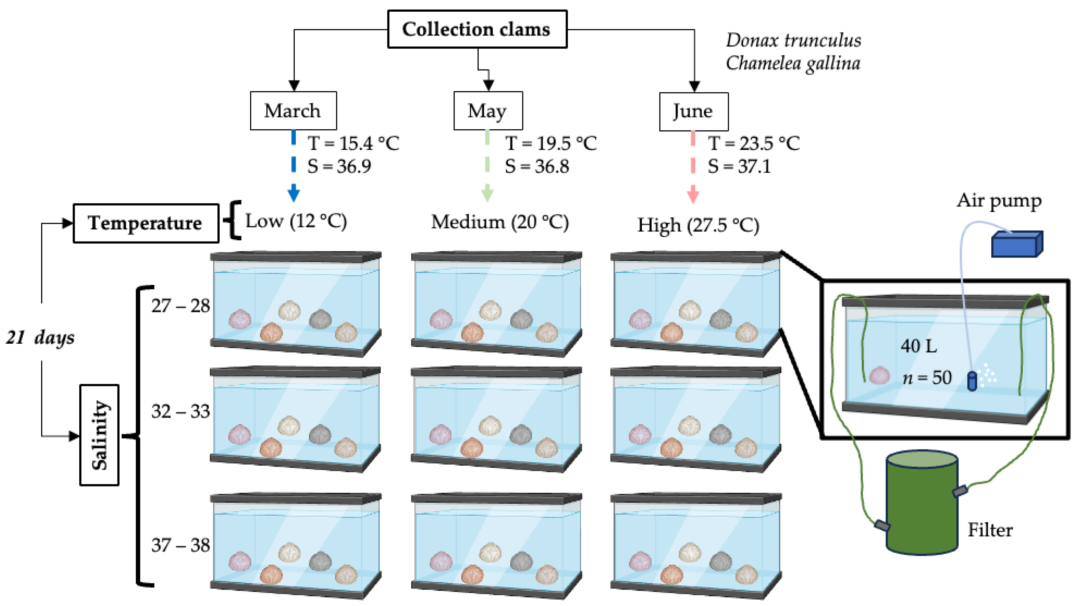
2.1. Collection of Clams and Maintenance

Specimens of D. trunculus and C. gallina were collected in the southern part of the Gulf of Valencia (SE Spain), between the beaches of l’Ahuir and Daimús. The organisms were collected with hand dredges at a depth of 0.5 to 2 m and 4 to 7 m, respectively. To avoid keeping individuals for long periods in laboratory conditions, it was decided to sample mollusks when the difference between the natural temperature in the field and the laboratory test temperature was minimal. Consequently, sampling of individuals was carried out during 2023 on three occasions: March (15.4 ° C and 36.9 of salinity), May (19.5 ° C and 36.8), and June (23.5 ° C and 37.1). The size range of D. trunculus clams was 16.1 to 28.5 mm, while it ranged from 12.8 to 24.5 mm for C. gallina in March, and from 20.8 to 31.4 mm in May and June.
The collected clams (around a 450 specimens of each species) were immediately transferred to the laboratory and maintained in aquariums filled with seawater collected from the sampling points. The aquariums were placed in environmental chambers with a controlled temperature and photoperiod (12 h light/12 h dark), constant aeration, and recirculating filtered seawater. Clams were fed daily with microalgae (Isochrysis galbana). The organisms were acclimated for 5–7 days to the experimental conditions by progressively decreasing or increasing the temperature or salinity by 1–2 (°C) per day. Organisms sampled in March, May, and June were acclimated to 12 ° C (low), 20 ° C (medium), and 27.5 ° C (high) temperatures, respectively (Figure 1). Experimental temperatures and salinities were chosen considering the characteristic conditions of the Mediterranean Sea, where values close to those tested in this study are recorded. During winter, water temperatures range between 13 and 16 ° C, while they are around 22 and 27 ° C in the summer [38]. Salinity generally varies between 36 and 38 [38], decreasing in places near river mouths [4].

2.2. Exposure to Experimental Temperatures and Salinities

Nine combined conditions of temperature and salinity were studied in both species. Animals were held for 21 days at three different temperatures (low (12 ° C), medium (20 ° C), and high (27.5 ° C)) and at three salinity ranges (27–28, 32–33, and 37–38) for each temperature condition (Figure 1). Experimental units for each environmental condition consisted of 40 L aquaria that contained 50 individuals. Seawater parameters (temperature, salinity, and dissolved oxygen) and mortality were monitored daily in every aquarium for both species. Temperature, salinity, and pH were measured using a calibrated WTW Model 340i multiprobe (WTW GmbH, Weilheim, Germany), while dissolved oxygen was measured using a proODO YSI (YSI Inc., Yellow Springs, OH, USA) optical probe. Salinity was corrected using distilled water and seawater. Mortality was determined by the number of clams with open shells that did not react to the mechanical stimulus. Organisms were exposed for 21 days as this period reflects the time frame of sustained exposure to different salinity/temperature regimes in the southern sector of the Gulf of Valencia.

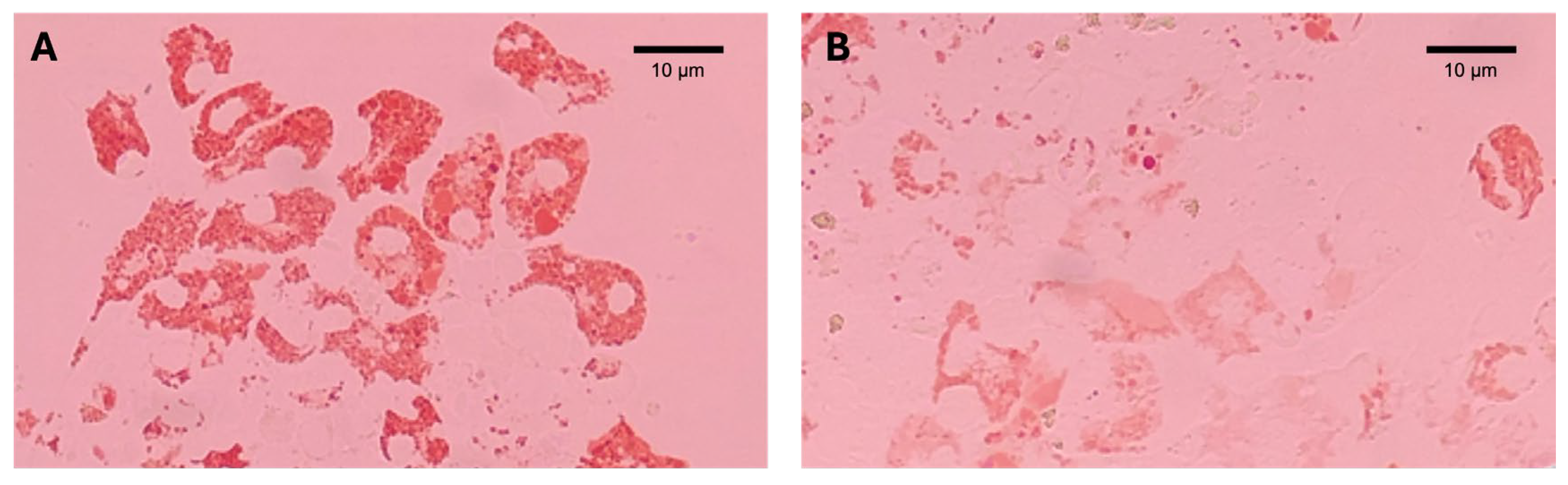
2.3. Lysosomal Membrane Stability (LMS)

After 21 days of exposure, hemolymph samples were obtained from 16 clams from each treatment and hemocyte monolayers were prepared as previously described in Martínez-Gómez et al. [39]. From each clam, 0.1 mL of hemolymph was extracted from the adductor muscle using a 1 mL hypodermic syringe with a 25-gauge needle and pre-loaded with 0.1 mL of filtered aquarium water (0.45 μ m) (corresponding with the seawater of the treatment). LMS was measured by using the in vivo cytochemical method named neutral red retention assay (NRRA), following the procedure described by Lowe et al. [40], with modifications as described in Martínez-Gómez et al. [39]. After 15 min of incubation with neutral red (NR) (CAS: 553-24-2; Labkem, Barcelona, Spain), hemocyte samples were observed under a microscope (×400 and ×200 magnification) at 15, 30, 60, 90, 120, and 180 min and examined for both structural abnormalities (lysosomal swelling, enlargement, and rounded-up fragmenting cells) and lysosomal NR leakage. Results of LMS were expressed as the neutral red retention time (NRRT) (minutes), corresponding to the last time period recorded when there was no evidence of dye loss or lysosomal abnormalities in more than 50% of the cells. To enhance the sensitivity of the LMS measurements, results were also expressed as %LMS, by applying the scoring procedure described in Martínez-Gómez et al. [39], which considers not only the neutral red retention time but also lysosomal alterations. In non-stressed cells, lysosomes will accumulate and retain the NR dye for a prolonged period and will not present lysosomal alterations, whereas in stressed cells, NR will leak into the cytosol and hemocytes will show lysosomal abnormalities. The size of the organisms used to analyze lysosomal membrane stability ranged from 13.9 to 23.9 mm (in March) and from 20.8 to 31.44 mm (in May and June) for C. gallina and from 16.1 to 28.5 mm for D. trunculus.

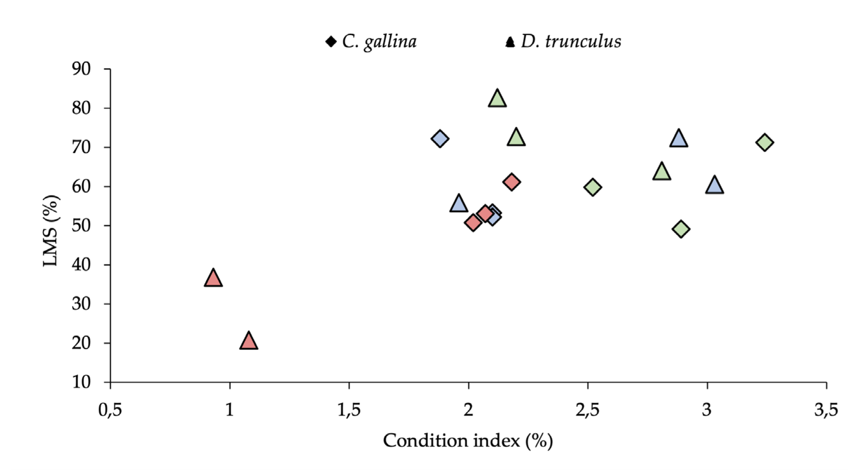
2.4. Condition Index (CI)

The CI provides information on the nutritional and physiological status of bivalves [24,41] and provides complementary information to interpret LMS results [42].
CI was determined before and after 21 days of exposure of the organisms (n = ~10 individuals) to the nine treatments (three temperatures ranges combined with three salinity ranges). Because CI varies according to organism age and physiological status [43], CI was standardized to certain size ranges. For D. trunculus, organisms were selected with a size range of 19–23 mm. In the case of C. gallina, because the organisms sampled in March were exceptionally small ones, the size range used was 13–16 mm, whereas for the May and June samplings, it was 17–20 mm.
The CI was calculated using the equation applied by Boscolo et al. [44], as follows:
%CI = [dry soft tissue weight (g)/shell length (cm)] · 100,
The weight of the dried soft tissue was obtained by drying the soft tissue individually at 100 °C until a constant weight was obtained. The shell length was measured with a digital vernier caliper.

2.5. Data Analysis

Values in the text are expressed as mean ± standard error (SE) if not otherwise specified. The 95% confident limits of the mean LMS values (expressed as minutes of NRRT) were assessed against their corresponding background assessment criteria (BAC) and environmental assessment criteria (EAC) developed for the technique [44]. Statistical analyses were carried out using the software packages IBM® SPSS Statistics v.17 and Statgraphics Centurion XVII. All data were preliminarily checked for normality (Shapiro–Wilk test) and homoscedasticity (Levene’s test); when necessary, they were transform using Arcsine x / 100 or Box–Cox transformation (lambda was found using the most likely estimate (MLE) approach) to meet the required assumptions [45]. Differences in NRRT, %LMS, and %CI among treatments were identified using multifactorial ANOVA (2-way ANOVA). The factors were temperature with 3 levels (10 °C, 20 °C and 27.5 °C) and salinity with 3 levels (27–28, 32–33, and 37–38). To obtain specific LMS information for all treatments and for each temperature and salinity condition, a pairwise comparison (Tukey’s HSD post hoc, p < 0.05) of 9 treatments was carried out: (i) the 3 ranges of salinity levels for each temperature and (ii) the 3 temperature levels for each salinity. Additionally, a pairwise comparison was performed to determine the differences between the CIs of the reference organisms in the three samplings (March, May, and June—on day 0) with the CI values of each treatment (after 21 days of exposure).
Finally, Pearson’s correlation coefficient was used to study the correlations between temperature, salinity, LMS, and %CI.

3. Results

Information on the experimental conditions for each treatment is presented in Table 1. The seawater temperature and salinity values of the treatments during the experiment differed slightly from the nominal values. Dissolved oxygen remained within the appropriate range (~100% oxygen saturation) at all experimental conditions. The pH remained fairly constant between 8.0 and 8.2.

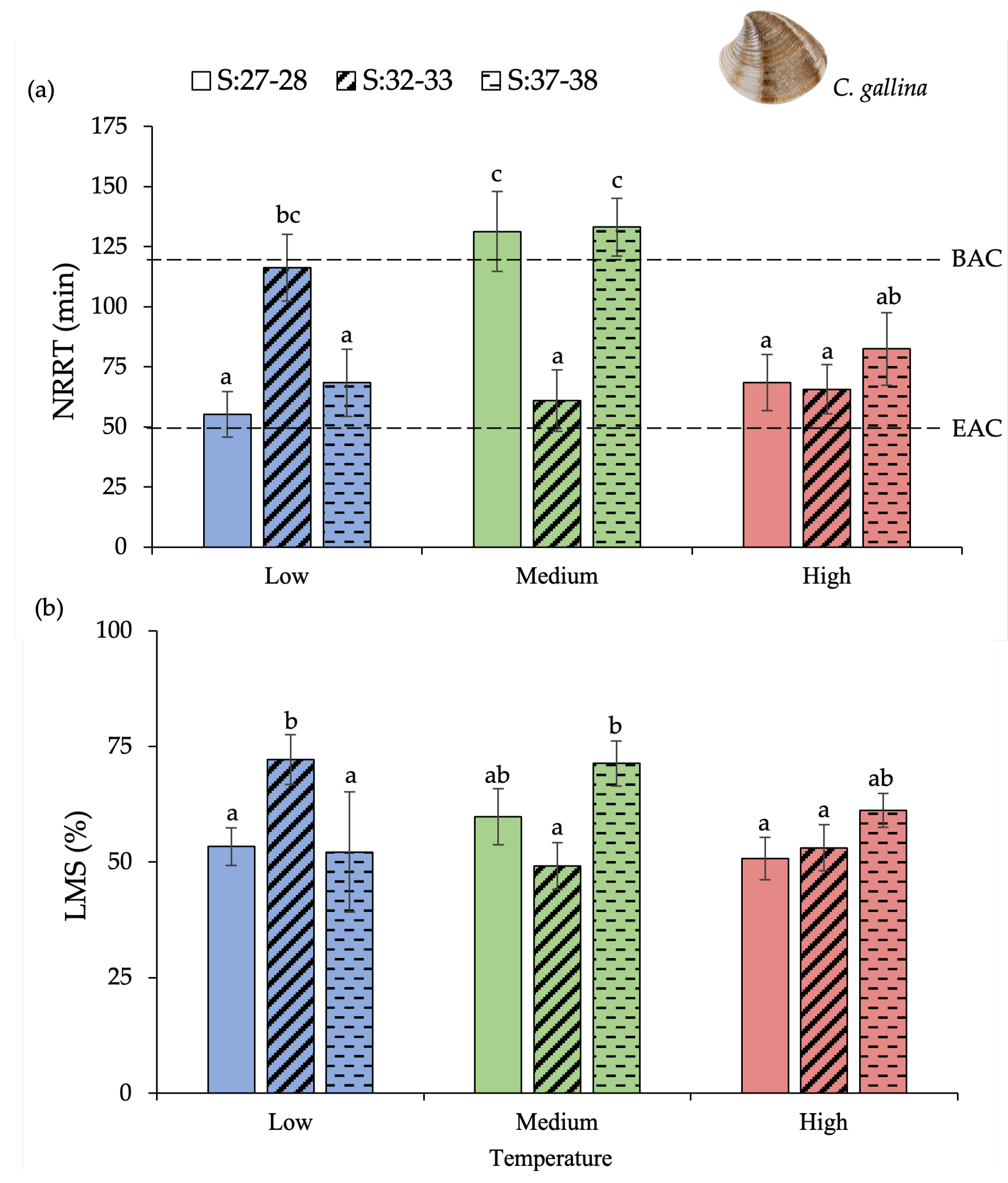
3.1. Chamelea Gallina

No mortality occurred in any of the treatments after 21 days of exposure, so survival was 100%.
The results of NRRT and %LMS values of C. gallina are presented in Figure 2 and Table S1. There was a significant difference in LMS (expressed as NRRT) as a result of temperature and the interaction of both factors (temperature and salinity) but no effect of salinity (2-way ANOVA; p-value < 0.05) (Table 2). LMS in C. gallina hemocytes (expressed as mean value of NRRT) was higher in organisms from the 20 ° C/27–28, 20 ° C/37–38, and 12 ° C/32–33 salinity treatments (Figure 2a), where hemocytes showed a longer retention time of neutral red within the lysosomal compartment (Figure 3A). Pairwise comparisons revealed three homogeneous subsets among the nine combined treatments (Tukey-b test; p-value < 0.05) (Table S2). At a low temperature (12 ° C), NRRT was significantly higher in animals exposed to salinity of 32–33, compared to those maintained at 27–28 and 37–38 (p-value = 0.003) (Figure 2a and Table S3). However, an opposite pattern was found at a medium temperature (20 ° C), with NRRTs being higher at salinities 27–28 (p-value = 0.002). When LMS was estimated by adding information of lysosomal alterations to the NRRTs (%LMS), no differences were observed due to temperature and salinity, but there was a significant interaction between these factors (2-way ANOVA; p-value 0.001) (Table 2). No differences were observed in %LMS between the three salinities tested within the three temperatures studied, except for the treatment comprising 12 ° C/32–33 of salinity, which was significantly higher (Figure 2b, Tables S2 and S3) (p-value = 0.014). Nevertheless, some organisms showed lysosomal alterations in the cells (Figure 3B).
There was a significant difference in the condition index for C. gallina due to temperature but there was no effect of salinity and no interaction between these factors (2-way ANOVA; p-value < 0.05) (Table 2). The CI of the organisms did not change significantly after 21 days of treatment, except in organisms maintained at the highest temperature (p-value = 0.007), where CI after 21 days was lower than at the beginning of the experiment (Table 3 and Table S4). Although the CIs obtained in the low-temperature treatments were similar to those obtained at a high temperature, they were not comparable due to the difference in size between the organisms studied (13–16 mm vs. 17–20 mm).
After 21 days of exposure to nine combined treatments, the CI in C. gallina (n = 9) did not correlate with the temperature or salinity conditions tested, nor with the LMS values measured in this species (Figure 4 and Table S5).

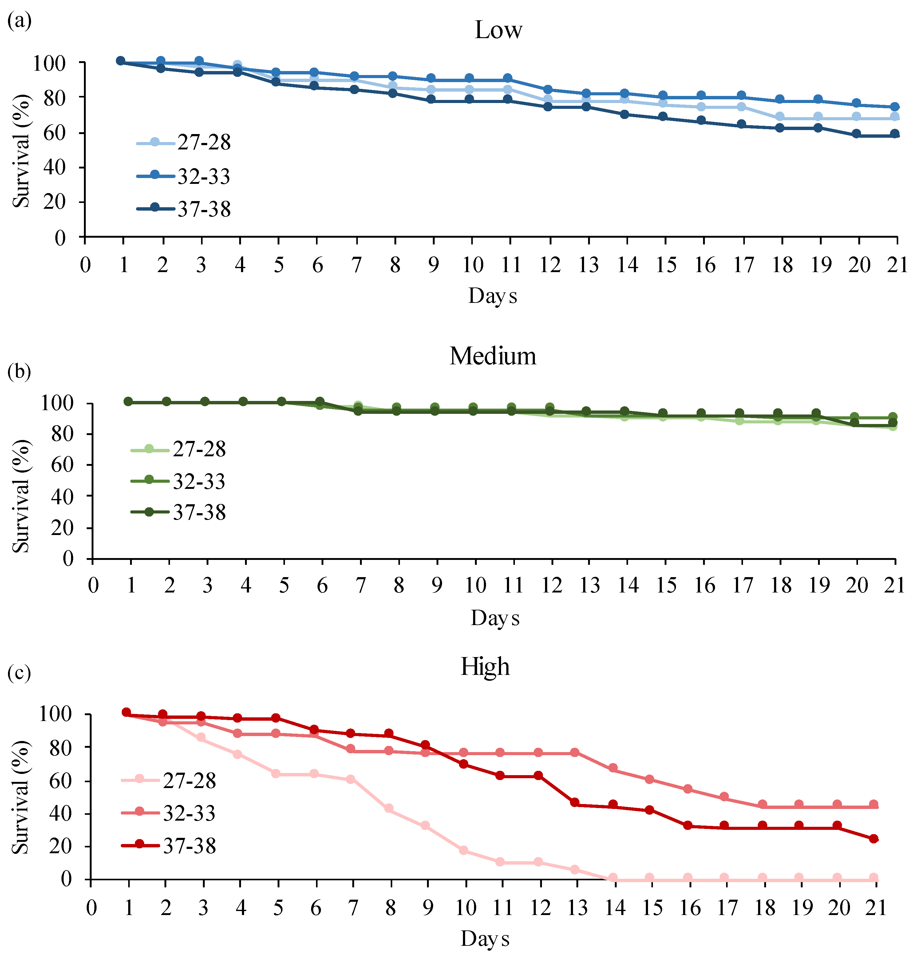
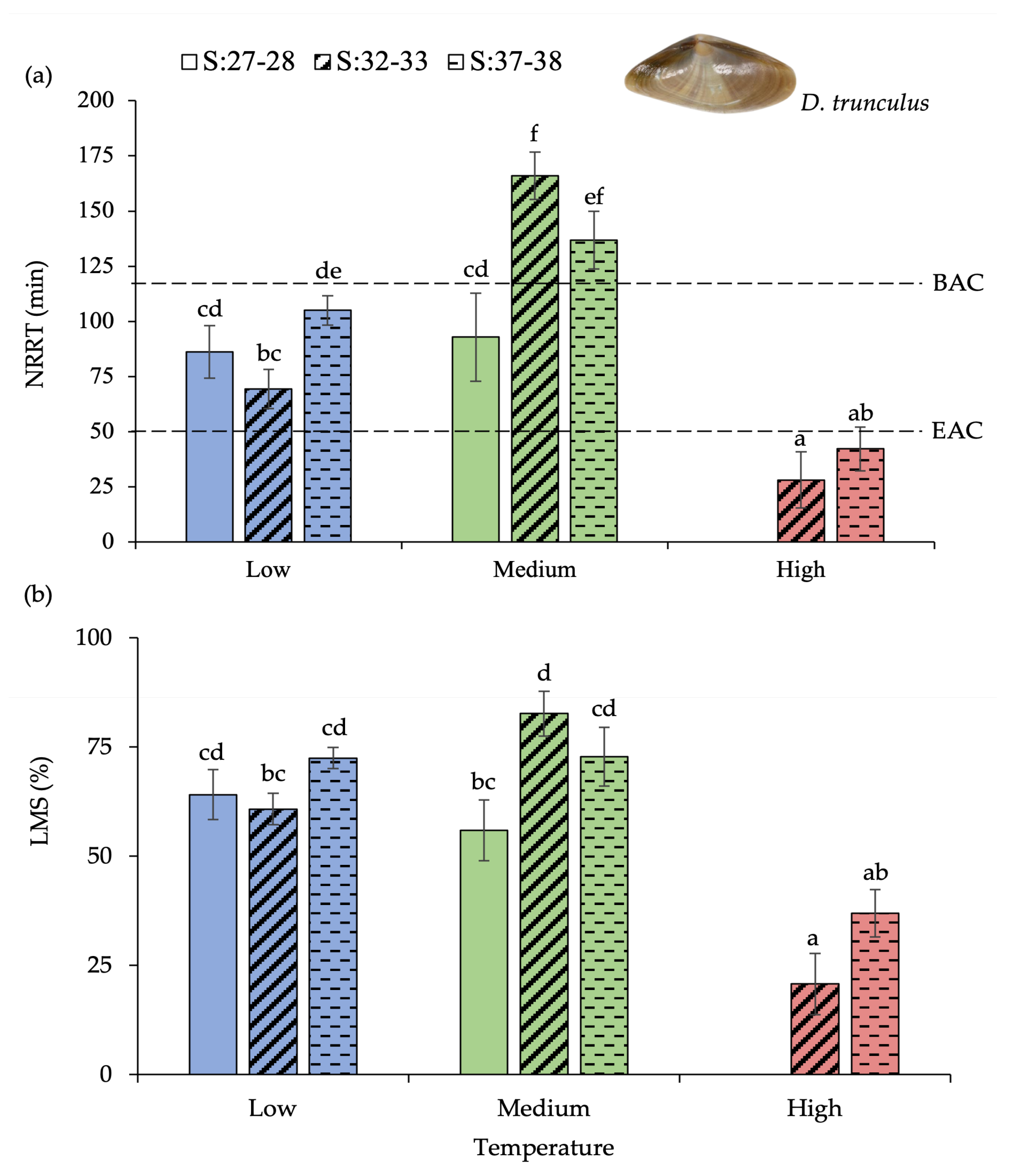
3.2. Donax Trunculus

In contrast from what was observed with C. gallina, there was acute mortality of individuals of D. trunculus during the experiments (Figure 5). Overall, the most notable decrease in percentage of survival was associated with the treatments conducted at the highest temperature, with a 100% mortality of individuals after 14 days at 27.5 °C/27–28 salinity (Figure 5c). The survival rate of the organism in the other treatments (low and medium temperatures and at all three salinity levels) ranged between 70 and 90% (Figure 5a,b).
There was a significant difference in LMS (expressed as NRRT and %LMS) as a result of both temperature and salinity, and there was a significant interaction between these factors (2-way ANOVA; p-value < 0.05) (Table 2). LMS in D. trunculus hemocytes (expressed as mean value of %LMS) was significantly lower in survival clams from the treatments conducted at 27.5 °C than in the other treatments (Figure 6). The highest mean values of LMS were observed in clams from the 20 °C/32–33 of salinity (NRRT = 165.9 ± 10.7 min; %LMS = 82.7 ± 5.1) and 20 °C/37–38 of salinity (NRRT = 136.9 ± 13.1 min; % LMS = 72.8 ± 6.7) treatments (Table S1).
Pairwise comparisons revealed the existence of LMS response subsets (6 subsets for NRRT and 4 subsets for %LMS) (Tukey’s post hoc test; p-value < 0.05) (Figure 6a,b, Tables S6 and S7). The treatments carried out at the same temperature but at different salinities had similar LMS results in %LMS (Figure 6b, Tables S6 and S7) (Tukey’s post hoc test; p-values > 0.05).
There was a significant difference in condition index due to temperature, but there was no effect of salinity or the interaction of both factors (Table 2). Unlike C. gallina, the CI of D. trunculus decreased with increasing temperature (Table 3). Organisms exposed to a low temperature exhibited the highest CI values, showing no significant differences due to salinity. At a medium temperature (20 °C), a lower CI was obtained compared to those exposed at a low temperature (12 °C). The reference CI (of 19.5 °C and 36.8 salinity) differed from the CI obtained at 27–28 and 32–33 (p-value = 0.007, Table 3). Finally, organisms exposed to a high temperature (27.5 °C) had the lowest CI values. At this temperature, significant differences were observed between the reference CI (23.5 °C and 37.1 salinity) and those exposed to salinities of 32–33 and 37–38 (p-value = 0.000, Table 3). It should be noted that, during exposure to a high temperature, spawning occurred in D. trunculus, coinciding with the low LMS and CI recorded, pointing to a temperature-induced spawning event in this species.
Correlation analysis also revealed positive correlations between the two “endpoints” (Table S8). In D. trunculus (n = 8), a negative correlation was observed between CI and treatment temperature (Pearson’s correlation Coeff. = −0.976; p-value = 0.000) and a moderate positive correlation between CI and %LMS (Pearson’s correlation Coeff. = 0.737; p-value = 0.037) (Figure 4).

4. Discussion

It is well known that changes in environmental conditions, such as temperature, salinity, dissolved oxygen, pH, food availability, and contamination, produce alterations in the immune and physiological functions of bivalves [20,46,47]. One of the most important environmental factors involved in immunomodulation is water temperature and salinity [48]. Several studies have shown that changes in temperature or salinity compromise different defense mechanisms of bivalves [20,49]. Earlier works with C. gallina and other bivalve species (e.g., Crassostrea gigas, Ostrea edulis, and Mytilus sp.) have found that LMS is sensitive to temperature fluctuations in both laboratory and field studies [50,51,52]. In contrast, few studies have evaluated the combined effects of both factors on LMS, in species such as Ostrea edulis, Ruditapes philippinarum and Anadara trapezia [53,54,55]. While it has been observed that phagocytic activity in C. gallina is altered differently depending on whether environmental parameters change individually or in combination [56], this study is the first to assess the combined effect of the environmental factors temperature and salinity on the stability of the lysosomal membrane in C. gallina and D. trunculus.
In this study, we found that certain combined conditions of seawater temperature and salinity maintained for at least 21 days are capable, by themselves, of causing significant cellular stress by affecting lysosomal membrane permeability in the clam species D. trunculus and C. gallina. We found a differential response to temperature, dependent on acclimation salinity, which has also been observed in other bivalves [53]. In C. gallina, at salinities of 32–33, the neutral red was retained within the lysosomal compartment for the shortest duration at 20 °C, increasing as the acclimation salinity was raised or lowered (Figure 2). However, an opposite pattern was observed in the treatments conducted at 12 °C, with longest retention times at 32–33 salinities. At the highest temperature (27.5 °C), no significant changes in lysosomal membrane stability were found at different salinities. Furthermore, our findings show that this differential response varied between the clam species under study. The lysosomal permeability to intermediate salinities (32–33) at 12 °C and 20 °C in C. gallina has opposite patterns in comparison to D. trunculus. Different patterns of variation in total hemocytes counts has been described between C. gallina and Mytilus galloprovincialis submitted to the same environmental conditions [56], which partially supports the differential interspecies responses found on LMS in our study. In both species, the LMS at 27.5 °C showed little response to changes in salinity. Consequently, our findings show that the adaptive capacity of the cellular processes in these species was reduced at the highest temperature tested. For D. trunculus, at the highest tested temperature (27.5 °C), the NR remained within the lysosomes for only a short time (Figure 6), signaling that the lipid membranes within the cell were less stable.
It is known that the interaction between two or more environmental variables may cause changes in other variables [57], such as a decrease in dissolved oxygen (DO) in the water, which may cause stress in organisms [58,59]. In our study, the DO concentration was kept under control (>7 mg/L and 100% oxygen saturation), and the observed effects cannot be attributed to significant changes in this variable (Table 1).
The neutral red retention assay has been and is widely used to determine lysosomal membrane stability in invertebrate hemocytes, with special reference to bivalves [60,61]. Threshold values used as assessment criteria for lysosomal membrane stability using the NRRA have been determined from Mytilus sp. on the bases of numerous studies performed around the UK and other European waters, including Mediterranean waters [39,62]. There is a limited number of studies investigating LMS in C. gallina hemocytes [52,54,63]. To our knowledge, there are no LMS data in the literature obtained by using the NRRA in the hemocytes of D. trunculus. In the absence of specific evaluation criteria for these species, absolute values for LMS measurements in Mytilus sp. could be assumed to be comparable. In this sense, NRRT shorter than the EAC level suggested the organism were severely stressed and probably exhibiting pathology. An NRRT shorter than the BAC level but longer than the EAC level was considered to represent stressed but compensating organism. Concerning C. gallina, neutral red retention times within the lysosomal compartment of the small granulocytes were in general longer (mean time higher than 50 min for all treatments) than the retention times reported on an earlier field study performed in Adriatic waters with the same species (NRRT ≤ 30 min in spring (20 ° C salinity of 35) and autumn (16 ° C salinity of 36)) [52]. According to the assessment criteria established for this biomarker, our results point out that the treatments performed with C. gallina caused less cellular stress compared to D. trunculus, since the organisms showed retention times below the environmental assessment criteria established for this biomarker (EAC < 50 min) in none of the treatments [62]. In general, for both species, the environmental conditions that led to greater LMS were those at a medium temperature (20 ° C) at the three studied salinity ranges. In other bivalves, temperatures close to 20 ° C have been found to be optimal for maintaining hemocyte lysosomal stability, with membrane stability being decreased both when increasing and decreasing the temperature [61,64]. In addition, in the case of C. gallina, organisms exposed to the lowest temperature (12 ° C) and salinity 32–33 also presented high stability. For D. trunculus at 20 ° C and salinity 32–33 and 37–38, the NRRTs were higher than the background assessment criteria value, while at the temperature 27.5 ° C, at these same salinities, the NRRTs obtained were lower than the EAC value.
Moore et al. [65] observed that mussels exposed to aquatic hyperthermia conditions (28 ° C) for 48 h were able to recover LMS values. In our current investigation, prolonged exposure to elevated temperatures (27.5 ° C) combined with a salinity of 27–28 resulted in 100% mortality in D. trunculus after 14 days. These results suggest that 27.5 ° C could be the threshold temperature level for this species at low salinities. Moreover, in the surviving treatments at that temperature (salinity 32–33 and 37–38), the lowest LMS and the lowest CI values of all treatments were observed. The cellular responses analyzed revealed extremely severe stress in the organisms of those treatments 27.5 ° C/32–33 and 27.5 ° C/37–38 (NRRT of 28.1 ± 12.7 min and 42.2 ± 9.9 min, respectively) and the lowest mean %LMS values (20.8 ± 7.0 and 36.9 ± 5.4, respectively). Overall, the results of this study indicated that LMS responses were more affected by temperature than salinity, as NRRTs greater than 50 min at 27.5 °C were never detected, regardless of salinity. In the case of C. gallina, although Moschino and Marin [24] reported alterations of physiological measurements, such as growth and condition index, as well as induction and alterations of sub-cellular biomarkers at temperatures higher than 25 ° C, our findings indicate that the values at 27.5 ° C were similar to others obtained at 12 and 20 ° C.
In addition to variations in environmental parameters, the lysosomes in the vascular system respond to physiological changes associated with gonadal maturation during the spawning period [66,67]. Spawning is also an important physiological stressor in bivalves, which significantly increases lysosomal membrane permeability [61,68,69]. There is a wide discussion among different authors on the impact of water temperature in relation to the spawning period of bivalves [67,70]. Concerning mussels, some researchers argue that lower NRRT values are obtained in the pre-spawning period due to the maintenance of their metabolic functions [36,71], while Harding et al. [72] suggest that these values are reached just after spawning. On the other hand, some indicate that NRRT is not affected by hormonal regulation during reproductive activity in mussels [34]. In our study, for D. trunculus, organisms collected in June (when this species would be in the spawning stage in our study area [73]) and exposed to high temperatures spawned, adding an additional stress factor to temperature and salinity. Thus, the lowest LMS and low CI values obtained in D. trunculus at 27.5 ° C could be at least partially due to the spawning observed in the aquaria.
Hemocytes represent the primary line of internal defense for bivalves (endocytosis, encapsulation, inflammation, wound repair, enzyme secretion, elimination by diapedesis), and changes in ambient temperature and salinity may enhance or decrease the hemocytes activity [74]. The LMS of hemocytes in D. trunculus was significantly affected at 27.5 ° C. In any case, temperature was a dominant factor in the physiological and immunological responses of D. trunculus, showing lower survival, lower stability of lysosomal membranes, and lower CI value when reaching a high temperature (27.5 ° C), thus indicating a higher level of stress. This fact is relevant considering the current situation of the Mediterranean Sea, where the effects of climate change are appreciated: marine heat waves are more frequent and intense with a longer duration so species such as Donax and Chamelea are subjected to extreme temperatures and for longer periods in the environment. After conducting this study, certain limitations were identified that could be addressed in future research. Kruft Welton et al. [20] states that infaunal species, such as ours, may not be negatively affected by climate change stressors due to their ability to burrow as a strategy to avoid high temperatures. However, Monari et al. [63] found that the C. gallina kept at 20 and 25 °C burrowed completely, whereas at 30 °C, the clams progressively emerged from the sediment and then remained on the surface. Nonetheless, including a sand bed in the aquaria during the experimental period would provide results ecologically more relevant concerning the effects of water temperature on the LMS in these species with burying behavior.

5. Conclusions

This study contributes to clarifying the cellular stress caused by the action of combined temperature and salinity conditions in the marine clams C. gallina and D. trunculus. This investigation demonstrates interspecies differences in the background response of the lysosomal membrane stability biomarker under similar environmental conditions. Given the importance of considering environmental variables when evaluating the toxicity of pollutants in aquatic organisms, our results have a relevant interest in the potential use of LMS as a contaminant-related biomarker in C. gallina and D. trunculus in field studies. Although both species can tolerate changes in either seawater salinity or temperature conditions, the tolerance range is narrower for D. trunculus, showing a significant cytotoxicity (NRRT < 50 min) at temperatures above 27.5 °C and salinities above 32, and causing 100% mortality at 27.5 °C and low salinities (27–28).This research contributes to consolidating the findings already obtained from other bivalve species that climate changes can affect hemocyte functionality, which is directly related to bivalve immune defense capabilities.

Supplementary Materials

The following supporting information can be downloaded at: https://www.mdpi.com/article/10.3390/app14072712/s1, Table S1: Descriptive variables of the organisms used in LMS of each species: size, neutral red retention time (NRRT), and percentage lysosomal membrane stability (%LMS). T: temperature; S: salinity; N: individuals. Data are expressed as mean ± SE., Table S2: Means for groups in homogeneous subsets of Tukey’s HSD (p < 0.05) test of neutral red retention time (NRRT) and percentage of lysosomal membrane stability (%LMS) among the 9 treatments for C. gallina (n = 16), Table S3: Pairwise comparisons of LMS (expressed as NRRT and %LMS) at three salinity (S) levels (27–28, 32–33, and 37–38) for each temperature (T) and at three temperature levels (low, medium, and high) for each salinity, for both species, Table S4: Pairwise comparisons of the condition index (CI) of the four salinities (T) levels (reference, 27–28, 32–33, and 37–38) for each temperature (S) (reference: low, medium, and high), Table S5: Correlation analysis between the different variables for C. gallina: salinity (S), temperature (T), condition index (CI), neutral red retention time (NRRT), and percentage of lysosomal membrane stability (%LMS) (n = 9), Table S6: Means for groups in homogeneous subsets of Tukey’s HSD (p < 0.05) test of neutral red retention time (NRRT) among the 9 treatments for D. trunculus (n = 16), Table S7: Means for groups in homogeneous subsets of Tukey’s HSD (p < 0.05) test of percentage of lysosomal membrane stability (%LMS) among the 9 treatments for D. trunculus (n = 16), Table S8: Correlation analysis between the different variables for D. trunculus: salinity, temperature, condition index (CI), neutral red retention time (NRRT), and percentage of lysosomal membrane stability (%LMS) (n = 8).

Author Contributions

Conceptualization, P.S.-M., C.M.-G., M.R., and S.F.; methodology, P.S.-M., C.M.-G., and M.R.; software, P.S.-M. and E.Z.; validation, C.M.-G., M.R., and S.F.; formal analysis, P.S.-M. and E.Z.; investigation, P.S.-M. and E.Z.; resources, M.R. and S.F.; data curation, P.S.-M.; writing—original draft preparation, P.S.-M., C.M.-G., and E.Z.; writing—review and editing, P.S.-M., C.M.-G., M.R., and S.F.; visualization, P.S.-M.; supervision, M.R. and S.F.; project administration, M.R.; funding acquisition, M.R. and S.F. All authors have read and agreed to the published version of the manuscript.

Funding

This research forms part of the ThinkInAzul program and was supported by MCIN (Ministerio de Ciencia, Inovación y Universidades) with funding from European Union NextGenerationEU (PRTR-C17.I1) and by Generalitat Valenciana (THINKINAZUL/2021/005).

Institutional Review Board Statement

No specific authorization is required for the use of Donax trunculus and Chamelea gallina in experimental applications according to the EC Directive/63/EU.

Informed Consent Statement

Not applicable.

Data Availability Statement

Data are contained within this article and the Supplementary Materials.

Acknowledgments

Authors would like to thank the anonymous reviewers for their comments and suggestions that improved this manuscript.

Conflicts of Interest

The authors declare that there are no conflicting interests regarding the publication of this work.

References

  1. Baeta, M.; Solís, M.A.; Ballesteros, M.; Defeo, O. Long-Term Trends in Striped Venus Clam (Chamelea gallina) Fisheries in the Western Mediterranean Sea: The Case of Ebro Delta (NE Spain). Mar. Policy 2021, 134, 104798. [Google Scholar] [CrossRef]
  2. Escrivá, J.; Rodilla, M.; Llario, F.; Falco, S. The Collapse of a Wedge Clam Fishery in the Spanish Mediterranean Coast and Recovery Problems. J. Shellfish Res. 2021, 40, 37–47. [Google Scholar] [CrossRef]
  3. La Valle, P.; Nicoletti, L.; Finoia, M.G.; Ardizzone, G.D. Donax trunculus (Bivalvia: Donacidae) as a Potential Biological Indicator of Grain-Size Variations in Beach Sediment. Ecol. Indic. 2011, 11, 1426–1436. [Google Scholar] [CrossRef]
  4. Escrivá, J.; Rodilla, M.; Martin-Diaz, J.P.; Estruch, V.D.; Sebastiá-Frasquet, M.T.; Llario, F.; Falco, S. Driving Forces That Structure Sublittoral Macrobenthic Communities in Sandy Beaches along Environmental Gradients. Estuar. Coast. Shelf Sci. 2020, 233, 106517. [Google Scholar] [CrossRef]
  5. Picard, J. Recherches qualitatives sur les biocoenoses marines des substrats meubles dragables de la región marseillaise. Recl. Trav. Stn. Mar. Endoume 1965, 36, 1–160. [Google Scholar]
  6. de la Huz, R.; Lastra, M.; López, J. The Influence of Sediment Grain Size on Burrowing, Growth and Metabolism of Donax trunculus L. (Bivalvia: Donacidae). J. Sea Res. 2002, 47, 85–95. [Google Scholar] [CrossRef]
  7. Pérès, J.M.; Picard, J. Nouveau Manuel de Bionomie Benthique de la Mer Méditerranée; Station Marine d’Endoume: Marseille, France, 1964; Volume 31. [Google Scholar]
  8. Morello, E.B.; Froglia, C.; Atkinson, R.J.; Moore, P.G. Impacts of Hydraulic Dredging on a Macrobenthic Community of the Adriatic Sea, Italy. Can. J. Fish. Aquat. Sci. 2005, 62, 2076–2087. [Google Scholar] [CrossRef]
  9. Zeichen, M.M.; Agnesi, S.; Mariani, A.; Maccaroni, A.; Ardizzone, G.D. Biology and Population Dynamics of Donax trunculus L. (Bivalvia: Donacidae) in the South Adriatic Coast (Italy). Estuar. Coast. Shelf Sci. 2002, 54, 971–982. [Google Scholar] [CrossRef]
  10. Baeta, M.; Solís, M.A.; Frias-Vidal, S.; Claramonte, L.; Sepouna, A.; Ballesteros, M. The Wedge Clam (Donax trunculus) Hand-Operated Fishery in the NW Mediterranean Sea: Landings, Catch Composition, Damage Rates and Impact of Fishing Activity. Ocean Coast. Manag. 2023, 237, 106534. [Google Scholar] [CrossRef]
  11. Martínez-Patiño, D.; Otero, J.; Louzán, A.; Ojea, J.; Nóvoa, S.; Álvarez-Salgado, X.A. Wedge Clam (Donax trunculus Linnaeus, 1758) Reproduction: Reproductive Traits and Environmental Influence in the NW Iberian Coast and Contrast across Atlantic and Mediterranean Waters. Hydrobiologia 2021, 848, 1347–1366. [Google Scholar] [CrossRef]
  12. Poloczanska, E.S.; Brown, C.J.; Sydeman, W.J.; Kiessling, W.; Schoeman, D.S.; Moore, P.J.; Brander, K.; Bruno, J.F.; Buckley, L.B.; Burrows, M.T.; et al. Global Imprint of Climate Change on Marine Life. Nat. Clim. Chang. 2013, 3, 919–925. [Google Scholar] [CrossRef]
  13. Tlili, S.; Mouneyrac, C. The Wedge Clam Donax trunculus as Sentinel Organism for Mediterranean Coastal Monitoring in a Global Change Context. Reg. Environ. Chang. 2019, 19, 995–1007. [Google Scholar] [CrossRef]
  14. Giangrande, A.; Pierri, C.; Del Pasqua, M.; Gravili, C.; Gambi, M.C.; Gravina, M.F. The Mediterranean in Check: Biological Invasions in a Changing Sea. Mar. Ecol. 2020, 41, e12583. [Google Scholar] [CrossRef]
  15. Lejeusne, C.; Chevaldonné, P.; Pergent-Martini, C.; Boudouresque, C.F.; Pérez, T. Climate Change Effects on a Miniature Ocean: The Highly Diverse, Highly Impacted Mediterranean Sea. Trends Ecol. Evol. 2010, 25, 250–260. [Google Scholar] [CrossRef] [PubMed]
  16. Gissi, E.; Manea, E.; Mazaris, A.D.; Fraschetti, S.; Almpanidou, V.; Bevilacqua, S.; Coll, M.; Guarnieri, G.; Lloret-Lloret, E.; Pascual, M.; et al. A Review of the Combined Effects of Climate Change and Other Local Human Stressors on the Marine Environment. Sci. Total Environ. 2021, 755, 142564. [Google Scholar] [CrossRef]
  17. Cheng, L.; von Schuckmann, K.; Abraham, J.P.; Trenberth, K.E.; Mann, M.E.; Zanna, L.; England, M.H.; Zika, J.D.; Fasullo, J.T.; Yu, Y.; et al. Past and Future Ocean Warming. Nat. Rev. Earth Environ. 2022, 3, 776–794. [Google Scholar] [CrossRef]
  18. Doney, S.C.; Fabry, V.J.; Feely, R.A.; Kleypas, J.A. Ocean Acidification: The Other CO2 Problem. Annu. Rev. Mar. Sci. 2009, 1, 169–192. [Google Scholar] [CrossRef]
  19. Harvey, B.P.; Gwynn-Jones, D.; Moore, P.J. Meta-Analysis Reveals Complex Marine Biological Responses to the Interactive Effects of Ocean Acidification and Warming. Ecol. Evol. 2013, 3, 1016–1030. [Google Scholar] [CrossRef]
  20. Kruft Welton, R.A.; Hoppit, G.; Schmidt, D.N.; Witts, J.D.; Moon, B.C. Reviews and Syntheses: The Clam before the Storm—A Meta-Analysis Showing the Effect of Combined Climate Change Stressors on Bivalves. Biogeosciences 2024, 21, 223–239. [Google Scholar] [CrossRef]
  21. Laubier, L. Mediterranean Sea and Humans: Improving a Conflictual Partnership. In The Mediterranean Sea; Saliot, A., Ed.; Handbook of Environmental Chemistry; Springer: Berlin/Heidelberg, Germany, 2005; Volume 5K, pp. 3–27. [Google Scholar] [CrossRef]
  22. Rivetti, I.; Fraschetti, S.; Lionello, P.; Zambianchi, E.; Boero, F. Global Warming and Mass Mortalities of Benthic Invertebrates in the Mediterranean Sea. PLoS ONE 2014, 9, e115655. [Google Scholar] [CrossRef]
  23. Pastor, F.; Khodayar, S. Marine Heat Waves: Characterizing a Major Climate Impact in the Mediterranean. Sci. Total Environ. 2023, 861, 160621. [Google Scholar] [CrossRef] [PubMed]
  24. Moschino, V.; Marin, M.G. Seasonal Changes in Physiological Responses and Evaluation of “Well-Being” in the Venus Clam Chamelea gallina from the Northern Adriatic Sea. Comp. Biochem. Physiol. Part A Mol. Integr. Physiol. 2006, 145, 433–440. [Google Scholar] [CrossRef]
  25. Widdows, J.; Staff, F. Biological Effects of Contaminants: Measurement of Scope for Growth in Mussels; Report; International Council for the Exploration of the Sea (ICES): Copenhagen, Denmark, 2006. [Google Scholar] [CrossRef]
  26. Froglia, C. II Contributo della Ricerca Scientifica Alia Gestio-Ne della Pesca dei Molluschi Bivalvi con Draghe Idrauliche. Biol. Mar. Medit. 2000, 7, 71–82. [Google Scholar]
  27. Ansell, A.D.; Barnett, P.R.O.; Bodoy, A.; Massé, H. Upper Temperature Tolerances of Some European Molluscs II. Donax vittatus, D. semistriatus and D. trunculus. Mar. Biol. 1980, 58, 41–46. [Google Scholar] [CrossRef]
  28. Sokolov, E.P.; Sokolova, I.M. Compatible Osmolytes Modulate Mitochondrial Function in a Marine Osmoconformer Crassostrea Gigas (Thunberg, 1793). Mitochondrion 2019, 45, 29–37. [Google Scholar] [CrossRef] [PubMed]
  29. Solan, M.; Whiteley, N. Stressors in the Marine Environment: Physiological and Ecological Responses; Societal Implications; Oxford University Press: Oxford, UK, 2016. [Google Scholar]
  30. Parada, J.M.; Molares, J.; Otero, X. Multispecies Mortality Patterns of Commercial Bivalves in Relation to Estuarine Salinity Fluctuation. Estuaries Coasts 2012, 35, 132–142. [Google Scholar] [CrossRef]
  31. Reyes-Martínez, M.J.; Martínez-Pita, I.; Soler-Navarro, D.; García-García, F.J. The Impact of Salinity Changes Associated with Size on the Wedge Clam Donax trunculus Linnaeus, 1758 (Mollusca: Bivalvia): A Laboratory Assay. Estuar. Coast. Shelf Sci. 2020, 241, 106838. [Google Scholar] [CrossRef]
  32. Matozzo, V.; Monari, M.; Foschi, J.; Serrazanetti, G.P.; Cattani, O.; Marin, M.G. Effects of Salinity on the Clam Chamelea gallina. Part I: Alterations in Immune Responses. Mar. Biol. 2007, 151, 1051–1058. [Google Scholar] [CrossRef]
  33. Pantea, E.-D.; Coatu, V.; Damir, N.-A.; Oros, A.; Lazar, L.; Rosoiu, N. Lysosomal Membrane Stability of Mussel (Mytilus galloprovincialis Lamarck, 1819) as a Biomarker of Cellular Stress for Environmental Contamination. Toxics 2023, 11, 649. [Google Scholar] [CrossRef]
  34. Castro, M.; Santos, M.M.; Monteiro, N.M.; Vieira, N. Measuring Lysosomal Stability as an Effective Tool for Marine Coastal Environmental Monitoring. Mar. Environ. Res. 2004, 58, 741–745. [Google Scholar] [CrossRef]
  35. OSPAR Commission. Protecting and Conserving the North-East Atlantic and Its Resources. Available online: https://www.ospar.org/ (accessed on 23 January 2024).
  36. Hagger, J.A.; Lowe, D.; Dissanayake, A.; Jones, M.B.; Galloway, T.S. The Influence of Seasonality on Biomarker Responses in Mytilus edulis. Ecotoxicology 2010, 19, 953–962. [Google Scholar] [CrossRef] [PubMed]
  37. Moore, M.N. 9 Background Document: Lysosomal Stability as a Global Health Status Indicator in Biomonitoring. In Integrated Marine Environmental Monitoring of Chemicals and Their Effects; ICES: Copenhagen, Denmark, 2012; p. 68. [Google Scholar]
  38. Nacef, L.; Bachari, N.E.I.; Bouda, A.; Boubnia, R. Variability and Decadal Evolution of Temperature and Salinity in the Mediterranean Sea Surface. Int. J. Eng. Geosci. 2016, 1, 24–33. [Google Scholar] [CrossRef]
  39. Martínez-Gómez, C.; Bignell, J.P.; Lowe, D. Lysosomal Membrane Stability in Mussels. ICES Tech. Mar. Environ. Sci. 2015, 56, 1–46. [Google Scholar]
  40. Lowe, D.M.; Moore, M.N.; Evans, B.M. Contaminant Impact on Interactions of Molecular Probes with Lysosomes in Living Hepatocytes from Dab Limanda limanda. Mar. Ecol. Prog. Ser. 1992, 91, 135–140. [Google Scholar] [CrossRef]
  41. Guerrero-Rios, R.J.; Hernández, N.T.; Espinoza-Rodríguez, N.; Barrios-Garrido, H.; Montiel, M.; Morales, F. Composición de tallas e Índice de Condición de la Almeja Estuarina en Curarire, Estado Zulia, Venezuela. Zootec. Trop. 2015, 33, 327–336. [Google Scholar]
  42. Hansson, T.; Thain, J.E.; Martínez-Gómez, C.; Hylland, K.; Gubbins, M.J.; Balk, L. Supporting Variables for Biological Effects Measurements in Fish and Blue Mussel; ICES Techniques in Marine Environmental Sciences: Denmark, Copenhagen, 2017; No. 60. [Google Scholar] [CrossRef]
  43. Zeng, Y.; Yang, H. Review of Molluscan Bivalve Condition Index Calculations and Application in Northern Quahogs Mercenaria mercenaria. Aquac. Res. 2021, 52, 23–36. [Google Scholar] [CrossRef]
  44. Boscolo, R.; Cornello, M.; Giovanardi, O. Condition Index and Air Survival Time to Compare Three Kinds of Manila Clam Tapes philippinarum (Adams & Reeve) Farming Systems. Aquac. Int. 2003, 11, 243–254. [Google Scholar] [CrossRef]
  45. Box, G.E.P.; Cox, D.R. An Analysis of Transformations. J. R. Stat. Soc. Ser. B Methodol. 1964, 26, 211–243. [Google Scholar] [CrossRef]
  46. de Almeida, J.M.B.; Gaspar, M.B.; Castro, M.; Rufino, M.M. Influence of Wind, Rainfall, Temperature, and Primary Productivity, on the Biomass of the Bivalves Spisula solida, Donax trunculus, Chamelea gallina and Ensis siliqua. Fish. Res. 2021, 242, 106044. [Google Scholar] [CrossRef]
  47. Munari, M.; Matozzo, V.; Chemello, G.; Riedl, V.; Pastore, P.; Badocco, D.; Marin, M.G. Seawater Acidification and Emerging Contaminants: A Dangerous Marriage for Haemocytes of Marine Bivalves. Environ. Res. 2019, 175, 11–21. [Google Scholar] [CrossRef]
  48. Fisher, M.R. Effect of Temperature and Salinity on Size at Maturity of Female Blue Crabs. Trans. Am. Fish. Soc. 1999, 128, 499–506. [Google Scholar] [CrossRef]
  49. Pourmozaffar, S.; Tamadoni Jahromi, S.; Rameshi, H.; Sadeghi, A.; Bagheri, T.; Behzadi, S.; Gozari, M.; Zahedi, M.R.; Abrari Lazarjani, S. The Role of Salinity in Physiological Responses of Bivalves. Rev. Aquac. 2020, 12, 1548–1566. [Google Scholar] [CrossRef]
  50. Camus, L.; Grøsvik, B.E.; Børseth, J.F.; Jones, M.B.; Depledge, M.H. Stability of Lysosomal and Cell Membranes in Haemocytes of the Common Mussel (Mytilus edulis): Effect of Low Temperatures. Mar. Environ. Res. 2000, 50, 325–329. [Google Scholar] [CrossRef]
  51. Hauton, C.; Hawkins, L.E.; Hutchinson, S. Response of Haemocyte Lysosomes to Bacterial Inoculation in the Oysters Ostrea edulis L. and Crassostrea gigas (Thunberg) and the Scallop Pecten maximus (L.). Fish Shellfish Immunol. 2001, 11, 143–153. [Google Scholar] [CrossRef] [PubMed]
  52. Carducci, F.; Biscotti, M.A.; Trucchi, E.; Giuliani, M.E.; Gorbi, S.; Coluccelli, A.; Barucca, M.; Canapa, A. Omics Approaches for Conservation Biology Research on the Bivalve Chamelea gallina. Sci. Rep. 2020, 10, 19177. [Google Scholar] [CrossRef] [PubMed]
  53. Hauton, C.; Hawkins, L.E.; Hutchinson, S. The Use of the Neutral Red Retention Assay to Examine the Effects of Temperature and Salinity on Haemocytes of the European Flat Oyster Ostrea edulis (L). Comp. Biochem. Physiol. B Biochem. Mol. Biol. 1998, 119, 619–623. [Google Scholar] [CrossRef]
  54. Munari, M.; Matozzo, V.; Marin, M.G. Combined Effects of Temperature and Salinity on Functional Responses of Haemocytes and Survival in Air of the Clam Ruditapes philippinarum. Fish Shellfish Immunol. 2011, 30, 1024–1030. [Google Scholar] [CrossRef] [PubMed]
  55. Taylor, A.M.; Maher, W.A.; Ubrihien, R.P. Mortality, Condition Index and Cellular Responses of Anadara trapezia to Combined Salinity and Temperature Stress. J. Exp. Mar. Biol. Ecol. 2017, 497, 172–179. [Google Scholar] [CrossRef]
  56. Matozzo, V.; Chinellato, A.; Munari, M.; Finos, L.; Bressan, M.; Marin, M.G. First Evidence of Immunomodulation in Bivalves under Seawater Acidification and Increased Temperature. PLoS ONE 2012, 7, e33820. [Google Scholar] [CrossRef]
  57. Soon, T.K.; Ransangan, J. Extrinsic Factors and Marine Bivalve Mass Mortalities: An Overview. J. Shellfish Res. 2019, 38, 223–232. [Google Scholar] [CrossRef]
  58. Matozzo, V.; Monari, M.; Foschi, J.; Papi, T.; Cattani, O.; Marin, M.G. Exposure to Anoxia of the Clam Chamelea gallina: I. Effects on Immune Responses. J. Exp. Mar. Biol. Ecol. 2005, 325, 163–174. [Google Scholar] [CrossRef]
  59. Andreyeva, A.Y.; Kladchenko, E.S.; Gostyukhina, O.L. Effect of Hypoxia on Immune System of Bivalve Molluscs. Mar. Biol. J. 2022, 7, 3–16. [Google Scholar]
  60. Lowe, D.M.; Soverchia, C.; Moore, M.N. Lysosomal Membrane Responses in the Blood and Digestive Cells of Mussels Experimentally Exposed to Fluoranthene. Aquat. Toxicol. 1995, 33, 105–112. [Google Scholar] [CrossRef]
  61. Zhao, C.; Li, X.; Luo, S.; Chang, Y. Assessments of Lysosomal Membrane Responses to Stresses with Neutral Red Retention Assay and Its Potential Application in the Improvement of Bivalve Aquaculture. Afr. J. Biotechnol. 2011, 10, 13968–13973. [Google Scholar]
  62. Vethaak, A.D.; Davies, I.M.; Thain, J.E.; Gubbins, M.J.; Martínez-Gómez, C.; Robinson, C.D.; Moffat, C.F.; Burgeot, T.; Maes, T.; Wosniok, W. Integrated Indicator Framework and Methodology for Monitoring and Assessment of Hazardous Substances and Their Effects in the Marine Environment. Mar. Environ. Res. 2017, 124, 11–20. [Google Scholar] [CrossRef] [PubMed]
  63. Monari, M.; Matozzo, V.; Foschi, J.; Cattani, O.; Serrazanetti, G.P.; Marin, M.G. Effects of High Temperatures on Functional Responses of Haemocytes in the Clam Chamelea gallina. Fish Shellfish Immunol. 2007, 22, 98–114. [Google Scholar] [CrossRef]
  64. Yu JinHa, Y.J.; Song JaeHee, S.J.; Choi MinChul, C.M.; Park SungWoo, P.S. Effects of Water Temperature Change on Immune Function in Surf Clams, Mactra veneriformis (Bivalvia: Mactridae). J. Invertebr. Pathol. 2009, 102, 30–35. [Google Scholar]
  65. Moore, M.N. Autophagy as a Second Level Protective Process in Conferring Resistance to Environmentally-Induced Oxidative Stress. Autophagy 2008, 4, 254–256. [Google Scholar] [CrossRef]
  66. Osada, M.; Matsumoto, T. Endocrine Control of Gametogenesis and Spawning in Bivalves. In Advances in Invertebrate (Neuro) Endocrinology. Volume 1: Phyla Other Than Arthropoda; Apple Academic Press: Palm Bay, FL, USA, 2021; pp. 179–219. [Google Scholar]
  67. Saucedo, P.E.; Southgate, P.C. Reproduction, Development and Growth. In Pearl Oyster; Elsevier: Oxford, UK, 2008; pp. 131–186. [Google Scholar]
  68. Moore, M.N.; Viarengo, A.G.; Somerfield, P.J.; Sforzini, S. Linking Lysosomal Biomarkers and Ecotoxicological Effects at Higher Biological Levels. In Ecological Biomarkers: Indicators of Ecotoxicological Effects; CRC Press Ltd.: Oxford, UK, 2012; pp. 107–130. [Google Scholar]
  69. Davies, I.M.; Gubbins, M.; Hylland, K.; Thain, J.; Maes, T.; Martínez-Gómez, C.; Giltrap, M.; Burgeot, T.; Wosniok, W.; Lang, T. 30 Technical Annex: Assessment Criteria for Biological Effects Measurements. In Integrated Marine Environmental Monitoring of Chemicals and Their Effects; ICES: Copenhagen, Denmark, 2012; p. 209. [Google Scholar]
  70. Bayne, B.L. Aspects of Reproduction in Bivalve Molluscs. In Estuarine Processes; Wiley, M., Ed.; Academic Press: Cambridge, MA, USA, 1976; pp. 432–448. [Google Scholar] [CrossRef]
  71. Song, L.; Li, X.; Clarke, S.; Wang, T.; Bott, K. The Application of Neutral Red Retention Assay to Evaluate the Differences in Stress Responses to Sexual Maturation and Spawning between Different Sizes of Pacific Oyster, Crassostrea gigas (Thunberg). J. Shellfish Res. 2007, 26, 493–499. [Google Scholar] [CrossRef]
  72. Harding, J.M.; Couturier, C.; Parsons, G.J.; Ross, N.W. Evaluation of the Neutral Red Retention Assay as a Stress Response Indicator in Cultivated Mussels (Mytilus Spp.) in Relation to Seasonal and Environmental Conditions. J. Shellfish Res. 2004, 23, 745–752. [Google Scholar]
  73. Ramón, M. Estudio de Las Poblaciones de Chamelea gallina (Linnaeus, 1758) y Donax trunculus Linnaeus, 1758 (Mollusca: Bivalvia) En El Golfo de Valencia (Mediterráneo Occidental); Universidad de Barcelona: Barcelona, Spain, 1993. [Google Scholar]
  74. Fisher, W.S. Environmental Influence on Host Response. Am. Fish. Soc. Spec. Publ. 1988, 18, 225–237. [Google Scholar]
Figure 1. Experimental design overview. A visual representation illustrating the arrangement and variables of the experimental design employed in this study.
Applsci 14 02712 g001
Figure 2. (a) Neutral red retention time (NRRT) and (b) Lysosomal membrane stability percentage (%LMS) in C. gallina for 21 days exposure to combined conditions of temperature (low, medium, and high) and salinity ranges (27–28, 32–33, and 37–38). Background assessment criteria (BAC). Environmental assessment criteria (EAC). Data are reported as mean ± SE, n = 16. The lowercase letters above the bars indicate inclusion within homogeneous subsets (2-way ANOVA; Tukey’s post hoc test; p-value < 0.05).
Applsci 14 02712 g002
Figure 3. Hemocytes (small granulocytes) from Chamelea gallina showing the retention of neutral red within the lysosomal compartment (A), and stressed cells showing loss of neutral red from pathologically enlarged lysosomes into the cytosol, enlargement, and leakage but colorless lysosomes (B).
Applsci 14 02712 g003
Figure 4. Correlation between CI and lysosomal membrane stability (%) of each treatment, for both species. The different colors represent the low—(blue), medium—(green), and high—(red) temperature treatments.
Applsci 14 02712 g004
Figure 5. Percentage of Donax trunculus’ survival as a function of salinity at each treatment temperature during 21 days of exposure at a (a) low water temperature (12 °C), (b) medium water temperature (20 °C), and (c) high water temperature (27.5 °C) at salinities of 27–28, 32–33, and 37–38, respectively.
Applsci 14 02712 g005
Figure 6. (a) Neutral red retention time (NRRT) and (b) lysosomal membrane stability percentage (%LMS) in D. trunculus after 21 days of exposure to combined conditions of temperature (low, medium, and high) and salinity ranges (27–28, 32–33, and 37–38). Background assessment criteria (BAC). Environmental assessment criteria (EAC). Data are reported as mean ± SE, n = 16. Lowercase letters above the bars indicate inclusion within homogeneous subsets (2-way ANOVA; Tukey’s post hoc test; p-value < 0.001).
Applsci 14 02712 g006
Table 1. Seawater parameters (mean ± SE) during the 21 days experiment conduced with Donax trunculus and Chamelea gallina. N: treatment number; T: temperature; S: salinity; DO: dissolved oxygen (mg/L and % oxygen saturation).
Test TreatmentDonax trunculusChamelea gallina
TST
( ° C)
SDO
(mg/L)
DO
(% sat.)
T
( ° C)
SDO
(mg/L)
DO
(% sat.)
1Low27–2812.30 ± 0.0527.09 ± 0.069.06 ± 0.01104.79 ± 1.4112.13 ± 0.0327.32 ± 0.049.12 ± 0.01106.01 ± 0.09
232–3312.00 ± 0.0332.56 ± 0.149.12 ± 0.01104.75 ± 1.4112.02 ± 0.0432.72 ± 0.089.14 ± 0.01106.10 ± 0.11
337–3812.17 ± 0.0338.12 ± 0.059.09 ± 0.02103.26 ± 0.9812.26 ± 0.0437.93 ± 0.049.10 ± 0.01106.19 ± 0.15
4Medium27–2820.11 ± 0.0527.35 ± 0.037.79 ± 0.01105.82 ± 0.8220.23 ± 0.0127.33 ± 0.017.76 ± 0.00105.64 ± 0.24
532–3319.95 ± 0.0432.70 ± 0.047.64 ± 0.04102.38 ± 0.6620.16 ± 0.0132.78 ± 0.017.77 ± 0.00103.49 ± 0.73
637–3820.34 ± 0.0537.63 ± 0.067.72 ± 0.02102.29 ± 0.8720.07 ± 0.0137.47 ± 0.017.78 ± 0.00105.51 ± 0.40
7High27–2827.13 ± 0.1027.59 ± 0.056.91 ± 0.01104.77 ± 0.2527.58 ± 0.1227.55 ± 0.186.79 ± 0.02105.96 ± 0.09
832–3326.94 ± 0.0932.73 ± 0.096.91 ± 0.01105.70 ± 0.0627.40 ± 0.1232.57 ± 0.076.86 ± 0.01105.97 ± 0.13
937–3826.98 ± 0.1137.71 ± 0.106.93 ± 0.01105.03 ± 0.1327.44 ± 0.1537.57 ± 0.086.82 ± 0.02105.95 ± 0.10
Table 2. Results of the two-way ANOVA, showing the effects of temperature (T), salinity (S), and their interaction in C. gallina and D. trunculus. Both factors were considered fixed and three dependent variables were examined: lysosomal membrane stability (expressed as NRRT and %LMS) and condition index (%CI).
FactorNRRT%LMS%CI
dfFp-ValuedfFp-ValuedfFp-Value
Chamelea gallinaT24.280.01621.01ns229.080.000
S20.65ns21.47ns22.29ns
T × S46.750.00044.920.00141.24ns
Donax trunculusT235.920.000233.370.0002135.270.000
S24.570.01223.670.02922.20ns
T × S44.490.00543.690.01440.83ns
NRRT: neutral red retention time. %LMS: percentage of lysosomal membrane stability. Statistically significant differences (p-value < 0.05). Ns: not significant (p-value > 0.05).
Table 3. Size and condition index (CI) of C. gallina and D. trunculus at the beginning and after 21 days exposure to nine combined treatments of temperature and salinity. Data are reported as mean ± SE.
Reference (Day 0)Salinity (Day 21)
27–2832–3337–38
T (°C)/SSize (mm)CI
(%)
Size
(mm)
CI
(%)
Size (mm)CI
(%)
Size (mm)CI
(%)
CI
Chamelea
gallina
TemperatureLow15.4/36.914.45
± 0.11
2.10 a
± 0.16
13.95
± 0.13
1.88 a
± 0.07
14.10
± 0.11
2.10 a
± 0.08
ns
Medium19.5/36.817.93
± 0.23
2.78 ab
± 0.10
18.91
± 0.35
2.52 a
± 0.16
19.03
± 0.29
2.89 ab
± 0.24
18.86
± 0.25
3.24 ab
± 0.32
0.006
High23.5/37.117.85
± 0.18
2.61 a
± 0.08
18.33
± 0.23
2.02 b
± 0.26
19.01
± 0.22
2.07 b
± 0.09
18.93
± 0.29
2.18 b
± 0.15
0.007
Donax trunculusLow15.4/36.922.16
± 0.14
2.81 a
± 0.09
22.15
± 0.17
3.03 a
± 0.08
22.04
± 0.16
2.88 a
± 0.20
ns
Medium19.5/36.820.66
± 0.20
2.34 a
± 0.08
21.51
± 0.15
1.96 c
± 0.07
22.08
± 0.11
2.12 bc
± 0.08
22.00
± 0.20
2.20 ab
± 0.06
0.007
High23.5/37.120.21
± 0.19
1.70 a
± 0.07
XX20.68
± 0.52
1.08 b
± 0.07
20.00
± 0.10
0.93 b
± 0.17
0.000
Lowercase letter superscripts indicate homogeneous subsets for each species and temperature treatment (Tukey’s post hoc). Ns: not significant (p-value > 0.05). “–” represents no data, and “X” indicates that there were no organisms due to mortality.
Disclaimer/Publisher’s Note: The statements, opinions and data contained in all publications are solely those of the individual author(s) and contributor(s) and not of MDPI and/or the editor(s). MDPI and/or the editor(s) disclaim responsibility for any injury to people or property resulting from any ideas, methods, instructions or products referred to in the content.

