
Do Continuous Rotating Endodontic Instruments Extrude Fewer Apical Debris Than Reciprocating Instruments in Non-Surgical Endodontic Retreatments? A Systematic Review

 ,
,  ,
, 
Abstract
1. Introduction
2. Materials and Methods
2.1. Protocol and Registration
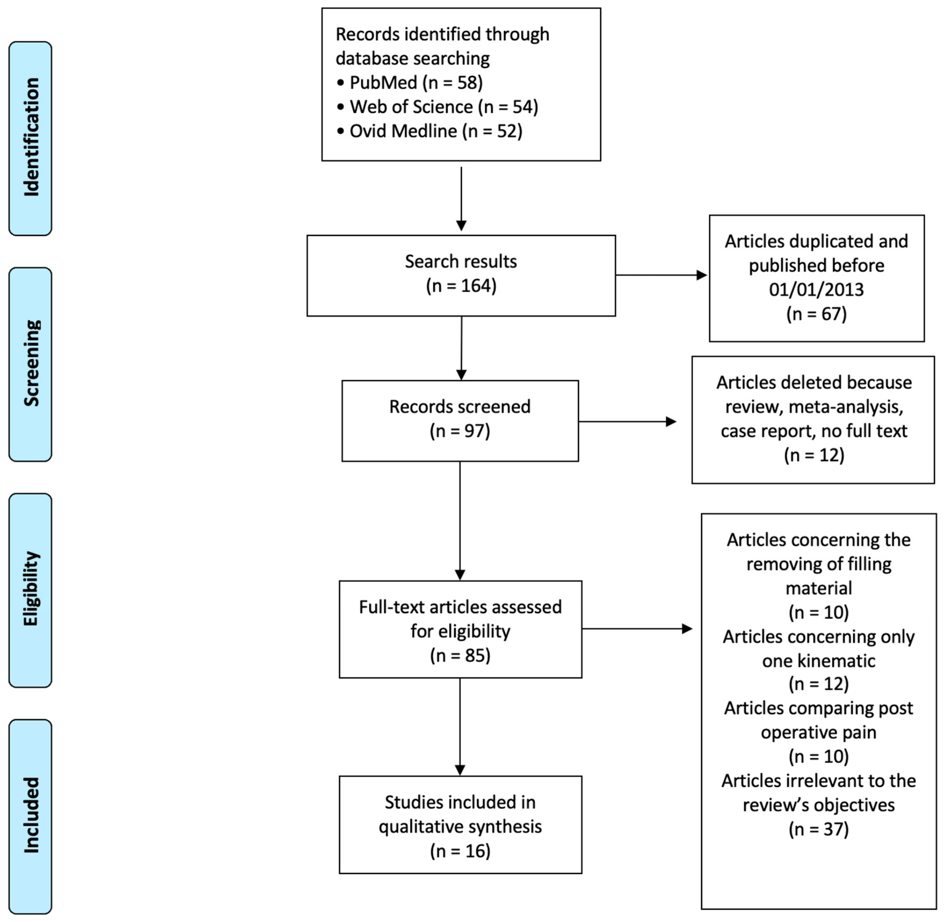
2.2. Search Strategy
2.3. Eligibility Criteria
- Articles comparing apical extrusion in endodontically treated elements using continuously rotating and reciprocating files.
- In vitro studies.
- The exclusion criteria considered were as follows:
- Research involving teeth with diseases.
- Research that evaluated apical extrusion using only one type of kinematics.
- Case reports, case series, reviews, and meta-analyses.
- Papers without the full text available.
- Papers not in the English language.
2.4. Risk of Bias Assessment
- If the power analysis was carried out in order to determine the minimum sample number
- If randomization of the division of the sample into groups was carried out
- If the randomization method is explained
- If it is explained who generated the random allocation sequence, who enrolled the patients, and who assigned the patients to intervention
- If it is explained who was blinded after assignment to the intervention
- If the statistical method that will be used to evaluate the results is explained
3. Results
3.1. Risk of Bias
3.2. Results of Individual Studies
4. Discussion
Limitation
5. Conclusions
Author Contributions
Funding
Conflicts of Interest
References
- Ng, Y.L.; Mann, V.; Rahbaran, S.; Lewsey, J.; Gulabivala, K. Outcome of primary root canal treatment: Systematic review of the literature—Part 2. Influence of clinical factors. Int. Endod. J. 2008, 41, 6–31. [Google Scholar] [CrossRef]
- Del Fabbro, M.; Corbella, S.; Sequeira-Byron, P.; Tsesis, I.; Rosen, E.; Lolato, A.; Taschieri, S. Endodontic procedures for retreatment of periapical lesions. Cochrane Database Syst. Rev. 2016, 10, CD005511. [Google Scholar] [CrossRef]
- Estrela, C.; Pécora, J.D.; Estrela, C.R.; Guedes, O.A.; Silva, B.S.; Soares, C.J.; Sousa-Neto, M.D. Common operative procedural errors and clinical factors associated with root canal treatment. Braz. Dent. J. 2017, 28, 179–190. [Google Scholar] [CrossRef] [PubMed]
- Torabinejad, M.; Corr, R.; Handysides, R.; Shabahang, S. Outcomes of nonsurgical retreatment and endodontic surgery: A systematic review. J. Endod. 2009, 35, 930–937. [Google Scholar] [CrossRef] [PubMed]
- Sjogren, U.; Hagglund, B.; Sundqvist, G.; Wing, K. Factors affecting the long-term results of endodontic treatment. J. Endod. 1990, 16, 498–504. [Google Scholar] [CrossRef]
- Stueland, H.; Ørstavik, D.; Handal, T. Treatment outcome of surgical and non-surgical endodontic retreatment of teeth with apical periodontitis. Int. Endod. J. 2023, 56, 686–696. [Google Scholar] [CrossRef]
- Tabassum, S.; Khan, F.R. Failure of endodontic treatment: The usual suspects. Eur. J. Dent. 2016, 10, 144–147. [Google Scholar] [CrossRef]
- Siqueira, J. Aetiology of root canal treatment failure: Why well-treated teeth can fail. Int. Endod. J. 2001, 34, 1–10. [Google Scholar] [CrossRef]
- Di Spirito, F.; Pisano, M.; Caggiano, M.; Bhasin, P.; Lo Giudice, R.; Abdellatif, D. Root Canal Cleaning after Different Irrigation Techniques: An Ex Vivo Analysis. Medicina 2022, 58, 193. [Google Scholar] [CrossRef]
- Takahashi, C.M.; Cunha, R.S.; De Martin, A.S.; Fontana, C.E.; Silveira, C.F.M.; Bueno, C.E.S. In vitro evaluation of the effectiveness of ProTaper Universal rotary retreatment system for gutta-percha removal with or without a solvent. J. Endod. 2009, 35, 1580–1583. [Google Scholar] [CrossRef]
- Giuliani, V.; Cocchetti, R.; Pagavino, G. Efficacy of ProTaper Universal retreatment files in removing filling materials during root canal retreatment. J. Endod. 2008, 34, 1381–1384. [Google Scholar] [CrossRef]
- Ünal, G.C.; Kaya, B.U.; Taç, A.G.; Keçeci, A.D. A comparison of the efficacy of conventional and new retreatment instruments to remove gutta-percha in curved root canals: An ex vivo study. Int. Endod. J. 2009, 42, 344–350. [Google Scholar] [CrossRef]
- Huang, X.; Ling, J.; Wei, X.; Gu, L. Quantitative evaluation of debris extruded apically by using ProTaper Universal Tulsa rotary system in endodontic retreatment. J. Endod. 2007, 33, 1102–1105. [Google Scholar] [CrossRef] [PubMed]
- Prada, I.; Micó-Muñoz, P.; Giner-Lluesma, T.; Micó-Martínez, P.; Collado-Castellano, N.; Manzano-Saiz, A. Influence of microbiology on endodontic failure. Literature review. Med. Oral Patol. Oral Cir. Bucal 2019, 24, e364–e372. [Google Scholar] [CrossRef] [PubMed]
- Tanalp, J.; Güngör, T. Apical extrusion of debris: A literature review of an inherent occurrence during root canal treatment. Int. Endod. J. 2014, 47, 211–221. [Google Scholar] [CrossRef] [PubMed]
- Nair, P. On the causes of persistent apical periodontitis: A review. Int. Endod. J. 2006, 39, 249–281. [Google Scholar] [CrossRef] [PubMed]
- Narayanan, L.L.; Vaishnavi, C. Endodontic microbiology. J. Conserv. Dent. 2010, 13, 233–239. [Google Scholar] [CrossRef]
- Kwang, S.; Abbott, P. The presence and distribution of bacteria in dentinal tubules of root filled teeth. Int. Endod. J. 2014, 47, 600–610. [Google Scholar] [CrossRef]
- Siqueira, J.F., Jr. Microbial causes of endodontic flareups. Int. Endod. J. 2003, 36, 453–463. [Google Scholar] [CrossRef]
- Siqueira, J.F.; Barnett, F. Interappointment pain: Mechanisms, diagnosis and treatment. Endod. Top. 2004, 7, 93–109. [Google Scholar] [CrossRef]
- Di Spirito, F.; Scelza, G.; Fornara, R.; Giordano, F.; Rosa, D.; Amato, A. Post-Operative Endodontic Pain Management: An Overview of Systematic Reviews on Post-Operatively Administered Oral Medications and Integrated Evidence-Based Clinical Recommendations. Healthcare 2022, 10, 760. [Google Scholar] [CrossRef]
- Bürklein, S.; Schäfer, E. Apically extruded debris with reciprocating single-file and full-sequence rotary instrumentation systems. J. Endod. 2012, 38, 850–852. [Google Scholar] [CrossRef]
- Sharma, A.; Sharma, R.; Sharma, M.; Jain, S.; Rai, A.; Gupta, S. Endodontic Flare-ups: An Update. Cureus 2023, 15, e41438. [Google Scholar] [CrossRef] [PubMed]
- Magar, S.S., Sr.; Alfayyadh, A.Y.; Alruwaili, K.K.; Almunahi, H.F.F.; Alsharari, A.H.L.; Magar, S.P. The Determination of Flare-Up Incidence and Associated Risk Factors During Endodontic Treatment: An Observational Retrospective Study. Cureus 2022, 14, e31424. [Google Scholar] [CrossRef]
- Sipavičiūtė, E.; Manelienė, R. Pain and flare-up after endodontic treatment procedures. Stomatologija 2014, 16, 25–30. [Google Scholar]
- Milani, A.S.; Froughreyhani, M.; Taghiloo, H.; Nouroloyouni, A.; Jafarabadi, M.A. The effect of antibiotic use on endodontic post-operative pain and flare-up rate: A systematic review with meta-analysis. Evid. Based Dent. 2022, 11. [Google Scholar] [CrossRef] [PubMed]
- Bassam, S.; El-Ahmar, R.; Salloum, S.; Ayoub, S. Endodontic postoperative flare-up: An update. Saudi Dent. J. 2021, 33, 386–394. [Google Scholar] [CrossRef]
- Seltzer, S.; Soltanoff, W.; Sinai, I. Biologic aspects of endodontics. 3. Periapical tissue reactions to root canal instrumentation. Oral Surg. Oral Med. Oral Pathol. 1968, 26, 534–546. [Google Scholar] [CrossRef]
- Torneck, C.D. Reaction of rat connective tissue to polyethylene tube implants, Part II. Oral Surg. Oral Med. Oral Pathol. 1967, 24, 674–683. [Google Scholar] [CrossRef] [PubMed]
- Naidorf, I.J. Endodontic flare-ups: Bacteriological and immunological mechanisms. J. Endod. 1985, 11, 462–464. [Google Scholar] [CrossRef]
- Torabinejad, M.; Eby, W.C.; Naidorf, I.J. Inflammatory and immunological aspects of the pathogenesis of human periapical lesions. J. Endod. 1985, 11, 479–488. [Google Scholar] [CrossRef]
- Souza, R.A. The importance of apical patency and cleaning of the apical foramen on root canal preparation. Braz. Dent. J. 2006, 17, 6–9. [Google Scholar] [CrossRef]
- Caviedes-Bucheli, J.; Rios-Osorio, N.; Gutiérrez de Pineres-Milazzo, C.; Jiménez-Peña, M.; Portigliatti, R.; Gaviño-Orduña, J.F.; Antúnez-Rivero, M.; Gomez-Sosa, J.F.; Munoz, H.R. Effectiveness, efficiency, and apical extrusion of 2 rotaries and 2 reciprocating systems in removing filling material during endodontic retreatment. A systematic review. J. Clin. Exp. Dent. 2023, 15, e250–e263. [Google Scholar] [CrossRef]
- Gunes, B.; Yeter, K.Y. The effect of cervical preflaring on the apical debris extrusion of single or multiple rotary Ni-Ti files. Niger. J. Clin. Pract. 2020, 23, 510–514. [Google Scholar] [CrossRef] [PubMed]
- Gambarini, G.; Rubini, A.G.; Al Sudani, D.; Gergi, R.; Culla, A.; De Angelis, F.; Di Carlo, S.; Pompa, G.; Osta, N.; Testarelli, L. Influence of different angles of reciprocation on the cyclic fatigue of nickel-titanium endodontic instruments. J. Endod. 2012, 38, 1408–1411. [Google Scholar] [CrossRef] [PubMed]
- De-Deus, G.; Brandão, M.C.; Barino, B.; Di Giorgi, K.; Fidel, R.A.; Luna, A.S. Assessment of apically ex-truded debris produced by the single-file ProTaper F2 technique under reciprocating movement. Oral Surg. Oral Med. Oral Pathol. Oral Radiol. Endod. 2010, 110, 390–394. [Google Scholar] [CrossRef]
- Varela-Patiño, P.; Ibañez-Párraga, A.; Rivas-Mundiña, B.; Cantatore, G.; Otero, X.L.; Martin-Biedma, B. Alternating versus continuous rotation: A comparative study of the effect on instrument life. J. Endod. 2010, 36, 157–159. [Google Scholar] [CrossRef]
- Ferreira, F.; Adeodato, C.; Barbosa, I.; Aboud, L.; Scelza, P.; Zaccaro Scelza, M. Movement kinematics and cyclic fatigue of NiTi rotary instruments: A systematic review. Int. Endod. J. 2017, 50, 143–152. [Google Scholar] [CrossRef]
- De Almeida-Gomes, F.; de Matos, H.R.; Nunes, R.F.; Arrais, A.M.; Ferreira-Maniglia, C.; de Morais Vitoriano, M.; Gurgel-Filho, E.D. Cyclic fatigue resistance of different continuous rotation and reciprocating endodontic systems. Indian J. Dent. Res. 2016, 27, 278–282. [Google Scholar] [CrossRef] [PubMed]
- Pedrinha, V.F.; Brandão, J.M.D.S.; Pessoa, O.F.; Rodrigues, P.A. Influence of File Motion on Shaping, Apical Debris Extrusion and Dentinal Defects: A Critical Review. Open Dent. J. 2018, 12, 189–201. [Google Scholar] [CrossRef] [PubMed]
- Moher, D.; Liberati, A.; Tetzlaff, J.; Altman, D.G.; PRISMA Group. Preferred reporting items for systematic reviews and meta-analyses: The PRISMA statement. PLoS Med. 2009, 6, e1000097. [Google Scholar] [CrossRef]
- Faggion, C.M. Guidelines for Reporting Pre-clinical In Vitro Studies on Dental Materials. J. Évid. Based Dent. Pract. 2012, 12, 182–189. [Google Scholar] [CrossRef]
- Koşar, T.; Çelik, D.; Taşdemir, T. Apically extruded debris of different file systems used with various kinematic movements during retreatment: An in vitro study. Aust. Endod. J. 2022, 49, 33–40. [Google Scholar] [CrossRef]
- Serefoglu, B.; Kandemir Demirci, G.; Miçooğulları Kurt, S.; Kaşıkçı Bilgi, İ.; Çalışkan, M.K. Impact of root canal curvature and instrument type on the amount of extruded debris during retreatment. Restor. Dent. Endod. 2020, 46, e5. [Google Scholar] [CrossRef]
- Solda, C.; Padoim, K.; Rigo, L.; Silva Sousa, Y.T.C.; Hartmann, M.S.M. Assessment of Apical Extrusion using Rotary and Reciprocating Systems during Root Canal Retreatment. J. Contemp. Dent. Pract. 2020, 21, 238–241. [Google Scholar] [CrossRef]
- Azim, A.A.; Wang, H.H.; Tarrosh, M.; Azim, K.A.; Piasecki, L. Comparison between Single-file Rotary Systems: Part 1—Efficiency, Effectiveness, and Adverse Effects in Endodontic Retreatment. J. Endod. 2018, 44, 1720–1724. [Google Scholar] [CrossRef]
- Delai, D.; Boijink, D.; Hoppe, C.B.; Grecca, A.S.; Kopper, P.M.P. Apically extruded debris in filling removal of curved canals using 3 NiTi systems and hand files. Braz. Dent. J. 2018, 29, 54–59. [Google Scholar] [CrossRef]
- Nevares, G.; Romeiro, K.; Albuquerque, D.; Xavier, F.; Fogel, H.; Freire, L.; Cunha, R. Evaluation of Apically Extruded Debris during Root Canal Retreatment Using ProTaper Next and Reciproc in Severely Curved Canals. Iran. Endod. J. 2017, 12, 323–328. [Google Scholar]
- Li, J.H.; Wu, T.T.; He, H.; Han, J.L. Evaluation of the efficacy of two kinds of NiTi retreatment instruments for removing filling material during root canal retreatment. Shanghai Kou Qiang Yi Xue 2017, 26, 268–271. (In Chinese) [Google Scholar]
- Kaşıkçı Bilgi, I.; Köseler, I.; Güneri, P.; Hülsmann, M.; Çalışkan, M.K. Efficiency and apical extrusion of debris: A comparative ex vivo study of four retreatment techniques in severely curved root canals. Int. Endod. J. 2017, 50, 910–918. [Google Scholar] [CrossRef] [PubMed]
- Yılmaz, K.; Özyürek, T. Apically Extruded Debris after Retreatment Procedure with Reciproc, ProTaper Next, and Twisted File Adaptive Instruments. J. Endod. 2017, 43, 648–651. [Google Scholar] [CrossRef]
- Çanakçi, B.C.; Ustun, Y.; Er, O.; Genc Sen, O. Evaluation of Apically Extruded Debris from Curved Root Canal Filling Removal Using 5 Nickel-Titanium Systems. J. Endod. 2016, 42, 1101–1104. [Google Scholar] [CrossRef]
- Altunbas, D.; Kutuk, B.; Toyoglu, M.; Kutlu, G.; Kustarci, A.; Er, K. Reciproc versus Twisted file for root canal filling removal: Assessment of apically extruded debris. J. Istanb. Univ. Fac. Dent. 2016, 50, 31–37. [Google Scholar] [CrossRef]
- Uzunoglu, E.; Turker, S.A. Impact of different file systems on the amount of apically extruded debris during endodontic retreatment. Eur. J. Dent. 2016, 10, 210–214. [Google Scholar] [CrossRef]
- Dincer, A.N.; Er, O.; Canakci, B.C. Evaluation of apically extruded debris during root canal retreatment with several NiTi systems. Int. Endod. J. 2015, 48, 1194–1198. [Google Scholar] [CrossRef] [PubMed]
- Silva, E.J.; Orlowsky, N.B.; Herrera, D.R.; Machado, R.; Krebs, R.L.; de Souza Coutinho-Filho, T. Effectiveness of rotatory and reciprocating movements in root canal filling material removal. Braz. Oral Res. 2015, 29, 1–6. [Google Scholar] [CrossRef]
- Leal Silva, E.J.N.; Sa, L.; Belladonna, F.G.; Neves, A.A.; Accorsi-Mendonςa, T.; Vieira, V.T.; De-Deus, G.; Moreira, E.J. Reciprocating versus rotary systems for root filling removal: Assessment of the apically extruded material. J. Endod. 2014, 40, 2077–2080. [Google Scholar] [CrossRef]
- Rios, M.A.; Villela, A.M.; Cunha, R.S.; Velasco, R.C.; De Martin, A.S.; Kato, A.S. Efficacy of 2 reciprocating systems compared with a rotary retreatment system for gutta-percha removal. J. Endod. 2014, 40, 543–546. [Google Scholar] [CrossRef]
- Akbulut, M.B.; Akman, M.; Terlemez, A.; Magat, G.; Sener, S. Efficacy of Twisted File Adaptive, Reciproc and ProTaper Universal Retreatment instruments for root canal filling removal: A cone-beam computed tomography study. Dent. Mater. J. 2016, 35, 126–131. [Google Scholar] [CrossRef]
- Monguilhott Crozeta, B.; de Souza-Neto, M.D.; Bianchi Leoni, G.; Mazzi-Chavez, J.F.; Correa Silva-Sousa, Y.T.; Baratto-Filho, F. A micro-computed tomography assessment of the efficacy of rotary and reciprocating techniques for filling material removal in root canal retreatment. Clin. Oral Investig. 2016, 20, 2235–2240. [Google Scholar] [CrossRef]
- Özyürek, T.; Demiryürek, E.O. Efficacy of different nickel-titanium instruments in removing gutta-percha during root canal retreatment. J. Endod. 2016, 42, 646–649. [Google Scholar] [CrossRef] [PubMed]
- Ahn, S.Y.; Kim, H.C.; Kim, E. Kinematic Effects of Nickel-Titanium Instruments with Reciprocating or Continuous Rotation Motion: A Systematic Review of In Vitro Studies. J. Endod. 2016, 42, 1009–1017. [Google Scholar] [CrossRef]
- Nevares, G.; Xavier, F.; Gominho, L.; Cavalcanti, F.; Cassimiro, M.; Romeiro, K.; Alvares, P.; Queiroz, G.; Sobral, A.P.; Gerbi, M.; et al. Apical extrusion of debris produced during continuous rotating and reciprocating motion. Sci. World J. 2015, 2015, 267264. [Google Scholar] [CrossRef]
- Surakanti, J.R.; Venkata, R.C.; Vemisetty, H.K.; Dandolu, R.K.; Jaya, N.K.; Thota, S. Comparative evaluation of apically extruded debris during root canal preparation using ProTaper™, Hyflex™ and Waveone™ rotary systems. J. Conserv. Dent. 2014, 17, 129–132. [Google Scholar] [CrossRef]
- Roda-Casanova, V.; Pérez-González, A.; Zubizarreta-Macho, A.; Faus-Matoses, V. Influence of Cross-Section and Pitch on the Mechanical Response of NiTi Endodontic Files under Bending and Torsional Conditions—A Finite Element Analysis. J. Clin. Med. 2022, 11, 2642. [Google Scholar] [CrossRef]
- Karataş, E.; Ersoy, İ.; Gündüz, H.A.; Uygun, A.D.; Kol, E.; Çakıcı, F. Influence of instruments used in root canal preparation on amount of apically extruded debris. Artif. Organs 2016, 40, 774–777. [Google Scholar] [CrossRef]
- Puleio, F.; Bellezza, U.; Torre, A.; Giordano, F.; Lo Giudice, G. Apical Transportation of Apical Foramen by Different NiTi Alloy Systems: A Systematic Review. Appl. Sci. 2023, 13, 10555. [Google Scholar] [CrossRef]
- Puleio, F.; Lo Giudice, G.; Militi, A.; Bellezza, U.; Lo Giudice, R. Does Low-Taper Root Canal Shaping Decrease the Risk of Root Fracture? A Systematic Review. Dent. J. 2022, 10, 94. [Google Scholar] [CrossRef]
- Borges, M.F.; Miranda, C.E.; Silva, S.R.; Marchesan, M. Influence of apical enlargement in cleaning and extrusion in canals with mild and moderate curvatures. Braz. Dent. J. 2011, 22, 212–217. [Google Scholar] [CrossRef]
- Aydin, U.; Zer, Y.; Zorlu Golge, M.; Kirkgoz Karabulut, E.; Culha, E.; Karataslioglu, E. Apical extrusion of Enterococcus faecalis in different canal geometries during the use of nickel titanium systems with different motion types. J. Dent. Sci. 2017, 12, 1–6. [Google Scholar] [CrossRef] [PubMed]
- Teixeira, J.M.; Cunha, F.M.; Jesus, R.O.; Silva, E.J.; Fidel, S.R.; Sassone, L.M. Influence of working length and apical preparation size on apical bacterial extrusion during reciprocating instrumentation. Int. Endod. J. 2015, 48, 648–653. [Google Scholar] [CrossRef] [PubMed]
- De Moura, J.D.M.; Bueno, C.E.; Fontana, C.E.; Pelegrine, R.A. Extrusion of debris from curved root canals instrumented up to different working lengths using different reciprocating systems. J. Endod. 2019, 45, 930–934. [Google Scholar] [CrossRef] [PubMed]
- Chang, J.W.; Cheung, A.W.; Cheung, G.S. Effect of root canal dimensions, injection rate, and needle design on the apical extrusion of an irrigant: An in vitro study. J. Investig. Clin. Dent. 2015, 6, 221–227. [Google Scholar] [CrossRef] [PubMed]
- Silva, E.J.; Teixeira, J.M.; Kudsi, N.; Sassone, L.M.; Krebs, R.L.; Coutinho-Filho, T.S. Influence of apical preparation size and working length on debris extrusion. Braz. Dent. J. 2016, 27, 28–31. [Google Scholar] [CrossRef]
- Cuellar, M.R.; Velásquez-Espedilla, E.G.; Pedrinha, V.F.; Vivan, R.R.; Duarte, M.A.; Andrade, F.B. Can kinematics, file diameter, and PUI influence the intracanal decontamination and apical bacterial extrusion? Braz. Oral Res. 2020, 35, e003. [Google Scholar] [CrossRef]
- Keskin, C.; Sivas Yilmaz, Ö.; Inan, U. Apically extruded debris produced during glide path preparation using R-Pilot, WaveOne Gold Glider and ProGlider in curved root canals. Aust. Endod. J. 2020, 46, 439–444. [Google Scholar] [CrossRef]
- Plotino, G.; Nagendrababu, V.; Bukiet, F.; Grande, N.M.; Veettil, S.K.; De-Deus, G.; Aly Ahmed, H.M. Influence of Negotiation, Glide Path, and Preflaring Procedures on Root Canal Shaping-Terminology, Basic Concepts, and a Systematic Review. J. Endod. 2020, 46, 707–729. [Google Scholar] [CrossRef]
- Topçuoğlu, H.S.; Üstün, Y.; Akpek, F.; Aktı, A.; Topçuoğlu, G. Effect of coronal flaring on apical extrusion of debris during root canal instrumentation using single-file systems. Int. Endod. J. 2016, 49, 884–889. [Google Scholar] [CrossRef]
- Barbosa-Ribeiro, M.; Arruda-Vasconcelos, R.; Fabretti, F.L.; Silva, E.J.; De-Deus, G.; Gomes, B.P. Evaluation of apically extruded debris using positive and negative pressure irrigation systems in association with different irrigants. Braz. Dent. J. 2018, 29, 184–188. [Google Scholar] [CrossRef]
- Psimma, Z.; Boutsioukis, C.; Kastrinakis, E.; Vasiliadis, L. Effect of needle insertion depth and root canal curvature on irrigant extrusion ex vivo. J. Endod. 2013, 39, 521–524. [Google Scholar] [CrossRef]
- Yeter, K.Y.; Evcil, M.S.; Ayranci, L.B.; Ersoy, I. Weight of apically extruded debris following use of two canal instrumentation techniques and two designs of irrigation needles. Int. Endod. J. 2013, 46, 795–799. [Google Scholar] [CrossRef]
- Azim, A.A.; Aksel, H.; Jefferson, M.M.; Huang, G.T. Comparison of sodium hypochlorite extrusion by five irrigation systems using an artificial root socket model and a quantitative chemical method. Clin. Oral Investig. 2018, 22, 1055–1061. [Google Scholar] [CrossRef]
- Kirchhoff, A.L.; Fariniuk, L.F.; Mello, I. Apical extrusion of debris in flat-oval root canals after using different instrumentation systems. J. Endod. 2015, 41, 237–241. [Google Scholar] [CrossRef]
- Shah, T.; Ramesh, S.; Sugumaran, S.; Choudhari, S. Endodontic retreatment efficacy with and without solvents: A systematic review. J. Conserv. Dent. Endod. 2023, 26, 610–615. [Google Scholar]
- Caviedes-Bucheli, J.; Castellanos, F.; Vasquez, N.; Ulate, E.; Munoz, H.R. The influence of two reciprocating single-file and two rotary-file systems on the apical extrusion of debris and its biological relationship with symptomatic apical periodontitis. A systematic review and meta-analysis. Int. Endod. J. 2016, 49, 255–270. [Google Scholar] [CrossRef]
- Sun, C.; Sun, J.; Tan, M.; Hu, B.; Gao, X.; Song, J. Pain after root canal treatment with different instruments: A systematic review and meta-analysis. Oral Dis. 2018, 24, 908–919. [Google Scholar] [CrossRef]
- Duncan, H.F.; Chong, B.S. Removal of root filling materials. Endod. Top. 2008, 19, 33–57. [Google Scholar] [CrossRef]
- Di Lauro, A.E.; Valletta, A.; Aliberti, A.; Cangiano, M.; Dolce, P.; Sammartino, G.; Gasparro, R. The Effectiveness of Autologous Platelet Concentrates in the Clinical and Radiographic Healing after Endodontic Surgery: A Systematic Review. Materials 2023, 16, 7187. [Google Scholar] [CrossRef] [PubMed]
- Iandolo, A.; Abdellatif, D.; Barbosa, A.F.A.; Scelza, G.; Gasparro, R.; Sammartino, P.; Silva, E.J.N.L. Confocal laser scanning microscopy evaluation of roots subjected to activation protocol in endodontic microsurgery. Aust. Endod. J. 2022, 48, 77–81. [Google Scholar] [CrossRef]
- Boutsioukis, C.; Arias-Moliz, M.T.; de Paz, L.E.C. A critical analysis of research methods and experimental models to study irrigants and irrigation systems. Int. Endod. J. 2022, 55, 295–329. [Google Scholar] [CrossRef]
- Laslami, K.; Khaldoune, S.; Sy, A.; Drouri, S.; Benkiran, I. Apical Extrusion: Is It an Inherent Occurrence during Every Endodontic Treatment? Cureus 2023, 15, e45211. [Google Scholar] [CrossRef]
- Altundasar, E.; Nagas, E.; Uyanik, O.; Serper, A. Debris and irrigant extrusion potential of 2 rotary systems and irrigation needles. Oral Surg. Oral Med. Oral Pathol. Oral Radiol. Endod. 2011, 112, 31–35. [Google Scholar] [CrossRef] [PubMed]
- Lu, Y.; Wang, R.; Zhang, L.; Li, H.L.; Zheng, Q.H.; Zhou, X.D.; Huang, D.M. Apically extruded debris and irrigant with two NiTi systems and hand files when removing root fillings: A laboratory study. Int. Endod. J. 2013, 46, 1125–1130. [Google Scholar] [CrossRef] [PubMed]


| Inclusion Criteria | Exclusion Criteria |
|---|---|
| Articles comparing apical extrusion in endodontically treated elements using continuously rotating and reciprocating files. | Research involving teeth with diseases |
| In vitro studies | Research that evaluated apical extrusion using only one type of kinematics. |
| Case reports, case series, reviews, and meta-analyses. | |
| Papers without the full text available. | |
| Papers not in the English language |
| Author | File Utilizzato | Evaluation Methods | Object of the Research | Conclusion |
|---|---|---|---|---|
| T. Koşar 2022 [43] | Genius; ProTaper Next; Reciproc Blue; Tango-Endo; Twisted File Adaptive | pre-weighed Eppendorf tubes. | 100 central incisor | No significant differences were found between reciprocant or continuous file. |
| B Serefoglu 2021 [44] | H-file; R-Endo; Reciproc; ProTaper Retreatment | microbalance | 104 severely curved and 104 straight root canals of maxillary molar teeth | No significant differences were found. |
| C. Solda 2020 [45] | ProTaper retreatment; WaveOne | microbalance | 34 mandibular premolar with single root canal | No significant differences were found between the groups |
| A.A. Azim 2018 [46] | WaveOne Gold; Hyflex EDM; XP Shaper | pre-weighed vials | 60 mandibular incisor | No difference was found regarding the amount of extruded debris among the 3 groups |
| D. Delai 2018 [47] | WaveOne Gold; ProTaper Universal Retreatment; D-RaCe Retreatment; Hand File | pre-weighed Eppendorf tubes. | 40 maxillary first molars. | WaveOne Gold produced significantly less debris compared with hand file and D-RaCe Retreatment, and similar to ProTaper Retreatment |
| G Nevares 2017 [48] | Reciproc; ProTaper Next | pre-weighed Eppendorf tubes. | 26 mesial canals of lower molars | No statistical difference |
| J. H. Li 2017 [49] | hand files; Mtwo Retreatment; Reciproc | microbalance | 45 mandibular central incisors | There was no significant difference among 3 groups for debris extrusion |
| I.Kaşıkçı Bilgi 2017 [50] | H-files; R-Endo; Reciproc; ProTaper Universal Retreatment | microbalance | 96 severely curved molars | The reciprocating systems were associated with significantly less extruded debris when compared with H-file; no differences were found between reciprocant instrument and continuous rotation instrument. |
| K. Yılmaz 2017 [51] | ProTaper Next; Reciproc; Twisted File Adaptive | pre-weighed Eppendorf tubes | 90 upper incisor | The RCP file system led to higher levels of apical extrusion in proportion to the PTN file system. Non cè significatività nella quantià di detirti estrusi confrontando Reciproc con Twisted File Adaptive |
| B.C. Çanakçi 2016 [52] | ProTaper; MTwo; D-Race; R-Endo; Reciproc | pre-weighed Eppendorf tubes. | 100 human mandibular premolars with curved root canals | Reciproc produced significantly (p < 0.001) more debris than the other systems. ProTaper R and Mtwo R produced significantly (p < 0.001) more debris than D-Race and R-Endo. |
| D. Altunbas 2016 [53] | Reciproc; Twisted File; H-files | pre-weighed Eppendorf tubes. | 60 human mandibular premolar | no significant difference was found between the Reciproc and TF systems |
| E. Uzunoglu 2016 [54] | D-RaCe; EdgeFile XR; Reciproc | pre-weighed Eppendorf tubes. | 36 single rooted | Reciproc caused significantly less debris extrusion compared to D-RaCe and EdgeFile XR (p < 0.05). |
| A. N. Dincer 2015 [55] | ProTaper Universal Retreatment; Mtwo; Reciproc; Gates-Glidden burs and H-files | pre-weighed glass vials weighed | 60 mandibular incisor | The Reciproc system produced significantly smaller amounts of apical extruded debris than the other groups |
| E. J. Silva 2015 [56] | ProTaper Retreatment System; WaveOne | visually observed using an operating microscope | 40 straight and oval single-rooted premolars | No difference in the amount of debris extruded |
| E. J. Silva 2014 [57] | ProTaper Universal Retreatment system; Reciproc; WaveOne | Glass vial and microbalance | 45 mandibular premolars with a single canal | The ProTaper Universal Retreatment system produced significantly more debris compared with the Reciproc and WaveOne systems |
| Item | T. Koşar 2022 [43] | B Serefoglu 2021 [44] | C. Solda 2022 [45] | A.A. Azim 2018 [46] | D. Delai 2018 [47] | G Nevares 2017 [48] | J. H. Li 2017 [49] | I.Kaşıkçı Bilgi 2017 [50] | K. Yılmaz 2017 [51] | B.C. Çanakçi 2016 [52] |
|---|---|---|---|---|---|---|---|---|---|---|
| 1 Abstract | Yes | Yes | Yes | Yes | Yes | Yes | Yes | Yes | Yes | Yes |
| 2a Background and objectives | Yes | Yes | Yes | Yes | Yes | Yes | Yes | Yes | Yes | Yes |
| 2b Background and objectives | Yes | Yes | Yes | Yes | Yes | Yes | Yes | Yes | Yes | Yes |
| 3 Intervention | Yes | Yes | Yes | Yes | Yes | Yes | Yes | Yes | Yes | Yes |
| 4 Outcomes | Yes | Yes | Yes | Yes | Yes | Yes | Yes | Yes | Yes | Yes |
| 5 Sample size | No | No | Yes | No | No | No | No | Yes | No | No |
| 6 Randomization: Sequence generation | Yes | Yes | Yes | Yes | Yes | Yes | No | Yes | No | Yes |
| 7 Allocation concealment mechanism | No | No | No | No | Yes | Yes | No | Yes | No | No |
| 8 Implementation | No | No | No | No | Yes | No | No | No | No | No |
| 9 Blinding | Yes | Yes | No | No | Yes | No | No | Yes | No | No |
| 10 Statistical methods | Yes | Yes | Yes | Yes | Yes | Yes | Yes | Yes | Yes | Yes |
| 11 Results, outcomes, and estimation | Yes | Yes | Yes | Yes | Yes | Yes | Yes | Yes | Yes | Yes |
| 12 Discussion Limitations | No | No | No | Yes | Yes | No | No | Yes | No | No |
| 13 Other information Funding | No | No | No | No | No | No | No | No | No | No |
| 14 Protocol | Yes | Yes | Yes | Yes | Yes | Yes | Yes | Yes | Yes | Yes |
| Item | D. Altunbas 2016 [53] | E. Uzunoglu 2016 [54] | A. N. Dincer 2015 [55] | E. J. Silva 2015 [56] | E. J. Silva 2014 [57] | |||||
| 1 Abstract | Yes | Yes | Yes | Yes | Yes | |||||
| 2a Background and objectives | Yes | Yes | Yes | Yes | Yes | |||||
| 2b Background and objectives | Yes | Yes | Yes | Yes | Yes | |||||
| 3 Intervention | Yes | Yes | Yes | Yes | Yes | |||||
| 4 Outcomes | Yes | Yes | Yes | Yes | Yes | |||||
| 5 Sample size | No | No | No | No | No | |||||
| 6 Randomization: Sequence generation | Yes | Yes | Yes | Yes | Yes | |||||
| 7 Allocation concealment mechanism | No | No | No | Yes | No | |||||
| 8 Implementation | No | No | No | No | No | |||||
| 9 Blinding | No | No | No | Yes | No | |||||
| 10 Statistical methods | Yes | Yes | Yes | Yes | Yes | |||||
| 11 Results, outcomes, and estimation | Yes | Yes | Yes | Yes | Yes | |||||
| 12 Discussion Limitations | Yes | Yes | Yes | Yes | Yes | |||||
| 13 Other information Funding | No | No | No | No | No | |||||
| 14 Protocol | Yes | Yes | Yes | Yes | Yes | |||||
Disclaimer/Publisher’s Note: The statements, opinions and data contained in all publications are solely those of the individual author(s) and contributor(s) and not of MDPI and/or the editor(s). MDPI and/or the editor(s) disclaim responsibility for any injury to people or property resulting from any ideas, methods, instructions or products referred to in the content. |
© 2024 by the authors. Licensee MDPI, Basel, Switzerland. This article is an open access article distributed under the terms and conditions of the Creative Commons Attribution (CC BY) license (https://creativecommons.org/licenses/by/4.0/).
Share and Cite
Puleio, F.; Giordano, F.; Bellezza, U.; Rizzo, D.; Coppini, V.; Lo Giudice, R. Do Continuous Rotating Endodontic Instruments Extrude Fewer Apical Debris Than Reciprocating Instruments in Non-Surgical Endodontic Retreatments? A Systematic Review. Appl. Sci. 2024, 14, 1621. https://doi.org/10.3390/app14041621
Puleio F, Giordano F, Bellezza U, Rizzo D, Coppini V, Lo Giudice R. Do Continuous Rotating Endodontic Instruments Extrude Fewer Apical Debris Than Reciprocating Instruments in Non-Surgical Endodontic Retreatments? A Systematic Review. Applied Sciences. 2024; 14(4):1621. https://doi.org/10.3390/app14041621
Chicago/Turabian StylePuleio, Francesco, Francesco Giordano, Ugo Bellezza, David Rizzo, Valentina Coppini, and Roberto Lo Giudice. 2024. "Do Continuous Rotating Endodontic Instruments Extrude Fewer Apical Debris Than Reciprocating Instruments in Non-Surgical Endodontic Retreatments? A Systematic Review" Applied Sciences 14, no. 4: 1621. https://doi.org/10.3390/app14041621
APA StylePuleio, F., Giordano, F., Bellezza, U., Rizzo, D., Coppini, V., & Lo Giudice, R. (2024). Do Continuous Rotating Endodontic Instruments Extrude Fewer Apical Debris Than Reciprocating Instruments in Non-Surgical Endodontic Retreatments? A Systematic Review. Applied Sciences, 14(4), 1621. https://doi.org/10.3390/app14041621