Article

Derivatives of Betulin and Betulinic Acid Containing a Phosphonate Group—In Silico Studies and Preliminary In Vitro Assessment of Antiviral Activity

1
Department of Organic Chemistry, Faculty of Pharmaceutical Sciences in Sosnowiec, Medical University of Silesia in Katowice, 4 Jagiellońska Str., 41-200 Sosnowiec, Poland
2
Zagórze Pharmacy, 108 Teofila Lenartowicza Str., 41-219 Sosnowiec, Poland
3
Laboratory of Virology, Department of Immunology of Infectious Diseases, Hirszfeld Institute of Immunology and Experimental Therapy, Polish Academy of Sciences, 12 Rudolfa Weigla Str., 53-114 Wrocław, Poland
*
Authors to whom correspondence should be addressed.
Appl. Sci. 2024, 14(4), 1452; https://doi.org/10.3390/app14041452
Submission received: 15 December 2023 / Revised: 6 February 2024 / Accepted: 7 February 2024 / Published: 9 February 2024
(This article belongs to the Special Issue Plant-Based Compounds or Extractions for Medical Applications)

Abstract

:
Viral diseases affecting both humans and animals are a serious public problem. Chemical modifications of the structure of compounds of natural origin, e.g., betulin, seem to be a promising model in the search for new antiviral agents. The subject of our work was to conduct preliminary tests on the antiviral activity of phosphonic derivatives of betulin and betulinic acid and to assess the pharmacokinetic profile of target compounds. Human (HHV-1, HAdV-5) and animal viruses (BEV, VSV) were used in the in vitro tests. Additionally, this paper presents the results of research using in silico methods (ADMET and molecular docking). Two compounds (betulin 29-phosphonate 3 and 3-(3′,3′-dimethylsuccinyl)betulin acid 29-phosphonate 8a) showed antiviral activity against BEV, and compound 3 was also active against HAdV-5. For compound 3, which showed advantageous pharmacokinetic parameters, molecular docking was performed to determine possible interactions with the cellular target HAdV-5 endopeptidase, which plays an important role in various functions of the virus. Selecting the most active derivatives makes it possible to plan tests on an animal model.

1. Introduction

Compounds derived from plants or other organisms are characterized by diverse structures and a broad range of biological activities, including antiviral properties [1,2,3,4,5,6]. Viruses and viral diseases have been at the center of interest of researchers in the field of medicine and agriculture for decades. Viral infections in humans and animals are the cause of various acute and chronic diseases. Regardless of significant medical progress, such as the development of many vaccines, there are still viral diseases, such as acquired immunodeficiency syndrome (AIDS), respiratory syndromes (severe acute respiratory syndrome—SARS; Middle East respiratory syndrome—MERS; or coronavirus disease 2019—COVID-19) and inflammation of the liver (hepatitis A, B and C), which are causes of high mortality [7,8,9]. Viruses also cause serious diseases of plants (e.g., tobacco mosaic virus, tomato yellow leaf curl virus, plum pox virus, and potato virus Y) and livestock (e.g., foot and mouth disease, rabies, rinderpest, bovine viral diarrhea, equine influenza, African swine fever, and avian influenza), negatively affecting agriculture and the livestock industry [2,10,11,12].
Many families of viruses have been characterized, differing in the type of nucleic acid, virulence and type of diseases caused in humans. Among them, there are adeno-viruses responsible for viral conjunctivitis, keratitis and respiratory infections or herpesviruses that cause cold sores, chickenpox, shingles, and Kaposi’s sarcoma [13]. Mild colds or severe infections (e.g., SARS, MERS, or COVID-19) are caused mostly by the coronavirus family or respiratory influenza virus belonging to the orthomyxoviruses [9,14]. Papillomaviruses are the cause of warts on the skin and also cervical cancer [15]. Picornaviruses from the rhinovirus group cause, among other conditions, colds, and hepatoviruses cause hepatitis A [16]. Smallpox virus belongs to the group of poxviruses and the rabies virus, which can be transmitted to humans by animals, belongs to rhabdoviruses [17]. Retroviruses are also an important group—they have the ability to carry out the reverse transcription process, which allows them to integrate their DNA with the host genome and create latent reservoirs of viruses, e.g., the human immunodeficiency virus (HIV) responsible for AIDS [7,18].
Viruses show different strategies for invading the body’s cells due to their genetic variability, the unique structure of molecular surface, and the course of the replication process. The unique nature of viruses poses challenges in implementing effective antiviral chemotherapy. Therefore, there is a continuous quest for novel antiviral agents characterized by optimal potency, selectivity, in vivo stability, and minimal toxicity. The reduction in conventional drugs used to treat viral infections and the emergence of specific viral resistance has increased the interest in plant-derived antivirals [19].
Triterpenoids play an important role in plant defense mechanisms against infections caused by various pathogens. The antiviral activity of triterpenoids is often associated with preventing virus adsorption and invasion into the host cells at an early stage. This leads to an inhibition of the viral replication process after the cell is infected [20].
Betulin and betulinic acid (Figure 1) belonging to the lupane-type pentacyclic triterpenes isolated from the bark of Betulaceae family have significant potential as antiviral agents [21].
The search for natural plant products as anti-AIDS agents has led to the testing of betulin and betulinic acid for anti-HIV activity. Betulin has been shown to inhibit both HIV-1 replication in H9 lymphocytes with an EC50 = 23 µM (effective concentration refers to the concentration of the tested substance that induces a protective effect against the virus in 50% of cells) and the growth of uninfected H9 cells with an IC50 (concentration of compound which is cytotoxic for 50% of cells) value of 45 µM [22]. Studies investigating the antiviral activity of betulin confirmed its action against various common viruses presented in Table 1.
The betulin oxidation product, betulinic acid, was modified by introducing a dimethylsuccinyl group into the C3 position, resulting in a derivative that, under the name bevirimat, qualified for phase II clinical trials as an anti-HIV-1 agent with a new mechanism of action, the so-called virus maturation inhibitor [33].
Compounds containing a phosphorus atom, including phosphonates, possesses antiviral and antimicrobial activity. Among them, there is an important antiviral drug called cidofovir, an anti-HSV-1 and HSV-2 agent. Other phosphorus-containing antivirals are adefovir and pradefovir, which are used in the treatment of hepatitis B virus (HBV); tenofovir, used against HIV-1, 2, and HBV; as well as foscarnet, which is active against herpes virus and HIV [34,35,36].
In a previously published paper, the results of studies on the anti-HIV-1 activity of bevirimat analogues containing a phosphorus substituent at the C30 and C29 positions were described [37]. As a continuation of our previous research, we determined the activity of phosphonate derivatives of betulin and betulinic acid against clinically important viruses from the Picornaviridae (BEV, ECBO; cytopathogenic bovine orphan virus), Rhabdoviridae (VSV; vesicular stomatitis virus) and Herpesviridae (HHV-1; herpes simplex virus type I) and Adenoviridae (HAdV-5; human adenovirus type-5) families. By conducting structure–activity studies, an important aspect of the research was also to determine the influence of the phosphonate group and its position relative to the double bond in the isopropenyl substituent on the effect of the obtained derivatives. In addition, using in silico methods, an analysis of ADMET (absorption, distribution, metabolism, excretion, and toxicity) parameters and an assessment of the drug similarity of the studied structures were carried out. Moreover, for the compound that showed activity against the HAdV-5 virus, the quantum chemical properties were calculated and molecular docking was performed.

2. Materials and Methods

2.1. Tested Compounds

The triterpenes studied in this work were synthesized based on the previously described procedures [37,38]. The general scheme (Figure S1) and procedure (Supplementary Materials Section S1) of the synthesis is shown in Supplementary Materials.
Based on the procedure described previously, the compounds were dissolved in DMSO (dimethyl sulfoxide) at a concentration of 20 mg/mL [30].

2.2. Biological Activity

2.2.1. Applied Cells and Viruses

The phosphonate derivatives were tested against cytopathogenic bovine orphan virus (BEV, ECBO–Enteric, strain LCR-4; ATTC VR-248™, Picornaviridae); VSV—vesicular stomatitis virus (strain Indiana; ATTC VR-1238™, Rhabdoviridae); HHV-1—herpes simplex virus type-1 (HHV-1 strain MacIntyre; ATTC VR-539™, Herpesviridae); HAdV-5—human adenovirus type-5 (strain Adenoid 75; ATTC VR-5™, Adenoviridae). All tests outlined below were conducted in A549 (ATCC® CCL-185™)—human lung adenocarcinoma epithelial cell line culture maintained in Dulbecco’s Modified Eagle’s Medium (DMEM) (Thermo Fisher Scientific, Waltham, MA, USA) supplemented with 5% fetal bovine serum (FBS), antibiotics (100 U/mL penicillin and 100 µg/mL streptomycin), and 2 mM L-glutamine (all from Sigma Aldrich, St. Louis, MO, USA).
The characteristics and classification of the applied viruses are shown in Table 2.

2.2.2. Cytotoxic Assay

The cytotoxicity of betulin derivatives on A549 (human lung cancer) cells was determined by the enzymatic MTT method using the 3-(4,5-dimethylthiazol-2-yl)-2,5-diphenyltetrazolium bromide as a reductive coloring agent [40]. The A549 cells were seeded at 2 × 10⁴ cell/well (100 µL per well) and incubated with solutions of the compounds ranging from 100 µg/mL to 0.5 µg/mL, in triplicate, for 48 h at 37 °C in a humidified atmosphere containing 5% CO2. Next, 20 µL of MTT solution (Promega, Madison, WI, USA) was added to each well, and incubation was continued at 37 °C/5% CO2 for 3 h. The 100 µL solubilization/stop solution containing SDS-HCl (0.01 M HCl in 10% sodium dodecyl sulfate) was added to each well [41]. The absorbance values of the wells were measured at 570 nm using a microplate reader (Multiskan RC spectrophotometer, Thermo Labsystems, Waltham, MA, USA). The CC20 values were determined. The 20% cytotoxic concentration (CC20) is defined as the concentration that reduced cell viability by 20% with respect to controls without compounds. The CC20 values were calculated from the dose–response curve as a percentage of the control values, with wells containing cultures without compounds serving as negative controls. Additionally, plates were examined under a microscope to assess cell appearance.

2.2.3. Virucidal Activity

The following viruses (VSV−1 × 10⁷ TCID50/mL, HHV-1 × 10⁶ TCID50/mL, BEV × 105 TCID50/mL, HAdV−5 1 × 10⁵ TCID50/mL) were incubated for 2 h at 20 °C with a non-toxic (CC20) concentration of the tested betulin derivatives. The same amount of virus was incubated with the culture medium as a control. The viruses were serially diluted (10−1 to 10−8) and applied on A549 cell monolayers previously seeded in 96-well plates 24 h prior the experiment (2 × 10⁴ cell/well). The plates were incubated for 48 h (VSV, HHV-1) and 72 h (BEV, HAdV-5) at 37 °C in an atmosphere containing 5% CO2. The viral cytopathic effects (CPE) such total and subtotal destruction, focal degeneration, swelling and clumping, or cell fusion were observed under an inverted microscope (Zeiss Axio Vert. A1, Oberkochen, Germany). The CPE effects were assessed using the scale: 0 (no CPE), 1 (25% cell destruction), 2 (50% cell destruction, 3 (75% cell destruction), and 4 (100% cell destruction). A CPE value of 2 was assumed to correspond to 50% viable cells, which is the cut-off value for determining the TCID50 of each virus.
The antiviral activity of compounds was expressed as the difference between the initial and final titers (LRV log reduction value) and converted into percentage values where 1 log reduction = 90% reduction of virus infectivity, and subsequently 2 log reduction = 99% reduction, 3 log reduction = 99.9% reduction, and 4 log reduction = 99.99%.
The method of administration is shown in Table 3, including preventive administration, premix administration, and therapeutic administration [42,43]. Doses of compounds in the range of 2.5–100.0 µg/mL were used in the studies.

2.2.4. Pretreatment Assays (Mode I)

To determine the effect of betulin derivatives on A549 cells, they were treated for 24 h at 37 °C with the CC20 of each compound (three replicates). After removal of the medium, the cell layer was infected with 100 TCID50/well of each virus. The viral inoculum was removed after 30 min for VSV and HHV-1 and 1 h for BEV and HAdV-5. The plates were washed and finally overlaid with 2% DMEM (100 µL per well), and then incubated for 48 h (VSV, HHV-1) and 72 h (BEV, HAdV-5) at 37 °C in an atmosphere containing 5% CO2. The cytopathic effect (CPE) was observed under a microscope. Finally, the MTT method was applied.

2.2.5. Time of Addition Assay (TOA, Mode II and III)

To determine the effect of betulin derivatives on adsorption and penetration or replication, a monolayer of A549 cells in 96-well plates was grown (24 h incubation, 37 °C, 5% CO2). Then, the cells were infected with 100 TCID50/well of each virus in the presence (Mode II) of each compound. In mode III, compounds were added to the cells after infection with the viruses. After adsorption time (30 min for VSV and HHV-1 and 1 h for BEV, HAdV-5), cells were washed and incubated with fresh Dulbecco’s culture fluid with 2% FBS and designated concentrations of the compounds. The viral cytopathic effect was assessed after 48 (VSV, HHV-1) and 72 (BEV, HAdV-5) hours under the microscope and using the MTT method. Reference compounds (acyclovir, ribavirin) and betulin were used in all experiments. Tests were performed in triplicate for each sample. Then, the Selectivity Index (SI) was calculated for all preparations that showed activity in option III. The Selectivity Index is the ratio of CC50/EC50 where CC50—Cytotoxic Concentration, the concentration of the tested substance causing cytopathic changes in healthy cells [44].
In silico Study
The methodology for in silico testing is included in the Supplementary Materials:
-
Assessment of ADME Properties and Drug-likeness of the Screened Molecules (Supplementary Materials Section S2.1) [45,46].
-
Calculations of Quantum Descriptors (Supplementary Materials Section S2.2) [47,48,49].
-
Method for Molecular Docking (Supplementary Materials Section S2.3) [50,51,52,53,54].

3. Results and Discussion

In previously published works, we have shown that triterpenoids with phosphorous substituents (phosphate and phosphonate) also contain carboxyacyl groups, making them similar to bevirimat exhibit anti-HIV-1 activity [37,55]. In order to more fully assess the antiviral potential of phosphonate derivatives 39, tests were carried out against various RNA and DNA viruses in this study. These viruses, spanning different taxonomic groups and featuring diverse genome types, include cytopathogenic bovine orphan virus (BEV), vesicular stomatitis virus (VSV), herpes simplex virus type-1 (HHV-1), and human adenovirus type-5 (HAdV-5).
The procedure for assessing the antiviral activity included assessment of the cytotoxicity of the tested compounds, antiviral activity, virucidal activity, and impact on virus replication.

3.1. Tested Compounds

Our team’s search for compounds with antiviral activity among pentacyclic triterpene derivatives resulted in the synthesis of a series of mono- and di- triazole derivatives of betulin, which were tested against viruses BEV, ECBO, VSV, HHV-1, and HAdV-5 viruses [30].
Literature information on the antiviral activity of compounds with substituents containing a phosphorus atom was the premise for synthesizing a series of 3-carboxyl phosphorus derivatives [34,35,36]. Anti-HIV activity tests showed the activity of only phosphorus analogues of bevirimate [37,38]. The presence of three pharmacophore groups in the structure of the synthesized derivatives (phosphorus, carboxyacyl and carboxyl) may affect the activity against other viruses. The derivatives of betulin and betulinic acid tested in this work are characterized by the presence of diethylphosphonate moiety at the allylic position C30 (compounds 4 and 5) or the vinylic position C29 (compounds 3 and 6). The second group of compounds consisted of 3-carboxyacyl derivatives 7a, 7b (obtained from compound 5) and 8a, 8b (obtained from compound 6) (Table 4).

3.2. Biological Activity

3.2.1. Cytotoxicity of the Tested Compounds

Evaluation of cytotoxicity of new chemical compounds is an important part of re-search on substances with potential pharmaceutical use. The MTT test is a quick and simple way to assess the cytotoxic effect of individual chemical compounds on cell lines in vitro. In the current study, the purpose of the test was to determine the maximum non-toxic concentration (CC20) of the tested compounds, which ensures the lack of antiviral effect resulting from their cytotoxicity [56,57].
The cytotoxic properties of the betulin 1 and its derivatives 39 were evaluated against the A549 cell line (human non-small cell lung cancer) used in antiviral tests. Acyclovir and ribavirin, which were selected as reference drugs for antiviral trials, were also studied. The maximum concentration tested was 100 µg/mL. In conjunction with the MTT assay, a visual inspection was conducted to identify morphologically altered cells, noting any observable cytopathic effects (CPE). The microscopic reading, which also considered cells detached from the substrate under the influence of the tested compounds, served to enhance the accuracy of the results, as cells, despite detachment, might still exhibit metabolic activity. The negative control was a cell culture without compounds. Values of CC20 [µM] for the compounds 39 and reference substances (betulin, acyclovir, ribavirin) are presented in Table 5.
The reference compounds (acyclovir, ribavirin) were characterized by significantly lower cytotoxicity towards A549 cells compared to compounds 39. The CC20 value for betulin 1 is higher than for its phosphonate analogue (compound 3).

3.2.2. Antiviral Activity

Due to the ability of viruses to mutate and develop drug resistance, the search for new antiviral substances remains relevant. To assess the antiviral potential of phosphonate betulin derivatives, various types of viruses were selected.
Bovine enteroviruses (BEV) belong to the Picornaviridae family, and they are very stable in various environmental conditions due to their resistance to pH, temperature, and salinity fluctuations and exposure to disinfectants. Therefore, this virus has been utilized as model virus to test the virucidal activity of different compounds by Polish Standard PN-EN 14675 [58]. This virus is an environmental pollutant. BEV has a wide spectrum of hosts, including humans; however, it mainly poses a threat to cattle populations worldwide. In neonates and young animals, under certain conditions, it can cause severe and even fatal respiratory, digestive, and reproductive disorders. Among humans, it is assumed that every third person is exposed to this virus regardless of where they live (whether in urban or rural areas in close contact with domestic animals), but no association has been observed between BEV and any clinical disorder in humans [59].
Vesicular stomatitis virus (VSV) is a member of the genus Vesiculovirus of the family Rhabdoviridae. It causes vesicular lesions in the oral mucosa, tongue, lips, nostrils, hooves, and nipples of infected animals. The diseases it causes are limited to its natural hosts such as horses, cattle, and pigs. In humans, the infection is mild and asymptomatic [60].
The herpes simplex viruses HHV-1 and HHV-2 are members of the human Herpes-viridae family. In 2016, it was found that worldwide among people aged 49 and under, approximately 491 million people live with HHV type 2 infection and 3752 million with HHV type 1 infection [61]. The symptoms of infection can range from mild to severe (from lesions to skin or mucous membranes of the mouth, lips, nose, genitals, or eyes to herpetic encephalitis) [62].
Among the adenoviruses (AdV), there are serotypes responsible for many different clinical symptoms and syndromes, such as conjunctivitis, otitis media, rhinitis, keratitis, pharyngitis, and gastroenteritis. Often, infections caused by adenoviruses are asymptomatic, which facilitates and increases the spread of the virus among the human population. HAdV5 belongs to endemic serotypes and mainly infects children [2].
The first stage of the research was to determine the virucidal activity of betulin phosphonate derivatives of selected viruses. Then, the effect of the tested compounds on the replication of the RNA and DNA viruses used in the research was examined.
  • Virucidal activity
Virucidal activity assays against BEV, VSV, HHV-1, and HAdV-5 were performed using the A549 cell line. The tested compounds were incubated with viruses for 2 h at room temperature with a non-toxic (CC20) concentration of each compound and then the mixtures were titrated on the A549 cell line. Simultaneously, the same amount of virus was incubated with culture medium as a control. After 48 h incubation (VSV and HHV-1) or 72 h for BEV and HAdV-5 virus-mediated cell death (cytopathic effect, CPE) was assessed using a microscope. All derivatives showed no virucidal activity against the tested viruses. Comparing the viral titers of controls and samples incubated with the tested betulin derivatives, the differences were found to be no more than one logarithm [42].
  • Effect on viral replication
The effect of the tested compounds on virus replication was assessed depending on the time of their addition to the cell cultures. The tested compounds did not show a protective effect on cells A549 treated with the virus (Mode I) and did not affect the adsorption and penetration of the tested viruses (no change in virus titer—Mode II). The results obtained are shown in Figure 2 and Figure 3.
Mode III assay infection of cells with a virus is followed by incubation with compounds and allows us to assess whether the active compounds affect the stage of DNA synthesis, virion assembly, and maturation.
The results (Figure 4) show that derivative 3 reduced the EBCO and the HADV-5 titers by 0.5 log TCID50 and 1.5 logTCID50, respectively, compared to the virus control group. The reduction in virus infectivity for compound 3 corresponds to a percent reduction close to 90%. Compound 8a reduced the EBCO titer by more than 2 log TCID50, indicating a 99% reduction in virus infectivity.
All tested compounds were inactive against VSV and HHV-1 viruses. Among all the tested compounds, only 3 (EC50—1.73 µM; SI—8.7) and 8a (EC50—13.59 µM; SI—9.4) showed activity against the BEV virus. Additionally, derivative 3 had an effect on virus HAdV-5 and the EC50 calculated for this compound was 2.59 µM (SI—5.8). The studied compounds have shown activity at the stage of DNA and protein synthesis, virion assembly, and maturation. This result is consistent with the data from the literature on the effects of natural compounds which inhibited HAdV-5 replication in the post-adsorption phase [2].
Compound 8a likely interferes with the replication machinery involved in the synthesis of positive-sense RNA in BEV. It may inhibit key steps in the viral life cycle that are essential for the production of new virus particles. This disruption hampers the ability of BEV to replicate and spread. Compound 3 likely interferes with the replication processes associated with the synthesis of double-stranded DNA in HAdV-5. The absence of an envelope in both viruses indicates that the compound’s action is more focused on intracellular processes rather than targeting an envelope structure. In summary, the compounds may act by specifically targeting the replication mechanisms of these viruses, disrupting key steps in the viral life cycle associated with their respective genomic and structural characteristics.
Analyzing the structure of the tested derivatives, it can be seen that both betulin 1 and its 30-diethylphosphonic derivative 4 showed no activity against the tested viruses. The obtained results indicate that the introduction of a phosphonate group to the isopropenyl substituent of the betulin molecule has a positive effect on the virucidal activity. However, the position of this group is crucial; the activity is ensured by the vinyl position (29-substituted derivative 3) while the allyl isomer (30-substituted derivative 4) remains inactive. Conversion of compound 3 to the carboxylic derivative 6 resulted in inactivity. Similarly, esterification of the C3 hydroxyl group with dimethylglutaric anhydride created the inactive derivative 8b. The introduction of a dimethylsuccinyl group at the C3 position with a shorter carbon chain (compound 8a) provided a derivative active against BEV.

3.3. In Silico Study

3.3.1. Assessment of ADMET Properties and Drug Likeness of the Screened Molecules

Modern research aimed at searching for new medicinal substances uses many computational methods that enable preliminary prediction of properties essential for assessing the probability that a chemical compound may become a drug [63]. As part of our work, for the tested compounds, pharmacokinetics and toxicity were assessed using in silico methods. Calculations of ADMET parameters for the tested compounds were performed using the pkCSM (2015) software and are presented in Table 6.
In order to initially assess the ease of absorption of a substance, various parameters should be considered. Both human intestinal absorption (HIA), human intestinal permeability (Caco-2), and the ability to interact with P-glycoprotein, the biological barrier responsible for the excretion of toxins and xenobiotics (PgP parameter), determine potential drug absorption difficulties. The HIA values obtained for the tested compounds 1, 39, defining the percentage of the substance that will be absorbed by the human intestine, range from 52.222 to 100.000 and indicate good absorption (a value less than 30% indicates poor absorption). The highest Caco-2 permeability expired as logPapp values were calculated for compounds 1 and 3. They indicate high and good permeability (respectively, the values equal 1.321 for betulin 1 and 0.742 for compound 3). According to the pkCSM prediction model, high Caco-2 permeability can be expected with a predicted value > 0.90. The logPapp value for the remaining compounds is in the range 0.228–0.660, which defines moderate Caco-2 permeability [45].
The tested derivatives may have an inhibitory effect on P-glycoprotein, while only compound 3 can be both its substrate and inhibitor. P-gp transport protein participates in the protection of cells against the effects of potentially harmful factors; however, the therapeutic effect of orally taken drugs that are Pgp substrates is also reduced. By reducing its activity, Pgp inhibitors may increase the bioavailability of other drugs. This causes the concentration of these drugs to increase to toxic levels, resulting in the occurrence of many side effects. Knowledge of this type of relationship requires caution and monitoring of the applied therapy [64].
Viral diseases often attack patients with weakened immunity, who are already receiving treatment for other ailments. Therefore, with the applied pharmacotherapy, one can expect not only side effects from the drug used, but also the possibility of drug–drug interactions. Mutual drug interactions also depend on the activity of cytochrome P450 enzymes responsible for the metabolism of a given drug (mainly the CYP3A4 isoenzyme) [65]. All tested compounds may be substrates of the CYP3A4 isoenzyme; however, their inhibitory effect on CYP3A4 and other isoforms (CYP1A2, CYP2C19, CYP2C9, CYP2D6) is not expected.
The blood–brain barrier (BBB) not easily penetrated by tested compounds. The calculated logBB values range from −0.083 for compound 9 to −1.373 for the derivative 7b (log BB < −1 are poorly distributed to the brain, logBB > 0.3 indicates readily cross the blood–brain barrier).
The predicted total clearance (expressed as log (mL/min/kg) defines the volume of plasma that is completely cleared of drug per unit of time by the organ that eliminates the drug from the body. Knowledge of this parameter is essential for determining the size of the maintenance dose of the drug. The highest value was calculated for betulin 1, the values for the other compounds are in the range −0.199 to −0.556.
The toxicity of the tested compounds was determined based on the Ames test, inhibition of the potassium channels (hERG; human ether-a-go-go gene), calculation of the maximum human-tolerated dose (MRTD)) and oral rat acute toxicity (LD50—the amount of compound administered at a time that causes death to 50% of a group of test animals). For all tested compounds, the result of the Ames test, which evaluated the mutagenic potential of compounds using bacteria, was negative, which suggests a lack of carcinogenic activity. These compounds are also not hERG I and II inhibitors, so they should not be the cause of the development of acquired long QT syndrome, which leads to lethal ventricular arrhythmias. The maximum recommended tolerated dose (MRTD) of the test compound less than or equal to 0.477 log (mg/kg/day) is considered low and high above 0.477 log (mg/kg/day). For the compounds tested, the calculated values range from -0.433 to 0.533. The MRTD determines the size of the maximum recommended starting dose for pharmaceuticals in phase I clinical trials. Lethal dose values (LD50) are a standard measure of acute toxicity. For compounds 1, 29, the LD50 ranged from 2.348 to 3.267 (mol/kg).
Drug-induced liver damage is a major safety concern for drugs. pkCSM classifies a compound as hepatotoxic if it has shown at least one pathological or physiological event strongly associated with disruption of normal liver function [45]. Compounds 39 showed no hepatotoxicity.
For compounds 3 and 8a, which showed activity in in vitro studies, and for the starting betulin 1, physicochemical parameters helpful in assessing bioavailability were determined (Table 7) and drug similarity was analyzed [46].
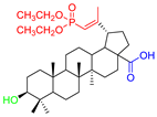
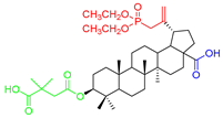
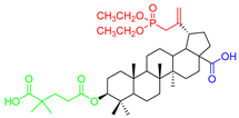
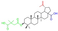
Considering two in vitro active semi-synthetic betulin derivatives, 3 and 8a, and the initial betulin 1, it can be seen that the introduction of new functional groups resulted in an increase in molar mass (MW; g/mol) from 442.72 (for betulin 1) to the value of 578.80 and 720.91, respectively. Another consequence was to increase the number of rotational bonds (nRt) and the number of acceptors (nHA) of hydrogen binding as well as a topologic polar surface (TPSA; Å2). The introduced structural changes did not affect the number of hydrogen bond donors (nHD).
The theoretical values of the lipophilicity of the tested compounds calculated using various algorithms are in the range of 5.29–9.74 and all go beyond the criteria of drug-likeness formulated by Lipinski, Weber, and Egan. Compound 8a showed the most violations of these rules. Betulin 1 and its derivative 3 met the Veber criteria without any violations. The number and type of violated criteria for the tested compounds are summarized in Table 8.
Compounds with a high molecular weight and higher lipophilicity are characterized by poor solubility in water, which results in low oral bioavailability. The choice of a compound that meets the criteria of drug similarity increases the chances of success in further stages of research (clinical trials). On the other hand, 16% of orally administered drugs do not meet at least one of Lipinski’s rule of five criteria; moreover, 6% of them violate more than two [66]. Moreover, new methods are currently being introduced to reduce the difficulties resulting from the limited oral bioavailability of drugs of natural origin. For this purpose, various types of carriers are used: polymeric nanoparticles, liposomes, dendrimers, micelles, and inorganic nanoparticles [67,68].
Based on the analysis of the pharmacokinetic profile (ADMET), as well as the drug-likeness, compound 3 was chosen for the next few studies.

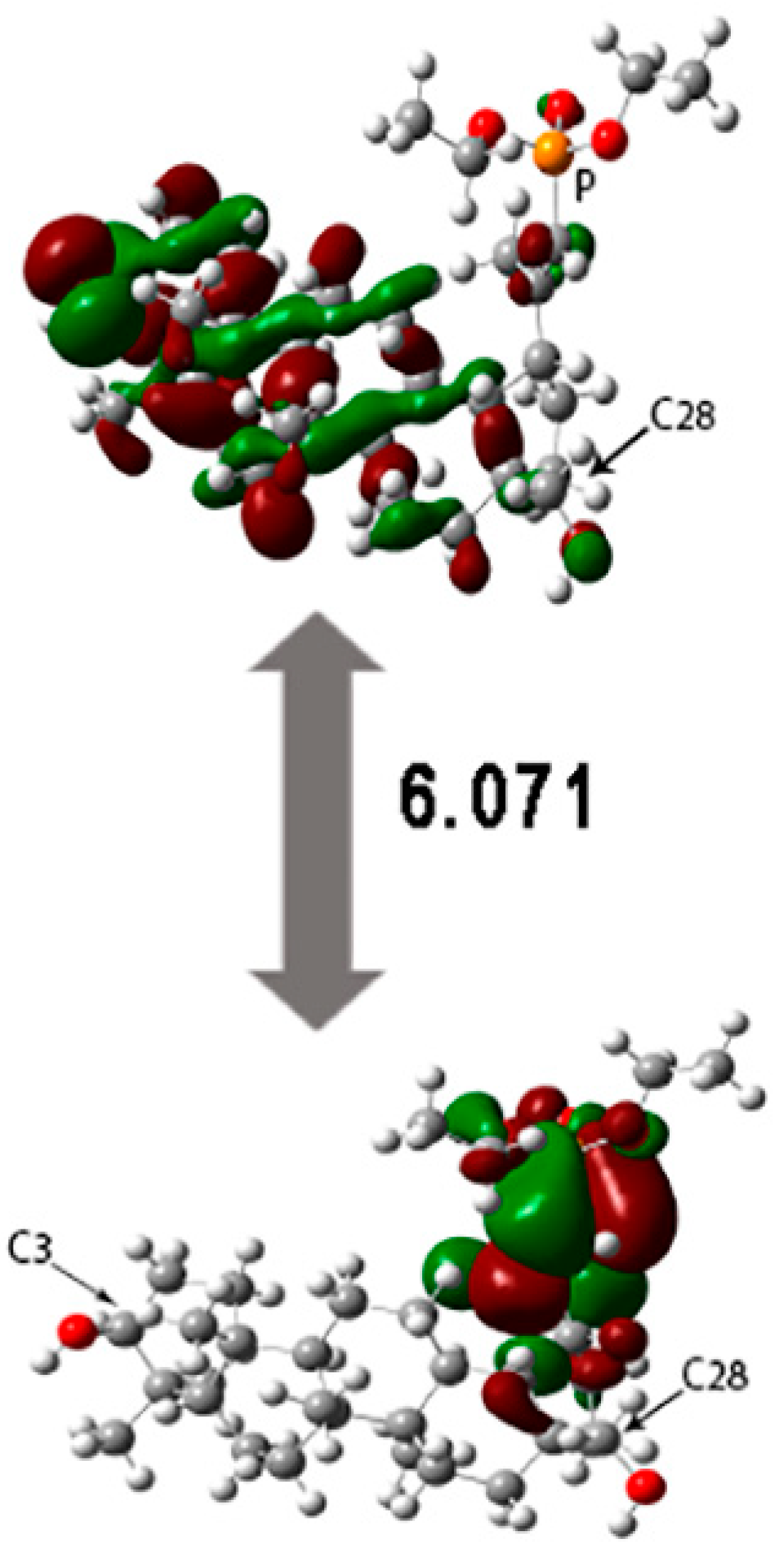
3.3.2. Study on Quantum Descriptors

The energy of HOMO (highest occupied molecular orbital) and LUMO (lowest unoccupied MO) orbital determines the quantum chemical properties of molecules [69]. The energy gap (ELUMO-EHOMO) makes it possible to designate the kinetic stability and reactivity of a compound [69,70,71,72]. The energy gap (ELUMO-EHOMO) makes it possible to designate the kinetic stability and reactivity of a compound [69,70,71,72]. The HOMO and LUMO orbitals and their energy gap were calculated using the Gaussian09 package at the B3LYP level using the 6-311G + (d,p) basis set [47].
As seen in Figure 5, the HOMO orbital is delocalized near the phosphonate group and E ring of betulin 1, while the LUMO orbital is delocalized on betulin scaffold.
The influence of the energy gap (ΔE) on the reactivity of the compound; decreasing the ΔG value increased the reactivity of the compound. Comparing the energy gap of compound 3 and betulin 1 [73] shows that the introduction of a phosphonate group led to an increase in the reactivity of the compound.
The energies of the HOMO-LUMO orbital enable the calculation of the global reactivity descriptors, such as the ionization potential (I), electron affinity (A), hardness (η), softness (s), chemical potential (µ), electronegativity (χ), and electrophilicity index (ω) [74]. Global reactivity descriptors are presented in Table 9.
The values of ionization potential (I) and electron affinity (A) show that the derivatives have low reactivity with the electrophilic molecules. The high values of electronegativity (χ) and electrophilicity index (ω) indicate that compounds are good acceptors for the nucleophilic target [69,74].
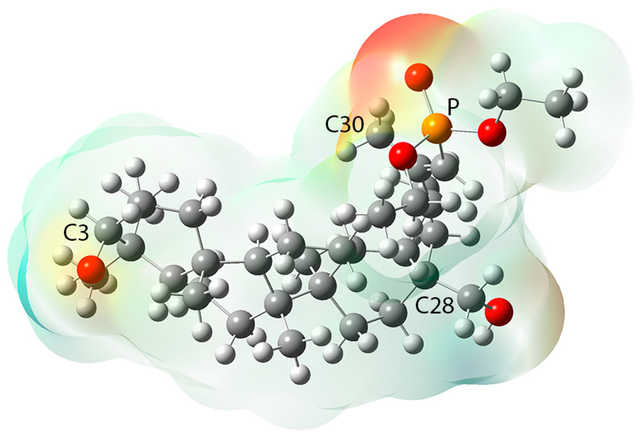
The molecular electrostatic potential map (MEP) is a useful tool to designate the electrophilic and nucleophilic region of the compound [75]. Surfaces with different charges are marked with different colors (negative—red, positive—blue, and neutral—green) (Figure 6) [76].
For compound 3, the nucleophilic regions are localized in three main areas. The first is localized near the phosphonate group. The second and third area include the oxygen atoms at positions C28 and C3, respectively. The electrophilic regions are localized near the hydrogen atom of the hydroxyl group at positions C3 and C28, respectively. The betulin scaffold is neutral (green color). Determination of the potential indicating the places or regions of the molecule to which an approaching electrophile is initially attracted can be useful in studying interactions related to the relative orientation of reactants; for example, between a drug and its cell receptor [76].

3.3.3. Molecular Docking

Molecular docking was performed to propose a probable mechanism of action of active compounds. The use of this method allows for the determination of possible interactions with selected cellular proteins without the need for expensive and time-consuming experimental methods. The obtained results may allow for further optimization of the structure of active compounds in order to improve biological activity [54].
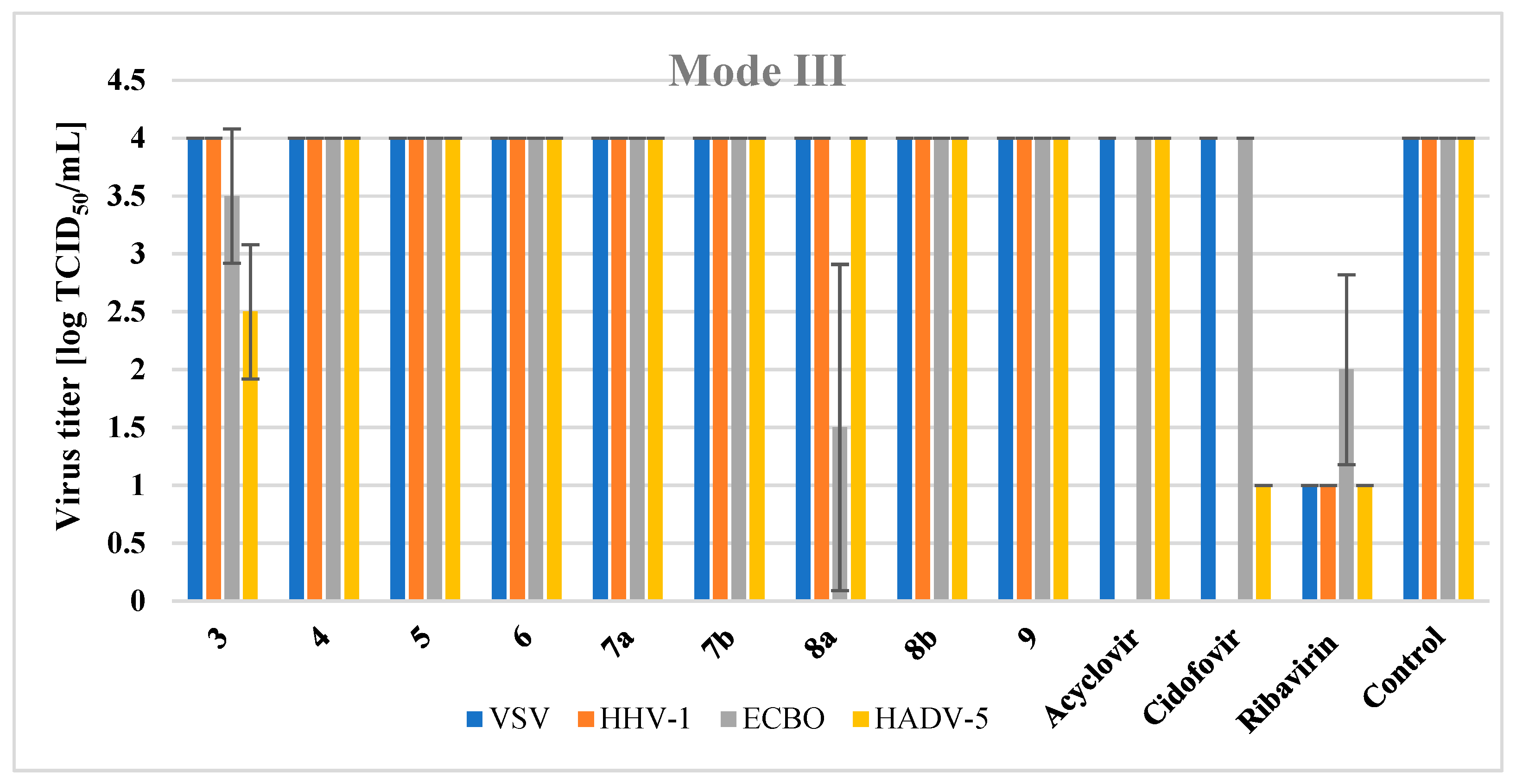
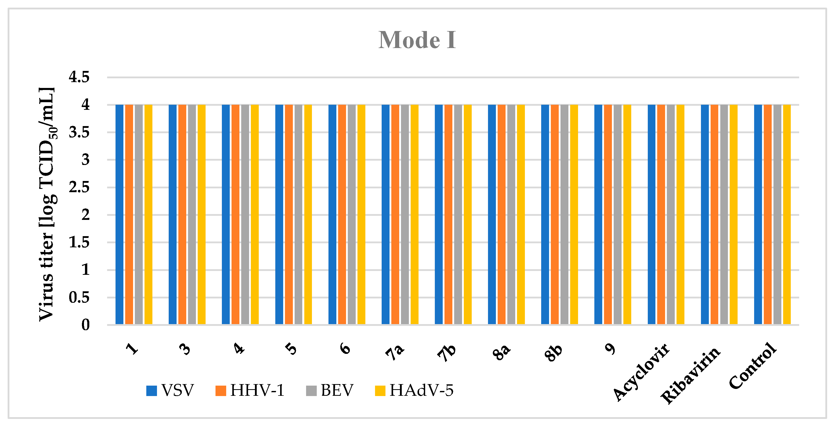
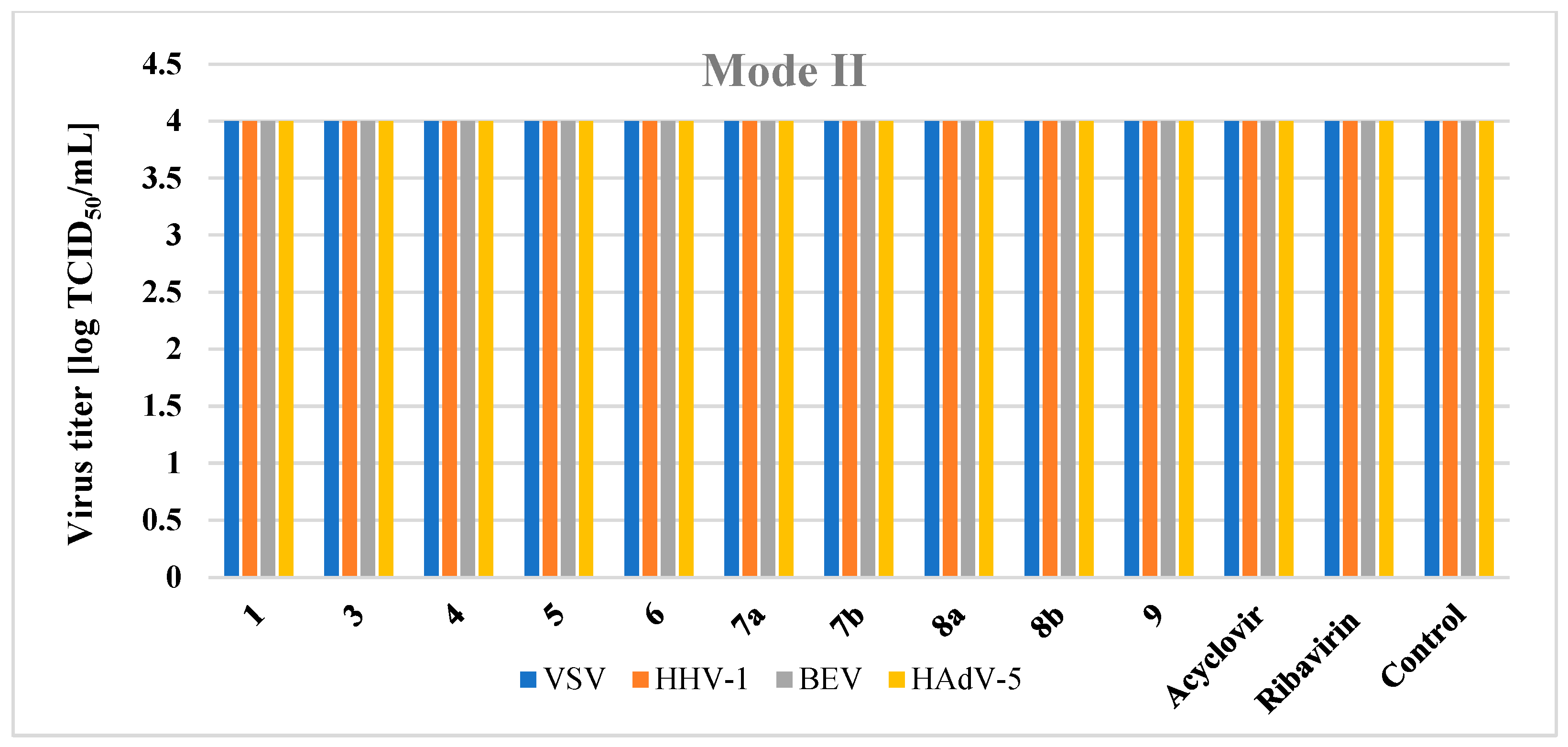
The time-of-addition experiment showed the activity of compound 3 in the third mode, where the final stages of assembly and maturation of new virions take place. One of the key replication enzymes of adenoviruses is adenaine cysteine protease. It plays a critical role in the formation of important core and capsid proteins, which then form the mature, infectious virus particles [77].
The target crystal structure of adenain was obtained from Protein Data Bank (PDB ID: 4PIE) [50]. Betulin 1 and active compound 3 were ranked by their binding energy with protein (Table 10). The lower binding energy, represented as ΔG, corresponds to a stronger binding affinity. Compound 3 exhibited a lower ΔG value compared with reference betulin (–4.8 and –3.4 kcal/mol, respectively).
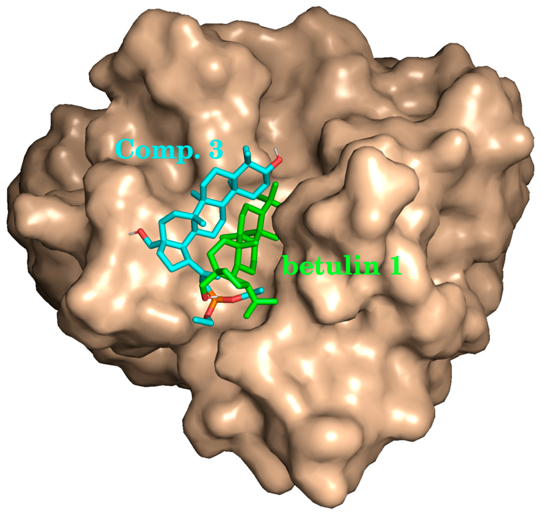
Visual inspection of the docking modes reveals that botulin 1, due its relative smaller hydrophilicity, binds deeper inside the protein pocket than compound 3 (Figure 7).
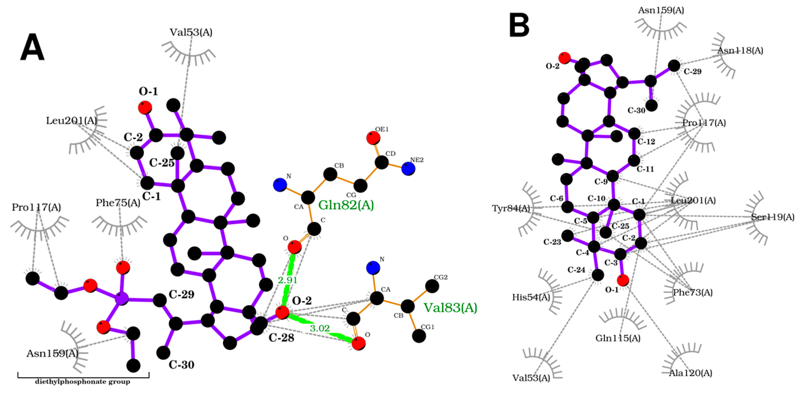
To acquire a more in-depth insight into the binding model, an analysis of protein–ligands bonding has been performed (Figure 8). Compound 3 forms two hydrogen bonds between the oxygen atom at position C-28 of the betulin core and Gln82 and Val83 residues of protein. On the other hand, the betulin molecule lacks a hydrogen bond. Additionally, both ligands form numerous hydrophobic bonds with various residues. The introduction of a diethylphosphonate group to the betulin molecule changes the structure and influences the way compound 3 interacts with the receptor, increasing the stability of the formed complex.
In general, in silico analysis confirms in vitro data. The most active compound 3 exhibited stronger binding affinity both in terms of ΔG and hydrogen bonding. Further exploration of the molecular mechanism of action could lead to more potent anti-adenovirus compounds being obtained from phosphonate derivatives of betulin.

4. Conclusions

The aim of this research was to initially assess the impact of the substituents introduced into the betulin system and their location on the antiviral activity of the tested derivatives.
30-Phosphonate (allyl isomers) derivatives of betulin, betulinic acid (4 and 5, respectively), and 3-carboxyacyl betulinic acid (7a, 7b) do not show activity against the tested viruses. From a series of derivatives containing a diethylphosphonate group at position C29 (vinylic isomer), two compounds were active at the stage of DNA and protein synthesis, virion assembly and maturation; 29-diethoxyphosphorylbetulin (3) against HAdV-5 and BEV, 29-diethoxyphosphoryl-3-(3′,3′)-dimethylsuccinyl betulinic acid (8a) against BEV.
The most active in vitro compound 3 in molecular docking to HadV-5 virus protease showed a stronger binding affinity in terms of both ΔG and hydrogen bonds compared to betulin 1. The obtained results indicate that the introduction of a diethylphosphonic group into the vinyl position (C29) of the betulin molecule has a beneficial effect on the activity against HAdV-5. They thus confirm the antiviral potential of betulin derivatives and encourage further development of research in this direction.

Supplementary Materials

The following supporting information can be downloaded at https://www.mdpi.com/article/10.3390/app14041452/s1, Figure S1. Scheme of the synthesis of the tested compounds 29. Section S1: Procedure for the synthesis of compounds 29. Section S2: Methodology of in silico studies: Section S2.1: Assessment of ADME Properties and Drug likeness of the Screened Molecules; Section S2.2: Calculations of Quantum Descriptors; Section S2.3: Method for Molecular Docking.

Author Contributions

Conceptualization, E.C. and E.B.; methodology, B.O.; software, M.K.-T. and P.P.; validation, B.O., E.B. and E.C.; writing—original draft preparation, E.B., E.C., P.P. and M.K.-T.; writing—review and editing, E.C., E.B. and B.O.; funding acquisition, E.B. and E.C.; supervision, E.C. All authors have read and agreed to the published version of the manuscript.

Funding

This research was funded by the Medical University of Silesia grant number [BNW-1-013/N/3/F].

Institutional Review Board Statement

Not applicable.

Informed Consent Statement

Not applicable.

Data Availability Statement

The data presented in this study are available in the article and Supplementary Materials.

Acknowledgments

The authors are grateful to Stanisław Boryczka for his valuable contribution to the creation of the presented study.

Conflicts of Interest

The authors declare no conflicts of interest.

References

  1. Ali, S.I.; Sheikh, W.M.; Rather, M.A.; Venkatesalu, V.; Muzamil Bashir, S.; Nabi, S.U. Medicinal plants: Treasure for antiviral drug discovery. Phytother. Res. 2021, 35, 3447–3483. [Google Scholar] [CrossRef]
  2. Musarra-Pizzo, M.; Pennisi, R.; Ben-Amor, I.; Mandalari, G.; Sciortino, M.T. Antiviral Activity Exerted by Natural Products against Human Viruses. Viruses 2021, 13, 828. [Google Scholar] [CrossRef]
  3. Perera, W.P.R.T.; Liyanage, J.A.; Dissanayake, K.G.C.; Gunathilaka, H.; Weerakoon, W.M.T.D.N.; Wanigasekara, D.N.; Fernando, W.S.K.; Rajapaksha, R.M.H.; Liyanage, R.P.; Perera, B.T. Antiviral Potential of Selected Medicinal Herbs and Their Isolated Natural Products. Biomed. Res. Int. 2021, 2021, 7872406. [Google Scholar] [CrossRef]
  4. Mukherjee, S.; Chouhan, K.B.S.; Tandey, R.; Yadav, N.; Dhobi, M.; Mandal, V. A status report with critical analysis of research trends in exploring medicinal plants as antiviral: Let us dig into the history to predict the future. Phytother. Res. 2021, 35, 4284–4296. [Google Scholar] [CrossRef]
  5. Kraus, J.L. Natural Products as Potential Antiviral Drugs: The Specific Case of Marine Biotoxins. Russ. J. Bioorg. Chem. 2021, 47, 1127–1132. [Google Scholar] [CrossRef]
  6. Saifulazmi, N.F.; Rohani, E.R.; Harun, S.; Bunawan, H.; Hamezah, H.S.; Nor Muhammad, N.A.; Azizan, K.A.; Ahmed, Q.U.; Fakurazi, S.; Mediani, A.; et al. A Review with Updated Perspectives on the Antiviral Potentials of Traditional Medicinal Plants and Their Prospects in Antiviral Therapy. Life 2022, 12, 1287. [Google Scholar] [CrossRef]
  7. World Health Organization (WHO). Global Progress Report on HIV, Viral Hepatitis and Sexually Transmitted Infections; World Health Organization: Geneva, Switzerland, 2021; Volume 53, pp. 1689–1699. Available online: https://www.who.int/publications/i/item/9789240027077 (accessed on 21 February 2023).
  8. Cui, J.; Li, F.; Shi, Z.L. Origin and evolution of pathogenic coronaviruses. Nat. Rev. Microbiol. 2019, 17, 181–192. [Google Scholar] [CrossRef] [PubMed]
  9. Tang, G.; Liu, Z.; Chen, D. Human coronaviruses: Origin, host and receptor. J. Clin. Virol. 2022, 155, 105246. [Google Scholar] [CrossRef] [PubMed]
  10. Burrell, C.J.; Howard, C.R.; Murphy, F.A. Epidemiology of Viral Infections. In Fenner and White’s Medical Virology, 5th ed.; Academic Press: Cambridge, MA, USA, 2017; Chapter 13; pp. 185–203. [Google Scholar] [CrossRef]
  11. Hilaire, J.; Tindale, S.; Jones, G.; Pingarron Cardenas, G.; Bačnik, K.; Ojo, M.; Frewer, L.J. Risk perception associated with an emerging agri-food risk in Europe: Plant viruses in agriculture. Agric. Food Secur. 2022, 11, 21. [Google Scholar] [CrossRef] [PubMed]
  12. Murcia, P.; Donachie, W.; Palmarini, M. Viral pathogens of domestic animals and their impact on biology, medicine and agri-culture. Encycl. Microbiol. (Third Ed.) 2009, 805–819. [Google Scholar] [CrossRef]
  13. Shieh, W.J. Human Adenovirus Infections in Pediatric Population—An Update on Clinico–Pathologic Correlation. Biomed. J. 2022, 45, 38–49. [Google Scholar] [CrossRef] [PubMed]
  14. Puig-Barberà, J.; Mira-Iglesias, A.; Burtseva, E.; Cowling, J.; Serhat, U.; Ruiz-Palacios, G.M.; Launay, O.; Kyncl, J.; Koul, P.; Siqueira, M.M.; et al. Influenza epidemiology and influenza vaccine effectiveness during the 2015–2016 season: Results from the Global Influenza Hospital Surveillance Network. BMC Infect. Dis. 2019, 19, 415. [Google Scholar] [CrossRef] [PubMed]
  15. de Martel, C.; Georges, D.; Bray, F.; Ferlay, J.; Clifford, G.M. Global Burden of Cancer Attributable to Infections in 2018: A Worldwide Incidence Analysis. Lancet Glob. Health 2020, 8, e180–e190. [Google Scholar] [CrossRef] [PubMed]
  16. Andino, R.; Kirkegaard, K.; Macadam, A.; Racaniello, V.R.; Rosenfeld, A.B. The Picornaviridae Family: Knowledge gaps, animal models, countermeasures, and prototype pathogens. J. Infect. Dis. 2023, 228, S427–S445. [Google Scholar] [CrossRef] [PubMed]
  17. Shepherd, J.G.; Davis, C.; Streicker, D.G.; Thomson, E.C. Emerging Rhabdoviruses and Human Infection. Biology 2023, 12, 878. [Google Scholar] [CrossRef]
  18. Siegel, R.D. Classification of Human Viruses. In Principles and Practice of Pediatric Infectious Diseases; Long, S.S., Prober, C.G., Fischer, M., Eds.; Elsevier Inc.: Amsterdam, The Netherlands, 2018; pp. 1044–1048. [Google Scholar] [CrossRef]
  19. Denaro, M.; Smeriglio, A.; Barreca, D.; De Francesco, C.; Occhiuto, C.; Milano, G.; Trombetta, D. Antiviral activity of plants and their isolated bioactive compounds: An update. Phytother. Res. 2020, 34, 742–768. [Google Scholar] [CrossRef]
  20. Liu, Y.; Yang, L.; Wang, H.; Xiong, Y. Recent Advances in Antiviral Activities of Triterpenoids. Pharmaceuticals 2022, 15, 1169. [Google Scholar] [CrossRef]
  21. Kazakova, O.B.; Smirnova, I.E.; Baltina, L.A.; Boreko, E.I.; Savinova, O.V.; Pokrovskii, A.G. Antiviral Activity of Acyl Derivatives of Betulin and Betulinic and Dihydroquinopimaric Acids. Russ. J. Bioorg. Chem. 2018, 44, 740–744. [Google Scholar] [CrossRef]
  22. Fujioka, T.; Kashiwada, Y.; Kilkuskie, R.E.; Cosentino, L.M.; Ballas, L.M.; Jiang, J.B.; Janzen, W.P.; Chen, I.S.; Lee, K.H. Anti-AIDS agents, 11. Betulinic acid and platanic acid as anti-HIV principles from Syzigium claviflorum, and the anti-HIV activity of structurally related triterpenoids. J. Nat. Prod. 1994, 57, 243–247. [Google Scholar] [CrossRef]
  23. Pavlova, N.I.; Savinova, O.V.; Nikolaeva, S.N.; Boreko, E.I.; Flekhter, O.B. Antiviral activity of betulin, betulinic and betulonic acids against some enveloped and non-enveloped viruses. Fitoterapia 2003, 74, 489–492. [Google Scholar] [CrossRef]
  24. Gong, Y.; Raj, K.M.; Luscombe, C.A.; Gadawski, I.; Tam, T.; Chu, J.; Gibson, D.; Carlson, R.; Sacks, S.L. The synergistic effects of betulin with acyclovir against herpes simplex viruses. Antivir. Res. 2004, 64, 127–130. [Google Scholar] [CrossRef]
  25. Navid, M.H.; Laszczyk-Lauer, M.N.; Reichling, J.; Schnitzler, P. Pentacyclic triterpenes in birch bark extract inhibit early step of herpes simplex virus type 1 replication. Phytomedicine 2014, 25, 1273–1280. [Google Scholar] [CrossRef]
  26. Muhammad, A.; Carlson, R.M.; Krasutsky, P.; Reza-Ul Karim, M. Inhibition of Epstein-Barr Virus by the Triterpenoid Betulin Diphosphate and Uvaol. J. Microbiol. Biotechnol. 2004, 14, 1086–1088. [Google Scholar]
  27. Karagöz, A.Ç.; Leidenberger, M.; Hahn, F.; Hampel, F.; Friedrich, O.; Marschall, M.; Kappes, B.; Tsogoeva, S.B. Synthesis of new betulinic acid/betulin-derived dimers and hybrids with potent antimalarial and antiviral activities. Bioorg. Med. Chem. 2019, 27, 110–115. [Google Scholar] [CrossRef]
  28. Pohjala, L.; Alakurtti, S.; Ahola, T.; Yli-Kauhaluoma, J.; Tammela, P. Betulin-derived compounds as inhibitors of alphavirus replication. J. Nat. Prod. 2009, 72, 1917–1926. [Google Scholar] [CrossRef]
  29. Kamińska, T.; Kaczor, J.; Rzeski, W.; Wejksza, K.; Kandefer-Szerszeń, M.; Witek, M. A comparison of the antiviral activity of three triterpenoids isolated from Betula alba bark. Ann. Univ. Mariae Curie-Sklodowska. Sect. C Biol. 2004, 59, 7–13. [Google Scholar]
  30. Bębenek, E.; Jastrzębska, M.; Kadela-Tomanek, M.; Chrobak, E.; Orzechowska, B.; Zwolińska, K.; Latocha, M.; Mertas, A.; Czuba, Z.; Boryczka, S. Novel Triazole Hybrids of Betulin: Synthesis and Biological Activity Profile. Molecules 2017, 22, 1876. [Google Scholar] [CrossRef] [PubMed]
  31. Alhadrami, H.A.; Sayed, A.M.; Sharif, A.M.; Azhar, E.I.; Rateb, M.E. Olive-Derived Triterpenes Suppress SARS-CoV-2 Main Protease: A Promising Scaffold for Future Therapeutics. Molecules 2021, 26, 2654. [Google Scholar] [CrossRef] [PubMed]
  32. Peyrat, L.A.; Eparvier, V.; Eydoux, C.; Guillemot, J.; Litaudon, M.; Stien, D. Betulinic Acid, The First Lupane-Type Triterpenoid Isolated from Both a Phomopsis sp. and Its Host Plant Diospyros carbonaria Benoist. Chem. Biodivers. 2017, 14, e1600171. [Google Scholar] [CrossRef] [PubMed]
  33. Wang, D.; Lu, W.; Li, F. Pharmacological intervention of HIV-1 maturation. Acta Pharm. Sin. B 2015, 5, 493–499. [Google Scholar] [CrossRef] [PubMed]
  34. Biron, K.K. Antiviral drugs for cytomegalovirus diseases. Antivir. Res. 2006, 71, 154–163. [Google Scholar] [CrossRef] [PubMed]
  35. Dracínský, M.; Krecmerová, M.; Holý, A. Study of chemical stability of antivirally active 5-azacytosine acyclic nucleoside phosphonates using NMR spectroscopy. Bioorg. Med. Chem. 2008, 16, 6778–6782. [Google Scholar] [CrossRef] [PubMed]
  36. Broganelli, P.; Chiaretta, A.; Fragnelli, B.; Bernengo, M.G. Intralesional cidofovir for the treatment of multiple and recalcitrant cutaneous viral warts. Dermatol. Ther. 2012, 25, 468–471. [Google Scholar] [CrossRef]
  37. Chrobak, E.; Marciniec, K.; Dąbrowska, A.; Pęcak, P.; Bębenek, E.; Kadela-Tomanek, M.; Bak, A.; Jastrzębska, M.; Boryczka, S. New Phosphorus Analogs of Bevirimat: Synthesis, Evaluation of Anti-HIV-1 Activity and Molecular Docking Study. Int. J. Mol. Sci. 2019, 20, 5209. [Google Scholar] [CrossRef]
  38. Chrobak, E.; Bębenek, E.; Kadela-Tomanek, M.; Latocha, M.; Jelsch, C.; Wenger, E.; Boryczka, S. Betulin Phosphonates; Synthesis, Structure, and Cytotoxic Activity. Molecules 2016, 21, 1123. [Google Scholar] [CrossRef]
  39. Kuhn, J.H. Virus taxonomy. Encycl. Virol. 2021, 28–37. [Google Scholar] [CrossRef]
  40. Tada, H.; Shiho, O.; Kuroshima, K.-I.; Koyama, M.; Tsukamoto, K. An improved colorimetric assay for interleukin 2. J. Immunol. Methods 1986, 93, 157–165. [Google Scholar] [CrossRef]
  41. Septisetyani, E.P.; Ningrum, R.A.; Romadhani, Y.; Wisnuwardhani, P.H.; Santoso, A. Optimization of sodium dodecyl sulphate as a formazan solvent and comparison of 3-(4,-5-dimethylthiazo-2-yl)-2,5-diphenyltetrazolium bromide (MTT) assay with WST-1 assay in MCF-7 cells. Indones. J. Pharm. 2014, 25, 245. [Google Scholar] [CrossRef]
  42. Pęcak, P.; Orzechowska, B.; Chrobak, E.; Boryczka, S. Novel betulin dicarboxylic acid ester derivatives as potent antiviral agents: Design, synthesis, biological evaluation, structure-activity relationship and in-silico study. Eur. J. Med. Chem. 2021, 225, 113738. [Google Scholar] [CrossRef]
  43. Lai, Y.; Han, T.; Zhan, S.; Jiang, Y.; Liu, X.; Li, G. Antiviral Activity of Isoimperatorin Against Influenza A Virus in vitro and its Inhibition of Neuraminidase. Front. Pharmacol. 2021, 12, 657826. [Google Scholar] [CrossRef]
  44. Al-Majed, A.A. Profiles of Drug Substances. Excip. Relat. Methodol. 2023, 48, 2–218. Available online: https://www.sciencedirect.com/bookseries/profiles-of-drug-substances-excipients-and-related-methodology (accessed on 27 September 2023).
  45. Pires, D.; Blundell, T.; Ascher, D. pkCSM: Predicting small-molecule pharmacokinetic and toxicity properties using graph-based signatures. J. Med. Chem. 2015, 58, 4066–4072. [Google Scholar] [CrossRef]
  46. Daina, A.; Michielin, O.; Zoete, V. SwissADME: A free web tool to evaluate pharmacokinetics, drug-likeness and medicinal chemistry friendliness of small molecules. Sci. Rep. 2017, 7, 42717. [Google Scholar] [CrossRef]
  47. Frisch, M.J.; Trucks, G.W.; Schlegel, H.B.; Scuseria, G.E.; Robb, M.A.; Cheeseman, J.R.; Scalmani, G.; Barone, V.; Mennucci, B.; Petersson, G.A.; et al. Gaussian 16, Revision, A. 03. 2016; Gaussian Inc.: Wallingford, CT, USA, 2016.
  48. Dennington, R.; Keith, T.; Millam, J. GaussView, Version 5; Semichem Inc.: Shawnee Mission, KS, USA, 2009.
  49. Foresman, J.B.; Frisch, A.E. Exploring Chemistry with Electronic Structure Methods: A Guide to Using Gaussian; Gaussian Inc.: Wallingford, CT, USA, 1996. [Google Scholar]
  50. Mac Sweeney, A.; Grosche, P.; Ellis, D.; Combrink, K.; Erbel, P.; Hughes, N.; Sirockin, F.; Melkko, S.; Bernardi, A.; Ramage, P.; et al. Discovery and structure-based optimization of adenain inhibitors. ACS Med. Chem. Lett. 2014, 5, 937–941. [Google Scholar] [CrossRef] [PubMed]
  51. Morris, G.M.; Huey, R.; Lindstrom, W.; Sanner, M.F.; Belew, R.K.; Goodsell, D.S.; Olson, A.J. AutoDock4 and AutoDockTools4: Automated docking with selective receptor flexibility. J. Comput. Chem. 2009, 16, 2785–2791. [Google Scholar] [CrossRef] [PubMed]
  52. Biovia, D.S. Discovery Studio Modeling Environment, Dessault Systemes. 2020. Available online: https://www.3dsbiovia.com/products/collaborative-science/ (accessed on 20 August 2023).
  53. Laskowski, R.A.; Swindells, M.B. LigPlot+: Multiple ligand-protein interaction diagrams for drug discovery. J. Chem. Inf. Model. 2011, 51, 2778–2786. [Google Scholar] [CrossRef]
  54. Trott, O.; Olson, A.J. AutoDock Vina: Improving the speed and accuracy of docking with a new scoring function, efficient optimization, and multithreading. J. Comput. Chem. 2010, 31, 455–461. [Google Scholar] [CrossRef]
  55. Marciniec, K.; Chrobak, E.; Dąbrowska, A.; Bębenek, E.; Kadela-Tomanek, M.; Pęcak, P.; Boryczka, S. Phosphate Derivatives of 3-Carboxyacylbetulin: Synthesis, In Vitro Anti-HIV and Molecular Docking Study. Biomolecules 2020, 10, 1148. [Google Scholar] [CrossRef]
  56. López-García, J.; Lehocký, M.; Humpolíček, P.; Sáha, P. HaCaT Keratinocytes Response on Antimicrobial Atelocollagen Substrates: Extent of Cytotoxicity, Cell Viability and Proliferation. J. Funct. Biomater. 2014, 5, 43–57. [Google Scholar] [CrossRef] [PubMed]
  57. Perryman, A.L.; Patel, J.S.; Russo, R.; Singleton, E.; Connell, N.; Ekins, S.; Freundlich, J.S. Naïve Bayesian Models for Vero Cell Cytotoxicity. Pharm. Res. 2018, 35, 170. [Google Scholar] [CrossRef]
  58. PN-EN 14675; Chemical Disinfectants and Antiseptics–Quantitative Suspension Test for the Evaluation of Virucidal Activity of Chemical Disinfectants and Antiseptics Used in the Veterinary Area–Test Method and Requirements. Polish Committee for Standardization: Warszawa, Poland, 2006.
  59. Gür, S.; Gürçay, M.; Seyrek, A. A study regarding bovine enterovirus type 1 infection in domestic animals and humans: An evaluation from the zoonotic aspect. J. Vet. Med. Sci. 2019, 81, 1824–1828. [Google Scholar] [CrossRef]
  60. Bishnoi, S.; Tiwari, R.; Gupta, S.; Byrareddy, S.N.; Nayak, D. Oncotargeting by Vesicular Stomatitis Virus (VSV): Advances in Cancer Therapy. Viruses 2018, 10, 90. [Google Scholar] [CrossRef]
  61. James, C.; Harfouche, M.; Welton, N.J.; Turner, K.M.; Abu-Raddad, L.J.; Gottlieb, S.L.; Looker, K.J. Herpes simplex virus: Global infection prevalence and incidence estimates, 2016. Bull. World Health Organ. 2020, 98, 315–329. [Google Scholar] [CrossRef]
  62. Aliabadi, N.; Jamalidoust, M.; Pouladfar, G.; Ziyaeyan, A.; Ziyaeyan, M. Antiviral activity of triptolide on herpes simplex virus in vitro. Immun. Inflamm. Dis. 2022, 10, e667. [Google Scholar] [CrossRef]
  63. Wu, F.; Zhou, Y.; Li, L.; Shen, X.; Chen, G.; Wang, X.; Liang, X.; Tan, M.; Huang, Z. Computational Approaches in Preclinical Studies on Drug Discovery and Development. Front. Chem. 2020, 8, 726. [Google Scholar] [CrossRef]
  64. Martinec, O.; Huliciak, M.; Staud, F.; Cecka, F.; Vokral, I.; Cerveny, L. Anti-HIV and antihepatitis C virus drugs inhibit P-glycoprotein efflux activity in Caco-2 cells and precision-cut rat and human intestinal slices. Antimicrob. Agents Chemother. 2019, 63, e00910-19. [Google Scholar] [CrossRef] [PubMed]
  65. Zhou, S.F.; Xue, C.C.; Yu, X.Q.; Li, C.; Wang, G. Clinically Important Drug Interactions Potentially Involving Mechanism-based Inhibition of Cytochrome P450 3A4 and the Role of Therapeutic Drug Monitoring. Ther. Drug Monit. 2007, 29, 687–710. [Google Scholar] [CrossRef]
  66. Lee, K.; Jang, J.; Seo, S.; Lim, J.; Kim, W.Y. Drug-likeness scoring based on unsupervised learning. Chem. Sci. 2022, 13, 554–565. [Google Scholar] [CrossRef]
  67. Dehelean, C.A.; Marcovici, I.; Soica, C.; Mioc, M.; Coricovac, D.; Iurciuc, S.; Cretu, O.M.; Pinzaru, I. Plant-Derived Anticancer Compounds as New Perspectives in Drug Discovery and Alternative Therapy. Molecules 2021, 26, 1109. [Google Scholar] [CrossRef] [PubMed]
  68. Madej, M.; Kurowska, N.; Strzalka-Mrozik, B. Polymeric Nanoparticles—Tools in a Drug Delivery System in Selected Cancer Therapies. Appl. Sci. 2022, 12, 9479. [Google Scholar] [CrossRef]
  69. Choudhary, V.; Bhatt, A.; Dash, D.; Sharma, N. DFT calculations on molecular structures, HOMO–LUMO study, reactivity descriptors and spectral analyses of newly synthesized diorganotin(IV) 2-chloridophenylacetohydroxamate complexes. J. Comput. Chem. 2019, 40, 2354–2363. [Google Scholar] [CrossRef]
  70. Kadela-Tomanek, M.; Pawełczak, B.; Jastrzębska, M.; Bębenek, E.; Chrobak, E.; Latocha, M.; Kusz, J.; Książek, M.; Boryczka, S. Structural, vibrational and quantum chemical investigations for 6,7-dichloro-2-methyl-5,8-quinolinedione. Cytotoxic and molecular docking studies. J. Mol. Struct. 2018, 1168, 73–83. [Google Scholar] [CrossRef]
  71. Kadela-Tomanek, M.; Jastrzębska, M.; Chrobak, E.; Bębenek, E.; Latocha, M.; Kusz, J.; Boryczka, S. Structural and spectral characterisation of 2-amino-2H-[1,2,3]triazolo[4,5-g]quinoline-4,9-dione polymorphs. Cytotoxic activity and molecular docking study with NQO1 enzyme. Spectrochim. Acta A Mol. Biomol. Spectrosc. 2020, 230, 118038–118050. [Google Scholar] [CrossRef]
  72. Rauk, A. Orbital Interaction Theory of Organic Chemistry, 2nd ed.; Wiley & Sons John: New York, NY, USA, 2001. [Google Scholar]
  73. Kazachenko, A.S.; Akman, F.; Vasilieva, N.Y.; Issaoui, N.; Malyar, Y.N.; Kondrasenko, A.A.; Borovkova, V.S.; Miroshnikova, A.V.; Kazachenko, A.S.; Al-Dossary, O.; et al. Catalytic sulfation of betulin with sulfamic acid: Experiment and DFT calculation. Int. J. Mol. Sci. 2022, 23, 1602. [Google Scholar] [CrossRef]
  74. Ali, B.; Khalid, M.; Asim, S.; Khan, M.U.; Iqbal, Z.; Hussain, A.; Hussain, R.; Ahmed, S.; Ali, A.; Hussain, A.; et al. Key electronic, linear and nonlinear optical properties of designed disubstituted quinoline with carbazole compounds. Molecules 2021, 26, 2760. [Google Scholar] [CrossRef]
  75. Chandrasekaran, K.; Kumar, R. Structural, spectral, thermodynamical, NLO, HOMO, LUMO and NBO analysis of fluconazole. Spectrochim. Acta A 2015, 150, 974–991. [Google Scholar] [CrossRef]
  76. Politzer, P.; Laurence, P.; Jayasuriya, K. Molecular electrostatic potentials: An effective tool for the elucidation of biochemical phenomena. Environ. Health Perspect. 1985, 61, 191–202. [Google Scholar] [CrossRef]
  77. Weber, J.M. Adenain, the adenovirus endoprotease (a review). Acta Microbiol. Immunol. Hung. 2003, 50, 95–101. [Google Scholar] [CrossRef]
Figure 1. Structure of betulin and betulinic acid.
Applsci 14 01452 g001
Figure 2. Mode I: effect of the studied compounds on the sensitivity of the cells to infection with VSV, HHV-1, BEV, and HAdV-5, at a dose of 100 TCID50 (with a median of three repetitions).
Applsci 14 01452 g002
Figure 3. Mode II: the activity of the studied compounds at the virus entry step of replication at a dose of 100 TCID50 (with a median of three repetitions).
Applsci 14 01452 g003
Figure 4. Mode III: effect of the studied compounds on reduction of the titer of tested viruses in the post-adsorption phase. Each compound was tested in triplicate.
Applsci 14 01452 g004
Figure 5. The HOMO and LUMO orbitals of compound 3.
Applsci 14 01452 g005
Figure 6. The MEP for compound 3.
Applsci 14 01452 g006
Figure 7. Docked poses of betulin 1 (green) and compound 3 (light blue) in the active site of adenain.
Applsci 14 01452 g007
Figure 8. Analysis of bonding between ligands ((A)—compound 3; (B)—betulin 1) and adenain. Hydrogen bonds are marked in green, hydrophobic in gray.
Applsci 14 01452 g008
Table 1. Antiviral properties of betulin.
VirusReferences
Herpes simplex virus type I (HSV-1-also known as HHV-1)[23,24,25]
Herpes simplex virus type II (HSV-2; HHV-2)[24]
Influenza FPV (H7N1)[23]
Enteric cytopathic human orphan virus 6 (ECHO 6)[23]
Epstein–Barr virus (EBV)[26]
Human cytomegalovirus (HCMV)[27]
Semliki Forest Virus (SFV)[28]
Vesicular stomatitis virus (VSV)[29]
Encephalomyocarditis virus (EMCV)[29]
Cytopathogenic bovine orphan virus (ECBO also known as BEV)[30]
SARS CoV-2 Mpro[31]
Dengue virus (DENV-NS5 RdRp)[32]
Table 2. The characteristics and classification of the applied viruses [39].
Viruses (The Baltimore Virus Classifications)
BEV (Picornaviridae, BC IV: (+) ssRNA viruses (+) sense RNA)
VSV (Rhabdoviridae, BC V: (−) ssRNA viruses (−) sense RNA)
HHV-1 (Herpesviridae, BC I: dsDNA viruses)
HAdV-5 (Adenoviridae, BC I: dsDNA viruses)
ss—single-stranded, ds—double-stranded.
Table 3. Different administration modes applied for tested compounds. Time of addition assay.
Mode IMode IIMode III
A549 cells were preincubated with the phoshonate derivatives then infected by tested virusesA549 cells were incubated simultaneously with the phoshonate
derivatives and viruses
A549 cells were infected with viruses then incubated with the phoshonate derivatives
Table 4. Structures of the tested compounds 39.
30-Phosphonate Derivatives29-Phosphonate Derivatives
Compound 4
Applsci 14 01452 i001
Compound 3
Applsci 14 01452 i002
Compound 5
Applsci 14 01452 i003
Compound 6
Applsci 14 01452 i004
Compound 7a
Applsci 14 01452 i005
Compound 7b
Applsci 14 01452 i006
Compound 8a
Applsci 14 01452 i007
Compound 8b
Applsci 14 01452 i008
Compound 9 (BVM)
Applsci 14 01452 i009
Table 5. Cytotoxicity of compounds 39 and reference substances on human lung cancer cell line A549.
CompoundCC20 [µM]
34.72
48.1
51.0
648.6
7a33.4
7b26.7
8a60.22
8b38.2
925.3
Betulin 128.22 a
Acyclovir196.08 a
Ribavirin409.84 a
a [42].
Table 6. Selected ADMET parameters of the tested compounds (pkCSM).
ParameterCompound
134567a7b8a8b9
Water solubility−5.492−5.343−5.504−3.366−3.325−2.921−2.918−2.919−2.915−3.04A
Caco-2 permeability1.3210.7420.660.4640.4980.2280.2430.2610.2770.547
HIA10097.79994.52883.4796.18952.22258.51764.9471.23570.09
pgP-substratenoyesnononononononono
pgP-I-inh.yesyesnononononononono
pgP-II-inh.yesyesyesyesyesyesyesyesyesyes
BBB permeability−0.467−0.105−0.541−0.71−0.686−1.351−1.373−1.327−1.348−0.083D
CYP2D6 substr.nonononononononononoM
CYP3A4 substr.yesyesyesyesyesyesyesyesyesyes
CYP1A2 inh.nononononononononono
CYP2C19 inh.nononononononononono
CYP2C9 inh.nononononononononono
CYP2D6 inh.nononononononononono
CYP3A4 inh.nononononononononono
Total
clearance
0.236−0.466−0.259−0.199−0.410−0.226−0.344−0.437−0.556−0.272E
AMESnonononononononononoT
hERG Inononononononononono
hERG IInononononononononono
Max tolerated dose (human)
Log mg/kg/day
−0.433−0.0950.1130.550.4890.5330.4940.5170.4830.32
Oral rat acute LD50 (mol/kg)3.2673.1553.0392.4712.4852.3482.3662.3482.3662.791
Hepatotoxicityyesnonononononononono
inh.—inhibitor; water solubility logS (log mol/L); Caco-2 permeability (log Papp in 10−6 cm/s); HIA intestinal absorption (human) (% Absorbed); BBB permeability (log BB); CNS permeability (log PS); total clearance log ml/min/kg; oral rat acute toxicity (LD50).
Table 7. Physicochemical parameters and predicted lipophilicity values of the selected compounds (SwissADME).
CompoundMWCsp3nRtnHAnHDTPSAWLOGPMLOGP
1442.720.9322240.467.006.00
3578.800.9475285.808.595.29
8a720.910.881292146.249.745.34
Table 8. Predicted drug likeness as the number of violated criteria for the selected compounds (SwissADME).
CompoundDrug-Likeness
LipinskiVeberEgan
1101
MLOGP > 4.15 WLOG > 5.88
3201
MW > 500, MLOGP > 4.15 WLOG > 5.88
8a222
MW > 500, MLOGP > 4.15nRt > 10, TPSA > 140WLOG > 5.88, TPSA > 131.6
Table 9. The global reactivity descriptors of 3.
ΔE (eV)I (eV)A (eV)η (eV)µ (eV)χ (eV)ω (eV)
6.0716.8830.8113.036−3.8473.8472.438
Table 10. Binding affinities of betulin 1 and derivative 3 in complexes with adenain.
CompoundΔG [kcal/mol]
1−3.4
3−4.8
Disclaimer/Publisher’s Note: The statements, opinions and data contained in all publications are solely those of the individual author(s) and contributor(s) and not of MDPI and/or the editor(s). MDPI and/or the editor(s) disclaim responsibility for any injury to people or property resulting from any ideas, methods, instructions or products referred to in the content.

