Article

Emergent and Known Honey Bee Pathogens through Passive Surveillance in the Republic of Kosovo

1
Veterinary Laboratory, Food and Veterinary Agency, Industrial Zone, 10000 Prishtina, Kosovo
2
National Reference Laboratory for Honey Bee Health, Istituto Zooprofilattico Sperimentale delle Venezie, Viale dell’Università 10, 35020 Legnaro, Italy
3
Agricultural and Veterinary Faculty, University of Prishtina, Bulevardi Bill Clinton P.N., 10000 Prishtina, Kosovo
4
UBT-Higher Education Institution, Lagjja Kalabria, 10000 Prishtina, Kosovo
*
Author to whom correspondence should be addressed.
These authors contributed equally to this work.
Appl. Sci. 2024, 14(3), 987; https://doi.org/10.3390/app14030987
Submission received: 27 December 2023 / Revised: 20 January 2024 / Accepted: 23 January 2024 / Published: 24 January 2024

Abstract

:
In recent years, honey bee colony losses in the Republic of Kosovo remained largely unknown. From 2019 to 2021, 81 apiaries with different disease suspicions were investigated in the framework of honey bee disease passive surveillance. Fifty-nine of the eighty-one apiaries were tested for Vairimorpha ceranae, Vairimorpha apis, trypanosomatids Lotmaria passim, and Crithidia mellificae. All samples were positive for V. ceranae (100%) whereas L. passim was found with a lower frequency (11.9%). V. apis and C. mellificae were not found. Thirteen of the eighty-one apiaries were tested for seven viruses (ABPV, CBPV, DWV, BQCV, SBV, IAPV, KBV) and five of them were found (ABPV, CBPV, DWV, BQCV, SBV). The most frequently detected viruses in honey bees and Varroa mites were DWV (100%) followed by BQCV, ABPV, SBV, and CBPV (92.3%, 69.2%, 30.8%, and 7.7%, respectively). Varroa mite samples had different degrees of co-infection by viruses. Nine of the eighty-one apiaries consisted of brood combs with larvae, eight of them were AFB positive, ERIC I genotype, and one EFB positive. This paper represents the first molecular investigation (PCR) and detection of the honey bee viruses ABPV, CBPV, DWV, BQCV, and SBV as well as V. ceranae, L. passim, and M. plutonius in the Republic of Kosovo.

1. Introduction

Honey bee health has recently become a major topic due to the important role that bees play in pollination and food production [1]. In the last ten years, some regions of the world have suffered from a significant reduction in honey bee colonies [2]. It is believed that the reduction in honey bee populations is caused by a number of different biotic and abiotic factors, in particular pests, genetic factors, bee management, including beekeeping practices and breeding, climatic changes, malnutrition, agricultural practices, and the use of pesticides [2]. From emergent bee pathogens, the microsporidian species Vairimorpha (formerly Nosema) apis and Vairimorpha (formerly Nosema) ceranae [3] have been identified in European honey bees, Apis mellifera. V. ceranae is the most common gut pathogen in adult honey bees [4] and its infection could induce a degeneration of gut epithelial cells [5], a significant reduction in honey production [6], and a reduction in bee lifespan [7,8,9,10].
Two trypanosomatids Lotmaria passim and Crithidia mellificae (Kinetoplastea: Trypanosomatidae) are capable of colonizing the digestive tract of honey bees [11,12]. Both pathogens are considered to alter bee physiology, behaviour, immune responses, and lifespan [13,14,15,16].
Several bee viruses, like acute bee paralysis virus (ABPV), black queen cell virus (BQCV), chronic bee paralysis virus (CBPV), deformed wing virus (DWV), Kashmir bee virus (KBV), Sacbrood virus (SBV), and Israeli acute paralysis virus (IAPV) have been documented to be transmitted and activated by Varroa mites in field conditions [17,18,19,20,21,22,23,24,25,26,27]. ABPV, BQCV, KBV, and IAPV belong to the Dicistroviridae family [28] while DWV and SBV are classified in the Iflaviridae family [28]. CBPV has not yet been classified into any taxa [29].
ABPV and IAPV cause trembling, inability to fly, rapidly progressing paralysis, and death of honey bees [30,31,32], whereas KBV in natural infections commonly persists within apparently healthy broods and adults [33,34,35]. CBPV causes massive worker bee losses, mostly in strong colonies, and its infection appears in two groups of clinical signs: one including inability to fly, clustering, trembling, and crawling; the other consisting of black hairless individuals with shortened abdomens [36]. BQCV and SBV are the most widely distributed of all honey bee viruses. BQCV does not cause visible symptoms in infected adult bees but it could lead queen larvae and pupae to death by turning their cells black. Two-day-old larvae appear to be most vulnerable to SBV infection and, once they are fed with contaminated larval food, they fail to pupate [37,38], acquiring a sac-like appearance. DWV causes deformed wings in honey bees and induces colony weakening and mortality. Typical disease symptoms of DWV infection include shrunken, crumpled wings, decreased body size, and discoloration in adult bees. Three genetic variants of DWV were discovered and identified as types A, B, and C, but DWV-A and -B are the most widespread variants [39,40].
Two bacterial pathogens affecting honey bee larvae but not adult bees are Paenibacillus larvae, the causative agent of American Foulbrood (AFB), and Melissococcus plutonius, responsible for European Foulbrood (EFB) [41]. The clinical symptoms of AFB are darkened, sunken, and perforated cell caps containing diseased larvae, a characteristic unpleasant odour, and sticky larval remains when drawn out with a matchstick. Instead, EFB usually affects young larvae that die while still coiled before they are capped. The younger larvae affected cover the bottom of the cell and are almost transparent, with a visible trachea and midgut; the latter, full of bacteria, appears as a yellow spot. Dead, flaccid discoloured larvae in uncapped cells show colour changes from pearly white to yellow to yellowish brown [42]. AFB and EFB are both widely distributed and potentially lethal to infected colonies [41,42,43,44].
According to the data from the Beekeepers’ Association in the Republic of Kosovo, the beekeeping industry consists of around 135,750 honey bee colonies and 6453 beekeepers [45], with mild fluctuations over the years. In the last two years, there have been frequent reports from beekeepers about Varroa destructor mite infestation and increased requests for investigation due to colony losses supposedly caused by some honey bee pathogens, such as viruses, fungi, or other parasites, often positively correlated with Varroa infestation. Due to the lack of information about the presence of viral disease, we aimed to determine the presence and distribution of known and emergent honey bee pathogens such as ABPV, KBV, IAPV, DWV, BQCV, CBPV, SBV, V. apis, V. ceranae, L. passim, and C. mellificae in apiaries of the Republic of Kosovo based on reporting from beekeepers and veterinarians.

2. Materials and Methods

2.1. Sample Collection

From 2019 to 2021, based on complaints of beekeepers concerning weakened colonies, the presence of Varroa mites, honey bees with deformed wings, sac brood, and sticky larvae, sampling was performed on 89 apiaries suspected of disease by beekeepers and veterinarians and adult bee specimens, brood combs with larvae and pupae, as well as Varroa mites, were collected and sent to the Kosovo Food and Veterinary Agency, Veterinary Laboratory for analyses (Table 1, Figure S1).
Group 1: fifty-nine apiary samples taken from 22 municipalities (Table 1). For each apiary, a pool of 30 adult worker honey bees collected from 3 different colonies was prepared to be tested for Vairimorpha spp. since suspicion of infection was raised based on a weak colony condition. Sampling was carried out upon the request of beekeepers for suspected cases of weak colonies during the spring, summer, and autumn periods. For each of the three colonies of the apiary, 30–60 bees were taken randomly from the flight board and inside of the hive, and, once pooled, the samples were sent by the beekeepers to the laboratory for Vairimorpha spp., L. passim, and C. mellificae detection. All the samples were stored at −20 °C until analysis.
Group 2: thirteen apiary samples (adult and/or larvae and/or pupae and/or Varroa) taken from 10 municipalities (Table 1). Sampling was carried out after suspicion of viral infections due to the presence of Varroa, lack of body hair on the thorax, deformed wings, and sac brood in weak or lost bee colonies. Each colony sample consisted of a full frame with different stages of bee development (adult and/or larvae and/or pupae and/or Varroa). Each apiary sample consisted of a pool of frames from 3 to 6 colonies belonging to the same apiary. In the laboratory, samples were collected from the honey bee combs with disposable forceps, placed in 10 mL plastic vials, and stored at −80 °C until analysis.
Group 3: nine apiary samples taken from 8 municipalities (Table 1) of which eight were analysed for AFB and only one for EFB. For each apiary, the sample consisted of a pool of several brood combs (10 × 10 cm with larvae) with suspicion of AFB or EFB infection.

2.2. Clinical Inspection of Samples

Thirty honey bee abdomens from Group 1 specimens were homogenized and prepared on the same day for Vairimorpha spp. spore detection by light microscopy at 400× magnification.
Adult bees, larvae, and pupae as well as Varroa mites collected from Group 2 were examined for the presence of sac-brood-like larvae, bees with deformed wings, or Varroa mites.
Samples from Group 3, consisting of brood combs, were clinically examined for symptoms of AFB and EFB, and then eight suspected larvae were tested for AFB and only one for EFB with rapid immunochromatography and microscopy.

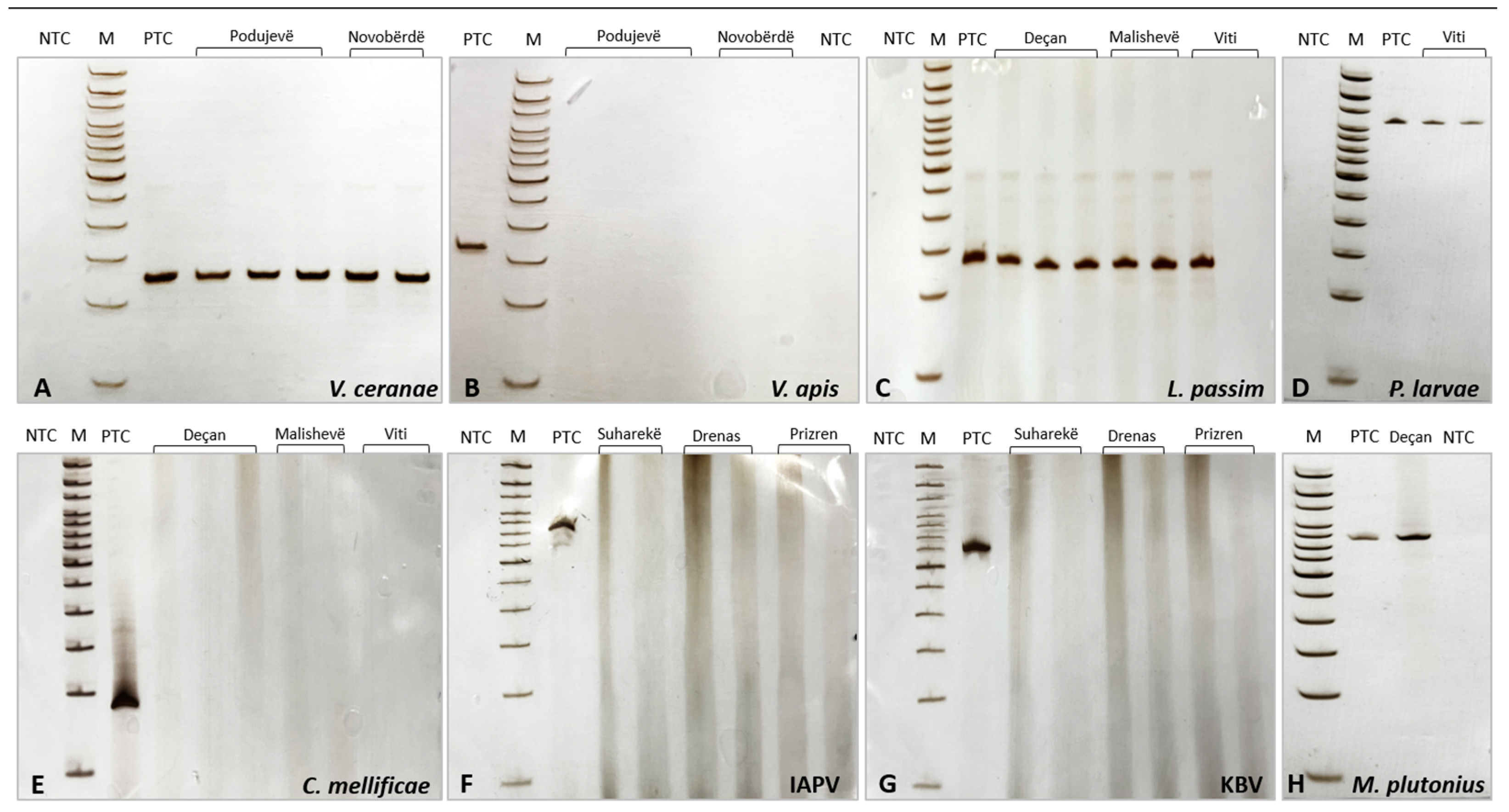
2.3. Vairimorpha spp. Spore Detection and Vairimorpha apis/Vairimorpha ceranae Species Identification

The abdomens of 30 honey bees were separated and macerated in about 2.5 mL of distilled water using a mortar and pestle and successively 27.5 mL of distilled water was added to a final volume of 1 mL per bee. A drop of this suspension was examined under a light microscope at 400× magnification to evaluate the presence of Vairimorpha spp. spores. Vairimorpha spp.-positive samples were analysed for V. apis and V. ceranae species identification after DNA extraction from 1 mL homogenate using the QIAamp® DNA Mini Kit (Qiagen, Hilden, Germany) according to the manufacturer’s instructions, with a lysozyme pre-incubation step. The yield and purity (260/280 and 260/230 nm absorbance ratios) of DNA were determined using the Nanodrop™ OneC (Thermo Fisher Scientific, Waltham, MA, USA) spectrophotometer. DNA was stored at −20 °C until use. Negative controls (negative process control—NPC: water for molecular biology applications instead of the sample) were included in each extraction session.
For V. apis and V. ceranae identification, two different sets of primers described by Martín-Hernández et al. [46] were used and PCR analysis was carried out as previously described by Bordin et al. [47]. Negative (negative template control—NTC: water for molecular biology applications instead of the DNA template) and positive (positive template control—PTC: DNA from V. apis- or V. ceranae-positive samples) controls were included in each PCR.

2.4. Lotmaria passim and Crithidia mellificae Detection

For L. passim and C. mellificae detection, two pairs of primers described by Bartolomé et al. [48] were used and PCR analysis was carried out as previously described by Bordin et al. [47]. Negative (NTC: water for molecular biology applications instead of the DNA template) and positive (PTC: DNA from L. passim- or C. mellificae-positive samples) controls were included in each PCR.

2.5. Honey Bee Viruses

Thirteen honey bee (adult and/or larvae and/or pupae) and eight Varroa mite samples were analysed for seven honey bee viruses relevant with respect to colony health status: ABPV, CBPV, DWV, SBV, BQCV, KBV, and IAPV.
For each sample, a pool of five specimens per development stage (adult, larvae, or pupae) or a pool of up to ten specimens of Varroa mites was homogenized by Tissue Lyser II (Qiagen, Hilden, Germany) (2 cycles, 1 min each, at 30 Hz) in the presence of a 5 mm stainless steel bead. Total RNA extraction was performed using the NucleoSpin RNA kit (Macherey Nagel GmbH & Co. KG, Dueren, Germany) according to the manufacturer’s instructions. The yield and purity of RNA (260/280 and 260/230 nm absorbance ratios) were assessed with a Nanodrop™ OneC spectrophotometer. RNA was stored at −80 °C until use. Negative controls (NPC: buffer RA1 from the extraction kit instead of the homogenized sample) were included in each extraction session.
To detect viral RNA, real-time RT-PCR (rRT-PCR), or end-point RT-PCR were performed as described by Bordin et al. [47]. To detect the presence of DWV the molecular protocol described by Martinello et al. [49] was used. Negative (NTC: water for molecular biology applications instead of the RNA template) and positive (PTC: viral RNA from positive sample) controls were included in each PCR.

2.6. Paenibacillus larvae (AFB) Detection by Field Tests, Isolation, and PCR

A first visual inspection of brood combs was carried out to search for symptoms of AFB: irregular and patchy brood pattern, unpleasant odour, perforated and sunken cell caps, dark-coloured larvae, ropy larvae, and stickiness. Larvae with AFB symptoms were then analysed with the AFB lateral flow test—Diagnostic Test Kit (Vita Europe, London, UK) according to the manufacturer instructions (https://www.vita-europe.com/beehealth/products/afbdiagnostic-test-kit/) (accessed on 20 January 2024). The samples were stored at −20 °C for further analyses.
The presence of P. larvae in the eight samples of suspected larvae was also determined by the culture method and PCR.
The isolation of P. larvae and the following colony identification (catalase test and Gram staining) was performed on Columbia sheep-blood agar (CSA), supplemented with nalidixic acid and pipemidic acid, according to the WOAH Manual of Diagnostic Tests and Vaccines for Terrestrial Animals, chapter 3.2.2 (2023) [50].
To assess the presence of P. larvae by the PCR method, DNA extraction was performed from a larvae homogenate using the QIAamp® DNA Mini Kit (Qiagen, Hilden, Germany) according to the manufacturer’s instructions, with a lysozyme pre-incubation step. The yield and purity (260/280 and 260/230 nm absorbance ratios) of DNA were determined using a Nanodrop™ OneC spectrophotometer. DNA was stored at −20 °C until use. Negative controls (NPC: water for molecular biology applications instead of the sample) were included in each extraction session.
For P. larvae detection, the primers described by Dobbelaere et al. [51], targeting a 1096 bp region of the 16S rRNA gene, were used. PCR was carried out on a Veriti™ 96-Well Thermal Cycler (Applied Biosystems™, Waltham, MA, USA), using the AmpliTaq™ Gold kit (Applied Biosystems™, Waltham, MA, USA). In a final volume of 50 µL, 200 ng of DNA was amplified using a final concentration of 2 mM MgCl2, 1 µM for each primer, 0.2 mM of dNTPs, and 1 U AmpliTaq Gold DNA polymerase. The thermal cycling profile consisted of an initial activation step at 95 °C for 10 min followed by 35 cycles at 93 °C for 60 s, 55 °C for 30 s, 72 °C for 60 s, and a final elongation step at 72 °C for 5 min. Negative (NTC: water for molecular biology applications instead of the DNA template) and positive (PTC: P. larvae DNA) controls were included in each run of PCR. The presence and the size of amplification products (1096 bp) were evaluated by electrophoresis in 7% acrylamide gel after silver staining or capillary electrophoresis on a LabChip® GX Touch HT Nucleic Acid Analyzer (Perkin Elmer, Waltham, MA, USA).

2.7. Genotyping of Paenibacillus larvae Isolates

For P. larvae genotyping, DNA was extracted from catalase-negative and Gram-positive colonies using the QIAamp® DNA Mini Kit (Qiagen, Hilden, Germany) according to the manufacturer’s protocol for Gram-positive bacteria, and the DNA was eluted in a final volume of 120 μL. The yield and purity of DNA were determined using the Nanodrop™ OneC spectrophotometer and the nucleic acid was stored at −20 °C until use. ERIC-typing PCR was performed using the primers published by Genersch et al. [52]. The PCR was carried out on a Veriti™ 96-Well Thermal Cycler (Applied Biosystems™, Waltham, MA, USA) in a final volume of 50 μL using the AmpliTaq™ Gold kit and containing a final concentration of 2.5 mM MgCl2, 800 μM of dNTP mix, 1 μM of each primer, 2.5 U AmpliTaq Gold DNA polymerase, and 50–100 ng of DNA. The thermal cycling amplification profile consisted of an activation step at 95 °C for 10 min, followed by 50 cycles at 94 °C for 1 min, 53 °C for 1 min, 72 °C for 2.5 min, and a final elongation step at 72 °C for 10 min. Negative (NTC: water for molecular biology applications instead of the DNA template) and positive (PTC: P. larvae DNA) controls for ERIC I, II, and IV genotypes were included in each PCR. After amplification, about 16 µL of the PCR reaction was electrophoresed in a 1.7% SeaKem® Gold Agarose gel (Lonza Rockland, Inc., Rockland, ME, USA) and the PCR products were visualized after ethidium bromide staining on a UV trans-illuminator.

2.8. Melissococcus plutonius Detection by Field Tests and PCR
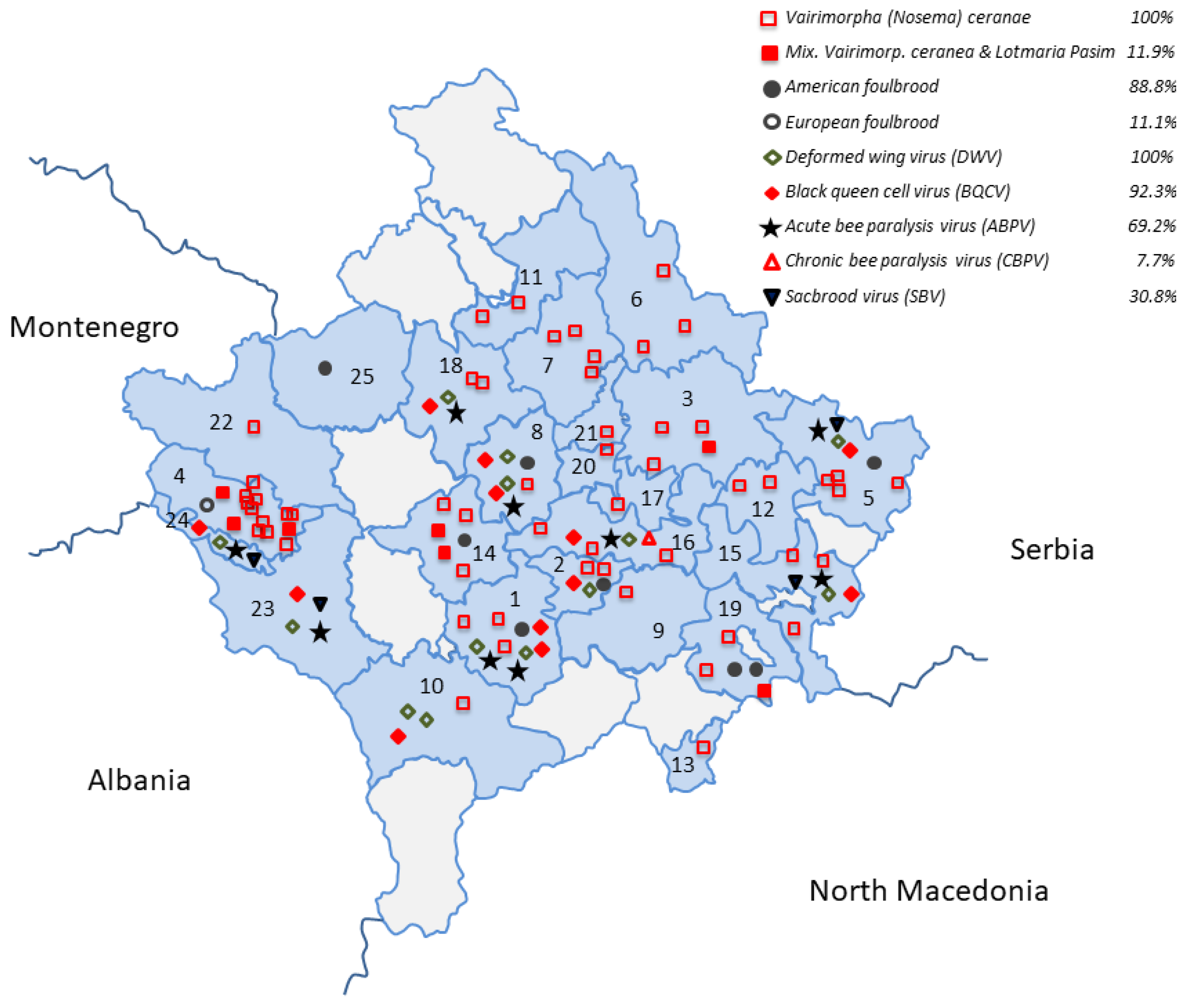
Brood combs were examined for EFB signs such as dead, flaccid discoloured larvae in uncapped cells or changes in their colour (from pearly white to yellow, yellowish to brown), and the presence of dry and dark brown scales that can easily be removed from the cells.
Larvae with EFB symptoms were tested with the EFB lateral flow test—Diagnostic Test Kit (Vita Europe, London, UK) according to the manufacturer’s instructions (https://www.vita-europe.com/beehealth/products/efb-diagnostic-test-kit/) (accessed on 20 January 2024). After analysis, all samples were stored at −20 °C until further examination.
A pool of larvae was analysed to detect the presence of M. plutonius by the PCR method, using the primers described by Govan et al. [53], targeting an 832 bp region of the 16S rRNA gene. DNA extraction was performed using the QIAamp® DNA Mini Kit (Qiagen, Hilden, Germany) according to the manufacturer’s instructions (protocol for bacteria—isolation of genomic DNA from Gram-positive bacteria). The yield and purity of DNA were determined using the Nanodrop™ OneC spectrophotometer and the nucleic acid was stored at −20 °C until use. Negative controls (NPC: water for molecular biology applications instead of the sample) were included in the extraction session.
The PCR was carried out as described above in a final volume of 50 μL containing a final concentration of 2.5 mM MgCl2, 0.2 mM dNTP mix, 0.5 μM of each primer, 2 U AmpliTaq Gold DNA polymerase, and 50–100 ng of DNA. The thermal cycling amplification profile consisted of an activation step at 95 °C for 10 min, followed by 35 cycles at 94 °C for 30 s, 60 °C for 30 s, 72 °C for 30 s, and a final elongation step at 72 °C for 10 min. Negative (NTC: water for molecular biology applications instead of the DNA template) and positive (PTC: M. plutonius DNA) controls were included in each PCR. The presence and the size of the amplification product (832 bp) were evaluated by electrophoresis in 7% acrylamide gel after silver staining.

3. Results

The results from 89 investigated apiaries are shown in Table 1 and Figure 1 and Figure 2.

3.1. Group 1—Vairimorpha spp. and Trypanosomatids L. passim and C. mellificae

Of 59 investigated apiaries from 22 municipalities, V. ceranae infections were found in all apiaries (100%), whereas all were negative for V. apis. The trypanosomatid L. passim was detected in seven apiaries (11.9%) from four municipalities, while C. mellificae was never detected (Table 1, Figure 1 and Figure 3A–C,E).

3.2. Group 2—Honey Bee Viruses (ABPV, CBPV, DWV, SBV, BQCV, KBV, and IAPV)

The following honey bee viruses were detected in samples (adults/larvae/pupae) from the 13 apiaries in the 10 municipalities: DWV in all thirteen apiaries (100%), BQCV in twelve apiaries (92.3%), ABPV in nine apiaries (69.2%), SBV in four apiaries (30.8%), and CBPV in one apiary only (7.7%). IAPV and KBV were not detected in any apiary (Figure 3F,G). Viruses were found in all the developmental stages of honey bees and in particular, adults specimens tested positive for DWV, BQCV, ABPV, CBPV, and SBV; pupae for DWV, BQCV, ABPV, and SBV; and larvae for DWV, BQCV, and ABPV (Table 1 and Figure 1).
In the eight Varroa samples derived from eight apiaries from six municipalities, four out of seven viruses (ABPV, CBPV, DWV, BQCV) and different co-infections were detected. In particular, only the sample from one municipality (Shtime) was found to be co-infected with all four viruses, and four Varroa mite samples from three municipalities (Gjilan, Lipjan, Suharekë) tested positive for three viruses (ABPV, DWV, BQCV), whereas two samples from Prizren and one sample from Drenas tested positive for ABPV and DWV, and for DWV and BQCV, respectively (Table 1 and Figure 2).

3.3. Group 3—Paenibacillus larvae and Melissococcus plutonius Detection

In the third group, all 8 larvae samples from the brood comb (1, Suharekë; 1, Kamenicë; 1, Drenas; 1, Malishevë; 1, Shtime; 2, Viti; 1, Istog) investigated for the presence of the causative agent of AFB tested positive for P. larvae on an AFB lateral flow test—Diagnostic Test Kit (Vita Europe, London, UK), PCR, and the culture method (Table 1, Figure 1 and Figure 3D). All P. larvae isolates were identified as the ERIC I genotype. The only sample from the Deҫan municipality analysed for M. plutonius presence was positive for EFB on a lateral flow test—Diagnostic Test Kit (Vita Europe, London, UK) and PCR (Table 1, Figure 1 and Figure 3H).

4. Discussion

In the last decade, a reduction in honey bee colonies has been frequently reported around the world [2,54]. Among the causes for mortality and global threat for honey bee colonies are several pathogens and parasites [55].
Our study was based on passive surveillance on weak honey bee colonies aiming to determine the occurrence and distribution of known and emergent honey bee pathogens in the Republic of Kosovo. An investigation based on passive surveillance was already carried out by Hulaj et al. [45] focusing on AFB.
In all the 59 apiaries tested for Vairimorpha spp., a high percentage of infection (100%) with V. ceranae was found, while no samples tested positive for V. apis. These data confirm what has already been reported in Italy [47,56] and in the neighbouring countries to the Republic of Kosovo, such as Serbia, Croatia, Bosnia–Herzegovina, Montenegro, North Macedonia, and Bulgaria, where V. ceranae dominates in microsporidia infections in honey bees [57,58,59,60,61]. Furthermore, this finding confirmed the previously reported higher diffusion of V. ceranae in other European countries and worldwide [62,63,64,65]. The presence of V. ceranae infection could reduce honey bee lifespan [7], significantly increase honey bee worker mortality, and could be one stressor responsible for elevated colony losses [66].
In the Republic of Kosovo, L. passim was only detected in seven of fifty-nine apiaries (11.9%) from four municipalities during the passive surveillance. In the Veneto region (northern Italy), L. passim was detected in almost all of the apiaries analysed with an overall positivity rate of 48.8% in 2020, while an increase was observed in 2021 with an overall value of 62.2% [47]. In Serbia, L. passim was detected during 2007–2015 every year. In the Republic of Kosovo, C. mellificae was never reported, as well as in neighbouring Serbia [67].
In our study, five honey bee viruses were found (ABPV, CBPV, DWV, BQCV, SBV) and the most frequent were DWV and BQCV with 100% and 92.3% prevalence in the 13 apiaries analysed, respectively. ABPV was detected in nine apiaries (69.2%), SBV in four apiaries (30.8%) from four municipalities, and CBPV in only one apiary (7.7%). No positivity for IAPV and KBV was observed in the investigated apiaries.
When comparing our research on honey bee viruses with other countries, we found that the same viruses (ABPV, CBPV, DWV, BQCV, and SBV) have also been detected in neighbouring Serbia, Austria, Greece, and other countries [68,69,70,71], whereas KBV was also detected in France [20]. The presence of DWV is often associated with V. destructor infestation, and the role of this mite in viral transmission has already been experimentally demonstrated [72]. It is known that DWV, vectored by the Varroa mite, adversely affects humoral and cellular immune responses and promotes the reproduction of this parasitic mite [27].
Both BQCV and SBV are not known to be transmitted by V. destructor [73]. However, a significantly higher prevalence of BQCV and SBV in colonies infested with V. destructor mites has been detected, hypothesizing the role of V. destructor in their transmission [74]. In our study, considering not only honey bee but also Varroa mite samples, the virus frequency was increased for ABPV (from 69.2% to 84.6%) and CBPV (from 7.7% to 15.4%).
Furthermore, larvae collected from the brood comb tested for AFB were positive for the P. larvae ERIC I genotype in eight out of nine apiaries. This finding is in line with the results of the passive survey carried out during 2007–2019 that revealed a wide diffusion of AFB of the ERIC I and II genotypes in the country [45], thus suggesting the necessity of applying active disease surveillance strategies. The single positivity detected for EFB suggests that the disease is present in the country but further investigations are needed to understand its true distribution and possible impact on honey bee colonies.

5. Conclusions

Although there have been clinical cases of suspected emergent honey bee diseases in the Republic of Kosovo, they have never been investigated; this paper represents the first molecular detection of honey bee viruses (ABPV, CBPV, DWV, BQCV, SBV), the microsporidian V. ceranae, the trypanosomatid L. passim, and the bacterium M. plutonius. Furthermore, we also confirmed the presence of P. larvae in the apiaries of the Republic of Kosovo.
We hope that the results presented herein could open the path to further investigations in order to consolidate and update the knowledge on the health status of honey bee colonies in the Republic of Kosovo.

Supplementary Materials

The following supporting information can be downloaded at https://www.mdpi.com/article/10.3390/app14030987/s1, Figure S1: Map with the involved municipalities and the type of beehive matrices that have been sampled.

Author Contributions

Conceptualization, B.H., I.G., X.M. and A.C.; sample collection, B.H., X.M. and A.C.; sample analysis, B.H., X.M., A.G., F.B., L.Z., M.C. and R.C.; result interpretation, B.H., A.G., F.B. and L.Z.; writing—original draft preparation, B.H., I.G., X.M. and A.C.; writing—review and editing, F.M., A.G. and F.B. All authors have read and agreed to the published version of the manuscript.

Funding

This research received no external funding.

Institutional Review Board Statement

Not applicable.

Informed Consent Statement

Not applicable.

Data Availability Statement

The data presented in this study are available on request from the corresponding author. The data are not publicly available due to restrictions concerning privacy.

Conflicts of Interest

The authors declare no conflicts of interest.

References

  1. Klein, A.-M.; Vaissiere, B.E.; Cane, J.H.; Steffan-Dewenter, I.; Cunningham, S.A.; Kremen, C.; Tscharntke, T. Importance of pollinators in changing landscapes for world crops. Proc. R. Soc. B 2007, 274, 303–313. [Google Scholar] [CrossRef] [PubMed]
  2. Hristov, P.; Shumkova, R.; Palova, N.; Neov, B. Factors Associated with Honey Bee Colony Losses: A Mini-Review. Vet. Sci. 2020, 7, 166. [Google Scholar] [CrossRef] [PubMed]
  3. Tokarev, Y.S.; Huang, W.-F.; Solter, L.F.; Malysh, J.M.; Becnel, J.J.; Vossbrinck, C.R. A formal redefinition of the genera Nosema and Vairimorpha (Microsporidia: Nosematidae) and reassignment of species based on molecular phylogenetics. J. Invert. Pathol. 2020, 169, 107279. [Google Scholar] [CrossRef] [PubMed]
  4. Chen, Y.; Evans, J.; Zhou, L.; Boncristiani, H.; Kimura, K.; Xiao, T.; Litkowski, A.; Pettis, J.S. Asymmetrical coexistence of Nosema ceranae and Nosema apis in honey bees. J. Invert. Pathol. 2009, 101, 204–209. [Google Scholar] [CrossRef] [PubMed]
  5. Galajda, R.; Valenčáková, A.; Sučik, M.; Kandráčová, P. Nosema Disease of European Honey Bees. J. Fungi 2021, 7, 714. [Google Scholar] [CrossRef] [PubMed]
  6. Botías, C.; Martín-Hernández, R.; Barrios, L.; Meana, A.; Higes, M. Nosema spp. infection and its negative effects on honey bees (Apis mellifera iberiensis) at the colony level. Vet. Res. 2013, 44, 25. [Google Scholar] [CrossRef] [PubMed]
  7. Higes, M.; Garcia-Palencia, P.; Martin-Hernandez, R.; Meana, A. Experimental infection of Apis mellifera honey bees with Nosema ceranae (Microsporidia). J. Invert. Pathol. 2007, 94, 211–217. [Google Scholar] [CrossRef] [PubMed]
  8. Paxton, R.J.; Klee, J.; Korpela, S.; Fries, I. Nosema ceranae has infected Apis mellifera in Europe since at least 1998 and may be more virulent than Nosema apis. Apidologie 2007, 38, 558–565. [Google Scholar] [CrossRef]
  9. Mayack, C.; Naug, D. Energetic stress in the honey bee Apis mellifera from Nosema ceranae infection. J. Invert. Pathol. 2009, 100, 185–188. [Google Scholar] [CrossRef]
  10. Martín-Hernández, R.; Botías, C.; Barrios, L.; Martínez-Salvador, A.; Meana, A.; Mayack, C.; Higes, M. Comparison of the energetic stress associated with experimental Nosema ceranae and Nosema apis infection of honey bees (Apis mellifera). Parasitol. Res. 2011, 109, 605–612. [Google Scholar] [CrossRef]
  11. Schwarz, R.S.; Bauchan, G.R.; Murphy, C.A.; Ravoet, J.; de Graaf, D.C.; Evans, J.D. Characterization of Two Species of Trypanosomatidae from the Honey Bee Apis mellifera: Crithidia mellificae Langridge and McGhee, and Lotmaria passim n. gen., n. sp. J. Eukaryot. Microbiol. 2015, 62, 567–583. [Google Scholar] [CrossRef]
  12. Arismendi, N.; Caro, S.; Castro, M.P.; Vargas, M.; Riveros, G.; Venegas, T. Impact of Mixed Infections of Gut Parasites Lotmaria passim and Nosema ceranae on the Lifespan and Immune-related Biomarkers in Apis mellifera. Insects 2020, 11, 420. [Google Scholar] [CrossRef] [PubMed]
  13. Runckel, C.; Flenniken, M.L.; Engel, J.C.; Ruby, J.G.; Ganem, D.; Andino, R.; De Risi, J.L. Temporal Analysis of the Honey Bee Microbiome Reveals Four Novel Viruses and Seasonal Prevalence of Known Viruses, Nosema, and Crithidia. PLoS ONE 2011, 6, e20656. [Google Scholar] [CrossRef]
  14. Ravoet, J.; Maharramov, J.; Meeus, I.; De Smet, L.; Wenseleers, T.; Smagghe, G.; de Graaf, D.C. Comprehensive Bee Pathogen Screening in Belgium Reveals Crithidia mellificae as a New Contributory Factor to Winter Mortality. PLoS ONE 2013, 8, e72443. [Google Scholar] [CrossRef]
  15. Schwarz, R.S.; Evans, J.D. Single and mixed-species trypanosome and microsporidia infections elicit distinct, ephemeral cellular and humoral immune responses in honey bees. Dev. Comp. Immunol. 2013, 40, 300–310. [Google Scholar] [CrossRef]
  16. Strobl, V.; Yañez, O.; Straub, L.; Albrecht, M.; Neumann, P. Trypanosomatid parasites infecting managed honey bees and wild solitary bees. Int. J. Parasitol. 2019, 49, 605–613. [Google Scholar] [CrossRef] [PubMed]
  17. Ball, B.V.; Allen, M.F. The prevalence of pathogens in honey bee colonies infested with the parasitic mite Varroa jacobsoni. Ann. Appl. Biol. 1988, 113, 237–244. [Google Scholar] [CrossRef]
  18. Allen, M.; Ball, B. The incidence and world distribution of the honey bee viruses. Bee World 1996, 77, 141–162. [Google Scholar] [CrossRef]
  19. Martin, S.J. The role of Varroa and viral pathogens in the collapse of honeybee colonies: A modeling approach. J. Appl. Ecol. 2001, 38, 1082–1093. [Google Scholar] [CrossRef]
  20. Tentcheva, D.; Gauthier, L.; Zappulla, N.; Dainat, B.; Cousserans, F.; Colin, M.E.; Bergoin, M. Prevalence and seasonal variations of six bee viruses in Apis mellifera L. and Varroa destructor mite populations in France. Appl. Environ. Microbiol. 2004, 70, 7185–7191. [Google Scholar] [CrossRef]
  21. Shen, M.; Cui, L.; Ostiguy, N.; Cox-Foster, D. Intricate transmission routes and interactions between picorna-like viruses (Kashmir bee virus and sacbrood virus) with the honey bee host and the parasitic Varroa mite. J. Gen. Virol. 2005, 86, 2281–2289. [Google Scholar] [CrossRef]
  22. Chantawannakul, P.; Ward, L.; Boonham, N.; Brown, M. A scientific note on the detection of honey bee viruses using real-time PCR (TaqMan) in Varroa mites collected from a Thai honey bee (Apis mellifera) apiary. J. Invert. Pathol. 2006, 91, 69–73. [Google Scholar] [CrossRef] [PubMed]
  23. Celle, O.; Blanchard, P.; Olivier, V.; Schurr, F.; Cougoule, N.; Faucon, J.-P.; Ribiere, M. Detection of Chronic bee paralysis virus (CBPV) genome and its replicative RNA form in various hosts and possible ways of spread. Virus Res. 2008, 133, 280–284. [Google Scholar] [CrossRef] [PubMed]
  24. Francis, R.M.; Nielsen, S.L.; Kryger, P. Varroa-virus interaction in collapsing honey bee colonies. PLoS ONE 2013, 8, e57540. [Google Scholar] [CrossRef] [PubMed]
  25. Mondet, F.; de Miranda, J.R.; Kretzschmar, A.; Le Conte, Y.; Mercer, A.R. On the front line: Quantitative virus dynamics in honeybee (Apis mellifera L.) colonies along a new expansion front of the parasite Varroa destructor. PLoS Pathog. 2014, 10, e1004323. [Google Scholar] [CrossRef] [PubMed]
  26. Chen, Y.P.; Pettis, J.S.; Corona, M.; Chen, W.P.; Li, C.J.; Spivak, M.; Visscher, P.K.; DeGrandi-Hoffman, G.; Boncristiani, H.; Zhao, Y.; et al. Israeli acute paralysis virus: Epidemiology, pathogenesis and implications for honey bee health. PLoS Pathog. 2014, 10, e1004261. [Google Scholar] [CrossRef] [PubMed]
  27. Di Prisco, G.; Annoscia, D.; Margiotta, M.; Ferrara, R.; Varricchio, P.; Zanni, V.; Caprio, E.; Nazzi, F.; Pennacchio, F. A mutualistic symbiosis between a parasitic mite and a pathogenic virus undermines honey bee immunity and health. Proc. Nat. Acad. Sci. USA 2016, 113, 3203–3208. [Google Scholar] [CrossRef]
  28. Valles, S.M.; Chen, Y.P.; Firth, A.E.; Guerin, D.M.A.; Hashimoto, Y.; Herrero, S.; de Miranda, J.R.; Ryabov, E.; ICTV Report Consortium. ICTV virus taxonomy profile: Iflaviridae. J. Gen. Virol. 2017, 98, 527–528. [Google Scholar] [CrossRef]
  29. McMenamin, A.J.; Flenniken, M.L. Recently identified bee viruses and their impact on bee pollinators. Curr. Opin. Insect. Sci. 2018, 26, 120–129. [Google Scholar] [CrossRef]
  30. Bailey, L.; Gibbs, A.J.; Woods, R.D. Two viruses from adult honey bees (Apis mellifera Linnaeus). Virology 1963, 21, 390–395. [Google Scholar] [CrossRef]
  31. Maori, E.; Lavi, S.; Mozes-Koch, R.; Gantman, Y.; Peretz, Y.; Edelbaum, O.; Tanne, E.; Sela, I. Isolation and characterization of Israeli acute paralysis virus, a dicistrovirus affecting honey bees in Israel: Evidence for diversity due to intra- and inter-species. J. Gen. Virol. 2007, 88, 3428–3438. [Google Scholar] [CrossRef] [PubMed]
  32. Ribière, M.; Ball, B.; Aubert, M. Natural history and geographical distribution of honey bee viruses. In Virology and the Honey Bee; Aubert, M., Ball, B., Fries, I., Moritz, R., Milani, N., Bernardinelli, I., Eds.; European Communities: Luxembourg, 2008; pp. 15–84. [Google Scholar]
  33. Anderson, D.L.; Gibbs, A.J. Inapparent virus infections and their interactions in pupae of the honey bee (Apis mellifera L.) in Australia. J. Gen. Virol. 1988, 69, 1617–1625. [Google Scholar] [CrossRef]
  34. Dall, D.J. Inapparent infection of honey bee pupae by Kashmir and Sacbrood bee viruses in Australia. Ann. Appl. Biol. 1985, 106, 461–468. [Google Scholar] [CrossRef]
  35. De Miranda, J.; Cordoni, G.; Budge, G. The Acute bee paralysis virus–Kashmir bee virus–Israeli acute paralysis virus complex. J. Invert. Pathol. 2010, 103 (Suppl. S1), S30–S47. [Google Scholar] [CrossRef] [PubMed]
  36. Bailey, L. Recent research on honey bee viruses. Bee World 1975, 56, 55–64. [Google Scholar] [CrossRef]
  37. Bailey, L.; Gibbs, A.J.; Woods, R.D. Sacbrood virus of the larval honey bee (Apis mellifera Linnaeus). Virology 1964, 23, 425–429. [Google Scholar] [CrossRef] [PubMed]
  38. Chen, Y.P.; Siede, R. Honey bee viruses. Adv. Virus Res. 2007, 70, 33–80. [Google Scholar] [CrossRef]
  39. McMahon, D.P.; Natsopoulou, M.E.; Doublet, V.; Fürst, M.; Weging, S.; Brown, M.J.F.; Gogol-Döring, A.; Paxton, R.J. Elevated virulence of an emerging viral genotype as a driver of honeybee loss. Proc. Biol. Sci. 2016, 283, 20160811. [Google Scholar] [CrossRef]
  40. Paxton, R.J.; Schäfer, M.O.; Nazzi, F.; Zanni, V.; Annoscia, D.; Marroni, F.; Bigot, D.; Laws-Quinn, E.R.; Panziera, D.; Jenkins, C.; et al. Epidemiology of a major honey bee pathogen, deformed wing virus: Potential worldwide replacement of genotype A by genotype B. Int. J. Parasitol. Parasites Wildl. 2022, 18, 157–171. [Google Scholar] [CrossRef]
  41. Bailey, L. Ætiology of European foulbrood; a disease of the larval honey-bee. Nature 1956, 178, 1130. [Google Scholar] [CrossRef]
  42. Bailey, L.; Ball, B.V. Honey Bee Pathology; Academic Press: Cambridge, MA, USA, 1991. [Google Scholar]
  43. Ellis, J.D.; Munn, P.A. The worldwide health status of honey bees. Bee World 2005, 86, 88–101. [Google Scholar] [CrossRef]
  44. Genersch, E. Honey bee pathology: Current threats to honey bees and beekeeping. Appl. Microbiol. Biot. 2010, 87, 87–97. [Google Scholar] [CrossRef] [PubMed]
  45. Hulaj, B.; Goga, I.; Cana, A.; Merovci, X.; Rossi, F.; Crudele, S.; Ricchiuti, L.; Mutinelli, F. Passive surveillance of American foulbrood in the Republic of Kosovo: Geographic distribution and genotype characterization. J. Apicul. Res. 2023, 62, 320–325. [Google Scholar] [CrossRef]
  46. Martín-Hernández, R.; Meana, A.; Prieto, L.; Salvador, A.M.; Garrido-Bailón, E.; Higes, M. Outcome of colonization of Apis mellifera by Nosema ceranae. Appl. Environ. Microbiol. 2007, 73, 6331–6338. [Google Scholar] [CrossRef] [PubMed]
  47. Bordin, F.; Zulian, L.; Granato, A.; Caldon, M.; Colamonico, R.; Toson, M.; Trevisan, L.; Biasion, L.; Mutinelli, F. Presence of Known and Emerging Honey Bee Pathogens in Apiaries of Veneto Region (Northeast of Italy) during Spring 2020 and 2021. Appl. Sci. 2022, 12, 2134. [Google Scholar] [CrossRef]
  48. Bartolomé, C.; Buendía-Abad, M.; Benito, M.; De la Rua, P.; Ornosa, C.; Martín-Hernández, R.; Higes, M.; Maside, X. A new multiplex PCR protocol to detect mixed trypanosomatid infections in species of Apis and Bombus. J. Invert. Pathol. 2018, 154, 37–41. [Google Scholar] [CrossRef] [PubMed]
  49. Martinello, M.; Baratto, C.; Manzinello, C.; Piva, E.; Borin, A.; Toson, M.; Granato, A.; Boniotti, M.B.; Gallina, A.; Mutinelli, F. Honey bee spring mortality in the north-east of Italy: Detection of pesticides and viruses in dead honey bees and other matrices. J. Apicult. Res. 2017, 56, 239–254. [Google Scholar] [CrossRef]
  50. World Organisation for Animal Health (WOAH). Manual of Diagnostic Tests and Vaccines for Terrestrial Animals. 2023. American Foulbrood of Honey Bees (Infection of Honey Bees with Paenibacillus larvae), Chapter 3.2.2 (Version Adopted in 2023). Available online: https://www.woah.org/fileadmin/Home/eng/Health_standards/tahm/3.02.02_AMERICAN_FOULBROOD.pdf (accessed on 18 May 2023).
  51. Dobbelaere, W.; de Graaf, D.C.; Peeters, J.E. Development of a fast and reliable diagnostic method for American foulbrood disease (Paenibacillus larvae subsp. larvae) using a 16S rRNA gene based PCR. Apidologie 2001, 32, 363–370. [Google Scholar] [CrossRef]
  52. Genersch, E.; Forsgren, E.; Pentikäinen, J.; Ashiralieva, A.; Rauch, S.; Kilwinski, J.; Fries, I. Reclassification of Paenibacillus larvae subsp. pulvifaciens and Paenibacillus larvae subsp. larvae as Paenibacillus larvae without subspecies differentiation. Int. J. Syst. Evol. Microbiol. 2006, 56, 501–511. [Google Scholar] [CrossRef]
  53. Govan, V.A.; Brozel, V.; Allsopp, M.H.; Davison, S. A PCR detection method for rapid identification of Melissococcus pluton in honey bee larvae. Appl. Environ. Microbiol. 1998, 64, 1983–1985. [Google Scholar] [CrossRef]
  54. Genersch, E.; Von Der Ohe, W.; Kaatz, H.; Schroeder, A.; Otten, C.; Büchler, R.; Berg, S.; Ritter, W.; Mühlen, W.; Gisder, S. The German Bee Monitoring Project: A Long Term Study to Understand Periodically High Winter Losses of Honey Bee Colonies. Apidologie 2010, 41, 332–352. [Google Scholar] [CrossRef]
  55. Genersch, E.; Aubert, M. Emerging and Re-Emerging Viruses of the Honey Bee (Apis mellifera L.). Vet. Res. 2010, 41, 54. [Google Scholar] [CrossRef] [PubMed]
  56. Papini, R.; Mancianti, F.; Canovai, R.; Cosci, F.; Rocchigiani, G.; Benelli, G.; Canale, A. Prevalence of the microsporidian Nosema ceranae in honeybee (Apis mellifera) apiaries in Central Italy. Saudi J. Biol. Sci. 2017, 24, 979–982. [Google Scholar] [CrossRef] [PubMed]
  57. Tlak Gajger, I.; Vugrek, O.; Grilec, D.; Petrinec, Z. Prevalence and distribution of Nosema ceranae in Croatian honey bee colonies. Vet. Med. 2010, 55, 457–462. [Google Scholar] [CrossRef]
  58. Stevanovic, J.; Stanimirovic, Z.; Genersch, E.; Kovacevic, S.R.; Ljubenkovic, J.; Radakovic, M.; Aleksic, N. Dominance of Nosema ceranae in honey bees in the Balkan countries in the absence of symptoms of colony collapse disorder. Apidologie 2011, 42, 49–58. [Google Scholar] [CrossRef]
  59. Stevanovic, J.; Simeunovic, P.; Gajic, B.; Lakic, N.; Radovic, D.; Fries, I.; Stanimirovic, Z. Characteristics of Nosema ceranae infection in Serbian honey bee colonies. Apidologie 2013, 44, 522–536. [Google Scholar] [CrossRef]
  60. Shumkova, R.; Georgieva, A.; Radoslavov, G.; Sirakova, D.; Dzhebir, G.; Neov, B.; Bouga, M.; Hristov, P. The first report of the prevalence of Nosema ceranae in Bulgaria. PeerJ 2018, 6, e4252. [Google Scholar] [CrossRef]
  61. Matović, K.; Vidanović, D.; Manić, M.; Stojiljković, M.; Radojićić, S.; Debeljak, Z.; Šekler, M.; Ćirić, J. Twenty-five-year study of Nosema spp. in honey bees (Apis mellifera) in Serbia. Saudi J. Biol. Sci. 2020, 27, 518–523. [Google Scholar] [CrossRef]
  62. Fries, I. Nosema ceranae in European honey bees (Apis mellifera). J. Invert. Pathol. 2010, 103 (Suppl. S1), 73. [Google Scholar] [CrossRef]
  63. Bromenshenk, J.J.; Henderson, C.B.; Wick, C.H.; Stanford, M.F.; Zulich, A.W.; Jabbour, R.E.; Deshpande, S.V.; McCubbin, P.E.; Seccomb, R.A.; Welch, P.M.; et al. Iridovirus and microsporidian linked to honey bee colony decline. PLoS ONE 2010, 5, e13181. [Google Scholar] [CrossRef]
  64. Alaux, C.; Folschweiller, M.; McDonnell, C.; Beslay, D.; Cousin, M.; Dussaubat, C.; Brunet, J.L.; Le Conte, Y. Pathological effects of the microsporidium Nosema ceranae on honey bee queen physiology (Apis mellifera). J. Invert. Pathol. 2011, 106, 380–385. [Google Scholar] [CrossRef] [PubMed]
  65. Grupe, A.C.; Quandt, C.A. A Growing Pandemic: A Review of Nosema Parasites in Globally Distributed Domesticated and Native Bees. PLoS Pathog. 2020, 16, e1008580. [Google Scholar] [CrossRef] [PubMed]
  66. Williams, G.R.; Shutler, D.; Burgher-MacLellan, K.L.; Rogers, R.E.L. Infra-Population and community dynamics of the parasites Nosema apis and Nosema ceranae, and consequences for honey bee (Apis mellifera) hosts. PLoS ONE 2014, 9, e99465. [Google Scholar] [CrossRef] [PubMed]
  67. Stevanovic, J.; Schwarz, R.S.; Vejnovic, B.; Evans, J.D.; Irwin, R.E.; Glavinic, U.; Stanimirovic, Z. Species-specific diagnostics of Apis mellifera trypanosomatids: A nine-year survey (2007–2015) for trypanosomatids and microsporidians in Serbian honey bees. J. Invert. Pathol. 2016, 139, 6–11. [Google Scholar] [CrossRef] [PubMed]
  68. Berényi, O.; Bakonyi, T.; Derakhshifar, I.; Köglberger, H.; Nowotny, N. Occurrence of Six Honey bee Viruses in Diseased Austrian Apiaries. Appl. Environ. Microbiol. 2006, 72, 2414–2420. [Google Scholar] [CrossRef] [PubMed]
  69. Bacandritsos, N.; Granato, A.; Budge, G.; Papanastasiou, I.; Roinioti, E.; Caldon, M.; Falcaro, C.; Gallina, A.; Mutinelli, F. Sudden Deaths and Colony Population Decline in Greek Honey Bee Colonies. J. Invert. Pathol. 2010, 105, 335–340. [Google Scholar] [CrossRef]
  70. Cirkovic, D.; Stevanovic, J.; Glavinic, U.; Aleksic, N.; Djuric, S.; Aleksic, J.; Stanimirovic, Z. Honey bee viruses in Serbian colonies of different strength. PeerJ 2018, 6, e5887. [Google Scholar] [CrossRef]
  71. Milićević, V.; Radojičić, S.; Kureljušić, J.; Šekler, M.; Nešić, M.; Veljović, L.; Zorić, J.M.; Radosavljević, V. Molecular detection of black queen cell virus and Kashmir bee virus in honey. AMB Express 2018, 8, 128. [Google Scholar] [CrossRef]
  72. Bowen-Walker, P.L.; Martin, S.J.; Gunn, A. The transmission of deformed wing virus between honey bees (Apis mellifera L.) by the ectoparasitic mite Varroa jacobsoni Oud. J. Invert. Pathol. 1999, 73, 101–106. [Google Scholar] [CrossRef]
  73. Beaurepaire, A.; Pior, N.; Doublet, V.; Antunez, K.; Campbell, E.; Chantawannakul, P.; Chejanovsky, N.; Gajda, A.; Heerman, M.; Panziera, D.; et al. Diversity and Global Distribution of Viruses of the Western Honey Bee, Apis mellifera. Insects 2020, 11, 239. [Google Scholar] [CrossRef]
  74. Al Naggar, Y.; Shafiey, H.; Paxton, R.J. Transcriptomic Responses Underlying the High Virulence of Black Queen Cell Virus and Sacbrood Virus following a Change in Their Mode of Transmission in Honey Bees (Apis mellifera). Viruses 2023, 15, 1284. [Google Scholar] [CrossRef] [PubMed]
Figure 1. Distribution of detected pathogens by municipality in the Republic of Kosovo. The numbers correspond to the 25 involved municipalities (1—Suharekë; 2—Shtime; 3—Prishtinë; 4—Deҫan; 5—Kamenicë; 6—Podujevë; 7—Vushtrri; 8—Drenas; 9—Ferizaj; 10—Prizren; 11—Mitrovicë; 12—Novobërdë; 13—Hani i Elezit; 14—Malishevë; 15—Gjilan; 16—Lipjan; 17—Graqanicë; 18—Skenderaj; 19—Viti; 20—Fushë Kosovë; 21—Obiliq; 22—Pejë; 23—Gjakovë; 24—Junik; 25—Istog). Each symbol indicates the presence of a different pathogen individually or in combination. Their percentage of detection is also indicated.
Applsci 14 00987 g001
Figure 2. Distribution of detected honey bee viruses in Varroa mites by municipality in the Republic of Kosovo. The numbers correspond to the 25 involved municipalities. Each symbol indicates the presence of a different virus detected in Varroa specimens. Their percentage of detection is also indicated.
Applsci 14 00987 g002
Figure 3. PCR products of eight bee pathogens after 7% acrylamide gel electrophoresis and silver nitrate staining. In this figure, PCR products from some of the analysed apiaries are shown: Suharekë, Drenas, Prizren, Podujevë, Novobërdë, Deçan, Malishevë, Viti. Eight bee pathogens were amplified by PCR: (A) V. ceranae (218 bp); (B) V. apis (321 bp); (C) L. passim (254 bp); (D) P. larvae (1096 bp); (E) C. mellificae (177 bp); (F) IAPV (767 bp); (G) KBV (659 bp); (H) M. plutonius (832 bp). M: 100 bp DNA Ladder (Invitrogen™); PTC: pathogen positive control; NTC: no template control.
Applsci 14 00987 g003
Table 1. Municipalities involved in the passive surveillance, number of sampled apiaries per group, pathogens investigated, and results of laboratory investigations.
MunicipalityGroup 1 Group 2Group 3
Vairimorpha spp. and TrypanosomatidsHoney Bee VirusesAmerican and European Foulbrood
N. of Sampled Apiaries N. of Apiaries with Positive ResultsN. of Sampled Apiaries N. of Apiaries with Positive Results (Honey Bee Samples at Different Stages of Development)N. of Sampled ApiariesN. of Apiaries with Positive Results
(Varroa Mite Samples)
N. of Sampled ApiariesN. of Apiaries with Positive Results
Vairimorpha ceranaeLotmaria passimABPVCBPVDWVBQCVSBVABPVCBPVDWVBQCVAFBEFB
1Suharekë 3302202202202211nt
2Shtime 2201001101111111nt
3Prishtinë 441ns ns ns
4Deҫan 14143ns ns 1nt1
5Kamenicë 440110111ns 11nt
6Podujevë 330ns ns ns
7Vushtri 440ns ns ns
8Drenas 1102102201001111nt
9Ferizaj 110ns ns ns
10Prizren 11020021021020ns
11Mitrovicë 220ns ns ns
12Novobërdë 220ns ns ns
13Hani i Elezit 110ns ns ns
14Malishevë 332ns ns 11nt
15Gjilan 33011011111011ns
16Lipjan 33011111011011ns
17Graqanicë 110ns ns ns
18Skenderaj 220110110ns ns
19Viti 221ns ns 22nt
20Fushë Kosovë 110ns ns ns
21Obiliq 110ns ns ns
22Pejë 110ns ns ns
23Gjakovëns 110111ns ns
24Junikns 110111ns ns
25Istogns ns ns 11nt
Total apiaries sampled per group59 13 8 9
Total pathogens detected per group 597 9113124 6186 81
% of infections 10011.9 69.27.710092.330.8 7512.510075
% of infection in honey bees and Varroa together 84.615.410092.330.8
nt = not tested. ns= not sampled.
Disclaimer/Publisher’s Note: The statements, opinions and data contained in all publications are solely those of the individual author(s) and contributor(s) and not of MDPI and/or the editor(s). MDPI and/or the editor(s) disclaim responsibility for any injury to people or property resulting from any ideas, methods, instructions or products referred to in the content.

