Article

The Development and Reliability of a Surface Electromyography-Based Index for Quantifying Knee Muscle Coactivation During the Lower Quarter Y-Balance Test

1
Physiotherapy Research Laboratory, University Centre of Physiotherapy and Rehabilitation, Faculty of Physiotherapy, Wroclaw Medical University, 50368 Wroclaw, Poland
2
Center of Orthopaedics and Traumatology, University Hospital Brandenburg an der Havel, Brandenburg Medical School Theodor Fontane, 14770 Brandenburg an der Havel, Germany
3
Faculty of Health Sciences Brandenburg, Brandenburg Medical School Theodor Fontane, 14770 Brandenburg an der Havel, Germany
4
Department of Orthopedics, Traumatology and Hand Surgery, Faculty of Medicine, Wroclaw Medical University, 50367 Wroclaw, Poland
5
Clinical Department of Orthopedics, Traumatology and Hand Surgery, Jan Mikulicz-Radecki University Hospital, 50367 Wroclaw, Poland
6
Department of Physiotherapy, Faculty of Health Sciences, Jagiellonian University Medical College, 31008 Krakow, Poland
7
Upper Limb Therapy Center, 50443 Wroclaw, Poland
8
Orthopaedic and Trauma Surgery Department, Independent Public Healthcare Center in Rypin, 87500 Rypin, Poland
9
eMKaMED Medical Center, 53110 Wroclaw, Poland
*
Author to whom correspondence should be addressed.
Appl. Sci. 2024, 14(21), 9788; https://doi.org/10.3390/app14219788
Submission received: 18 September 2024 / Revised: 12 October 2024 / Accepted: 23 October 2024 / Published: 26 October 2024
(This article belongs to the Special Issue New Advances in Physiotherapy and Rehabilitation)

Abstract

:
This study aimed to develop and evaluate the reliability of a surface electromyography-based (sEMG) index to quantify knee muscle coactivation in healthy recreational athletes during the Lower Quarter Y-Balance Test (YBT-LQ). A prospective observational repeated measures design was used to assess both intra- and inter-rater reliability. Forty males completed three trials, with two raters independently conducting assessments. sEMG signals from the vastus medialis, vastus lateralis, biceps femoris, and semitendinosus muscles were collected to calculate four coactivation indices (CoAIs) for different muscle pairs. The reliability evaluation of these CoAIs was based on intraclass correlation coefficients. The results revealed that the reliability of the CoAIs varied depending on the muscle pair, direction, and limb examined. The highest intra- and inter-rater reliability was noted for the left lower limb in the posterolateral direction. The lowest reliability was found for the right lower limb in the anterior direction. Statistical analyses indicated significant differences in specific CoAIs across different YBT-LQ directions. These findings highlight the potential of sEMG-based CoAIs for assessing knee muscle activity during the YBT-LQ in healthy male recreational athletes. However, choosing CoAIs is critical for reliable clinical and research assessments of knee stability and neuromuscular control.

1. Introduction

The knee joint is crucial in lower limb biomechanics but, at the same time, is often prone to injuries [1,2,3,4]. Therefore, developing comprehensive methods that could be useful for understanding knee biomechanics and being applied for the primary and secondary injury prevention of lower extremities is crucial in sports medicine. These assessments and considering various factors are vital in monitoring rehabilitation and evaluating return to sport to ensure patients representing different levels of sports engagement have regained the necessary abilities [5,6,7,8,9,10,11]. Implementing injury prevention programs and systematic approaches can significantly mitigate injury risks, for example, in football players [12,13]. Effective knee injury prevention and rehabilitation strategies depend on thoroughly understanding neuromuscular coordination and knee stability during dynamic movement [14,15]. One valuable approach to assessing these factors can be considered analyzing muscle coactivation, which provides useful insights into the simultaneous activation of agonist and antagonist muscles around the studied joint.
The so-called SEBT group, including the Star Excursion Balance Test, its modified version, and the Lower Quarter Y-Balance Test (YBT-LQ), is widely used to assess stability limits, an essential aspect of postural stability [16]. These limits are defined by the body’s ability to shift its center of gravity in various directions without losing balance [17]. The YBT-LQ, in particular, is a widely recognized measure used to evaluate an individual’s ability to maintain stability while performing reaching tasks in three directions: anterior, posteromedial, and posterolateral. The results from this test are used for assessing injury risk, evaluating intervention outcomes, and determining criteria for returning to sports after musculoskeletal injuries [18,19,20]. The YBT-LQ has been shown to have excellent inter- and intra-rater reliability in healthy adults [21]. This means it consistently produces repeatable results whether assessed by different evaluators (inter-rater reliability) or at other times by the same evaluator (intra-rater reliability). This makes it a reliable tool for clinical decision-making.
Surface electromyography (sEMG) is a non-invasive method of assessing muscle activity, including the evaluation of muscle coactivation [22,23,24]. The main advantage of sEMG is its ability to provide real-time data, which is particularly useful for assessing the activity of muscles during dynamic tasks, such as the YBT-LQ. While sEMG is commonly used to analyze the amplitude of muscle activation, it can also assess muscle fatigue, an important aspect that can impact muscle function during dynamic activities. Fatigue is commonly assessed through the median frequency or zero-crossing rate of the EMG signal [25].
An sEMG index is a quantifiable measure derived from sEMG data to assess the coactivation between muscles surrounding a joint. Coactivation refers to the simultaneous activation of agonist and antagonist muscles, which is crucial for joint stability during movement. In this study, an sEMG-based index was designed to capture the coactivation, sometimes called co-contraction or co-working, between knee extensor and flexor muscle groups, offering a way to evaluate knee muscle function. Establishing a reliable sEMG-based index would ensure consistent and objective measurements of muscle coactivation. This study seeks to address this gap by developing a reproducible metric that can be used in clinical and research settings, especially for prevention in athletes.
Improving the reliability of measurement tools used in orthopedics, traumatology, sports medicine, and rehabilitation measures is crucial for raising evaluation standards in clinical studies, especially now, when technology is advancing rapidly in diagnostic and patient monitoring devices in these areas. Reliability always has to be established for newly developed measurement tools [26,27,28,29] as well as when applying a commonly used tool according to a newly established procedure [30] and when there is no information in the literature about the reliability of a widely utilized tool according to a well-known methodology [31]. As Prill et al. (2023) emphasized, enhancing the consistency and accuracy of measurements is critical in ensuring that clinical outcomes are reliable and meaningful [32]. This statement was echoed by Królikowska et al. (2023), who highlighted the need for better evaluation standards in clinical research to ensure that findings are reproducible and applicable in everyday practice [33].
Reliability refers to the extent to which a measurement can be replicated [34,35], meaning it should consistently yield the same results under similar conditions. There are three types of reliability: intra-rater, inter-rater, and test–retest. Intra-rater reliability is the variation in obtained data when one examiner records measurements across at least two trials [36]. In turn, intra-rater reliability (within-observer consistency) concerns whether there is agreement between assessments of the same participants by the same rater on a minimum of two different occasions [37].
On the other hand, to assess inter-rater reliability, a sample of n participants should be examined using the same tool and according to the same methodology with a minimum of two examiners [38]. Distinct types of inter-rater reliability can be distinguished; however, in terms of our study, the most proper is the type of reliability that involves selecting a random sample of k examiners from a larger group, where each examiner evaluates every participant, meaning that each examiner rates a total of n participants. From a practical point of view, inter-rater reliability seems more valuable than intra-rater reliability. However, in determining whether or how much the measure is user-dependent, comparing intra-rater and inter-rater reliability is crucial [39].
Therefore, this study aimed to develop an sEMG-based index for quantifying knee muscle coactivation purposes in healthy male recreational athletes during the YBT-LQ and determine its reliability.

2. Materials and Methods

2.1. Setting and Ethical Considerations

This study was conducted in 2021–2022 in a university laboratory. It followed the Declaration of Helsinki’s ethics guidelines and principles and was approved by the Bioethics Committee at the Medical University of Wroclaw, Wroclaw, Poland (approval number KB-351/2021). All study participants were informed of the purpose and approach to be used and signed an informed consent form.

2.2. Studied Participants

This study involved recreational athletes who willingly participated in this research and met all the inclusion criteria and were students at the university where this study was conducted. The participants were recruited through advertising on university social media. The specific inclusion criteria were as follows: (1) age 18–30 years; (2) no injury and/or disease in the lower limb(s) or lumbar spine; (3) no present pain or any other symptoms in the lower limbs or lumbar spine; (4) no present systematic disease; and (5) recreational athletes, defined as individuals who regularly engage in sports or physical activities at least three times per week for a minimum of one year but do not compete at a professional or elite level. Recreational activities included sports such as football, running, basketball, or similar physical activities performed for fitness, leisure, or enjoyment rather than for competition.
Before the tests, age (years), body weight (kg), and height (m) data were collected. Consecutively, the Body Mass Index, BMI (kg∗m−2), was calculated.

2.3. Study Design

This study had a prospective, observational design. Both intra-rater and inter-rater reliability were determined. The participants underwent three separate trials carried out independently by two raters [38,40]. Both raters were physiotherapists who were well experienced with the equipment and test protocol.
Rater 1 conducted the first and third trials one week apart, while Rater 2 performed the second trial on the first day. There was a break of over 90 min between the trials conducted by Rater 1 and the session carried out by Rater 2 on the first day.
The reason for determining the duration of the breaks between the three trials should be that it allows the participants to fully recover and be in their best condition for the test without feeling tired. However, they should also be short enough to ensure that the participants’ condition does not change between the measurements. Regarding Rater 1, who performed two trials, a more extended break was taken to reduce the possibility of remembering the results from the first trial.
Rater 1 did not have any insight into the previous results when performing the subsequent trials. The raters were also blinded; they did not know the results obtained by the other raters.
All the measurements were performed bilaterally, starting from a randomly selected limb. The raters and setting in a reliability study reflected the actual conditions in which the measurement would be employed for research and clinical purposes. Participants were instructed to wear sports clothes and to maintain their regular training routines during the experimental period while refraining from vigorous physical activity between the three trials.

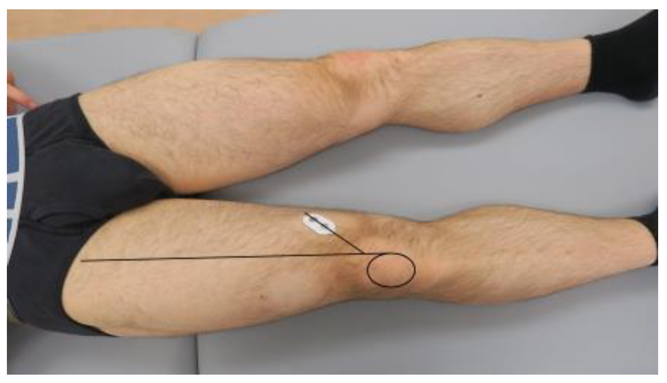
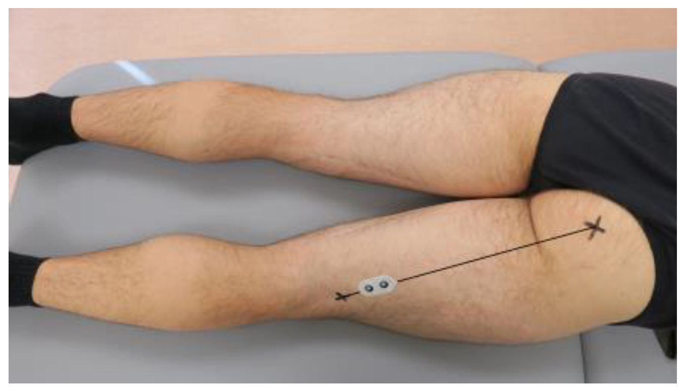
2.4. Surface Electromyography

The sEMG signal was recorded with 16-bit accuracy at a sampling rate of 1500 Hz using the sEMG TeleMyo™ Direct Transmission System (DTS), Noraxon, Scottsdale, AZ, USA. The sEMG signal was processed using MyoResearch 3.14.38 software (Noraxon USA, Inc., Scottsdale, AZ, USA). Dual-electrodes for Noraxon sEMG (21 × 40 mm) were used for this study.
During the YBT-LQ, the sEMG signals were collected from the vastus medialis (VM), vastus lateralis (VL), biceps femoris (BF), and semitendinosus (ST) muscles on the lower limb examined (standing limb).
The VM and VL from the quadriceps and the BF and ST from the hamstrings were selected because they are key antagonist muscle groups that stabilize the knee. The quadriceps (VM and VL) control knee extension, while the hamstrings (BF and ST) manage knee flexion. Measuring their coactivation provides valuable insights into how these muscles work together to maintain knee stability during dynamic tasks, such as the YBT-LQ. The activation of the rectus femoris was not investigated due to controversies raised in the literature concerning crosstalk occurring during the assessment of this muscle [41].
The area over the investigated muscle was prepared before the electrodes were placed on the skin. Hair in the area was removed using a disposable razor to improve the electrodes’ adhesion. The skin at the electrode placement sites was cleansed by rubbing with a cotton swab soaked in 2% salicylic acid.
The electrodes were placed following Beretta Piccoli et al.’s (2014) study [42]. To collect VM muscle activation data, the dual-electrode was placed within the first 38 mm of the line on the distal portion of the muscle belly and oriented 50° with respect to the reference line between the medial side of the patella and the anterior superior iliac spine (Figure 1).
The dual-electrode for collecting VL muscle activation data was placed within the first 43 mm of the line on the distal portion of the muscle belly and oriented 20° with respect to the reference line between the lateral side of the patella and the anterior superior iliac spine (Figure 2).
The dual-electrode for collecting BF muscle activation data was placed between 72% and 100% of the line between the ischial tuberosity and the lateral side of the popliteal cavity (Figure 3).
The dual-electrode for collecting ST activation data was placed between 74% and 100% of the line between the ischial tuberosity and the medial side of the popliteal cavity (Figure 4).
To perform MVC normalization, maximum voluntary contractions (MVCs) were conducted for each targeted muscle before the YBT-LQ, following Surface Electromyography for Non-Invasive Assessment of Muscles (SENIAM) project recommendations [42]. Participants were instructed to perform specific muscle contractions to elicit the maximum VM, VL, BF, and ST activation. For the VM and VL, participants performed an isometric knee extension against a fixed resistance while seated, ensuring that the hip and knee joints were at a 90-degree angle. Participants executed an isometric knee flexion for the BF and ST while lying prone with the knee flexed at 30 degrees. Each MVC was held for 3–5 s, and the highest stable value of the sEMG signal was recorded. This process was repeated three times for each muscle, with sufficient rest intervals between trials to avoid fatigue.

2.5. Lower Quarter Y-Balance Test

The YBT-LQ was conducted using the Y Balance Test Kit™. Participants began with a standardized warm-up of five minutes of light aerobic exercise and dynamic stretching. Clear instructions and a demonstration of the YBT-LQ were provided, followed by a practice trial to ensure familiarity with the test procedure. The test was carried out according to standard methodology [43].

2.6. The Calculation of the Surface Electromyography-Based Index for Quantifying Knee Muscle Coactivation During the Lower Quarter Y-Balance Test

For sEMG analysis and coactivation index (CoAI) calculation, the repetition with the best result was selected from each trial. The best result was the repetition, where the participant achieved the longest reach distance in the respective direction, providing a measure of the maximum performance under optimal conditions.
The raw sEMG signals from the VM, VL, BF, and ST muscles underwent processing using a band-pass filter (20–450 Hz). The root mean square (RMS) of the filtered sEMG signals was then calculated, representing the intensity of muscle activation. These RMS values were normalized against the MVC values obtained for each muscle, as described in Section 2.4. The normalized sEMG values were expressed as a percentage of the MVC (%MVC).
The CoAI was calculated by taking the normalized activation level of the antagonist muscle, dividing it by the normalized activation level of the corresponding agonist muscle, and then multiplying the result by 100. This provided a percentage value indicating the degree of antagonist muscle activation relative to the agonist muscle.
This study calculated four different coactivation indices (CoAIs) for each pair of muscles: CoAI1 (ST/VM), CoAI2 (ST/VL), CoAI3 (BF/VM), and CoAI4 (BF/VL). The calculations were performed separately for each YBT-LQ direction, the right and left lower limbs, and each of the three trials.
High CoAI values indicate a greater activation of the antagonist muscles (ST or BF) compared to the agonist muscles (VM or VL), while low CoAI values suggest that the agonist muscles (VM or VL) are more dominant during the task.

2.7. Statistical Analysis

SPSS Statistics Version 28.0.1.0 (142) (IBM® SPSS® Statistics, Armonk, NY, USA) and Microsoft Office Excel 365 Personal (Microsoft Corporation, Redmond, WA, USA) were used for the statistical analysis.
According to Bujang and Baharun’s guidelines, considering the necessity for two measurements per participant to determine intra-rater and inter-rater reliability separately, setting a statistical power of 80%, and establishing a minimum intraclass correlation coefficient (ICC) of 0.50, the number of participants in a sample for analysis needs to be at least 22 [44]. On the other hand, Koo and Li suggest obtaining at least 30 heterogeneous targets for reliability study purposes [45]. Considering possible missing data and dropouts, the studied sample consisted of 40 participants [46].
For the statistical analysis, the arithmetic mean (x) and standard deviation (SD, ±) were calculated for the age, body mass, body height, and BMI of the studied participants, as were the four coactivation indices’ values separately for each direction of the YBT-LQ for both the left and right lower limbs and across the three separate trials. All the studied features were normally distributed according to the Shapiro–Wilk test performed.
The reliability assessment was based on the ICC calculation according to commonly used guidelines [38]. For intra-rater test reliability assessment purposes, a two-way mixed effects model, single measurement type, and absolute agreement definition were used. The inter-rater test reliability was assessed using the ICC with a two-way random effects model, single rater type, and absolute agreement definition [36]. The ICC values ranged from 0.0 to 1.0, with values closer to 1 indicating better reliability. The level of reliability was interpreted on the reported 95% confidence interval (CI) of the estimated ICC, as the estimated ICC is only an expected value of the true ICC [36]. When interpreting the ICC values, the authors followed Cicchetti and Sparrow’s (1981) recommendations: ICC < 0.40 poor reliability; ICC 0.40–0.59 fair reliability; ICC 0.60–0.74 good reliability; and ICC ≥ 75 excellent reliability [47].
Consecutively comparative statistics were calculated based on the results obtained during the third trial, including parametric tests for dependent samples to compare the CoAIs between the right and left lower limbs and a repeated measures ANOVA to compare CoAIs across different directions of the YBT-LQ. The statistical significance was set at p < 0.050 [48].

3. Results

The study group consisted of the first 40 males with a mean age of 23.56 ± 2.33 years, body height of 1.79 ± 0.04 m, body mass of 80.33 ± 11.43 kg, and BMI of 25.05 ± 3.36 who met all the inclusion criteria. Initially, there were 45 volunteers. However, five were not included due to their low level of sports activity.

3.1. Intra-Rater and Inter-Rater Reliability

Table 1 presents the intra-rater and inter-rater reliability of the sEMG CoAI1 (ST/VM) for different directions of the YBT-LQ.
Considering the lower bound of the 95% CI, the lowest intra-rater reliability was noted for the left lower limb in the posterolateral direction, which ranged from poor to good with ICC = 0.495, 95% CI (0.227, 0.695). Then, the highest level of reliability could be considered for the right lower limb in the anterior direction, which ranged from fair to excellent, ICC = 0.742, 95% CI (0.561, 0.855).
Also, based on the lower bound of the 95% CI, the lowest inter-rater reliability was noted for the right lower limb in the anterior direction, which was from poor to fair, with ICC = 0.315, 95% CI (0.002, 0.570). On the other hand, the highest reliability was for the left lower limb for the posterolateral direction and was from good to excellent, ICC = 0.790, 95% CI (0.633, 0.884).
Table 2 presents the intra-rater and inter-rater reliability of the sEMG-based CoAI2 (ST/VL) for different directions of the YBT-LQ.
Based on the lower bound of the 95% CI, the lowest intra-rater reliability was noted for the left lower limb in the posterolateral direction, which ranged from poor to good with ICC = 0.567, 95% CI (0.317, 0.744). Consecutively, the highest level of reliability was noted for the right lower limb in the anterior direction, which ranged from good to excellent, ICC = 0.765, 95% CI (0.598, 0.869).
Also, considering the lower bound of the 95% CI, the lowest inter-rater reliability was noted for the right lower limb in the anterior direction, which ranged from poor to good with ICC = 0.503, 95% CI (0.227, 0.703). The highest level of reliability was noted for the right lower limb in the posterolateral direction, which ranged from good to excellent, ICC = 0.779, 95% CI (0.620, 0.877).
Consecutively, Table 3 presents the intra-rater and inter-rater reliability of the sEMG-based CoA3 (BF/VM) for different directions of the YBT-LQ.
Considering the lower bound of the 95% CI, the lowest intra-rater reliability was noted for the right lower limb in the posterolateral direction, which ranged from poor to fair with ICC = 0.279, 95% CI (−0.026, 0.538). In turn, the highest level of reliability could be considered for the left lower limb in the posteromedial direction, which ranged from good to excellent, ICC = 0.829, 95% CI (0.700, 0.905).
Based on the lower bound of the 95% CI, the lowest inter-rater reliability was noted for the right lower limb in the anterior direction, which ranged from poor to good with ICC = 0.488, 95% CI (0.208, 0.693). Consecutively, the highest level of reliability was noted for the left lower limb in the posterolateral direction, which ranged from good to excellent, ICC = 0.793, 95% CI (0.642, 0.885).
Finally, Table 4 presents the intra-rater and inter-rater reliability of the sEMG-based CoAI4 (BF/VL) for different directions of the YBT-LQ.
Considering the lower bound of the 95% CI, the lowest intra-rater reliability was noted for the right lower limb in the posterolateral direction, which ranged from poor to fair with ICC = 0.303, 95% CI (−0.004, 0.559). In turn, the highest level of reliability was noted for the left lower limb in the posteromedial direction, which ranged from good to excellent, ICC = 0.818, 95% CI (0.682, 0.900).
Based on the lower bound of the 95% CI, the lowest inter-rater reliability was noted for the right lower limb in the anterior direction, which ranged from poor to good with ICC = 0.515, 95% CI (0.243, 0.712). Consecutively, the highest level of reliability was noted for the left lower limb in the posterolateral direction, which ranged from fair to excellent, ICC = 0.713, 95% CI (0.519, 0.837).

3.2. Between-Limb Comparison

As Table 5 shows, no statistically significant differences were found in the four CoAIs between the right and left lower limbs of the participants.

3.3. Between-Direction Comparison

A repeated measures ANOVA with a Greenhouse–Geisser correction determined that CoAI1 (ST/VM) in the right lower limb differed statistically significantly between the three YBT-LQ directions (F = 3.394, p = 0.049). However, post hoc analysis with a Bonferroni correction revealed that CoAI1 (ST/VM) was comparable between the anterior and posteromedial (p = 0.116), anterior and posterolateral (p = 0.181), and posteromedial and posterolateral (p = 1.000) directions. A repeated measures ANOVA with a Greenhouse–Geisser correction for the left lower limb did not show statistically significant CoAI1 (ST/VM) differences between the three YBT-LQ directions (F = 2.022, p = 0.146).
A repeated measures ANOVA with a Greenhouse–Geisser correction determined that CoAI2 (ST/VL) in the right lower limb differed statistically significantly between the three YBT-LQ directions (F = 13.442, p < 0.001). Post hoc analysis with a Bonferroni correction revealed that CoAI2 (ST/VL) for the anterior direction was statistically significantly larger than that for the posteromedial (p < 0.001) and posterolateral (p = 0.001) directions. At the same time, CoAI2 (ST/VL) was comparable between the posteromedial and posterolateral directions (p = 1.000). A repeated measures ANOVA with a Greenhouse–Geisser correction in the left lower limb revealed that CoAI2 (ST/VL) also differed statistically significantly between the three YBT-LQ directions (F = 5.651, p = 0.009). Like the right lower limb, post hoc analysis with a Bonferroni correction revealed that CoAI2 (ST/VL) for the anterior direction was statistically significantly larger than that for the posterolateral direction (p = 0.024). Although CoAI2 (ST/VL) for the anterior direction was larger than that for the posteromedial direction, the difference was not statistically significant (p = 0.077). Again, CoAI2 (ST/VL) was comparable between the posteromedial and posterolateral directions (p = 1.000).
A repeated measures ANOVA with a Greenhouse–Geisser correction determined that CoAI3 (BF/VM) in the right lower limb differed statistically significantly between the three YBT-LQ directions (F = 4.114, p = 0.026). Post hoc analysis with a Bonferroni correction revealed that CoAI3 (BF/VM) for the posteromedial direction was statistically significantly larger than that for the anterior direction (p = 0.034). No statistically significant differences existed between the anterior and posterolateral (p = 0.331) and posteromedial and posterolateral (p = 0.633) directions. In the left lower limb, a repeated measures ANOVA with a Greenhouse–Geisser correction also revealed statistically significant CoAI3 (BF/VM) differences between the three YBT-LQ directions (F = 3.549, p = 0.041). However, post hoc analysis with a Bonferroni correction did not show any statistically significant differences between the anterior and posteromedial (p = 0.094), anterior and posterolateral (p = 0.083), and posteromedial and posterolateral (p = 1.000) directions.
Regarding CoAI4 (BF/VL), a repeated measures ANOVA with a Greenhouse–Geisser correction did not show any statistically significant differences between the three YBT-LQ directions in either the right lower limb (F = 1.848, p = 0.169) or the left lower limb (F = 0.510, p = 0.594).

4. Discussion

In this study, CoAIs were calculated and evaluated in terms of reliability to quantify how knee flexor muscles (antagonists) and extensor muscles (agonists) co-contract during the YBT-LQ. Specifically, the activation of flexor muscles (ST and BF) was compared with that of extensor muscles (VM and VL) to assess the contribution of the investigated muscles during the given task.
Four distinct CoAIs were calculated for each muscle pair, accounting for different directions, limbs, and trials. The results revealed that the reliability of these sEMG-based CoAIs varied depending on the muscle pairs and movement directions. Notably, CoAIs involving the ST, precisely ST/VL and ST/VM, demonstrated good to excellent reliability, while those involving the BF exhibited lower reliability. These findings highlight the need to consider specific muscle pairs, test directions, and rater consistency when using CoAIs in clinical and research settings.
For instance, the high reliability observed in the ST and VL muscle pair could suggest that these muscles effectively coactivate to stabilize the knee during anterior, posteromedial, and posterolateral movements. In contrast, the lower reliability in the BF and VM pair may indicate inconsistent activation, potentially reflecting variability in knee stabilization strategies or individual biomechanical differences. These insights can potentially emphasize the critical role of specific muscle pairs in maintaining dynamic knee stability.
The reliability analysis of the CoAIs during the YBT-LQ varied across directions and muscle pairs. CoAI2 (ST/VL) had the highest intra-rater reliability in the anterior direction, reflecting strong consistency, while CoAI3 (BF/VM) showed the lowest reliability in the posterolateral direction. This suggests that the investigated muscle pairs co-work differently for different YBT-LQ directions. The anterior direction likely places greater demand on the knee extensors, explaining the higher reliability of CoAI2 (ST/VL). In contrast, the posterolateral direction requires more complex coordination between the knee flexors and extensors, contributing to the lower reliability of CoAI3 (BF/VM). This highlights the need for direction-specific considerations when assessing knee muscle coactivation using CoAIs.
No significant differences were observed between limbs in the four CoAIs, suggesting symmetrical muscle coactivation patterns in healthy recreational athletes. While this indicates balanced neuromuscular control, relying solely on CoAIs may not fully capture the complexity of lower limb symmetry, as muscle coordination involves more than coactivation indices. In clinical populations, such as those recovering from knee injuries, asymmetry is often a marker of impaired neuromuscular function. Exploring how coactivation patterns change in injured individuals could enhance this study’s clinical relevance, especially for injury prevention and rehabilitation. Future research could investigate the potential of these indices to monitor asymmetry and guide targeted rehabilitation strategies.
An interesting issue was the diversity of the developed indices in terms of their comparison between different directions. For example, CoAI2 showed significantly higher values in the anterior direction compared to the posteromedial and posterolateral directions, which could potentially indicate the dominant role of the ST and VL muscles in stabilizing the knee during anterior motion. In contrast, CoAI4 (BF/VL) demonstrated no significant differences across directions, indicating a stable coactivation pattern. The reason for the variability observed in CoAI1 (ST/VM) and CoAI3 (BF/VM) should be further explored.
The developed set of four sEMG-based indices to quantify knee muscle coactivation during the YBT-LQ could potentially be a valuable tool for assessing neuromuscular control and stability. This could be particularly beneficial for populations at risk of injury or re-injury, such as athletes prone to anterior cruciate ligament (ACL) injuries or those recovering from ACL reconstruction. For example, these indices could identify imbalances or deficits in neuromuscular control predisposing individuals to injury. In clinical practice, the indices could be applied to evaluate neuromuscular coordination during rehabilitation, helping clinicians detect persistent abnormalities in muscle coactivation patterns that may indicate compromised knee stability, a known risk factor for ACL injuries. By monitoring the coactivation of key muscle groups like the ST and VL (CoAI2), clinicians can assess the effectiveness of interventions to improve dynamic stability and adjust rehabilitation protocols accordingly.
It is worth highlighting that reliability is not assigned to any specific instrument of measurement but rather to the particular testing design and methodology that employ the instrument [33]. Therefore, it should also be emphasized that the testing procedure for the YBT-LQ allows for considerable flexibility in various aspects, which can significantly impact the results [16]. It also has to be highlighted that while this study did not specifically investigate the optimal rest period between stimuli, it is acknowledged that rest duration can influence muscle performance and coactivation patterns, especially during dynamic tasks. Future research could explore the effects of varying rest intervals to determine the most appropriate rest time for minimizing fatigue and ensuring accurate assessments. This would provide a more comprehensive understanding of how rest duration impacts neuromuscular performance in similar contexts. Also, while this study did not account for individual factors such as height, foot size, or limb dominance, these variables could influence the results, particularly in dynamic tasks like the YBT-LQ. Future research should consider these factors.
The present study has limitations that must be considered when analyzing the findings. First, the studied sample was restricted to healthy male recreational athletes, so the findings may not apply widely to other groups such as females, older or younger individuals, those with lower limb disorders due to injury or disease, or professional athletes. The available literature shows in the example of other movement tasks that muscle activation patterns differ based on sex and age in healthy individuals [49,50,51]. Muscle activation patterns during different movement tasks also differ between healthy individuals and individuals suffering from various diseases and disorders [52,53,54].
Secondly, using sEMG is associated with challenges, including signal crosstalk from nearby muscles, variability in electrode placement, and skin impedance, which can impact the accuracy of muscle activation measurements. Only four muscles from the knee joint area were examined in this study. However, knee stability relies on activating a broader set of muscles. Significant contributions to knee coactivation may have been overlooked by not including other important muscles like gastrocnemius.
Although this study focuses on knee muscle coactivation during the YBT-LQ, it is important to acknowledge that muscle fatigue can significantly impact coordination and stability, especially during dynamic tasks. Fatigue has been shown to alter coactivation patterns, reducing neuromuscular control and increasing injury risk. However, this study did not directly assess fatigue using EMG measures such as median frequency or zero-crossing rate, which are, as already mentioned, commonly used indicators for detecting muscle fatigue [25]. Future studies could incorporate fatigue analysis to gain more comprehensive insights into how fatigue alters muscle coordination.
Furthermore, while the YBT-LQ was utilized to evaluate dynamic balance and knee coactivation, it is important to note that the specific nature of the test restricts the generalizability of the results to other functional movements, such as running, jumping, or cutting, which are common in athletic settings. Although the intra- and inter-rater reliabilities were considered acceptable, minor differences in electrode placement or participant movement patterns may still introduce some variability in the measurements. Moreover, due to the absence of kinematic data, this study could not directly link muscle activation patterns with joint angles or movement biomechanics during the YBT-LQ, which could limit the understanding of how coactivation contributes to dynamic knee stability.
Considering the above limitations, expanding the sample to include more diverse populations in future research is essential. This means introducing studies that include females, older and younger participants than those in the present study, individuals recovering from lower limb injuries, etc., which would improve the generalizability of the findings and provide greater insight into how muscle coactivation may vary across different groups. Also, incorporating more advanced methods, such as high-density sEMG, could help reduce the limitations of crosstalk and improve the spatial resolution of muscle activation data [55]. Additionally, including a broader range of muscles involved in knee joint stability would offer a more comprehensive understanding of coactivation patterns.
Future research could test the reliability and validity of the CoAIs across various functional tasks beyond the YBT-LQ, including running, jumping, and agility drills, to extend the applicability of the CoAIs. Longitudinal studies would also be valuable in assessing how knee muscle coactivation changes over time in response to training, fatigue, or rehabilitation. Moreover, integrating kinematic or motion capture data with sEMG analysis would offer deeper insights into the relationship between muscle activation and joint movement, helping to clarify the role of coactivation in maintaining dynamic knee stability during complex, multi-joint movements. These advancements would enhance the clinical applications of the CoAIs, providing a more robust tool for injury prevention and rehabilitation.
The described method of calculating the coactivation index by taking the normalized activation level of the antagonist muscle, dividing it by the normalized activation level of the corresponding agonist muscle, and then multiplying by 100 is a common approach and gives a sense of how much the antagonist muscle is activated relative to the agonist during a task. However, this method assumes that both muscles should ideally activate at similar levels, which may not always be the case. Therefore, future studies could be extended by analyzing different approaches that involve comparing the lower activation value to the higher activation value, as this accounts for the relative contribution of both muscles without assuming that one should always dominate. Some methods also involve taking the difference or the sum of the normalized activations to provide a different perspective on coactivation. When analyzing coactivation, it is important to consider muscle function, as agonist and antagonist muscles do not always work in direct opposition; in some cases, one muscle may need to dominate in activation depending on the phase of movement. The ideal coactivation balance can also vary based on the specific demands of the task or movement being performed, such as those related to balance, strength, or stability.

5. Conclusions

This study suggests that sEMG-based CoAIs for assessing knee muscle activity during the YBT-LQ demonstrate variable reliability depending on the muscle pair and direction assessed. While some CoAIs showed good to excellent reliability, others were less consistent. These results underscore the importance of selecting appropriate CoAIs for clinical and research applications, emphasizing those with higher reliability to ensure an accurate assessment of knee stability and neuromuscular control.

Supplementary Materials

The following supporting information can be downloaded at https://www.mdpi.com/article/10.3390/app14219788/s1, Table S1: Data generated and analyzed during the present study.

Author Contributions

Conceptualization, P.R., Ł.O. and A.K.; methodology, M.D. (Maciej Daszkiewicz), R.P., Ł.O., M.K. (Mateusz Kuźniecow) and A.K.; software, P.R. and Ł.O.; validation, M.D. (Maciej Daszkiewicz), R.P., M.K. (Mateusz Kuźniecow), M.L. and M.K. (Michał Kułakowski); formal analysis, P.R., R.B. and A.K.; investigation, M.D. (Maciej Daszkiewicz), M.K. (Mateusz Kuźniecow), M.K. (Monika Kentel), M.K. (Maciej Kentel), M.D. (Maciej Dejnek) and A.K.; resources, M.D. (Maciej Daszkiewicz), M.K. (Mateusz Kuźniecow), M.K. (Monika Kentel), M.K. (Maciej Kentel), M.D. (Maciej Dejnek) and A.K.; data curation, M.D. (Maciej Daszkiewicz), M.K. (Monika Kentel), M.K. (Maciej Kentel), M.K. (Mateusz Kowal), M.D. (Maciej Dejnek) and A.K.; writing—original draft preparation, M.D. (Maciej Daszkiewicz), R.P., P.R., R.B., Ł.O., M.K. (Mateusz Kuźniecow), M.L., M.K. (Michał Kułakowski), M.K. (Monika Kentel), M.K. (Maciej Kentel), M.K. (Mateusz Kowal), M.D. (Maciej Dejnek) and A.K.; writing—review and editing, M.D. (Maciej Daszkiewicz), R.P., P.R., R.B., Ł.O., M.K. (Mateusz Kuźniecow), M.L., M.K. (Michał Kułakowski), M.K. (Monika Kentel), M.K. (Maciej Kentel), M.K. (Mateusz Kowal), M.D. (Maciej Dejnek) and A.K.; visualization, M.D. (Maciej Daszkiewicz); supervision, R.P., P.R., R.B. and A.K.; project administration, R.P., P.R., R.B. and A.K.; funding acquisition, P.R. All authors have read and agreed to the published version of the manuscript.

Funding

This research was financed from a subsidy granted to the Wroclaw Medical University from the Ministry of Science in Poland with internal number SIMPLE: SUBZ.A470.24.075.

Institutional Review Board Statement

This study was conducted according to the guidelines of the Declaration of Helsinki and approved by the Bioethics Committee at the Medical University of Wroclaw, Wroclaw, Poland (approval number KB-351/2021), granted on 26 April 2021.

Informed Consent Statement

Informed consent was obtained from all subjects involved in this study. Written informed consent was obtained from the participants to publish this paper.

Data Availability Statement

The original contributions presented in this study are included in the Supplementary Materials; further inquiries can be directed to the corresponding author.

Acknowledgments

The authors would like to thank Bartosz Witkowski from the Department of Orthopedics, Traumatology, and Hand Surgery, Faculty of Medicine, Wroclaw Medical University, Wroclaw, Poland, for his support in the preparation of figures for the purposes of the present manuscript.

Conflicts of Interest

The authors declare no conflicts of interest.

References

  1. Sonesson, S.; Lindblom, H.; Hägglund, M. Higher age and present injury at the start of the season are risk factors for in-season injury in amateur male and female football players-a prospective cohort study. Knee Surg. Sports Traumatol. Arthrosc. 2023, 31, 4618–4630. [Google Scholar] [CrossRef] [PubMed]
  2. Torvaldsson, K.; Lindblom, H.; Sonesson, S.; Senorski, E.H.; Stigson, H.; Tamm, L.; Sandberg, J.; Hägglund, M. Swedish Olympic athletes report one injury insurance claim every second year: A 22-year insurance registry-based cohort study. Knee Surg. Sports Traumatol. Arthrosc. 2023, 31, 4607–4617. [Google Scholar] [CrossRef] [PubMed]
  3. Astur, D.C.; Margato, G.F.; Zobiole, A.; Pires, D.; Funchal, L.F.Z.; Jimenez, A.E.; Freitas, E.V.; Cohen, M. The incidence of anterior cruciate ligament injury in youth and male soccer athletes: An evaluation of 17,108 players over two consecutive seasons with an age-based sub-analysis. Knee Surg. Sports Traumatol. Arthrosc. 2023, 31, 2556–2562. [Google Scholar] [CrossRef] [PubMed]
  4. Paudel, Y.R.; Sommerfeldt, M.; Voaklander, D. Increasing incidence of anterior cruciate ligament reconstruction: A 17-year population-based study. Knee Surg. Sports Traumatol. Arthrosc. 2023, 31, 248–255. [Google Scholar] [CrossRef] [PubMed]
  5. Al-Gburi, M.; Kristiansen, J.B.; Christensen, K.B.; Krogsgaard, M.R. Functional performance tests, clinical measurements, and patient-reported outcome measures do not correlate as outcomes 1 year after anterior cruciate ligament reconstruction. Knee Surg. Sports Traumatol. Arthrosc. 2023, 31, 5905–5912. [Google Scholar] [CrossRef]
  6. Velasquez Garcia, A.; Iida, N.; Kuroiwa, T.; Hsu, K.L.; de Marinis, R.; Abdo, G.; Ekdahl, M. Substantial influence of psychological factors on return to sports after anterior shoulder instability surgery: A systematic review and meta-analysis. Knee Surg. Sports Traumatol. Arthrosc. 2023, 31, 5913–5923. [Google Scholar] [CrossRef]
  7. Królikowska, A.; Reichert, P.; Senorski, E.H.; Karlsson, J.; Becker, R.; Prill, R. Scores and sores: Exploring patient-reported outcomes for knee evaluation in orthopaedics, sports medicine and rehabilitation. Knee Surg. Sports Traumatol. Arthrosc. 2024. [Google Scholar] [CrossRef]
  8. Thorolfsson, B.; Piussi, R.; Snaebjornsson, T.; Karlsson, J.; Samuelsson, K.; Beischer, S.; Thomeé, R.; Hamrin Senorski, E. Greater self-efficacy, psychological readiness and return to sport amongst paediatric patients compared with adolescents and young adults, 8 and 12 months after ACL reconstruction. Knee Surg. Sports Traumatol. Arthrosc. 2023, 31, 5629–5640. [Google Scholar] [CrossRef]
  9. Tischer, T.; Martens, G.; Cabri, J.; Thoreux, P.; Tscholl, P.; Edouard, P.; Leclerc, S.; Le Garrec, S.; Delvaux, F.; Croisier, J.L.; et al. The awareness of injury prevention programmes is insufficient among French- and German-speaking sports medicine communities in Europe. Knee Surg. Sports Traumatol. Arthrosc. 2023, 31, 2563–2571. [Google Scholar] [CrossRef]
  10. Mir, B.; Vivekanantha, P.; Dhillon, S.; Cotnareanu, O.; Cohen, D.; Nagai, K.; de Sa, D. Fear of reinjury following primary anterior cruciate ligament reconstruction: A systematic review. Knee Surg. Sports Traumatol. Arthrosc. 2023, 31, 2299–2314. [Google Scholar] [CrossRef]
  11. Kaarre, J.; Zsidai, B.; Winkler, P.W.; Narup, E.; Horvath, A.; Svantesson, E.; Senorski, E.H.; Musahl, V.; Samuelsson, K. Different patient and activity-related characteristics result in different injury profiles for patients with anterior cruciate ligament and posterior cruciate ligament injuries. Knee Surg. Sports Traumatol. Arthrosc. 2023, 31, 308–315. [Google Scholar] [CrossRef] [PubMed]
  12. Obërtinca, R.; Meha, R.; Hoxha, I.; Shabani, B.; Meyer, T.; Aus der Fünten, K. Efficacy of a new injury prevention programme (FUNBALL) in young male football (soccer) players: A cluster-randomised controlled trial. Br. J. Sports Med. 2024, 58, 548–555. [Google Scholar] [CrossRef] [PubMed]
  13. Magaña-Ramírez, M.; Gallardo-Gómez, D.; Álvarez-Barbosa, F.; Corral-Pernía, J.A. What exercise programme is the most appropriate to mitigate anterior cruciate ligament injury risk in football (soccer) players? A systematic review and network meta-analysis. J. Sci. Med. Sport 2024, 27, 234–242. [Google Scholar] [CrossRef] [PubMed]
  14. Rommers, N.; Rössler, R.; Tassignon, B.; Verschueren, J.; De Ridder, R.; van Melick, N.; Longé, L.; Hendrikx, T.; Vaes, P.; Beckwée, D.; et al. Most amateur football teams do not implement essential components of neuromuscular training to prevent anterior cruciate ligament injuries and lateral ankle sprains. Knee Surg. Sports Traumatol. Arthrosc. 2022, 30, 1169–1179. [Google Scholar] [CrossRef]
  15. Kim, S.; Glaviano, N.R.; Park, J. Exercise-induced fatigue affects knee proprioceptive acuity and quadriceps neuromuscular function more in patients with ACL reconstruction or meniscus surgery than in healthy individuals. Knee Surg. Sports Traumatol. Arthrosc. 2023, 31, 5428–5437. [Google Scholar] [CrossRef]
  16. Zając, B.; Olszewski, M.; Mika, A. Influence of protocol variables on outcomes of the star excursion balance test group (SEBT, mSEBT, YBT-LQ) in healthy individuals: A systematic review. Front. Physiol. 2024, 15, 1415887. [Google Scholar] [CrossRef]
  17. Melzer, I.; Benjuya, N.; Kaplanski, J.; Alexander, N. Association between ankle muscle strength and limit of stability in older adults. Age Ageing 2009, 38, 119–123. [Google Scholar] [CrossRef]
  18. Gribble, P.A.; Hertel, J.; Plisky, P. Using the Star Excursion Balance Test to assess dynamic postural-control deficits and outcomes in lower extremity injury: A literature and systematic review. J. Athl. Train 2012, 47, 339–357. [Google Scholar] [CrossRef]
  19. Plisky, P.; Schwartkopf-Phifer, K.; Huebner, B.; Garner, M.B.; Bullock, G. Systematic Review and Meta-Analysis of the Y-Balance Test Lower Quarter: Reliability, Discriminant Validity, and Predictive Validity. Int. J. Sports Phys. Ther. 2021, 16, 1190–1209. [Google Scholar] [CrossRef]
  20. Chaabene, H.; Negra, Y.; Sammoud, S.; Moran, J.; Ramirez-Campillo, R.; Granacher, U.; Prieske, O. The Effects of Combined Balance and Complex Training Versus Complex Training Only on Measures of Physical Fitness in Young Female Handball Players. Int. J. Sports Physiol. Perform. 2021, 16, 1439–1446. [Google Scholar] [CrossRef]
  21. Powden, C.J.; Dodds, T.K.; Gabriel, E.H. The reliability of the star excursion balance test and lower quarter y-balance test in healthy adults: A systematic review. Int. J. Sports Phys. Ther. 2019, 14, 683–694. [Google Scholar] [CrossRef] [PubMed]
  22. Molina-Rueda, F.; Fernández-Vázquez, D.; Navarro-López, V.; López-González, R.; Carratalá-Tejada, M. Muscle Coactivation Index during Walking in People with Multiple Sclerosis with Mild Disability, a Cross-Sectional Study. Diagnostics 2023, 13, 2169. [Google Scholar] [CrossRef] [PubMed]
  23. Bandini, V.; Carpinella, I.; Marzegan, A.; Jonsdottir, J.; Frigo, C.A.; Avanzino, L.; Pelosin, E.; Ferrarin, M.; Lencioni, T. Surface-Electromyography-Based Co-Contraction Index for Monitoring Upper Limb Improvements in Post-Stroke Rehabilitation: A Pilot Randomized Controlled Trial Secondary Analysis. Sensors 2023, 23, 7320. [Google Scholar] [CrossRef]
  24. Rodrigues, C.; Correia, M.; Abrantes, J.; Benedetti, M.; Nadal, J. Muscle coactivation analysis for neuromuscular control assessment of lower limb stretch-shortening cycle. Gait Posture 2023, 106, S175–S176. [Google Scholar] [CrossRef]
  25. Kim, H.; Lee, J.; Kim, J. Electromyography-signal-based muscle fatigue assessment for knee rehabilitation monitoring systems. Biomed. Eng. Lett. 2018, 8, 345–353. [Google Scholar] [CrossRef]
  26. Czamara, A.; Królikowska, A.; Szuba, Ł.; Widuchowski, W.; Kentel, M. Single- vs. double-bundle anterior cruciate ligament reconstruction: A new aspect of knee assessment during activities involving dynamic knee rotation. J. Strength. Cond. Res. 2015, 29, 489–499. [Google Scholar] [CrossRef] [PubMed]
  27. Królikowska, A.; Mika, A.; Plaskota, B.; Daszkiewicz, M.; Kentel, M.; Kołcz, A.; Kentel, M.; Prill, R.; Diakowska, D.; Reichert, P.; et al. Reliability and Validity of the Athletic Shoulder (ASH) Test Performed Using Portable Isometric-Based Strength Training Device. Biology 2022, 11, 577. [Google Scholar] [CrossRef] [PubMed]
  28. Królikowska, A.; Maj, A.; Dejnek, M.; Prill, R.; Skotowska-Machaj, A.; Kołcz, A. Wrist motion assessment using Microsoft Azure Kinect DK: A reliability study in healthy individuals. Adv. Clin. Exp. Med. 2023, 32, 203–209. [Google Scholar] [CrossRef]
  29. Oleksy, Ł.; Królikowska, A.; Mika, A.; Reichert, P.; Kentel, M.; Kentel, M.; Poświata, A.; Roksela, A.; Kozak, D.; Bienias, K.; et al. A Reliability of Active and Passive Knee Joint Position Sense Assessment Using the Luna EMG Rehabilitation Robot. Int. J. Environ. Res. Public Health 2022, 19, 15885. [Google Scholar] [CrossRef]
  30. Królikowska, A.; Czamara, A.; Kentel, M. Does Gracilis Tendon Harvest During ACL Reconstruction with a Hamstring Autograft Affect Torque of Muscles Responsible for Shin Rotation? Med. Sci. Monit. 2015, 21, 2084–2093. [Google Scholar] [CrossRef]
  31. Czamara, A.; Markowska, I.; Krolikowska, A.; Szopa, A.; Domagalska Szopa, M. Kinematics of Rotation in Joints of the Lower Limbs and Pelvis during Gait: Early Results-SB ACLR Approach versus DB ACLR Approach. BioMed Res. Int. 2015, 2015, 707168. [Google Scholar] [CrossRef] [PubMed]
  32. Prill, R.; Królikowska, A.; Becker, R.; Karlsson, J. Why there is a need to improve evaluation standards for clinical studies in orthopaedic and sports medicine. Knee Surg. Sports Traumatol. Arthrosc. 2023, 31, 4–5. [Google Scholar] [CrossRef] [PubMed]
  33. Królikowska, A.; Reichert, P.; Karlsson, J.; Mouton, C.; Becker, R.; Prill, R. Improving the reliability of measurements in orthopaedics and sports medicine. Knee Surg. Sports Traumatol. Arthrosc. 2023, 31, 5277–5285. [Google Scholar] [CrossRef]
  34. Daly, L.; Bourke, G.J. Interpretation and Uses of Medical Statistics; Wiley: Hoboken, NJ, USA, 2008. [Google Scholar]
  35. Bruton, A.; Conway, J.H.; Holgate, S.T. Reliability: What is it, and how is it measured? Physiotherapy 2000, 86, 94–99. [Google Scholar] [CrossRef]
  36. Lee, K.M.; Lee, J.; Chung, C.Y.; Ahn, S.; Sung, K.H.; Kim, T.W.; Lee, H.J.; Park, M.S. Pitfalls and important issues in testing reliability using intraclass correlation coefficients in orthopaedic research. Clin. Orthop. Surg. 2012, 4, 149–155. [Google Scholar] [CrossRef]
  37. Peacock, J.; Peacock, P. Oxford Handbook of Medical Statistics; OUP: Oxford, UK, 2011. [Google Scholar]
  38. Shrout, P.E.; Fleiss, J.L. Intraclass correlations: Uses in assessing rater reliability. Psychol. Bull. 1979, 86, 420–428. [Google Scholar] [CrossRef]
  39. Schlüter, I.-M.; Prill, R.; Królikowska, A.; Cruysen, C.; Becker, R. A Pilot Study on the Reliability of Ultrasound-Based Assessment of Patella Diameter and Sulcus Angle. Diagnostics 2022, 12, 3164. [Google Scholar] [CrossRef]
  40. Wise, K.L.; Kelly, B.J.; Knudsen, M.L.; Macalena, J.A. Reliability Studies and Surveys. In Basic Methods Handbook for Clinical Orthopaedic Research: A Practical Guide and Case Based Research Approach; Musahl, V., Karlsson, J., Hirschmann, M.T., Ayeni, O.R., Marx, R.G., Koh, J.L., Nakamura, N., Eds.; Springer: Berlin/Heidelberg, Germany, 2019; pp. 343–358. [Google Scholar]
  41. Nene, A.; Byrne, C.; Hermens, H. Is rectus femoris really a part of quadriceps? Assessment of rectus femoris function during gait in able-bodied adults. Gait Posture 2004, 20, 1–13. [Google Scholar] [CrossRef] [PubMed]
  42. Hermens, H.J.; Freriks, B.; Disselhorst-Klug, C.; Rau, G. Development of recommendations for SEMG sensors and sensor placement procedures. J. Electromyogr. Kinesiol. 2000, 10, 361–374. [Google Scholar] [CrossRef]
  43. Plisky, P.J.; Rauh, M.J.; Kaminski, T.W.; Underwood, F.B. Star Excursion Balance Test as a predictor of lower extremity injury in high school basketball players. J. Orthop. Sports Phys. Ther. 2006, 36, 911–919. [Google Scholar] [CrossRef]
  44. Bujang, M.A.; Baharum, N. A simplified guide to determination of sample size requirements for estimating the value of intraclass correlation coefficient: A review. Arch. Orofac. Sci. 2017, 12, 1–11. [Google Scholar]
  45. Koo, T.K.; Li, M.Y. A Guideline of Selecting and Reporting Intraclass Correlation Coefficients for Reliability Research. J. Chiropr. Med. 2016, 15, 155–163. [Google Scholar] [CrossRef]
  46. Madjarova, S.J.; Pareek, A.; Eckhardt, C.M.; Khorana, A.; Kunze, K.N.; Ollivier, M.; Karlsson, J.; Williams, R.J., 3rd; Nwachukwu, B.U. Fragility Part I: A guide to understanding statistical power. Knee Surg. Sports Traumatol. Arthrosc. 2022, 30, 3924–3928. [Google Scholar] [CrossRef]
  47. Cicchetti, D.V.; Sparrow, S.A. Developing criteria for establishing interrater reliability of specific items: Applications to assessment of adaptive behavior. Am. J. Ment. Defic 1981, 86, 127–137. [Google Scholar] [PubMed]
  48. Madjarova, S.J.; Williams, R.J., 3rd; Nwachukwu, B.U.; Martin, R.K.; Karlsson, J.; Ollivier, M.; Pareek, A. Picking apart p values: Common problems and points of confusion. Knee Surg. Sports Traumatol. Arthrosc. 2022, 30, 3245–3248. [Google Scholar] [CrossRef]
  49. Bailey, C.A.; Corona, F.; Pilloni, G.; Porta, M.; Fastame, M.C.; Hitchcott, P.K.; Penna, M.P.; Pau, M.; Côté, J.N. Sex-dependent and sex-independent muscle activation patterns in adult gait as a function of age. Exp. Gerontol. 2018, 110, 1–8. [Google Scholar] [CrossRef] [PubMed]
  50. Santuz, A.; Janshen, L.; Brüll, L.; Munoz-Martel, V.; Taborri, J.; Rossi, S.; Arampatzis, A. Sex-specific tuning of modular muscle activation patterns for locomotion in young and older adults. PLoS ONE 2022, 17, e0269417. [Google Scholar] [CrossRef]
  51. Jeon, W.; Ramadan, A.; Whitall, J.; Alissa, N.; Westlake, K. Age-related differences in lower limb muscle activation patterns and balance control strategies while walking over a compliant surface. Sci. Rep. 2023, 13, 16555. [Google Scholar] [CrossRef]
  52. Srivastava, S.; Patten, C.; Kautz, S.A. Altered muscle activation patterns (AMAP): An analytical tool to compare muscle activity patterns of hemiparetic gait with a normative profile. J. NeuroEng. Rehabil. 2019, 16, 21. [Google Scholar] [CrossRef]
  53. Almeida, M.B.d.; Moreira, M.; Miranda-Oliveira, P.; Moreira, J.; Família, C.; Vaz, J.R.; Moleirinho-Alves, P.; Oliveira, R. Evolving Dynamics of Neck Muscle Activation Patterns in Dental Students: A Longitudinal Study. Sensors 2024, 24, 5689. [Google Scholar] [CrossRef]
  54. Newcomer, K.L.; Jacobson, T.D.; Gabriel, D.A.; Larson, D.R.; Brey, R.H.; An, K.-N. Muscle activation patterns in subjects with and without low back pain. Arch. Phys. Med. Rehabil. 2002, 83, 816–821. [Google Scholar] [CrossRef] [PubMed]
  55. Alcan, V.; Zinnuroğlu, M. Current developments in surface electromyography. Turk. J. Med. Sci. 2023, 53, 1019–1031. [Google Scholar] [CrossRef] [PubMed]
Figure 1. The placement of the dual-electrode to collect the activation data of the vastus medialis muscle.
Applsci 14 09788 g001
Figure 2. The placement of the dual-electrode to collect the activation data of the vastus lateralis muscle.
Applsci 14 09788 g002
Figure 3. The placement of the dual-electrode to collect the activation data of the biceps femoris muscle.
Applsci 14 09788 g003
Figure 4. The placement of the dual-electrode to collect the activation data of the semitendinosus muscle.
Applsci 14 09788 g004
Table 1. The intra-rater and inter-rater reliability of the surface electromyography-based coactivation index, precisely CoAI1 (ST/VM).
Surface Electromyography-Based CoAI1 (ST/VM)
Intra-Rater ReliabilityInter-Rater Reliability
YBT-LQ DirectionRight Lower LimbLeft Lower LimbRight Lower LimbLeft Lower Limb
Anterior0.742 (0.561, 0.855)0.613 (0.379, 0.774)0.315 (0.002, 0.570)0.660 (0.445, 0.804)
Posteromedial0.636 (0.406, 0.790)0.585 (0.339, 0.756)0.620 (0.385, 0.780)0.667 (0.451, 0.809)
Posterolateral0.720 (0.528, 0.842)0.495 (0.227, 0.695)0.756 (0.583, 0.863)0.790 (0.633, 0.884)
Values expressed as intraclass correlation coefficient, ICC, and 95% confidence interval (lower bound, upper bound) of ICC. CoAI, coactivation index; ST, semitendinosus; VM, vastus medialis; YBT-LQ, Lower Quarter Y-Balance Test.
Table 2. The intra-rater and inter-rater reliability of the surface electromyography-based coactivation index, precisely CoAI2 (ST/VL).
Surface Electromyography-Based CoAI2 (ST/VL)
Intra-Rater ReliabilityInter-Rater Reliability
YBT-LQ DirectionRight Lower LimbLeft Lower LimbRight Lower LimbLeft Lower Limb
Anterior0.765 (0.598, 0.869)0.707 (0.512, 0.833)0.503 (0.227, 0.703)0.675 (0.465, 0.813)
Posteromedial0.761 (0.591, 0.867)0.743 (0.565, 0.856)0.712 (0.520, 0.836)0.743 (0.563, 0.855)
Posterolateral0.690 (0.487, 0.823)0.567 (0.317, 0.744)0.779 (0.620, 0.877)0.732 (0.544, 0.849)
Values expressed as intraclass correlation coefficient, ICC, and 95% confidence interval (lower bound, upper bound) of ICC. CoAI, coactivation index; ST, semitendinosus; VL, vastus lateralis; YBT-LQ, Lower Quarter Y-Balance Test.
Table 3. The intra-rater and inter-rater reliability of the surface electromyography-based coactivation index, precisely CoA3 (BF/VM).
Surface Electromyography-Based CoA3 (BF/VM)
Intra-Rater ReliabilityInter-Rater Reliability
YBT-LQ DirectionRight Lower LimbLeft Lower LimbRight Lower LimbLeft Lower Limb
Anterior0.554 (0.296, 0.737)0.551 (0.293, 0.734)0.488 (0.208, 0.693)0.671 (0.455, 0.811)
Posteromedial0.703 (0.504, 0.831)0.829 (0.700, 0.905)0.629 (0.399, 0.784)0.662 (0.447, 0.805)
Posterolateral0.279 (−0.026, 0.538)0.707 (0.511, 0.834)0.568 (0.312, 0.746)0.793 (0.642, 0.885)
Values expressed as intraclass correlation coefficient, ICC, and 95% confidence interval (lower bound, upper bound) of ICC. BF, biceps femoris; CoAI, coactivation index; ST, semitendinosus; VM, vastus medialis; YBT-LQ, Lower Quarter Y-Balance Test.
Table 4. The intra-rater and inter-rater reliability of the surface electromyography-based coactivation index, precisely CoAI4 (BF/VL).
Surface Electromyography-Based CoAI4 (BF/VL)
Intra-Rater ReliabilityInter-Rater Reliability
YBT-LQ DirectionRight Lower LimbLeft Lower LimbRight Lower LimbLeft Lower Limb
Anterior0.370 (0.067, 0.610)0.544 (0.280, 0.730)0.515 (0.243, 0.712)0.559 (0.300, 0.741)
Posteromedial0.617 (0.384, 0.776)0.818 (0.682, 0.900)0.612 (0.378, 0.773)0.616 (0.381, 0.776)
Posterolateral0.303 (−0.004, 0.559)0.733 (0.547, 0.849)0.560 (0.301, 0.741)0.713 (0.519, 0.837)
Values expressed as intraclass correlation coefficient, ICC, and 95% confidence interval (lower bound, upper bound) of ICC. BF, biceps femoris; CoAI, coactivation index; VL, vastus lateralis; YBT-LQ, Lower Quarter Test.
Table 5. The between-limb comparison of the four studied CoAIs.
Surface Electromyography-Based CoAIs
CoAIYBT-LQ DirectionRight LimbLeft LimbBetween-Limb p-Value
CoAI1 (ST/VM)Anterior54.55 ± 29.1756.30 ± 46.450.826
Posteromedial43.44 ± 28.0848.69 ± 37.250.318
Posterolateral43.86 ± 27.5541.81 ± 24.540.624
CoAI2 (ST/VL)Anterior60.32 ± 31.7963.56 ± 41.810.681
Posteromedial40.79 ± 20.3546.64 ± 27.060.130
Posterolateral41.24 ± 25.4942.39 ± 29.860.839
CoAI3 (BF/VM)Anterior40.82 ± 27.5241.47 ± 27.150.906
Posteromedial48.95 ± 33.7454.27 ± 42.490.383
Posterolateral53.27 ± 27.9250.53 ± 35.500.565
CoAI4 (BF/VL)Anterior44.48 ± 27.5947.78 ± 31.230.523
Posteromedial45.88 ± 25.7551.87 ± 32.670.280
Posterolateral51.43 ± 27.6549.30 ± 32.220.708
Values expressed as arithmetic mean and standard deviation (±). BF, biceps femoris; CoAI, coactivation index; VL, vastus lateralis; YBT-LQ, Lower Quarter Y-Balance Test.
Disclaimer/Publisher’s Note: The statements, opinions and data contained in all publications are solely those of the individual author(s) and contributor(s) and not of MDPI and/or the editor(s). MDPI and/or the editor(s) disclaim responsibility for any injury to people or property resulting from any ideas, methods, instructions or products referred to in the content.

